HAL Id: hal-01562180
https://hal.archives-ouvertes.fr/hal-01562180
Submitted on 13 Jul 2017HAL is a multi-disciplinary open access
archive for the deposit and dissemination of sci-entific research documents, whether they are pub-lished or not. The documents may come from teaching and research institutions in France or abroad, or from public or private research centers.
L’archive ouverte pluridisciplinaire HAL, est destinée au dépôt et à la diffusion de documents scientifiques de niveau recherche, publiés ou non, émanant des établissements d’enseignement et de recherche français ou étrangers, des laboratoires publics ou privés.
Distributed under a Creative Commons Attribution| 4.0 International License
Cation binding to nucleic acids
Adrien Marchand, Clémence Rabin, Sandrine Livet, Solenne Delahaye,
Valentina d’Atri, Joséphine Abi-Ghanem, Frederic Rosu, Valérie Gabelica
To cite this version:
Adrien Marchand, Clémence Rabin, Sandrine Livet, Solenne Delahaye, Valentina d’Atri, et al.. Cation binding to nucleic acids. ASMS 2016 64th ASMS Conference on Mass Spectrometry and Allied Topics, Jun 2016, San Antonio, United States. �hal-01562180�
Cation Binding to Nucleic Acids
Adrien Marchand,1 Clémence Rabin,1 Sandrine Livet,1 Solenne Delahaye,1 Valentina D’Atri,1 Josephine Abi‐Ghanem,1 Frédéric Rosu,2 Valérie Gabelica1,*
1. Univ. Bordeaux / Inserm / CNRS (ARNA Laboratory) IECB Pessac France 2. CNRS UMS 3033 IECB Pessac France * [email protected] Introduction Metal cations such as magnesium or potassium are of prime importance for the structure and function of nucleic acids. Detecting cation and metal adducts to nucleic acids by mass spectrometry is easy. Actually, too easy. Cations present in solution (intentionally or not) indeed stick very well to nucleic acid multiply charged ions (Figure 1). Large concentrations of salts also give rise to clusters that eventually suppress the ion signal. In the negative ion mode, metal cations cannot be removed by collisional activation. What is difficult is therefore not to detect cation binding, but to distinguish specific cation binding at peculiar coordination sides (which are often relevant on the structural biology point of view), from nonspecific cation “adducts” at randomly distributed sites. Here we discuss strategies to distinguish specific from nonspecific binding, to quantify specifically bound cations, and to deduce their structural role. We will also discuss the origin of nonspecific adduct formation. Figure 1: ESI‐MS spectrum of a G‐quadruplex forming sequence (dTAGGGTTAGGGTTA GGGTTAGGG) in 100 mM trimethylammonium acetate and 10 mM KCl. Methods DNA sequences forming single‐stranded or G‐quadruplex structures (which require specifically bound potassium ions to form), and RNA sequences forming single strands, hairpins, duplexes or kissing loop complexes (which require specifically bound magnesium ions to form) were purchased from Eurogentec or IDT. Magnesium or manganese acetate, KCl, ammonium acetate or trimethylammonium acetate were used to fix ionic strength and cation concentration. Samples were analyzed by an Agilent 6560 electrospray‐IMS‐Q‐TOF, which allows to record drift tube ion mobility data from each m/z. The nonspecific adducts distributions were derived from control sequences, as a function of the charge state (including in supercharging conditions obtained with sulfolane). Then, the specific adducts distributions were quantified after subtraction of the nonspecific adducts contribution.
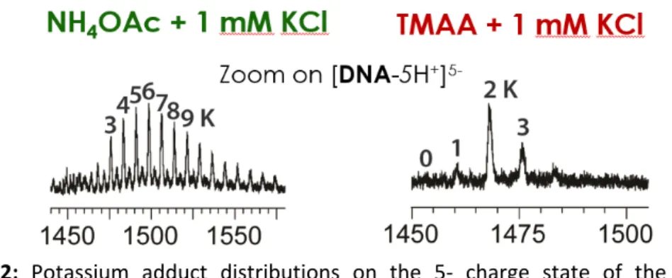
Results Here nonspecific adducts are defined as metal adducts formed upon binding to groups or structures that are not the specific structural motif under investigation. It does not mean that these adducts (or ion pairs) do not exist in solution. For example, for G‐quadruplexes, the specific adducts are the potassium adducts bound to the specific inter‐quartet locations, while the nonspecific adducts are those that would form in any single stranded sequence of the same length and base content. In the case of G‐ quadruplexes, we used trimethylammonium acetate (TMAA) instead of the traditionally used ammonium acetate, to achieve two objectives: first, TMA cannot coordinate in‐between the G‐quartets; second, TMA better suppresses than ammonium the nonspecific K+ adducts (Figure 2).
Figure 2: Potassium adduct distributions on the 5‐ charge state of the sequence
dTAGGGTTAGGGTTAGGGTTAGGG (forming a G‐quadruplex with 3 quartets and 2 specific potassium binding sites), recorded in 1 mM KCl in either 100 mM NH4OAc (left) or TMAA (right). We then used this buffer to study the K+ binding stoichiometry and equilibrium binding constants, on several telomeric DNA G‐quadruplex sequences. We found interesting differences among sequences, revealed by the preferred stoichiometry of potassium binding. Some sequences bind 2 K+ ions in a very cooperative manner, whereas others prefer the 1‐K+ binding stoichiometry and the second potassium ion comes only when adding large amounts of KCl (Figure 3). For magnesium complexes to RNA structures, TMAA could not be used because it prevented that structure (a kissing loop complex; or “kissing complex”) to form. We therefore doped ammonium acetate solutions with magnesium, and observed numerous adducts. A procedure to subtract the contribution of nonspecific adducts was necessary. For that, we used as a “control” a RNA duplex structure of the same length, base composition, and number of base pairs as our kissing complex. The only difference is the base pair arrangement and the absence of kissing loop motif. “Nonspecific adducts” are therefore defined as adducts that are not specific to the kissing loop motif (with no assumption on whether these adducts pre‐exist in solution or are formed during the electrospray process). Using a mathematical subtraction of the signal intensity of the adducts on the control from the signal intensity of the adducts on the structure of interest, we deduced the presence of two specific binding sites, one of higher affinity than the other. Preliminary molecular modelling simulations in solution confirm this trend, and lead us to assign the highest affinity binding site exactly at the kissing loop motif (Figure 4).
Figure 3: Mass spectrometry titrations (20°C) of 10 µM 22GT or 24TTG sequences, in 100 mM TMAA with increasing amounts of KCl (zooms on the 5‐ charge state). (right) Concentration of the complexes with 0, 1 or 2 specific potassium ions bound in green, blue and orange, respectively, as a function of total KCl concentrationThe error bars are the standard errors on the treatment of the three charge states. Figure 4: Mass spectrometry monitoring of the magnesium titration of the RNA*RNA kissing complex dimer TAR‐R06 (B), and of a control duplex sequence not containing the kissing complex motif (A). The intensity ratios allow to infer the distribution of abundances that pertain to specific adducts. On the right: molecular dynamics simulation of the kissing complex TAR‐R06 in the presence of two magnesium ions. One (violet) remains located in the kissing loop motif for the major duration of the simulation. The other (pink) switches between two preferred binding sites, indicating a lower affinity.