D
IOXYGENA
CTIVATION ANDS
UBSTRATEH
YDROXYLATION BY THEH
YDROXYLASEC
OMPONENT OFT
OLUENE/O-X
YLENEM
ONOOXYGENASEFROM PSEUDOMONAS SPORIUM OX1
by
Leslie Justin Murray B.A. Chemistry, B.A. Biology
Swarthmore College, 2002
SUBMITTED TO THE DEPARTMENT OF CHEMISTRY IN PARTIAL FULFILLMENT OF THE REQUIREMENTS FOR THE DEGREE OF
DOCTOR OF PHILOSOPHY OF INORGANIC CHEMISTRY AT THE
MASSACHUSETTS INSTITUTE OF TECHNOLOGY
SEPTEMBER 2007
© Massachusetts Institute of Technology All rights reserved
Signature of Author:
Department of Chemistry August 02, 2007
Certified by:
Stephen J. Lippard Arthur Amos Noyes Professor of Chemistry Thesis Supervisor
Accepted by:
Robert W. Field Haslem and Dewey Professor of Chemistry Chairman, Departmental Committee on Graduate Studies
This doctoral thesis has been examined by a Committee of the Department of Chemistry as follows:
Daniel G. Nocera W. M. Keck Professor of Energy and Professor of Chemistry Committee Chairman
Stephen J. Lippard Arthur Amos Noyes Professor of Chemistry Thesis Supervisor
Stuart S. Licht Assistant Professor of Chemistry
D
IOXYGENA
CTIVATION ANDS
UBSTRATEH
YDROXYLATION BY THEH
YDROXYLASEC
OMPONENT OFT
OLUENE/O-X
YLENEM
ONOOXYGENASEFROM PSEUDOMONAS SPORIUM OX1
by
Leslie Justin Murray
Submitted to the Department of Chemistry on 02 August, 2007
In Partial Fulfillment of the Requirements for the Degree of Doctor of Philosophy in Chemistry
ABSTRACT
Non-heme carboxylate-bridged diiron centers in the hydroxylase components of the bacterial multicomponent monooxygenases activate dioxygen at structurally homologous active sites. Catalysis requires the management of four substrates: electrons, protons, dioxygen, and hydrocarbons. Protein component complexes control the delivery of these substrates to the diiron center in the hydroxylase ensuring selective hydrocarbon oxidation. A detailed mechanistic understanding of structural and chemical consequences of such interactions is a significant challenge.
This thesis begins with an overview of our current understanding of these processes. The discussion is primarily on the methane monooxygenase systems (MMO) because these have been the most extensively studied BMMs to date. Recent results for the toluene/o-xylene monooxygenase (ToMO) and phenol hydroxylase systems from Pseudomonas sporium OX1 are also briefly summarized, the former being the research focus of this dissertation.
Restricting access to the diiron center in ToMOH and other non-heme carboxylate-bridged diiron proteins was proposed to facilitate observation of oxygenated intermediates. To examine this hypothesis, dioxygen activation in ToMOH mutants that were predicted to occlude this channel was investigated by rapid-freeze quench (RFQ) EPR, Mössbauer, and ENDOR spectroscopy and stopped-flow optical spectroscopy. For the I100W mutant, a transient species is observed with an absorption maximum at 500 nm. EPR and Mössbauer spectra of RFQ samples identified this species as a diiron(III,IV) cluster spin-coupled to a neutral W radical. ENDOR spectra of this intermediate confirmed the protonation state and type of the amino acid radical and also identified a labile terminal water or hydroxide on the diiron center. Decay of this intermediate results in hydroxylation of the W radical. A diamagnetic precursor to the mixed-valent diiron(III,IV) center was also observed at an earlier time-point, with Mössbauer parameters typical of high-spin FeIII. We have tentatively assigned this antiferromagnetically-coupled diiron(III) intermediate as a peroxo-bridged cluster.
A similar diiron(III) species is observed in RFQ Mössbauer samples from the reaction of reduced wild type hydroxylase with dioxygen. Substrate accelerates the decay rate of this species, providing evidence for the diiron(III) transient as the active oxidant. Under steady state conditions, hydrogen peroxide was generated in the absence of substrate. The oxidized hydroxylase also decomposed hydrogen peroxide to liberate dioxygen if no reducing equivalents were present. This
catalase activity suggests that dioxygen activation could be reversible. The linear free energy relationship determined from steady state hydroxylation of para substituted phenols has a negative slope. A value of ρ < 0 is indicative of electrophilic attack on the aromatic substrate by the oxidizing diiron(III) intermediate. The results from these steady state and pre-steady experiments provide compelling evidence that the diiron(III) transient is the active oxidant in ToMO and is a peroxodiiron(III) transient, despite differences between the optical and Mössbauer spectroscopic parameters and those of other peroxodiiron(III) centers.
Enzymatic oxidation of the radical clock substrate probe, norcarane, by ToMO gives rise to both desaturation and hydroxylation products, norcarenes and norcaranols respectively. Norcarenes are better substrates for this enzyme system than norcarane, producing additional oxidation products. In all, more than twenty oxidation products were characterized in these reaction mixtures, half of which arose from norcarene oxidation. Accounting for these secondary oxidation products, we determined that no substrate radical intermediates with a significant lifetime (τ < 25 ps) are formed during catalysis.
Thesis Supervisor: Stephen J. Lippard Title: Arthur Amos Noyes Professor of Chemistry
To my teachers
ACKNOWLEDGEMENTS
My completion of graduate school would have been impossible without my parents, my brother, and Sophia. Their emotional support through the early times at MIT and in the dash at the death was immensely important and uplifting. Their support and love have been like a puppeteer’s strings that have kept me standing upright when I’ve felt like collapsing to the floor. We did it!
Early in my graduate career, I was without a lab to call home and my advisor, Professor Stephen Lippard, placed his faith and trust in me to undertake a project that was far removed from my research interests at that time. Accidents invariably flower into beautiful experiences, and my time in his lab is an experience that I would never trade. Intellectually, I have gained a profound awe and respect of enzymatic catalysis. His weekly, monthly, or biannual intellectual or scientific nitpickings and his demand for perfection, or what he perceived it as, were invaluable and annoying all at once. I am indebted to him for his guidance and support over the past five years. Useful, interesting, and challenging discussions were always on the menu in Professor Daniel Nocera’s office. He instructed me during both of my oral examinations on photoinduced electron-transfer and charge separation. Now I know more than nothing! The remaining inorganic faculty, Professors Dick Schrock, Kit Cummins, Alan Davison, and Joseph Sadighi have provided me with the platform to understand, question, and appreciate the wealth of chemistry that falls under the veil of inorganic. And last, but not least, I thank Professor Stuart Licht for agreeing to sit on my thesis committee as I struggled to make the availabilities of various professors magically align.
Swarthmore College is a small place and it is odd to stay close to friends as one moves from undergraduate to graduate institutions. Tushar Parlikar has been a brother to me since my first days at Swarthmore and having him at MIT to share a drink, meal, ice cream, conversation, or anything else was wonderfully rejuvenating and relaxing. No matter how bad the day, a friendly face was just around the corner in electrical engineering. The walk to/from campus or Sunday morning dim sum was always in the company of Simon Kaufman, Aliki Bonarou, Russell Gordley, and Imo Akpan. I’m truly fortunate to have had their friendship while at MIT even if I’ve not been the most stellar correspondent.
For two summers, I worked at some iteration of Glaxo. I learned an enormous amount from Graham Robinett, Bill Zuercher, Stephen Reister, and Phil Turnbull, to name a few, while I was there. These experiences heightened my chemical interest and guided me toward graduate school. In my first summer, I worked for Sam Gerritz who always hoped secretly that I’d go to his graduate alma mater. He’s talked me through a few rough times at M.I.T., and it has been good to have a friendly ear who knows what life in MIT Chemistry is like. Day trips with Sophia in August to visit the Gerritz family at Manchester-by-Sea to sit by the water, chat about life, and relax have been eagerly anticipated off-days many a summer.
I’ve spent more time in lab than anywhere else in the last few years. Fittingly, the people with whom I’ve worked have helped me all along the way. Matt Sazinsky and Laurance Beauvais took me under their wings as I floundered through protein expressions and purifications. I watched Euro 2006 with Dr. Viviane Izzo, screaming our hearts out as our teams played good, then bad, then lost. Colleague, classmate, and friend equal Mi Hee Lim. These coworkers have been great friends who’ve shared multiple scorpion bowls, swigs of rum, and late night dinners. Woon Ju Song, who is taking up the reigns on ToMO, has been immensely helpful to me as I was writing. The remainder of my subgroup past and present – Jessica Blazyk, Lisa Chatwood, Erik Dill, Mike McCormick, and Christy Tinberg – for their invaluable intellectual support and camaraderie over these five years.
My teachers from primary and secondary school in Trinidad – Mrs. Farinha, Mrs. Mitchell, Mrs. Camille Warwick, Mrs. Judy Lee Pow, Mr. Clifford Pierre, and Mr. Rajnatsingh – and my professors at Swarthmore College – Bob Paley, Bob Pasternack, Ahamindra Jain, Michael Wedlock, Paul
Rablen, Tom Stephenson, and Liz Vallen – were instrumental. This thesis acknowledges their tireless efforts as educators.
My research in the Lippard lab would have been impossible without our collaborators. Professor Brian Hoffman, Roman Davydov, Sun-Hee Kim, and Judy Nocek at Northwestern University have been amazing coworkers throughout my MIT career. Brian and Roman, in particular, have always given freely of their time to ensure that I understood the science. On my eight or so visits to Emory, I’ve had multiple dinners and lunches where the discussion has ranged from personal to professional with Professor Boi Hanh Vincent Huynh. His postdoctoral associates, Ricardo García-Serres, Sunil Naik, Danilo Ortillo, have been the workhorses behind data collection, processing, and analysis for the rapid freeze quench studies. Their contribution to my graduate work is immeasurable. Ioannis Papayannopoulos in the Proteomics Core Facility in the CCR at MIT examined my samples over and over and over again. Although we weren’t able to get the beautiful data for which we were hoping, I’m very grateful to him for patient conversations regarding the experiments and subsequent data analysis.
My undergraduates, Jessica Lee and Eric Brown, who worked under my supervision have contributed greatly to the work that’s presented here. Moreover, every time graduate school wore me down and made me question if this chemistry thing was worth it, along came Jessica with questions about her project, and biscotti to boot. Those discussions rejuvenated me on those long days or weeks.
Lastly, the Lippard Lab is filled with fantastic coworkers such as Andy Tennyson, Loi Do, Liz Nolan, Lindsey McQuade, K. Summer Lovejoy, Katie Barnes, Emily Carson, Sungho Yoon, Dong Xu, Dong Wang, Yongwon Jung, Evan Guggenheim, and Lippardites past and present. Cheers to you all!
I’m DONE!
TABLE OF CONTENTS ABSTRACT 3 DEDICATION 5 ACKNOWLEDGEMENTS 6 TABLE OF CONTENTS 8 LIST OF TABLES 12 LIST OF SCHEMES 13 LIST OF FIGURES 14 ABBREVIATIONS 17
CHAPTER 1. Substrate Trafficking & Dioxygen Activation in Bacterial Multicomponent
Monooxygenases 19
Introduction 20
BMM Protein Components 22
Component Interactions 22
Electron Transfer to the Oxidized Hydroxylase 28
Formation and Reactivity of Activated Dioxygen Intermediates 30
Steady-state studies 30
Transient kinetic studies 31
Organization & Scope of Thesis 33
References 37
CHAPTER 2. Oxidation of a Proximal Residue in the I100W Variant of ToMOH 41
Context 42
Introduction 42
Experimental Methods 45
General Considerations 45
Site Directed Mutagenesis of the ToMOH α-Subunit 45
Steady-State Activity Assays and Product Determination for ToMOH Variants 46
Stopped-Flow Optical Spectroscopy 47
RFQ Sample Preparation 49
Mössbauer Spectroscopy 50
X-Band EPR Spectroscopy 50
ENDOR Spectroscopy 50
Enzymatic Digestion and Mass Spectrometry of ToMOH I100W 50
Results 52
Steady-State Product Distribution and Activity of ToMOH Variants for Phenol 52
Stopped-Flow Optical Study of the Reaction of ToMOH Variants with O2 52 Time-Dependent Mössbauer Spectra for the Reaction of ToMOHred I100W with O2 54
Time-Dependent EPR Spectra for the Reaction of ToMOH I100W with O2 57
Kinetic Activation Parameters and Kinetic Isotope Effects for Formation and
Decay of the Diiron(III,IV)–W• Species 60
Effect of pH on the Reaction of ToMOH I100W with O2-Saturated Buffer 62
Effect of Substrates on the Decay Rate of the ToMOH I100W Transient 63
1H- and 2H-ENDOR Spectra of the Mixed-Valent Diiron(III,IV)–W• couple 64
Tryptic Digestion and Mass Spectrometry Analyses of ToMOH I100W and its
Oxidation Product 67
Discussion 68
Mixed-Valent Diiron(III,IV)–W• Species as an Entry to Identifying ToMOH
Intermediates 68
Implications for Catalysis in Native ToMOH 72
Mechanism of Dioxygen Activation and W100 Oxidation 75
Conclusions 79
References 81
CHAPTER 3. Characterization of the Arene-Oxidizing Intermediate in ToMOH as a
Diiron(III) Species 85
Context 86
Introduction 86
Experimental Methods 88
General Considerations 88
Rapid-Freeze Quench Sample Preparation for Mössbauer Spectroscopy 89
Assay for Hydrogen Peroxide Release in the Steady-State 90
An Attempt to Initiate Catalysis by a Peroxide Shunt Pathway 91
Hammett Studies 91
Results 92
Formation of a Diiron(III) Intermediate by Oxygenation of ToMOHred 92
Kinetics of the Reaction of ToMOHred with O2 94
Effect of Phenol on Decay of the Diiron(III) Intermediate 96
Release of Hydrogen Peroxide by ToMO under Steady-State Conditions 99
Peroxide Shunt Pathway and Catalase Activity of ToMOHox 100
Hammett Relationship for Substrate Oxidation by ToMO 101
Discussion 102
Mechanism of Dioxygen Activation by ToMOH 102
Substrate Hydroxylation by ToMOH 107
Implications for the CBDI Protein Family 111
Conclusions 111
References 113
CHAPTER 4. Oxidation of Norcarane by ToMO 117
Context 118
Introduction 118
Experimental Methods 120
General Considerations 120
Enzymatic Oxidations by ToMO 121
Results & Discussion 122
Desaturase Reactions of Norcarane 122
Analytical Protocol for Oxidation Products 123
Product Yields from Norcarane Oxidations 124
Enzyme-Catalyzed Oxidations of Norcarenes 126
Desaturation Effects on Radical Clock Probe Oxidations 129
Implications for Catalysis in ToMO 130
Conclusions 132
Acknowledgements 132
References 133
APPENDIX A. Preliminary Studies of Photoinduced Electron Transfer
to Oxidized MMOH 135
Introduction 136
Experimental Methods 137
General Considerations 137
Synthesis of Labeled MMOB 137
Effect of MMOHox on Fluorescence of RuMMOB 138
EPR Sample Preparation 138
Results 138
Characterization of RuMMOB Constructs 138
Photoreduction of MMOH by RuMMOB 140
Discussion 140
Future Directions 142
References 143
APPENDIX B.Electron Transfer from Reduced ToMOC to the Oxidized Hydroxylase 146
Introduction 147
Experimental Methods 148
General Considerations 148
EPR Sample Preparation and Data Acquisition for Mixtures Containing ToMOCred 149
EPR Sample Preparation of Chemically Reduced Hydroxylase 149
Results 150
Reduction of ToMOHox by Sub-stoichiometric ToMOCred Results in Diiron(II)
Centers 150
Generation of Mixed-Valent Diiron(II,III) ToMOH by Chemical Reduction 150
Discussion 152
Reduction to Oxidized ToMOH Proceeds Through a Half-Sites Mechanism 152
Half-sites Reductive and Oxidative Phases in ToMO 154
Future Directions 154
References 156
Curriculum Vitae 159
LIST OF TABLES
Table 1.1 Spectroscopic Parameters for Peroxodiiron(III) Intermediates at Non-heme
Diiron Centers 21
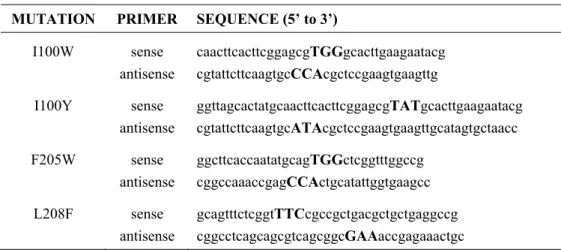
Table 2.1 Sequences for Mutagenic Primers for the α-Subunit of ToMOH 46
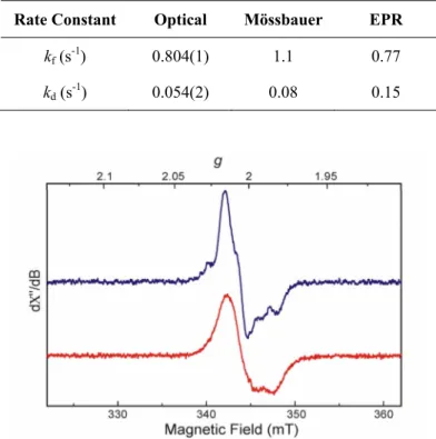
Table 2.2 Formation and Decay Rate Constants of the Mixed-Valent Diiron(III,IV)–W•
Measured by Optical, Mössbauer, and EPR Spectroscopy 58
Table 2.3 Activation Parameters for Formation and Decay of Species Observed in the
Reaction of ToMOH I100W:3ToMOD and MMOH:2MMOB with Dioxygen 61
Table 2.4 Formation and Decay Rate Constants for I100W Transient in H2O and D2O
Buffers 61
Table 2.5 Formation and Decay Rate Constants at Varying pH Values 62
Table 3.1 Mössbauer and Optical Spectroscopic Parameters for μ-1,2-Peroxodiiron(III)
Complexes 105
Table 4.1 Observed Retention Times for GC Elutions as a Function of Column
Temperature 125
Table 4.2 ProductYields from ToMO, PH, and MMO Catalyzed Oxidations of
Norcarane 127
Table 4.3 Product Yields from Oxidations of Norcarenes Catalyzyed by ToMO 127
Table A.1 Simulated g-values and Intensities of EPR Signals in MMOH Samples 139
LIST OF SCHEMES
Scheme 2.1 Proposed Mechanism for Formation of the Diiron(III,IV)–W• Transient. 76
Scheme 2.2 Proposed Mechanism for Decay of the Diiron(III,IV)–W• Transient. 77
Scheme 3.1 Dioxygen Activation at Iron-Heme and CBDI Centers. 87
Scheme 3.2 Proposed Pathways for Substrate Hydroxylation. 101
Scheme 3.3 Reaction of ToMOHox with Hydrogen Peroxide. 106
Scheme 3.4 Substrate Hydroxylation and Dioxygen Activation by ToMOH. 108
Scheme 4.1 Ring-Expansion Products Arising from Norcarane Oxidation. 119
Scheme 4.2 Products of Norcarane Oxidation. 120
Scheme 4.3 Hydride Abstraction Mechanism for ToMO Oxidation of Norcarane. 131
Scheme 4.4 Oxidation Pathways of 2- and 3-Norcarene by ToMO. 132
LIST OF FIGURES
Figure 1.1 Dioxygen activation at non-heme carboxylate-bridged diiron centers. 21
Figure 1.2 Substrate access channel and the conserved pore in ToMOH. 24
Figure 1.3 Crystal structure of PHH in complex with PHM shows the binding of the
regulatory protein in the canyon region. 24
Figure 1.4 PHM binds to PHH at the proposed ET pathway and interacts with
Asn204. 25 Figure 1.5 Redox-state-dependent conformations of residues in the active site pocket
of MMOH. 26
Figure 1.6 A surface-rendered representation of the pore in the α-subunit of PHH to
which PHM is not bound. 26
Figure 1.7 Possible scenarios for component interactions in the BMMs depicting a
half-sites mechanism and a non-interacting sites model. 29
Figure 1.8 Proposed mechanism for reaction of Q and Hperoxo with substrates. 32
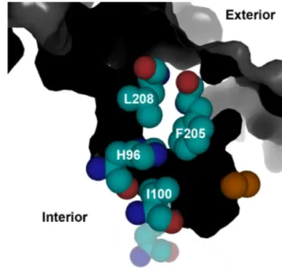
Figure 2.1 View of the active site pocket of ToMOH from the substrate access
channel. 44 Figure 2.2 A substrate access channel in ToMOH extends from the protein surface to
the diiron active site. 44
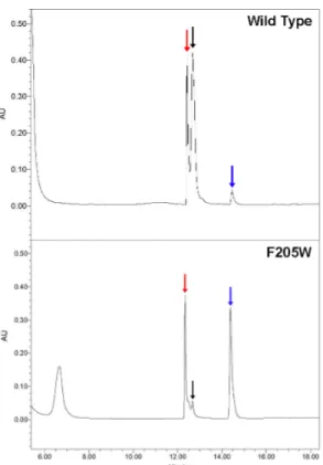
Figure 2.3 HPLC traces at 280 nm of steady-state reaction mixtures for wild-type and
the F205W variants of ToMOH. 53
Figure 2.4 Stopped-flow UV/visible spectra for the reaction of I100Wred:3 ToMOD
with O2-saturated buffer. 53
Figure 2.5 Mössbauer spectra of freeze-quenched samples from the reaction of
ToMOHred:3ToMOD with O2. 55
Figure 2.6 Mössbauer spectra of the sample quenched after 3.5 s recorded with an
applied field of 50 mT and 8T. 56
Figure 2.7 Speciation plot for the reaction of ToMOHred:3ToMOD with dioxygen. 56
Figure 2.8 Time-dependent EPR spectra for the transient formed during the reaction
of ToMOHred I100W:3ToMOD with dioxygen. 58
Figure 2.9 EPR spectra of the mixed-valent diiron(III,IV)–W• transient generated
during reaction of reduced ToMOH I100W with dioxygen. 58
Figure 2.10 X-band EPR specta of the diiron(III,IV)–W• at 20 K and 65 K. 60
Figure 2.11 Eyring Plots for formation and decay of the tryptophanyl radical generated
during reaction of reduced ToMOH I100W with oxygenated buffer. 60
Figure 2.12 SKIE for kf and kd of the ToMOH I100W transient. 61
Figure 2.13 Effect of proton concentration on formation and decay of the
diiron(III,IV)–W• species 62
Figure 2.14 Effect of substrates on the decay rate of the tryptophanyl radical. 63
Figure 2.15 2H-Mims ENDOR of the tryptophanyl radical in buffers containing H 2O
and D2O. 64
Figure 2.16 1H-Mims ENDOR spectra of the tryptophanyl radical. 65
Figure 2.17 2H-Mims ENDOR spectra of the diiron(III,IV)–W• species generated in
deuterated buffers. 66
Figure 2.18 MALDI-TOF spectra of tryptic peptides for the α-subunit of O2-reacted
and as-isolated ToMOH I100W. 67
Figure 2.19 ESI-MS/MS fragment ions arising from the tryptic peptide containing
W100. 68 Figure 3.1 Mössbauer spectra of freeze-quenched samples from the reaction of
reduced ToMOH:2ToMOD mixtures with O2. 93
Figure 3.2 High-field Mössbauer spectrum of a freeze-quench sample from the reaction of reduced ToMOH:2ToMOD with O2 frozen at 0.14 s after
mixing the reduced protein with O2. 94
Figure 3.3 Overlay of Mössbauer spectra of the diiron(III) intermediate and product. 95
Figure 3.4 Speciation plot for reaction of diiron(II) ToMOH:2ToMOD with
dioxygen. 95
Figure 3.5 Mössbauer spectra of double-mixing RFQ samples for reaction of the
diiron(III) intermediate with buffer containing phenol. 97
Figure 3.6 Speciation plot for reaction of the diiron(III) intermediate with phenol. 98 Figure 3.7 Overlay of Mössbauer spectra of the diiron(III) intermediate in the absence
and presence of phenol. 98
Figure 3.8 Optical spectrum of a sample following an RFQ double-mixing Mössbauer
experiment. 99 Figure 3.9 Concentration of hydrogen peroxide evolved under steady-state conditions
with and without phenol. 100
Figure 3.10 Change in the concentration of hydrogen peroxide with time after mixing
with ToMOH–ToMOD mixtures. 100
Figure 3.11 Hammett plots for oxidation of p-substituted phenols by ToMO. 101
Figure 4.1 Portions of GC traces (40 °C, DB-5 column) of norcarane before and after
reaction with ToMO. 123
Figure 4.2 SIM mode GC spectra obtained by monitoring 12 ion channels. 125
Figure 4.3 Portions of GC traces of products from oxidation of 2-norcarene and 3-norcarene with toluene monooxygenase from P. sporium OX1 (ToMO), soluble methane monooxygenase from M. capsulatus (Bath) (sMMO), and
cytochrome P450 2B1 (CYP2B1). 128
Figure A.1 LC trace and deconvoluted MS data for RuMMOB N101C. 139
Figure B.1 EPR specta for mixtures containing variable ratios of ToMOCred to
ToMOH:2ToMOD. 151 Figure B.2 EPR Spectra of ToMOH:2ToMOD mixtures reduced with one, two, three,
and four equivalents of electrons from sodium dithionite. 152
ABBREVIATIONS
ACP acyl-carrier protein
BMM bacterial multicomponent monooxygenase
CBDI non-heme carboxylate-bridged diiron
COSY correlation spectroscopy
Δ9D stearoyl-ACP Δ9 desaturase
DFT density functional theory
dNTP deoxynucleotide triphosphate
E. coli Escherichia coli
EDTA ethylenediamine-N,N,N’,N’-tetraacetic acid
ENDOR electron nuclear double resonance
EPR electron paramagnetic resonance
ESI electrospray ionization in either positive (+) or negative (-) modes
ET electron transfer
FAD flavin adenine dinucleotide
FID flame ionization detection
Ft ferritin
GC gas chromatography
Hmv diiron(II,III) hydroxylase component, prefaced with system abbreviation
Hox oxidized hydroxylase component, prefaced with system abbreviation
Hperoxo peroxodiiron(III) intermediate, prefaced with system abbreviation
Hred reduced hydroxylase component, prefaced with system abbreviation
HPLC high performance liquid chromatography
ITC isothermal titration calorimetry
LC/MS liquid chromatography/mass spectrometry
M. caps. Methylococcus capsulatus (Bath) M. trich. Methylosinus trichosporium OB3b
MALDI matrix-assisted laser desorption/ionization
mCPBA m-chloroperoxybenzoic acid
MMO soluble methane monooxygenase, enzyme system
MMOB regulatory protein of MMO
MMOH hydroxylase component of MMO
MMOR NADH oxidoreductase component of MMO
MMOR-Fd truncated MMOR containing only a ferredoxin domain
MOPS N-morphilinopropane sulfonic acid
MS/MS mass spectrometry/mass spectrometry
MWCO molecular weight cut-off
NADH reduced form of β-nicotinamide adenine dinucleotide
NHE normal hydrogen electrode
NMR nuclear magnetic resonance
NOE nuclear overhauser effect
PH phenol hydroxylase from Pseudomonas sporium OX1, enzyme system
PHH hydroxylase component of PH
PHM regulatory protein of PH
Q diiron(IV) intermediate formed in MMOH
RCS radical-clock substrate
RFQ rapid-freeze quench
RNR-R2 ribonucleotide reductase R2 subunit (Class 1)
rR resonance Raman
SDS-PAGE sodium dodecyl sulfate polyacrylamide gel electrophoresis
SKIE solvent kinetic isotope effect
T4MO toluene 4-monooxygenase from Pseudomonas mendocina KR1, enzyme system
T4MOH hydroxylase component of T4MO
TFA trifluoroacetic acid
TOF time of flight
ToMO toluene/o-xylene monooxygenase from Pseudomonas sp. OX1, enzyme system
ToMOC Rieske component of ToMO
ToMOD regulatory protein of ToMO
ToMOF NADH oxidoreductase component of ToMO
ToMOH hydroxylase component of ToMO
Tris tris(hydroxymethyl)aminomethane
X mixed-valent diiron(III,IV) intermediate formed in RNR-R2
XAS X-ray absorption spectroscopy
CHAPTER 1
SUBSTRATE TRAFFICKING AND DIOXYGEN ACTIVATION
IN BACTERIAL MULTICOMPONENT MONOOXYGENASES
* Reproduced in part with permission from Acc. Chem. Res. 2007, 40 (7), 466-474. Copyright 2007 American Chemical Society 19
INTRODUCTION
The biological activation of small substrates, such as dioxygen and dinitrogen, is carried out by large proteins comprising multiple subunits and containing metal cofactors. Examples of these remarkable enzymes include nitrogenase, cytochromes P450, tyrosinase, hydrogenase, cytochrome c oxidase, and the non-heme carboxylate-bridged diiron proteins. Members of the last family, which include the BMMs, RNR-R2, and Δ9D, activate dioxygen at structurally homologous diiron centers housed within a four-helix bundle. The metal atoms in this bundle are coordinated by the side-chains of two E(D/H)XXH motifs.1,2 The BMMs exquisitely couple the consumption of electrons and protons to dioxygen activation and substrate hydroxylation. Understanding the management of four substrates (electrons, protons, dioxygen, and hydrocarbons) in these enzyme systems is a central goal of ongoing research.
The mechanism of dioxygen activation in the carboxylate-bridged diiron family of enzymes is the subject of intense investigation. In the systems studied thus far, the resting state of the enzyme is a diiron(III) cluster with bridging oxo or hydroxo ligands, as observed in RNR-R2 and MMOH respectively. This state is unreactive toward dioxygen. Catalysis is initiated by two-electron reduction of the resting state to the reactive diiron(II) state. In systems where structures of the diiron(III) and diiron(II) forms of the enzymes, or analogues thereof, have been solved, reduction is accompanied by reorganization of the dimetallic cluster, whereby a dangling, or non-bonded, oxygen atom of a terminal carboxylate ligand shifts so that it bridges the metal ions. To maintain charge neutrality at the active site, proton transfers are predicted to occur during the reduction process. These protons may protonate the bridging hydroxo or oxo ligands, and the formed water dissociates from the dimetallic center.3,4 The resulting diiron(II) form rapidly reacts with dioxygen to form a peroxodiiron(III) intermediate, which has been characterized by a number of spectroscopic methods (Table 1.1).1 In MMO and RNR-R2, this intermediate evolves to higher-valent species Q and X,
respectively, which carry out methane hydroxylation or the one-electron oxidation of aromatic amino acid residues (Figure 1.1). In MMOH, both the peroxodiiron(III) (MMOHperoxo) and Q intermediates are reactive toward substrates,5 similar to the reactivity observed for oxygenated intermediates in the cytochrome P450 family6 and some dicopper systems.7 The catalytic cycle is closed as the
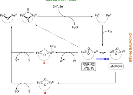
Figure 1.1. Dioxygen activation at non-heme carboxylate-bridged diiron
centers. Reaction of the reduced diiron(II) state with dioxygen affords a peroxide-bridged intermediate, which can evolve to a high-valent species or decay to the resting diiron(III) state. In MMOH, intermediate Q can oxidize substrates, denoted as S, or decay via other pathways to the resting state. The peroxo intermediate in RNR-R2 oxidizes Trp48 to form X, which then oxidizes Tyr122 to restore the resting diiron(III) state.
Table 1.1. Spectroscopic Parameters for Peroxodiiron(III) Intermediates at Non-heme Diiron Centers
Opticala Mössbauera Resonance Ramana λmax [nm] ε [M-1cm-1] δ [mm/s] ΔE Q [mm/s] O-O [cm-1] Fe-O [cm-1] MMO (M. caps.) 700 1800 0.66 1.51 - - MMO (M. trich.) 725 2500 0.67 1.51 - - RNR-R2b 700 1500 0.63 1.74 - - RNR-R2 D84E 700 1500 0.63 1.58 890 - RNR-R2 D84E/W48F 700 - - - 870 458 Δ9D 700 1200 0.68 1.90 898 442 0.64 1.06 Frog M Ft 650 - 0.62 1.08 851 458
cis-μ-1,2-peroxo Fe2III 694 2650 0.66 1.40 888
aAll parameters are reported from reference 1. b Reported in reference 42.
oxygenated intermediates return to the resting diiron(III) state following substrate hydroxylation or electron abstraction. In the absence of substrate, intermediate Q decays by a yet to be determined pathway to the resting state.
This discussion begins with a description of the protein components and their interactions. We then proceed through the stages of the catalytic cycle, examining first the ET events, followed by dioxygen activation and, finally, substrate hydroxylation. The role of protein component interactions in these processes is also highlighted.
BMM PROTEIN COMPONENTS
The BMMs are subdivided into different families, but all contain at least three components in the enzymatic system: a hydroxylase, an NADH oxidoreductase, and a regulatory protein. A fourth, Rieske protein, is also utilized in toluene/alkene monooxygenases.8 In MMO, ToMO, and PH, the hydroxylase is a dimer of three polypeptide chains, (αβγ)2, with each α-subunit housing the carboxylate-bridged diiron center, the site of substrate hydroxylation. The hydroxylase molecular mass varies from 220 kDa in ToMO and PH to 251 kDa in MMOH. The 38-kDa oxidoreductase component shuttles electrons from NADH through its bound FAD and [2Fe-2S] cofactors to the hydroxylase either directly, as in MMO and PH, or indirectly via the Rieske component. The 10 to 15-kDa cofactor-less regulatory proteins bind to the hydroxylase and couple electron consumption to hydrocarbon oxidation.9-11 The precise mechanistic consequences of these protein-protein interactions on ET and dioxygen activation are the subject of ongoing research. Very recently, the structure of PHH complexed to its regulatory protein, PHM, was solved, providing the first detailed picture of such an interaction.12
COMPONENT INTERACTIONS
The assembly of protein complexes during the reductive and oxidative phases of the catalytic
cycle must be finely orchestrated to ensure that electron consumption is linked to substrate hydroxylation. For example, reactive oxygenated diiron intermediates in the hydroxylase must not undergo adventitious reduction by the reductase or Rieske protein components.13 The regulatory protein is proposed to compete with the reductase for a shared binding site on the hydroxylase,10 thereby protecting oxygenated intermediates formed at the diiron center from undesired reductive quenching following dioxygen activation.12 On the other hand, ET from these reducing components to the resting, diiron(III) state of the hydroxylase must occur to initiate catalysis. In this section, we first discuss studies of the structures and interactions between the regulatory proteins and their respective hydroxylases and then turn our attention to the ternary system comprising the regulatory, hydroxylase, and reductase/Rieske components.
Crystal structures of BMM hydroxylases from MMO, ToMO, and PH have been solved. Three structurally conserved features of note are hydrocarbon access routes, a likely ET pathway to the diiron core, and a pore through the four-helix bundle housing the diiron center for possible dioxygen or proton translocation. Soaking or pressurizing crystals of these proteins with substrate or product analogues has revealed the first of the three shared features, a potential pathway for the entrance or egress of hydrocarbon substrates or products. Soaking ToMOH crystals with the product analogue p-bromophenol revealed a large channel that delineates an access pathway from the exterior of the protein through the α-subunit to the active site (Figure 1.2).14 When MMOH crystals were treated with xenon, bromomethanes, or iodoethane, these small hydrophobic species were detected in a series of adjacent hydrophobic pockets within the protein that trace a pathway from the protein surface to the diiron center.15 The distinct pockets are capable of forming a contiguous pathway, as evidenced by structures of crystals soaked with ω-halogenated alcohols. In these structures, several residues, including Leu110 and Leu289, shift to allow the substrate analogues to traverse the protein cavities.16 The structure of MMOH crystals soaked with 6-bromohexan-1-ol revealed another
property of note. Residues 212-216 in helix E of the α-subunit unwind to extend the π-character of this helix, increasing the active site volume and reorienting Thr213 and Asn214, residues strictly conserved among the BMMs. The structure of PHH complexed to PHM contains a similarly extended π-helix, providing strong evidence that the binding of BMM regulatory proteins to their respective hydroxylases gives rise to specific conformational changes in helix E that directly affect the configuration of the active site pocket.12 The environment around the diiron cluster was predicted from XAS studies of ToMOH and MMOH in complex with their regulatory proteins17 to undergo conformational changes, and the PHH-PHM structure provides the first evidence for the nature of these changes.
PHM binds in the canyon region of PHH, specifically on helices E and F, and covers a hydrogen-bonding network that extends from the exterior to the iron-coordinated histidine residues at the active site. This feature is the second one conserved among the BMMs (Figures 1.3 and 1.4). The
Figure 1.2. Substrate access channel and the conserved
pore in ToMOH. The access channel (magenta) delineates a path from the diiron center (orange atoms) to the protein surface through the α-subunit (grey). The conserved pore (green), which is gated by Asn202, extends from the active site pocket to the protein surface. One half of the dimer is depicted.
Figure 1.3. Crystal structure of PHH in complex with
PHM shows the binding of the regulatory protein in the canyon region. The surface of PHH is rendered translucent with the α-, β-, and γ-subunits depicted in grey, purple, and blue, respectively. The iron atoms are depicted as orange spheres and denoted by arrows. PHM (red) binds to the α-subunit in a location similar to that predicted for binding of other regulatory proteins to their hydroxylases.
locus on PHH where PHM binds, and the predominantly hydrophobic contacts between the proteins, agrees with the predictions of early NMR line-broadening studies carried out on MMOB and MMOH from M. caps.18 The binding site of the regulatory proteins in the BMMs may therefore be conserved throughout the family. The hydrogen-bonding network that is covered by PHM, and presumably MMOB, is spatially homologous to the proposed ET pathway in other non-heme diiron proteins.14 Binding of the BMM regulatory proteins to this surface, therefore, would interfere with ET for the reasons described above.13
The third structurally conserved feature among BMMs is a small pore through the four helix bundle, which is proximal to the proposed ET pathway and extends from the active site pocket to the protein surface. In most of the crystal structures of BMM hydroxylases, this pore is closed, with a strictly conserved Asn residue (214, 202, and 204 in MMOH, ToMOH, and PHH respectively) serving as a gate to the opening. Crystallographic studies reveal this Asn residue to shift in a redox-dependent manner. Its side chain is oriented away from the active site in the oxidized form and points inward in the reduced or Mn(II)-reconstituted forms of the hydroxylase. This motion correlates with
Figure 1.4. PHM binds to PHH at the proposed ET pathway and interacts with Asn204. (A) The surface-exposed
residues, Arg235 and Lys68, of the conserved hydrogen-bonding network in the α-subunit are highlighted in yellow on a surface-rendered diagram of PHH. The α-, β-, and γ-subunits are depicted in grey, purple, and blue, respectively. (B) Diagram A with PHM (red) included. PHM covers this network when complexed to PHH and may prevent the reductase from binding at this locus. (C) A cutaway view, perpendicular to that in A and B, shows that this network, which extends from the iron atoms (orange spheres) to the surface, lies at the interface between the two proteins. PHM also interacts with Asn204 (green sticks), the side chain of which is oriented away from the diiron center as in the oxidized form of the hydroxylase and forms a hydrogen bond with Ser72 of the latter.
the carboxylate shift that occurs upon reduction of the dimetallic center (Figure 1.5).3,4 DFT calculations predict that dioxygen activation at the diiron(II) core of MMOH is accompanied by dissociation ofthis carboxylate group from its bridging position.19 The Asn gate is open and the pore provides direct access to the diiron center in the bromohexanol-soaked structure of MMOH due to the increased π-character of helix E.16
In the α-subunits of PHH, helix E adopts π-character similar to that in the MMOH bromohexanol-soaked structure, orients Asn204 away from the active site, and opens the pore.12 PHM is bound only to one face of the PHH dimer and restricts access through this pore for the α−subunit to which it is bound, but for the other, the pore allows access to the diiron center (Figure 1.6). The opening of the pore by the shift in Asn204 on the face opposite that to which PHM binds may be an allosteric effect transmitted through the protein dimer interface, arise from interactions
Figure 1.5. Redox-state-dependent conformations of
residues in the active site pocket of MMOH. The amino acid residues depicted in gray correspond to the reduced state. Residues Glu243 and Asn214 shift upon oxidation (yellow) with Glu243 changing its coordination mode to the dimetallic center.
Figure 1.6. A surface-rendered representation of the
pore in the α-subunit of PHH to which PHM is not bound. Asn204 (blue) is oriented away from the diiron center, opens the pore (red), and allows access from the protein exterior to the diiron center (orange spheres). Four iron-binding ligands, Glu108, Glu139, His236, and His141, are identified. This structural change may arise from PHM binding at the opposing face, interactions with the N-terminus of the β-subunit from the opposing monomer (purple), or packing in the crystal.
with the N-terminus of the α-subunit of the adjacent monomer, or be a consequence of crystal packing. Residue Asn204 in PHH interacts with the hydroxyl side chain of a serine residue of the regulatory protein (Figure 1.4C), suggesting that mechanical strain created when the latter binds to the hydroxylase could promote rearrangement of the shifting carboxylate.12 The hypothesis20 that the regulatory protein binds to the MMO hydroxylase and provides a sieve for methane access to the active site at that position is inconsistent with the PHH-PHM structure. Moreover, we have not been able to rationalize this alternative proposal with the observation of CH2Br2, Xe, and other methane substrate analogues bound in the hydrophobic cavities of MMOH during structure determinations of crystals exposed to these agents. Finally, we note the possibility that dioxygen or protons may enter the active site via the pore. Additional structures of hydroxylases in complex with their respective regulatory proteins are required to evaluate further these possibilities.
Among the BMMs, component interactions in MMO isolated either from M. caps. or M. trich. have been the most extensively studied. Similar interactions were proposed for the binding of
MMOB and MMOR to MMOH from studies using chemically modified MMOH.21 The nature of
these similar interactions was proposed to be electrostatic in contrast to the NMR line-broadening experiments mentioned above and the crystal structure of PHH-PHM. Whereas the binding face may have predominantly hydrophobic contacts, important electrostatic interactions, possibly to Arg245 and Lys74 in MMOH, may be essential for catalysis. A higher resolution structure of PHH-PHM with greater occupancy of PHM would be invaluable in resolving this apparent contradiction. The efficiency of cross-linking MMOB to MMOH decreases in the presence of MMOR,9 suggesting that these components may share, either partially or completely, the same binding site on the hydroxylase. Both MMOR and MMOB bind to MMOH with a 2:1 stoichiometry in the M. caps. system, with
MMOR binding being an order of magnitude stronger to MMOH than MMOB.10 A 1:1
MMOB:MMOH complex has been proposed for the analogous M. trich. enzyme system.9 In M.
trich., MMOB binds thirty-fold more weakly to reduced versus oxidized MMOH.22 The Kd values determined from the formation and dissociation rate constants of these complexes disagree with the thermodynamic ones, indicative of rapid pre-equilibrium complex formation followed by a slower structural change,10,22 such as an allosteric effect being transmitted to the other binding site on the dimer. Component binding to one face of the hydroxylase could effect a structural change in the canyon region on the opposite half of the dimer, as noted previously in the PHH-PHM structure. In agreement with the stoichiometry mentioned above, a ratio of 2 MMOB:1 MMOH affords maximal activity for steady-state hydroxylation in the M. caps. system. Moreover, enzymatic inhibition occurs at higher ratios of MMOB, suggesting that MMOB saturates the binding sites on MMOH, inhibiting the association of MMOR with MMOH.
ELECTRON TRANSFER TO THE OXIDIZED HYDROXYLASE
An early study of ET from NADH to the diiron(III) centers in oxidized MMOH, premixed with MMOR and variable equivalents of MMOB, demonstrated that all of the ET events from the [2Fe-2S] cluster of MMOR to diiron(III) active sites of MMOH are enhanced by MMOB.10 More recently, an investigation of the reaction of chemically reduced MMOR or MMOR-Fd with MMOH-MMOB mixtures showed the opposite effect, in which MMOB served to inhibit ET.23 These two apparently contradictory effects of MMOB on ET may be a consequence of a slow structural change associated with MMOB or MMOR binding to MMOH.23 The conclusion that hysteresis may control the activity of MMOH was similarly drawn from an analysis of the product distributions when various substrates were hydroxylated by MMO from M. trich.24 The interaction of MMOH with either MMOR or MMOB may depend on the presence or absence of the other component. The two may bind either concurrently, on opposing canyon regions of MMOH, or in rapid succession at the same canyon region on one side of the hydroxylase.
These two scenarios for the interaction of the components during the catalytic cycle are
illustrated in Figure 1.7. In the half-sites reactivity mechanism, the reductase binds on one canyon surface for ET while the regulatory protein binds on the other canyon surface for dioxygen activation and substrate hydroxylation (Figure 1.7A). Hysteresis would reflect binding of the regulatory and the reductase components to the opposite faces of the hydroxylase, thereby predisposing each diiron center to traverse opposite segments of the catalytic cycle. RFQ Mössbauer experiments performed to investigate the reaction of reduced MMOH25 and ToMOH26-28 with dioxygen have recorded consistently a maximal conversion of ~ 50% of the initial diiron(II) protein to the oxygenated intermediates. This observation suggests that the oxidative phase of the catalytic cycle can occur only at one active site of the dimer, irrespective of whether both active sites are fully reduced. The reaction of reduced ToMOH with dioxygen studied by RFQ Mössbauer spectroscopy will be
Figure 1.7. Possible scenarios for component interactions in the BMMs depicting a half-sites
mechanism (A) and a non-interacting sites model (B). In (A), one active site is undergoing reduction while the second is activating dioxygen. In (B), a ternary complex may form but is not obligatory for catalysis and may have implications for ET and dioxygen activation. Adapted from reference 10.
presented in more detail in Chapters 2 and 3. Half-sites reactivity has also been proposed in other non-heme diiron proteins, in particular Δ9D29 and RNR-R2,30 and may be a conserved feature in these systems. A different scenario, depicted in Figure 1.7B, is that the regulatory protein binds loosely to the hydroxylase and is displaced partially or shifts in the presence of the reductase to form a ternary complex that is competent for ET and catalysis. The inhibitory effect of excess MMOB on steady-state catalysis in MMO is best simulated by a model in which MMOB and MMOR bind to MMOH non-competitively and form a transient ternary complex.10
FORMATION AND REACTIVITY OF ACTIVATED DIOXYGEN INTERMEDIATES
Steady-state studies. The mechanism of hydrocarbon hydroxylation by the BMMs under
steady-state conditions has been examined with radical clock substrate (RCS) probes. These hydrocarbons often contain strained rings that open with a characteristic lifetime following the loss of a hydrogen atom or ion. Such alternative substrates have been used to probe the mechanism of steady-state catalysis for a variety of enzyme systems including the BMMs.1,31 Radical-derived ring-opened products will be observed only if their recombination rate is slower than ring expansion, in which case the lifetime of the radical can be estimated from ratios of the rearranged to unrearranged product alcohols. In general, only unrearranged products arising from rapid recombination of bound radical species are observed for most of the RCS probes tested in MMO, and their lifetime has been estimated as being less than 150 fs.1,31 In addition to ring expansion from H-atom abstraction from the substrate, some RCS probes can ring-open following hydride abstraction to yield products that differ from the radical-derived ones. Norcarane, methylcubane, and 1,1-dimethylcyclopropane are examples of such RCS probes and the major products formed in steady-state catalysis by ToMO32, T4MO33, and MMO1 with these substrates are the unrearranged alcohols. Little or no rearranged products, either radical- or carbocation-derived, are observed for these substrates in reactions with T4MO and MMO, in agreement with the other RCS experiments mentioned above. For the oxidation
of norcarane by MMO, ring-expansion products from both intermediates formed and the product ratios placed a lower limit of 20 ps on the radical lifetime, significantly greater than the value determined from any other probe. Oxidation of this RCS by T4MO afforded a radical-rearranged product (4.5% of the total), and no carbocation-derived product could be detected, contrary to the results from the oxidation of both 1,1-dimethyl- and 1,1-diethylcyclopropane.33 A recent reexamination of the norcarane reaction for MMO and ToMO revealed that desaturation of the substrate by BMMs can explain these discrepancies.34 Evidence for both a hydride and an H-atom transfer mechanism suggests that more than one reactive oxygenated intermediate may be involved.
Transient kinetic studies. In the oxidative phase of the catalytic cycle (Figure 1.1), the regulatory
protein is required for the formation of detectable intermediates35 and tunes the regioselectivity of substrate hydroxylation.36 Transient, pre-steady-state kinetic studies have been performed with chemically reduced mixtures of the hydroxylases and their regulatory proteins to study the reactivity of individual intermediates during the reaction of dioxygen with the reduced BMM hydroxylases. In particular, we have utilized single- and double-mixing stopped-flow methodologies whereby a solution of reduced hydroxylase with its regulatory protein is mixed rapidly with dioxygen, allowed to age for a time period that maximizes the formation of an intermediate of interest, and the aged solution is subsequently mixed with buffer containing a substrate. Loss of an optical absorption band characteristic of the intermediate,5 or of an infrared band of a substrate,37 is measured as a function of substrate concentration. Among the BMMs, this strategy has been the most effective for investigating the reactions of MMOH with dioxygen since both MMOHperoxo and Q have characteristic absorption bands. The effect of substrates on the decay rate of each intermediate can be readily monitored by recording a change in these optical features. Initial studies of MMO in which propylene, methane, and acetylene were used as substrates indicated that MMOHperoxo reacts only with propylene whereas Q reacts with all three substrates.5 Previous steady-state studies revealed that terminal alkenes are epoxidized by MMO with no C–H bond activation.38,39 Although Q oxidizes propylene faster than
MMOHperoxo, a judiciously chosen substrate might react more rapidly with MMOHperoxo than Q to confirm that MMOHperoxo is a reactive species. The oxidation of alternate substrates by these intermediates was therefore explored. Two such substrates, ethyl vinyl ether and diethyl ether, reacted more rapidly with MMOHperoxo than with Q.40 A comparison of the nucleophilicity of the substrates investigated, and a detailed study of the oxidation of these particular substrates, indicated that MMOHperoxo is a more electrophilic oxidant than Q, preferring to react by a two-electron, or a hydride abstraction, pathway, whereas one-electron oxidation processes are preferred by Q (Figure 1.8). This difference in reactivity may explain the carbocation-derived products in the RCS probe experiments because attack of MMOHperoxo on these substrates is expected to form transient carbocations and Q would yield radical intermediates.
In contrast to what we observe for MMOHperoxo, peroxodiiron(III) intermediates in ferritin,41 RNR-R2,42-44 Δ9D,29 and synthetic model compounds have no observed reactivity toward hydrocarbons. These peroxo species have been characterized by rR spectroscopy, from which it
Figure 1.8. Proposed mechanism for reaction of Q and MMOHperoxo with substrates. Q reacts by
sequential one-electron oxidations (upper). Reaction of MMOHperoxo with ethyl vinyl ether
proceeds by a two-electron mechanism, in which a transient carbocation is stabilized by the
proximal oxygen atom (middle). Hydroxylation of diethyl ether by MMOHperoxo is also proposed
to proceed by a two-electron process, in which hydride abstraction forms a transient carbocation that recombines with the coordinated hydroxide (lower). Adapted from reference 40.
appears that the peroxide moiety bridges the dimetallic center in a μ-1,2 fashion. Although a rR spectrum has not yet been obtained for MMOHperoxo, computational methods predict that a μ-η2:η2 -peroxo butterfly structure is more stable than the alternative μ-1,2 geometry of the bound peroxide.19,40,45 The occurrence of such a binding mode in MMOH
peroxo may explain why it reacts with electron-rich hydrocarbons and why a Q-type species is only observed in MMO. The difference in reactivity between MMOHperoxo and Q parallels the known differences between (μ-η2:η2 -peroxo)dicopper(II) species and high-valent di(μ-oxo)dicopper(III) center in the dicopper complexes.7 The lower-valent complex reacts by two-electron processes compared to the higher-valent counterpart, which prefers sequential one-electron oxidations. Theoretical studies on MMOH revealed that compression of the diiron cluster by the protein scaffold at the active site facilitates formation of a (μ-η2:η2-peroxo)diiron(III) structure and its subsequent conversion to Q.45 It will be interesting to learn whether such an effect may be absent in other BMM hydroxylases which have different spectral parameters for the peroxo species and for which a diiron(IV) species has not yet been observed. A key difference between MMOB and the other BMM regulatory proteins is that the former has an unstructured 35 amino acid N-terminal domain.18 Truncation of this domain leads to uncoupling of hydroxylation during steady-state turnover.46 The presence of the N-terminal tail also correlates with the unique ability of MMOH to form Q, and binding of this domain to MMOH might further serve to compress the diiron core. Despite the structural homology between the active site of MMOH and ToMOH, no intermediates were observed by stopped-flow optical spectroscopy in studies of the latter.26,27
ORGANIZATION & SCOPE OF THESIS
This thesis addresses the oxidative and reductive phases of the catalytic cycle in the ToMO system. Investigations of dioxygen activation in mutant and native forms of ToMOH are presented in Chapters 2 and 3, respectively. Studies of the steady-state oxidation of the RCS probe norcarane are
described in Chapter 4. Appendix A discusses attempts to photoreduce the oxidized hydroxylase in MMO by tris(bipyridyl)ruthenium(III) complexes tethered to MMOB, and Appendix B details electron transfer from reduced ToMOC to oxidized ToMOH. The work in these appendices is preliminary in nature.
Until recently, no intermediates were reported during stopped-flow optical spectroscopic study of the reactions of reduced ToMOH or PHH, complexed with their respective regulatory proteins, with O2-saturated buffer. Comparison of the crystal structures of ToMOH and MMOH reveal that residue I100 in the former is analogous to a leucine in the latter that is proposed to gate substrate access to the diiron site during catalysis.47 As described in Chapter 2, mutations I100W/Y, L208F, and F205W were introduced in the α-subunit of ToMOH with the aim of retarding access at different points along the substrate access channel of any buffer components to the diiron site that might quench high-valent intermediates before they could accumulate to an observable level.14,47 The reactions of these reduced mutant hydroxylases in complex with ToMOD with dioxygen were studied by stopped-flow optical spectroscopy. This method revealed that, in the ToMOH I100W mutant, a transient species having a maximal absorption at 500 nm formed upon oxygenation. No optically active intermediates were observed for the native or any of the other mutant hydroxylases. The reaction of the I100W mutant with dioxygen was further interrogated by RFQ EPR, ENDOR, and Mössbauer spectroscopy. Two intermediates were observed: a mixed-valent diiron(III,IV) cluster coupled to a neutral tryptophanyl radical, the latter giving rise to the optical absorption at 500 nm, and a diiron(III) species, which has no distinct optical absorption features and forms prior to the mixed-valent diiron(III,IV).26
Chapter 3 details our studies of dioxygen activation in the native system under steady-state and pre-steady-state conditions. First, we present the reaction of chemically reduced native ToMOH in complex with ToMOD by RFQ Mössbauer spectroscopy. A diiron(III) intermediate with isomer
shift, δ, and quadrupole splitting, ΔEQ, parameters similar to those for the I100W diiron(III) transient was discovered.27 This intermediate is longer-lived than that in the I100W mutant, presumably because it is no longer being quenched by a nearby redox-active residue. The effect of phenol, a substrate for this system, on the decay rate of this oxygenated intermediate was investigated by double-mixing RFQ Mössbauer spectroscopy. The decay is enhanced by more than thirty-fold in the presence of substrate, strongly suggesting that this species is the active oxidant in this system. Steady-state experiments are also described that complement the pre-steady-state investigations. The ToMO enzyme system appears to be incapable of accessing the peroxide shunt pathway for substrate hydroxylation in the absence of reducing equivalents from NADH, unlike MMO.24 Second, the system generates hydrogen peroxide if substrate is absent from the reaction mixture. Third, the steady-state activity for the hydroxylation of a series of para-substituted phenols provided some insight into the nature of the key intermediate in this system. Taken together, these results indicate that a peroxodiiron(III) intermediate is the active oxidant in this system. The peroxide moiety is predicted to have a binding geometry and/or protonation state different from those previously reported in similar carboxylate-bridged diiron systems.
RCS probes have been extensively used to probe enzymatic oxidations under steady-state conditions. In most enzyme systems studied with these substrates, the calculated lifetimes of radical intermediates and the ratios of radical- and cation-derived products are consistent across a variety of different probes. Lifetime calculations from experiments employing norcarane as the RCS probe, however, are inconsistent in most enzyme systems studied thus far. The values obtained from product distributions for norcarane reactions are significantly greater that those from other substrates. A careful examination of the oxidation of norcarane by ToMO determined that more than twenty products form. Almost half of these products arise from oxidation of 2- and 3-norcarene, desaturation products of the parent compound. We therefore conclude that the lifetimes previously estimated in
the homologous T4MO, and for other CBDI enzymes such as MMO,48,49 greatly overestimated the amount of radical-derived product formed.32,34 By estimating the correct yields for the radical- and cation-derived products from our analysis, we determined lifetimes that agree with independent measurements carried out with different probes. Desaturation of substrates by this and other enzyme systems requires a judicious choice of RCS probe for which such reactivity is unfavorable.
No intermediates prior to MMOHperoxo have been observed in any carboxylate-bridged diiron proteins to date. DFT methods19 predict that a short-lived superoxodiiron(II,III) species forms initially after dioxygen binding but that the calculated relative energies of MMOHred, MMOHsuperoxo, and MMOHperoxo preclude the accumulation and observation of such an intermediate. Appendix A describes studies utilizing tris(bipyridyl)ruthenium(II) complexes tethered to MMOB as a method of photoreducing the diiron cluster in MMOH in the presence of dioxygen to search for spectroscopic evidence for early events of dioxygen activation.
Efficient ET from reduced ToMOC to the oxidized hydroxylase requires ToMOH and ToMOD to be mixed prior to the reduction event. Moreover, this reaction only yields ~ 50% of oxidized ToMOC as determined by the change in optical absorbance. Two- or three-exponential functions were required to model the kinetic traces obtained, suggesting that two or more processes are involved in the ET reaction. One possible explanation for this observation is that the electrons can be distributed statistically upon reacting with ToMOHox to yield mixtures of reduced, mixed-valent, and oxidized diiron sites. To address this question, the species formed after reaction of varying equivalents of ToMOCred with ToMOHox were determined by EPR spectroscopy. We observed formation of reduced diiron(II) centers at substoichiometric ratios of ToMOCred to ToMOHox, suggesting that reduction favors formation of a two-electron reduced active site as opposed to mixed-valent centers. These results, together with our dioxygen activation studies, lead us to propose that a half-sites reactivity mechanism governs both the oxidative and reductive phases of the catalytic cycle in ToMO.
REFERENCES
1. Merkx, M.; Kopp, D. A.; Sazinsky, M. H.; Blazyk, J. L.; Müller, J.; Lippard, S. J., Angew.
Chem., Int. Ed. 2001, 40 (15), 2782-2807, and references cited therein.
2. Kurtz, D. J., Jr., J. Biol. Inorg. Chem. 1997, 2 (2), 159-167.
3. Whittington, D. A.; Lippard, S. J., J. Am. Chem. Soc. 2001, 123 (5), 827-838.
4. McCormick, M. S.; Sazinsky, M. H.; Condon, K. L.; Lippard, S. J., J. Am. Chem. Soc. 2006, 128 (47), 15108-15110.
5. Valentine, A. M.; Stahl, S. S.; Lippard, S. J., J. Am. Chem. Soc. 1999, 121 (16), 3876-3887. 6. Nam, W.; Ryu, Y. O.; Song, W. J., J. Biol. Inorg. Chem. 2004, 9 (6), 654-660.
7. Decker, H.; Dillinger, R.; Tuczek, F., Angew. Chem., Int. Ed. 2000, 39 (9), 1591-1595 and references cited therein.
8. Notomista, E.; Lahm, A.; DiDonato, A.; Tramontano, A., J. Mol. Evol. 2003, 56 (4), 435-445. 9. Fox, B. G.; Liu, Y.; Dege, J. E.; Lipscomb, J. D., J. Biol. Chem. 1991, 266 (1), 540-550. 10. Gassner, G. T.; Lippard, S. J., Biochemistry 1999, 38 (39), 12768-12785.
11. Dalton, H., Philos. Trans. R. Soc. B 2005, 360 (1458), 1207-1222.
12. Sazinsky, M. H.; Dunten, P. W.; McCormick, M. S.; DiDonato, A.; Lippard, S. J., Biochemistry
2006, 45 (51), 15392-15404.
13. Sazinsky, M. H.; Lippard, S. J., Acc. Chem. Res. 2006, 39 (8), 558-566, and references cited therein.
14. Sazinsky, M. H.; Bard, J.; DiDonato, A.; Lippard, S. J., J. Biol. Chem. 2004, 279 (29), 30600-30610, and references cited therein.
15. Whittington, D. A.; Rosenzweig, A. C.; Frederick, C. A.; Lippard, S. J., Biochemistry 2001, 40 (12), 3476-3482.
16. Sazinsky, M. H.; Lippard, S. J., J. Am. Chem. Soc. 2005, 127 (16), 5814-5825.
17. Jackson Rudd, D.; Sazinsky, M. H.; Lippard, S. J.; Hedman, B.; Hodgson, K. O., Inorg. Chem.
2005, 44 (13), 4546-4554.
18. Walters, K. J.; Gassner, G. T.; Lippard, S. J.; Wagner, G., Proc. Natl. Acad. Sci. U.S.A. 1999, 96 (14), 7877-7882.
19. Gherman, B. F.; Baik, M.-H.; Lippard, S. J.; Friesner, R. A., J. Am. Chem. Soc. 2004, 126 (9), 2978-2990.
20. Zheng, H.; Lipscomb, J. D., Biochemistry 2006, 45 (6), 1685-1692.
21. Balendra, S.; Lesieur, C.; Smith, T. J.; Dalton, H., Biochemistry 2002, 41 (8), 2571-2579.
22. Zhang, J.; Wallar, B. J.; Popescu, C. V.; Renner, D. B.; Thomas, D. D.; Lipscomb, J. D.,
Biochemistry 2006, 45 (9), 2913-2926.
23. Blazyk, J. L.; Gassner, G. T.; Lippard, S. J., J. Am. Chem. Soc. 2005, 127 (49), 17364-17376. 24. Froland, W. A.; Andersson, K. K.; Lee, S.-K.; Liu, Y.; Lipscomb, J. D., J. Biol. Chem. 1992, 267
(25), 17588-17597.
25. Liu, K. E.; Valentine, A. M.; Wang, D.; Huynh, B. H.; Edmondson, D. E.; Salifoglou, A.; Lippard, S. J., J. Am. Chem. Soc. 1995, 117 (41), 10174-10185.
26. Murray, L. J.; García-Serres, R.; Naik, S.; Huynh, B. H.; Lippard, S. J., J. Am. Chem. Soc. 2006,
128 (23), 7458-7459.
27. Murray, L. J.; Naik, S.; Ortillo, D. O.; García-Serres, R.; Lee, J. K.; Huynh, B. H.; Lippard, S. J.,
J. Am. Chem. Soc. 2007, submitted.
28. Murray, L. J.; García-Serres, R.; McCormick, M. S.; Davydov, R.; Naik, S.; Hoffman, B. M.; Huynh, B. H.; Lippard, S. J., Biochemistry 2007, submitted.
29. Broadwater, J. A.; Ai, J.; Loehr, T. M.; Sanders-Loehr, J.; Fox, B. G., Biochemistry 1998, 37 (42), 14664-14671.
30. Sjöberg, B.-M.; Karlsson, M.; Jörnvall, H., J. Biol. Chem. 1987, 262 (20), 9736-9743.
31. Baik, M.-H.; Newcomb, M.; Friesner, R. A.; Lippard, S. J., Chem. Rev. 2003, 103 (6), 2385-2419.
32. Newcomb, M.; Lansakara-P., D. S. P.; Kim, H.-Y.; Chandrasena, R. E. P.; Lippard, S. J.; Beauvais, L. G.; Murray, L. J.; Izzo, V.; Hollenberg, P. F.; Coon, M. J., J. Org. Chem. 2007, 72 (4), 1128-1133.
33. Moe, L. A.; Hu, Z.; Deng, D.; Austin, R. N.; Groves, J. T.; Fox, B. G., Biochemistry 2004, 43 (50), 15688-15701.
34. Newcomb, M.; Chandrasena, R. E. P.; Lansakara-P., D. S. P.; Kim, H.-Y.; Lippard, S. J.; Beauvais, L. G.; Murray, L. J.; Izzo, V.; Hollenberg, P. F.; Coon, M. J., J. Org. Chem. 2007, 72 (4), 1121-1127.
35. Liu, Y.; Nesheim, J. C.; Lee, S.-K.; Lipscomb, J. D., J. Biol. Chem. 1995, 270 (42), 24662-24665.
36. Lipscomb, J. D., Annu. Rev. Microbiol. 1994, 48 371-399.
37. Muthusamy, M.; Ambundo, E. A.; George, S. J.; Lippard, S. J.; Thorneley, R. N. F., J. Am.
Chem. Soc. 2003, 125 (37), 11150-11151.
38. Colby, J.; Stirling, D. I.; Dalton, H., Biochem. J. 1977, 165 395-402. 39. Green, J.; Dalton, H., J. Biol. Chem. 1989, 264 (30), 17698-17703.
40. Beauvais, L. G.; Lippard, S. J., J. Am. Chem. Soc. 2005, 127 (20), 7370-7378, and references cited therein.
41. Bou-Abdallah, F.; Papaefthymiou, G. C.; Scheswohl, D. M.; Stanga, S. D.; Arosio, P.; Chasteen, N. D., Biochem. J. 2002, 364 (1), 57-63.
42. Yun, D.; García-Serres, R.; Chicalese, B. M.; An, Y. H.; Huynh, B. H.; Bollinger, J. M., Jr.,
Biochemistry 2007, 46 (7), 1925-1932.
43. Bollinger, J. M., Jr.; Edmondson, D. E.; Huynh, B. H.; Filley; Norton, J. R.; Stubbe, J., Science
1991, 253 (5017), 292-298.
44. Moënne-Loccoz, P.; Baldwin, J.; Ley, B. A.; Loehr, T. M.; Bollinger, J. M., Jr., Biochemistry
1998, 37 (42), 14659-14663.
45. Rinaldo, D.; Philipp, D. M.; Lippard, S. J.; Friesner, R. A., J. Am. Chem. Soc. 2007, 129 (11), 3135-3147.
46. Callaghan, A. J.; Smith, T. J.; Slade, S. E.; Dalton, H., Eur. J. Biochem. 2002, 269 1835-1843. 47. Rosenzweig, A. C.; Brandstetter, H.; Whittington, D. A.; Nordlund, P.; Lippard, S. J.; Frederick,
C. A., Proteins 1997, 29 (2), 141-152.
48. Newcomb, M.; Shen, R.; Lu, Y.; Coon, M. J.; Hollenberg, P. F.; Kopp, D. A.; Lippard, S. J., J.
Am. Chem. Soc. 2002, 124 (24), 6879-6996.
49. Brazeau, B. J.; Austin, R. N.; Tarr, C.; Groves, J. T.; Lipscomb, J. D., J. Am. Chem. Soc. 2001,
123 (48), 11831-11837.
CHAPTER 2
OXIDATION OF A PROXIMAL RESIDUE IN
THE I100W VARIANT OF TOMOH
*Reproduced in part with permission from J. Am.Chem. Soc. 2006, 128(23), 7458-7459 Copyright 2006 American Chemical Society 41