HAL Id: tel-01552304
https://tel.archives-ouvertes.fr/tel-01552304v2
Submitted on 6 Jul 2017HAL is a multi-disciplinary open access archive for the deposit and dissemination of sci-entific research documents, whether they are pub-lished or not. The documents may come from teaching and research institutions in France or abroad, or from public or private research centers.
L’archive ouverte pluridisciplinaire HAL, est destinée au dépôt et à la diffusion de documents scientifiques de niveau recherche, publiés ou non, émanant des établissements d’enseignement et de recherche français ou étrangers, des laboratoires publics ou privés.
Ca2+ handling in a mice model of CPVT
Yueyi Wang
To cite this version:
Yueyi Wang. Ca2+ handling in a mice model of CPVT. Cardiology and cardiovascular system. Université Paris Saclay (COmUE), 2016. English. �NNT : 2016SACLS156�. �tel-01552304v2�
NNT : 2016SACLS156
THESE DE DOCTORAT
DE
L’U
NIVERSITE PARIS-SACLAY
PREPAREE A
L’U
NIVERSITE PARIS SUD EN CHATENAY-MALABRY
E
COLED
OCTORALE N° 569
Innovation Thérapeutique : du fondamental à l’appliqué
Physiopathologie Moléculaire et Cellulaire
Par
Mme YueYi WANG
Ca
2+handling in a mouse model of
Catecholaminergic Polymorphic Ventricular Tachycardia
Thèse présentée et soutenue à Châtenay-Malabry, le 7 juillet 2016 : Composition du Jury :
M Pous Christian, Professeur, Université Paris-Saclay,Châtenay-Malabry, France,Président du Jury M Mangoni Matteo E., Directeur de Recherche, CNRS, Montpellier, France, Rapporteur
M Egger Marcel, Professeur, University of Bern, Suisse, Rapporteur
M Charpentier Flavien, Directeur de Recherche, Inserm, Nantes, Examinateur
Mme Zorio Esther, MD, Hospital Universitario y Politécnico La Fe, Valencia, Spain, Examinatrice Mme Gomez Ana-Maria, Directrice de Recherche, Inserm, Châtenay-Malabry, France,Directeur de thèse
I
Acknowledgment
My PhD is an amazing experience and it would not have been possible without the support and guidance that I received from many people.
I would like to express my deepest gratitude to my supervisor Dr. Ana Maria Gomez for her professional guidance, patience, constant encouragement and support. Her guidance helped me in all the time of research and writing of this thesis. Her advices on research, my career and my personal life have been priceless. I have learnt a great deal from her and I could not imagine having a better supervisor.
I sincerely thank Dr. Jean-Pierre Benitah and Dr. Rodolphe Fischmeister, who opened the door and accepted me into this big family about five years ago, making all these life-changing experiences possible. I also thank Dr. Jean-Pierre Benitah for the guidance, valuable comments and suggestions for my research, and help that he kindly offered during my PhD.
I greatly appreciate the contribution to my project by the authors in my paper. I thank Pietro Mesirca and Matteo Mangoni for the patch-clamp experiments and analysis, Elena Marqués-Sulé for the ECG arrhythmia analysis, Alexandra Zahradnikova Jr for the first ECGs implant surgery, recording and analysis, Olivier Villejouvert for some ECG recording and analysis, Esther Zorio for the clinical data and arrhythmia analysis, Jean-Pierre Benitah for supervising the work, and Ana Maria Gomez for supervising the work and writing the IDL programs. I also thank all the authors for edited the manuscript of the paper.
I am grateful to Alexandra Zahradnikova Jr and Ana Llach for their help in my experiments and my life. I am also grateful to Riccardo Rizzetto, Elena Marqués-Sulé, Olivier Villejouvert and Thassio Mesquita for their support and being great friends. I also thank Francoise Boussac for administrative assistance.
Many thanks to all my wonderful colleagues for offering precious help and letting my PhD life enjoyable.
II I gratefully acknowledge the financial support of China Scholarship Council (CSC) for the past four years.
Last but not the least, I would like to thank my family: my Mum Guirong Li, my Dad Xingxu WANG, my husband Yongchao XU, my mother-in-law Dongfeng HUANG and my father-in-law Jinxiang Xu.
I
Ca
2+manutention dans un model de souris de CPVT
La tachycardie ventriculaire catécholergique (CPVT) est une maladie héréditaire qui se manifeste par des syncopes voire une mort subite, lors de stress émotionnel ou physique en absence
d’altération morphologique du cœur. Bien qu’étant une maladie rare, qui touche 1 à 5 personnes sur 10 000 en Europe, cette maladie n’est malheureusement diagnostiquée qu’après le décès d’un enfant
ou jeune adulte dans une famille avec des causes inconnues. Une syncope ou la mort subite peuvent
apparaitre dès l’âge de 7 ans. Pour 10-20% des patients, la première manifestation est la mort
subite. Les arythmies les plus fréquentes sont la tachycardie ventriculaire bidirectionnelle, mais aussi la tachycardie supraventriculaire, la fibrillation atriale, et la dysfonction sinusale. Au repos, ces patients ont un électrocardiogramme normal, mais une tendance plus importante à la bradycardie.
Des mutations dans 4 gènes ont été identifiées comme étant à l’origine de la CPVT. La plupart des
mutations autosomiques dominantes identifiées (~60%, CPVT1) concernent le récepteur de la ryanodine type 2 (RyR2), canal de libération du Ca2+ du réticulum sarcoplasmique (RS) cardiaque.
D’autres mutations concernent des gènes codant pour des protéines régulatrices du RyR2 : la calséquestrine (CPVT2, autosomique récessive, qui apparait dans environ 2% de cas), la triadine (autosomique récessive) et la calmoduline1 (autosomique dominante).
Nos collaborateurs ont identifié la mutation RyR2R420Q chez une famille espagnole atteinte de CPVT. Ces patients ont une dysfonction du pacemaker (nœud sinusal, NSA) avec une bradycardie.
Nous avons construit une souris portant cette mutation, et étudié l’activité du NSA en analysant le
calcium intracellulaire, afin d’élucider les mécanismes impliqués dans cette bradycardie.
Toutes les expériences ont été faites sur des souris, mâles et femelles, d’environ 6 mois, hétérozygotes pour la mutation (KI), et sur leurs frères et sœurs ne portant pas la mutation (WT). Tout d’abord, pour caractériser les souris, des électrocardiogrammes (ECG) ont été enregistrés en
II télémétrie. Des capteurs ont été implantés chez la souris en subcutanée sous anesthésie sous isofluorane. Les enregistrements ont été faits au moins une semaine après la chirurgie, pour que les
animaux récupèrent de l’opération. Une injection d'épinéphrine et de caféine (β+1β0 mg/Kg) en
intra-péritonéal a été réalisée, afin de vérifier leur phénotype. Ceci a induit une tachycardie bidirectionnelle chez toutes les souris KI et aucune chez les WT, validant ainsi le modèle. Le
rythme cardiaque était similaire pendant la période d’activité (nuit) chez les souris KI et WT, mais
plus lent pendant la période de repos (jour) chez les femelles KI. Une stimulation sympathique avec
injection d’isoprénaline (ISO 1 mg/kg) a accéléré le rythme de toutes les souris, mais de manière plus importante chez les souris KI. D’autre part, la stimulation parasympathique avec injection de
carbachol (CCH 0,25 mg/kg) ralentit le rythme cardiaque de façon similaire chez toutes les souris.
En présence d’ISO, un plus grand nombre de souris présentent des échappements jonctionnels, indiquant que l’absence d’activité du NSA durant ces battements.
Afin d’analyser les mécanismes, nous avons disséqué le NSA et l'avons chargé avec un indicateur
fluorescent du Ca2+ (le fluo-4) puis analysé les mouvements calciques spontanés par microscopie confocale. Cette technique, développée dans le laboratoire, est très délicate mais nous permet
d’étudier les mouvements du Ca2+
dans la cellule du NSA dans son contexte naturel, sans dissocier le tissu. Ces données in-vitro ont montré une différence basale du rythme cardiaque entre mâles et femelles, avec une fréquence de transitoires [Ca2+]i plus lente chez les femelles. La présence de la mutation ralentie la fréquence des transitoires [Ca2+]i chez les deux sexes (WT : mâle 0,66 ± 0,03 s ; femelle 0,76 ± 0,03 s ; KI : mâle 0,76 ± 0,04 s ; femelle 0,94 ± 0,04 s). De plus, ces transitoires [Ca2+]i ont une plus faible amplitude et une prolongation du temps au pic (amplitude : WT 3,82 ± 0,11 ; KI 3,55 ± 0,10 ; temps au pic : WT 46,95 ± 1,63 ms ; KI 52,41 ± 1,50 ms).
Afin d’évaluer l’activité du RyR2, nous avons analysé les sparks Ca2+, qui sont des évènements
élémentaires produits par l’activation d’un cluster de RyR2s. Nos analyses en microscopie confocale sur des NSA disséqués ont montré que la fréquence des sparks Ca2+ n'était que légèrement augmentée (WT : mâle 5,92 ± 1,33 sparks×s-1×100µm-1; femelle 6,30 ± 1,07 sparks×s-1×100µm-1;
III KI : mâle 13,07 ± 2,32 sparks×s-1×100µm-1; femelle 8,03 ± 1,26 sparks×s-1×100µm-1). Par contre, la durée de ces sparks est fortement prolongée dans les cellules KI par rapport aux sparks Ca2+ enregistrés dans les cellules WT (WT : mâle 42,69 ± 0,47 ms ; femelle 36,52 ± 0,37 ms ; KI : mâle 52,98 ± 0,38 ms ; femelle 58,17 ± 0,47 ms). Ceci produit une libération plus importante de Ca2+ dans chaque spark en moyenne. Ainsi, les cellules KI ont une fuite de Ca2+ par unité du temps pendant la diastole plus importante que les cellules WT (WT : mâle 1749,06 ± 28,01 Ca2+ release ×s-1×100µm-1; femelle 1679,65 ± 25,09 Ca2+ release ×s-1×100µm-1; KI : mâle 5091,91 ± 56,82 Ca2+ release ×s-1×100µm-1; femelle 3300,64 ± 36,91 Ca2+ release ×s-1×100µm-1). Pour analyser la conséquence que cette augmentation de la libération du Ca2+ peut avoir, nous avons évalué la
charge calcique du RS, qui était réduite, ceci pouvant ainsi réduire l’automatisme. De plus, les
canaux calciques de type L du sarcolemme, aussi impliqués dans l’automatisme, sont inactivés par le calcium intracellulaire. L'analyse de ce courant par patch-clamp montre que les cellules isolées du NSA des souris KI ont une amplitude de courant plus faible. En revanche, cette diminution était dépendante du Ca2+intracellulaire car elle disparait lorsqu’on tamponne le Ca2+ intracellulaire par le BAPTA.
En résumé, la mutation RyR2R420Q produit des sparks Ca2+ plus longs dans le temps, suggérant des ouvertures du canal plus longues, ce qui est directement impliqué dans la bradycardie du fait de la
vidange du réservoir RS et l’inactivation des canaux calciques type L. En conséquence, les thérapies en cours d’étude qui favoriseraient la stabilisation du RyRβ à l’état fermé pourraient ne
pas être efficaces, et il faudrait plutôt essayer des thérapies qui faciliteraient la fermeture du canal, une fois qu'il est ouvert.
I
Table of contents
Chapter 1 Introduction ... 1
1.1 Overview of heart function, action potentials and CICR ... 1
Action potentials ... 2
1.2 Sinoatrial node (SAN) automaticity ... 5
1.2.1 Membrane clock theory ... 5
1.2.1.1 Ion channels ... 5 1.2.1.2 Membrane clock ... 18 1.2.2 Ca2+ clock theory ... 18 1.2.2.1 Sodium-calcium exchanger (NCX) ... 18 1.2.2.2 RyR2 in Ca2+ clock ... 20 1.2.2.3 Ca2+ clock ... 20
1.2.3 Coupled clock theory ... 22
1.2.4 Autonomic regulation of cardiac automaticity ... 23
1.2.4.1 Sympathetic regulation of cardiac automaticity ... 24
1.2.4.2 Parasympathetic regulation of cardiac automaticity ... 31
1.2.4.3 Additional regulator of cardiac automaticiy ... 33
1.3 CPVT and CPVT related sinus dysfunction ... 42
RyR2 related CPVT mutations ... 43
1.4 Ryanodine receptors (RyRs) ... 45
1.4.1 RyR isoforms and tissue distribution ... 46
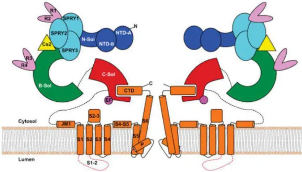
1.4.2 RyR structure ... 48
1.4.2.1 Overall mammalian RyR structure ... 48
1.4.2.2 RyR2 structure ... 49
1.4.2.3 Membrane topology and pore region ... 50
1.4.2.4 Ion permeation ... 52
1.4.2.5 Gating ... 53
1.4.2.6 N-terminal region and R420 site ... 54
1.4.2.7 RyR2 Ca2+ release events ... 57
1.4.2.8 RyR regulation by endogenous effectors ... 58
1.4.3 RyR accessory proteins ... 63
1.4.3.1 FK506-binding proteins (FKBP) ... 63
1.4.3.2 Calmodulin (CaM) ... 64
1.4.3.3 Calsequestrin (CASQ) ... 65
II
1.5 Objectives ... 68
Chapter 2 Materials and methods ... 69
2.1 Generation of RyR2 (R420Q) mutation mice (Performed by Institut Clinique de la Souris) and etics statement ... 69
Ethics statement ... 70
2.2 Genotyping ... 71
2.3 Telemetry ... 74
2.3.1 ECG and telemetry ... 74
2.3.2 ECG analysis ... 76
2.4 Ca2+ handling recording by confocal microscopy ... 78
2.4.1 SAN dissection ... 78
2.4.2 Ca2+ handling recording by confocal microscopy ... 79
2.4.3 Confocal microscope recording analysis ... 82
2.5 Protein measurement ... 83
2.6 SAN cell dissociation and patch clamp (Performed by Pietro Mesirca) ... 89
2.6.1 SAN cell dissociation ... 89
2.6.2 SAN cell patch clamp (Performed by Pietro Mesirca) ... 89
2.7 Statistics ... 91
2.8 Materials ... 91
Chapter 3 Results ... 92
3.1.1- Manuscript ... 92
Mechanisms of bradycardia in RyR2R420Q Catecholaminergic Polymorphic Ventricular Tachycardia mutation... 92
3.1.2. Additional data on RyR2R420Q SAN... 93
Chapter 4 Discussion ... 102
Reference: ... 109
Anexe ... 147
Reconciling depressed Ca2+ sparks occurrence with enhanced RyR2 activity in failing mice cardiomyocytes ... 147
1
Chapter 1 Introduction
The heart rhythm is initiated by the activity of sinoatrial node (SAN), which generates electrical impulses and spreads to the heart inducing heart contraction. Arrhythmias occur when SAN or spreading system has anomalies, or when the contraction is driven by a stimulus in another area (ectopic foci), referring abnormal rhythm of the heart beats.1 At intracellular levels, calcium (Ca2+) plays a key role in maintaining normal heart function, such as automatic pacing, action potential generation, excitation-contraction coupling (EC coupling). Thus, sick intracellular Ca2+ release in heart could cause cardiac arrhythmias.
This thesis seeks to elucidate the relationship between Ca2+ handling and cardiac arrhythmias, specifically, focusing on the role of the pathological changes in the intracellular Ca2+ regulation through Ca2+ release channel (ryanodine receptor 2, RyR2) modification contributing to the abnormal SAN function.
1.1 Overview of heart function, action potentials and CICR
The human heart beats about 105 times a day resulting in 2 billion heartbeats during a lifetime.2 Sinoatrial node (SAN) is described as the primary and physiological pacemaker since its discovery more than a century ago.3 Under physiological condition, it spontaneously initiates regular electricity. SAN anticipates to atrioventricular (AV) node and Purkinje fibers network (PFN) electrical activity, considering that the two can also generate pacemaker activity, although they only drive the heart when the SAN is in pathological condition.4-6 For instance, AV node can become dominant during SAN block or heart failure, and PFN can also lead a viable rhythm during AV block. Thereby this system can maintain heartbeat when the SAN fails.
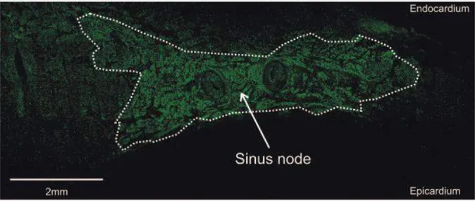
SAN is located near the entrance of the superior vena cava (SCV) in the right atrium (Figure 1), bordered by the crista terminalis. As the impulse-generating tissue, it initiates the electrical impulse, which spreads across both right and left atrium and induces the atria depolarization and contraction, known as atrial systole. This process corresponds to P wave (~0.08s in humans) on the surface electrocardiogram (ECG) (Figure 2). When the impulse reaches the AV node (located near the AV valve, in the interatrial septum), it slows down because of the conduction property of AV node. This delay makes sure all the blood has been ejected to the ventricles before ventricular contraction. It corresponds to PR segment (~0.04s in humans), a flat line following the P wave, while the PR interval (~ 0.12s in humans) represents the duration from the beginning of atrial depolarization until ventricular depolarization. The AV node passes the signal to AV bundles (also known as His bundles), which bifurcates into left and right bundle branches. The signal then passes to Purkinje fibers and through both ventricles, inducing ventricular depolarization and contraction, known as
2 ventricle systole. Thereby the blood is pumped throughout the body. On the ECG, it is represented by QRS complex (~0.12s in humans). Meanwhile, the atrial repolarization occurs, although it’s buried in the QRS complex. The Q wave depicts the septum depolarization, from left to right. When the depolarization travels throughout the ventricles, the R wave and S wave show up. Then another flat line, ST segment (~0.08s in humans), indicates no large electrical vector, while ventricle is contracting. The ventricle repolarization represented by T wave (0.16s in humans). As the epicardial cells repolarize before the endocardial cells, the T wave reflects positively.
Figure 1 The mammalian heart with the cardiac conduction system. SAN, sinoatrial node; SCV, superior vena cava; RA, right atrium; AVN, atrioventricular node; ICV, inferior vena cava; CVB, central fibrous body; TV, tricuspid valve; AVB, atrioventricular bundle; BB, bundle branches; PFN, Purkinje fibers network; LA, left atrium; PV, pulmonary veins; MV, mitral valve; RV, right ventricle; LV, left ventricle. (Panel A is adapted from Mangoni and Nargeot, 20086)
Action potentials
The cardiac action potential (AP) is the membrane potential (Em) waveform that is determined by a complex interplay of many ion channels and transporters, and the [Ca2+]i transient itself.7 The cardiac AP morphology between cardiomyocytes varies dramatically in different regions of the heart according to the physiological characteristics (Figure 2).
In order to maintain the heart rate, the SAN cells spontaneously generate regular and cyclic AP. Then, the electrical impulse propagates to atrium, AV node, Purkinje fiber and ventricle (Figure 2).
In ventricular myocytes, the AP contains four phases. Between 2 consecutives AP, the resting membrane is maintained by IK1 of the Kir2.1 potassium channels. When a ventricular myocyte is excited by adjacent cells, voltage-dependent NaV1.5 sodium channels are activated resulting in a rapid inward Na+ current (INa) and concomitantly rapid membrane depolarization constituting the upstroke of the AP (phase 0). The membrane depolarization induces the slow deactivation of NaV1.5 channel and activation of voltage-dependent K+ channels (KV4.3 and KV1.4), which generates a rapid repolarizating current (Ito) (phase 1). Then, the voltage-dependent Ca2+ channels
3 (Cav1.2) are activated, producing an inward Ca2+ current (ICa,L). The activation of outward rectifying K+ currents (IKur, IKr and IKs via KV1.4, KV11.1 and KV7.1, respectively) and ICa,L are at similar voltages, result in the plateau pase of AP (phase 2). Deactivation of Cav1.2 induces the predominance of K+ currents and further membrane depolarization (phase 3). In the end, Kir2.1 is re-activated, which generates the IK1 current and push the membrane potential back to the resting level.6, 8-10
Figure 2 Electro-activity of the adult heart. Top: schematic of a human heart with illustration of typical action potential waveforms in different regions of the heart. Bottom: schematic of a surface electrocardiogram. (Adapted from Weisbrod et al., 201611)
In myocytes, the membrane (including transverse tubules, t-tubules) depolarization during the action potential activates ICa,L inducing SR Ca2+ release through ryanodine receptor 2 (RyR2), which is known as calcium induced calcium release (CICR) leading to myocytes and heart contraction.12, 13 This process from electrical excitation of the myocytes to contraction of the heart is termed cardiac excitation-contraction coupling (EC coupling).7 The action potential and the following contraction are well studied in ventricular myocytes. However, the generation of electrical impulse in SAN cells is still under debate. As many ion channels expressed in ventricular myocytes are also expressed in SAN cell, the studies about SAN cells are always in comparison to ventricular myocytes.
4 Figure 3 Schematic of membrane clock and calcium clock mechanisms. Top: the red trace shows an example of a typical action potential of spontaneously beating rabbit SANC. The different phases of the AP are labeled, while phase 4 also represents diastolic depolarization (DD). MDP, maximum diastolic potential. Middle: schematic representation of the timing and magnitude of the different components of the “membrane clock”. Bottom: the timing and magnitude of the important components of the “Ca2+clock” are shown at the bottom. LCRs, local Ca2+ releases. L-type Ca2+ current, ICa,L. T-type Ca2+ current, ICa,T. Potassium current, IK. NCX current, INCX. (Adapted from Monfredi et al., 201314)
In automatic cells there is an additional phase of the AP, which is the slow depolarization (phase 4), which slowly depolarizes the membrane, until reaching threshold and producing another AP. In ventricular and atrial myocytes this phase does not exist and the myocytes are not automatic, as they need to be excited by other cells to generate an AP. The AP of SAN includes diastolic depolarization (DD, Figure 3, also referred to as phase 4), rapid depolarizing upstroke (phase 0) and repolarization (phase 2 and 3).15 Major ion currents involved in the AP are depicted in Figure 3, and further introduced in the next session. The morphology of AP in SAN cells is different from that of ventricular myocytes (Figure 3). The SAN cell doesn’t have real resting membrane potential. The more negative potential reached is named maximum diastolic potential. The phase 4 includes the slow increase in membrane potential toward an excitation threshold, at which the action potential fires.14, 16 Regarding the mechanisms of DD, one important factor involved is the funny current (If) carried by hyperpolarization-activated cyclic nucleotide-gated channels (HCN channels), which
5 depolarizes the membrane. Other currents also contribute to the progression of depolarization, such as ICa,T, INCX and ICa,L carried respectively by T-type Ca2+ channel (CaV3.1, CaV3.2), sodium-calcium exchanger (NCX), L-type Ca2+ channel (CaV1.2, CaV1.3). The phase 4 consists of two components, a linear component (the first two-thirds of DD) and a nonlinear, exponentially rising component (the last third of DD). The nonlinear component is proposed to occur along with the subsequent cytosolic Ca2+ increase induced by local Ca2+ release by SR and concomitant inward NCX activation.17 The rapid upstroke (phase 0, Figure 3) of SAN AP is much faster than diastolic depolarization, but still slower than that of ventricular myocytes as it’s carried out by slow Ca2+ current in SAN instead of the fast Na+ current in ventricular myocytes. The initial repolarization phase (phase 1) in ventricular myocytes is completely absent, and the plateau of phase 2 is replaced by a slow velocity repolarization phase. In the end, the Em is driven to maximum diastolic potential (MDP) by potassium channels. The AP of SAN has a relatively positive MDP of around -50mV due to the absence of IK1.6, 8, 9, 14, 16, 18
1.2 Sinoatrial node (SAN) automaticity
This session serves to introduce SAN action potential and the generation of pacemaker activity. For now, the mysterious ability of SAN generating the spontaneous rhythm is still under debate. There are two theories that can explain the SAN automaticity, the membrane clock theory, Ca2+ clock theory and the subsequent coupled clock theory, which are reviewed in this session.
1.2.1 Membrane clock theory
Before introducing the membrane clock, we will simply discuss the ion channels involved in membrane clock.
1.2.1.1 Ion channels 1.2.1.1.1 HCN channels HCN channels and If current
The hyperpolarization-activated cyclic nucleotide-gated (HCN) channels belong to the superfamily of voltage-gated pore loop channels, expressed widely in nervous system and in heart. They are located on plasma membrane. As a voltage-gated channel, HCNs can sense the changes of the electrical membrane potential, be activated by membrane hyperpolarization (around -50/-65 mV) and generate a unique inward current. Their activation could be facilitated by direct interaction with cyclic adenosine monophosphate (cAMP), via binding with the C-terminus of the channel. Other regulators have also been described, including phosphatidylinositol 4,5-bisphosphate (PIP2), kinases and phosphatases.19-22
6 HCN channels are tetramers (four subunits), and homologous to voltage-gated potassium channels. Like K+ channels, HCN channels are blocked by millimolar concentration of Cs+, and activated by excellular K+.23-27 But unlike K+ channels, they are not sensitive to Ba2+, tetraethylammonium (TEA) and 4-aminopyridine (4-AP), which are strong blockers of potassium channel.26
Each HCN channel subunit consists of six transmembrane helices (S1-S6), including the voltage sensor (S4) and a glycine-tyrosine-glycine (GYG) sequence in the P loop between S5-S6. In potassium channels, the GYG sequence forms the highly selective filter for K+. However, HCN channels conduct both Na+ and K+, in spite of the greater permeability for K+, and even display a small permeability for Ca2+.28-30 Until now, the mechanisms about the specific permeation properties of HCN channels are still unclear. It has been found that the sequences in the P-loop and before GYG are different in K+ channels and in HCN channels, which are TT-V/I-GYG and LC-I-GYG respectively.31-33 D’Avanzo et al. changed the residues closed to the selectivity filter in the HCN4 channel to match the K+ channels, and failed to increase K+ selectivity, suggesting the differences are outside the P-loop region.34 Macri et al. further investigated, showing that the
cysteine doesn’t contribute to permeation.35
It was also proposed that the GYG sequence is coordinated in a less rigid model in HCN channels than in K+ channels, allowing the entrance of cations of different sizes.36
Upon membrane hyperpolarization, HCN channels generate a unique and mixed inward current (termed Ih/If, Figure 3).26, 36-39 If is first described in 1979, and referred by some researchers as pacemaker current, in respect that it plays an important role in control of cardiac and neuronal rhythmicity.28, 40-42 If current widely presents in cardiac pacemaker cells of most species. However, it is absent in some SAN cells from monkey and rat, although it is questioned that this could be artificial effect produced by dialysis of the intracellular medium with the pipette solution.43, 44
The physiological contribution of If current in SAN pacemaker activity is still under debate. If is activated upon membrane hyperpolarization at the end of the repolarization phases of an action potential. When If is activated, it shifts the current flow from outward to inward (Figure 3), which reverses the action potential at maximum diastolic potential (MDP). It ends the repolarization process, and initiates the first part of depolarization process until the activation of voltage-dependent Ca2+ channels is achieved (~ -40mV). The fact that blockade of If causes the prolongation of cycle length in SAN cells indicates that If participates to the generation of pacemaker activity.45, 46 For instance, Cs+ (2mM) decreases beating rate of rabbit SAN cells by 30%.45 In human SAN cells, 2mM Cs+ is sufficient to slow DD slope and pacemaker activity (26% cycle length increase).47
7 However, it is notable that blockade of If only results in a modest prolongation of cycle length in SAN cell.48 The presence of Cs+ only induces slightly decrease of pacing rate, which has been seen as an indication that If is not the unique determinant for SAN automaticity.45, 49 Yet, this idea is challenged by DiFrancesco and his coworkers, who mentioned that Cs+ does not fully block If and the blockage is voltage dependent. Thus the partially unblocked If would still be able to drive the automaticity, and the membrane potential changes further unblock If.25 Although, Cs+ is also a K+ channel blocker which could indirectly affect If.25
Additions of other If blockers are also used to quantify whether this current is necessary for SAN automaticity generation, in which ivabradine is a specific organic blocker of If.50 The blockers access to the binding site within the open pore of HCN channel and cannot leave the binding site when the channel is closed.51-54 A low concentration of ivabradine (<γ μM) specifically inhibits If without suppressing ICa,L or other membrane ion currents.55, 56 Ivabradine slows automaticity by
~β0% at concentration of γ μM in isolated rabbit SAN cells, and reduces the heart rate in vivo in mice and conscious dogs, but doesn’t block pacemaking.46, 57-59
Therefore, these data again suggest that If is probably not an absolute prerequisite for SAN automaticity. However, some researchers point out that the block/unblock by the blockers (ivabradine, UL-FS 49, etc) is coupled to ionic flow rather than voltage alone.51-54 The blockers are removed from the blocking site by strong hyperpolarization inducing inward large If.51-54 It is also noticeable that γ μM ivabradine also perturbs Ca2+ clock with reduced SR Ca2+ load, slowed intracellular Ca2+ cycling kinetics, and prolonged the period of spontaneous LCRs occurrence during diastolic depolarization.60 A higher
concentration of ivabradine (10 μM) also suppresses ICa,L.55
The low activation voltage of If may also protect SAN cells from hyperpolarizing, bradycardiac effect of surrounding atrial myocardium.14 Thus, it was also proposed that If is responsible for a slower DD in the pacemaker range of < -65 mV of subsidiary pacemaker cells rather than of SAN cells, due to its low activation voltage and slow activation kinetics.61, 62
The activation of If could be facilitated by direct interaction with cyclic adenosine monophosphate (cAMP), produced at the sarcolemma from adenosine triphosphate (ATP) by adenylate cyclases (ACs) in basal condition and during sympathetic/ parasympathetic stimulation. Activation or inhibition of ACs/cAMP is a consequence of sympathetic and parasympathetic response of the autonomic nervous system controlling the pacemaker activity. A small augmentation of cAMP increases If open probability. Besides, the If activation curve shifts positively under -adrenergic stimulation (or a rise in cAMP), and negatively upon muscarinic stimulation.6, 50, 63 The shift indirectly indicates the important role of If on pacemaking and during sympathetic/ parasympathetic activation. However, the responses are questioned by the evidence that the full range of responses to autonomic agonists is possible in the partial absence of If or when
8 If function is deficient.64, 65 Cytosolic Ca2+ concentration and CaM (Calmodulin) are also reported to regulate If, but not CaMKII (Ca2+/CaM activates Ca2+/calmodulin-dependent protein kinase II).66 BAPTA (Ca2+ chelator) loading significant decreases the amplitude of If in guinea-pig SAN cells, and shifts the voltage-dependent activation to more negative potentials.66 Three CaM antagonists (W7, Calmidazolium and ophiobolin A) cause the same effect as BAPTA.66
Kinetics and contributions to SAN automaticity of HCN isoforms
Four members of this family (HCN 1-4) have been cloned so far.26, 27, 33, 67-69 HCN channels can be assembled into either homotetramers by identical (homomeric) type of subunits, or heterotetramers by different (heteromeric) types.70-75
HCN channels present distinct biophysical properties, such as activation kinetics, sensitivity to cyclic nucleotides.6, 37, 67, 69 HCN1 channel shows fastest opening kinetics and the lowest sensitivity to cAMP.37, 38 However, HCN4 channel is the most slowly activation isoform, and markedly sensitive to cAMP.27, 33, 37, 67 HCN2 and HCN3 represent intermediate properties between HCN1 and HCN4.27, 37, 76
All four HCN channel isoforms have been detected in the heart.77, 78 In the SAN, HCN4 is the predominant isoform and account for 80% of If, whereas the expression of HCN1 and HCN2 are also found and responsible for the remaining 20%.77, 79-81 HCN3 was recently observed in rodent ventricular myocytes.78 Both heteromeric HCN1-HCN4 and heteromeric HCN2-HCN4 channel exist in native SAN tissues.70, 74
In rabbit and mouse SAN, HCN1 was detected.77, 79, 80 HCN1 held >18% of the total HCN mRNA in rabbit SAN, although only a low level of HCN1 was found in mouse SAN.79, 81 Mice lacking HCN1 channel display congenital SAN dysfunction, characterized by bradycardia, sinus dysrhythmia, increased heart rate variability, prolonged sinoatrial node recovery time, increased sinoatrial conduction time, and recurrent sinus pauses.82 Therefore, HCN1 is required for stable heart rate and regular beat-to-beat variation.82
HCN2 is the dominant ventricular isoform.77 Only a detectable amount of HCN2 was found in rabbit SAN (HCN4>>HCN1>>HCN2), but a moderate level was found in mouse SAN (HCN4>>HCN2>>HCN1).79, 81 Overexpression of HCN2 leads to elevation of If current and increase in spontaneous firing rate, but it is not clear whether overexpression of any other inward current generator would produce the similar result.83, 84 HCN2-deficient mice display modest cardiac dysrhythmia, in which the mean heart rate is not changed, but the heart rate variability is increased.85 Isolated SAN cells from HCN2-deficient mice present reduced Ih and slightly hyperpolarized maximum diastolic potential.85 These data suggest that HCN2 may provide a safety mechanism for the correct setting of the MDP.85, 86 Besides, the heart rate in response to
9 isoproterenol is increased similarly in HCN2-deficient and wildtype mice, indicating HCN2 is not required for sympathetic stimulation. It is also proposed that HCN2 and HCN1 probably are not essential for the development of cardiac conduction systerm and generation of automaticity, but for prevention of arrhythmias.85, 87
Figure 4 Expression of HCN4 protein in human SAN. Low-power montage of immunolabeling of HCN4 protein (green signal). White dashed line highlights SAN. (Adapted from Chandler et al., 200988)
HCN4 is the predominant isoform in human, rabbit, dog and rodent SAN, and is often used as a marker of pacemaking tissue (Figure 4).88 It plays an important role in SAN pacemaker activity. Firstly, global or heart-specific deletion of HCN4 channels induced embryonic lethality, while mice died from embryonic days 5.89, 90 HCN4-deficient embryos had significantly slower heart rate, suppression of If and AV block compared with wild type, which also indicates the essential role of HCN4 for the development of cardiac conduction systerm.89, 90 Secondly, to overcome the embryonic lethality, Herrmann et al. used temporally controlled deletion of HCN4.64 Cells isolated from HCN4 deletion mice showed reduced If (about 75%) and a slight response to isoproterenol.64 The mutant mice also presented cardiac arrhythmia characterized by recurrent sinus pauses.64 But the heart rate challenged by isoproterenol had no change.64 This result was then confirmed by Hoesl et al. who used temporally controlled HCN4 gene deletion technique, and found that application of isoproterenol accelerate the knock-out mice and wild-type mice to similar levels.65 Thus, it was proposed that HCN4 is necessary for generating and maintaining a stable cardiac rhythm, but not critical for acceleration of the heart rate.6, 64, 90 Moreover, HCN4 gene mutations are associated with arrhythmias.42, 91, 92 For instance, patient carrying HCN4 mutation L573X exhibits severe sinus bradycardia and intermittent episodes of atrial fibrillation, chronotropic incompetence during exercise.93 Up to date, 13 HCN4 mutations from 14 families were reported.42, 91, 92 In summary, all these evidences indicate the important contribution of HCN4 to SAN pacemaker activity.
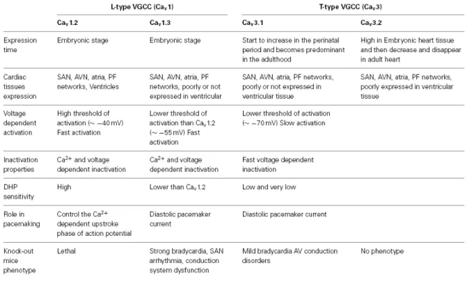
10 L-type Ca2+ channel is a voltage gated channel on sarcolemmal membrane expressed all over the heart.6, 13, 14, 94 LTCC is activated when membrane potential reaches ~ -50mV, and generates L-type Ca2+ current (ICa,L). It exhibits fast Ca2+- and slow voltage-dependent inactivation.95, 96 The fast Ca2+ dependent inactivation produces a negative feedback and limits its own Ca2+ entry, whereas the slow voltage-dependent inactivation results in a time dependent behavior and prevents the premature Ca2+ entry when cytosolic Ca2+ decreases.97 ICa,L is also regulated by PKA, CaMKII. The low activation voltage implicates the important role of ICa,L in generation and regulation of SAN automaticity by contributing to the diastolic depolarization, although ICa,L also participates in the rapid upstroke of the action potential (Figure 3).6, 98-101
LTCCs are highly sensitive to dihydropyridines (DHPs), such as nifedipine and BAY K8644.6, 102, 103
Blockade of ICa,L by nifedipine abolishes the spontaneous activity in both isolated SAN cells and in SAN center tissue without affecting T-type Ca2+ channel, but not in SAN periphery tissue.101, 104
In contrast, blockade of Na+ current by tetrodotoxin has no effect on SAN center tissue, but slows spontaneous activity in periphery tissue.101 These results indicate the obligatory role of L-type Ca2+ current in generation of SAN automaticity in SAN center region, while Na+ current plays a major role in pacemaking in periphery. Indeed, LTCC antagonist nicardipine could induce bradycardia in anesthetized mice in vivo.105 A recent research showed colocalization between RyR2 and LTCC (Cav1.2 or Cav1.3) in the SAN, which may be relevant to the functional role of RyR-mediated Ca2+ release in pacemaking, in respect that ICa,L could induce Ca2+ release through RyR2 in ventricular myocytes (CICR).12, 13 In ventricular myocytes from failing heart, structural alterations, such as altered T-tubular structure and increased orphaned RyRs, contribute to the defect on the efficacy of ICa,L to release Ca2+.106, 107 These results indicate the crucial role of LTCC to CICR and EC coupling in ventricular myocytes, and also suggest the potential importance of colocalization between RyR2 and LTCC in SAN.
LTCCs are formed by five subunits, α1, , and , α2, δ.6, 7, 13 The α1 subunit forms the ion conducting pore and bears the main functional characteristics.7, 13 The architecture of α1 subunit is similar to tetrameric K+ channels and voltage-dependent Na+ channel. Each α1 subunit consists of four homologous domains (І-IV), and each domain contains six transmembrane helices (S1-S6) and a P-loop between S5 and S6. The charged S4 contributes as a voltage sensor, and the α2, δ, and are auxiliary subunits.
Four α1 subunits have been cloned for LTCC (termed Cav1.1, Cav1.2, Cav1.3 and Cav1.4, corresponding to α1S, α1C, α1D and α1F).108 Cav1.1 subunit is expressed in the skeletal muscle, while Cav1.4 is widely expressed in the retina, adrenal gland, bone marrow, spinal cord, muscle, spleen and immune cells.13 Cav1.2 and Cav1.3 are expressed in the brain, cardiovascular system and neuroendocrine cells.12, 13, 88, 109, 110 Cav1.2 is highly expressed in the whole heart, and Cav1.3 is
11 preferentially expressed in supraventricular regions, although the expression of Cav1.3 in SAN is lower than Cav1.2.80, 111 Isolated SAN cells from Cav1.3 gene inactivation mice showed a ~70% reduction in ICa,L density, demonstrating the major contribution of Cav1.3 to total ICa,L in mouse SAN cells.6, 98, 112 CaV1.2-mediated ICa,L accounts for the residual ICa,L.6
The electrical characteristics of Cav1.2 and Cav1.3 mediated L-type Ca2+ currents are also different (Table 1). Cav1.3 mediated current is activated at more negative voltage and more rapidly, while inactivation during depolarization is slower than Cav1.2 mediated current113 and the dihydropyridine sensitivity for Cav1.3 is lower than Cav1.2.113
Table 1 Characteristics of the LTCC and TTCC isoforms involved in SAN automaticity. (Adapted from Mesirca et al., 201513)
Genetically modified mice are used to examine the role of Cav1.3 Ca2+ channel in SAN automaticity. Cav1.3 deficient mice show SAN dysfunction, including bradycardia and arrhythmia.114 Consisting with that, Matthes et al. also found bradycardia in isolated heart of Cav1.3-knockout mice.115 Isolated SAN cells from Cav1.3 deficient mice exhibit decreased pacemaking rate with shift in activation threshold of ICa,L.116 Similarly, Mangoni et al. investigated SAN cells from Cav1.3 gene inactivation mice, and found slower pacemaker activity and arrhythmia.98 Moreover, Torrente et al. further found slower pacemaker activity in SAN cells from genetic Cav1.3 knock-out mice, associated with impaired [Ca2+]i dynamics, reduced local [Ca2+]i release events, and deficient synchronization.112 These results indicate that in SAN cells Cav1.3 accounts for regulation of [Ca2+]i dynamics, triggering local [Ca2+]i release, and thus controlling
12 pacemaker activity. In the other hand, DHP (isradipine) reduced the heart rate in knock-in Cav1.2DHP-/- mice, in which the DHP sensitivity in Cav1.2 α1 was eliminated without effecting channel function and expression, indicating Cav1.3 is an important component of LTCC in SAN.117 The stronger colocalization of Cav1.3 with RyR2 also suggests the major impact of Cav1.3 on SAN.12 Besides SAN automaticity, Cav1.3 also plays functional roles in AVN pacemaking activity and conduction, and in atrial tissues.111, 115, 118, 119 AV conduction abnomalities were reported in CaV1.3-deficient mice.115 In support of that, Marger et al. also reported that CaV1.3 is required for AVN cells spontaneous automaticity.119 Zhang et al. isolated AV node from CaV1.3 null mice, and found a significant decrease in the firing rate and a depolarizing shift in ICa,L, but no significant difference in density peak indicating the potential existence of compensatory changes.118 In atrial myocytes, the lack of CaV1.3 channels in null mutant mice results in a depolarizing shift in the voltage-dependent activation of ICa,L.111
Cav1.2 also contributes the L-type Ca2+ current in SAN.12 BayK induced heart rate increase is absent in Cav1.2DHP-/- mice, indicating the contribution of Cav1.2 to SAN automaticity regulation.117 However, more research is needed.
1.2.1.1 3 T-type Ca2+ channel (TTCC)
T-type Ca2+ channel (TTCC) is also a voltage gated channel on sarcolemmal membrane as LTCC. It is activated at more negative voltage (~-70 mV) than LTCC, and generates T-type Ca2+ current (ICa,T, Figure 3).
6, 13, 95
TTCC is expressed in embryonic and adult heart, including SAN, the AVN and the Purkinje fiber network, but not in other working cardiomyocytes.95, 120-128 Comparation of the expression of CaV3.1 protein in human SAN, paranodal area and right atrium shows a significant higher expression of CaV3.1 in SAN and paranodal area than in right atrium (Figure 5).88 Comparation of ICa,T density in SAN between species indicates a reverse dependence upon the body size, i.e. ICa,T density is larger in small animals and become smaller as the body size increases.129 The ICa,T density sequence for different species is mouse>guinea pig>rabbit>pig, while it is substantial in mouse SAN cells and almost absent in porcine SAN cells.129
In contrast to LTCC, TTCC displays slower activation, faster voltage dependent inactivation and lower channel conductance.96, 130 Besides, unlike LTCC, TTCC is insensitive to dihydropyridines, but sensitive to micromolar Ni2+ ions. Application of Ni2+ to SAN cells slows pacemaking.95, 100, 131 However, the mechanism under the contribution of TTCC to SAN automaticity is not fully understood. The low activation voltage may indicate its involvement in initiation of diastolic depolarization and SAN automaticity. Moreover, a research suggested that ICa,T triggers Ca2+ sparks from the SR, which in turn stimulate NCX activation and depolarize the pacemaker potential to threshold in cat SAN cells.131, 132 Although, one study found Ni2+ only
13 slightly decreased cycle length, and did not decrease the number of Ca2+ sparks in rabbit SAN cells, while a species dependent role of TTCC may exist.133 Notably, Ni2+ is also used as an inhibitor of NCX.
Figure 5. Expression of CaV3.1 protein in human SAN, paranodal area and right atrium. The three high power
images show the immunolabelled CaV3.1 (red signal) in the SAN, paranodal area and right atrium. The bar graph
indicates the intensity of CaVγ.1 protein labeling in the three areas. Means+SEM (n=4) is shown. * and † represent significantly different (P<0.05) from SN (*) or PN (†) (one-way ANOVA); ‡ indicates significantly different (P<0.05) from PN (paired t-test). (Adapted from Chandler et al., 200988)
TTCC is structurally homologous to LTCC. Three α1 subunits have been cloned for TTCC, termed Cav3.1, Cav3.2 and Cav3.3 corresponding to α1G, α1H, α1I.108 Cav3.1 is expressed in cardiac muscle, skeletal muscle and neurons, while Cav3.2 is expressed in cardiac muscle and neurons, and Cav3.3 is only expressed in neurons.80, 108, 123, 124 The transcription level of Cav3.1 is higher than Cav3.2 in SAN.134 Genetically modified mice have been used to understand the roles of TTCC isoforms.
Genetic inactivation of Cav3.1 TTCC in mice causes bradycardia, delays atrioventricular conduction, and prolongs the SAN recovery time.123 Isolated SAN cells present slower pacemaker activity by 37% and a related reduction of the slope of the diastolic depolarization, and isolated AVN cells also show slower pacemaker activity.119, 123 Le Quang et al. used radiofrequency AVN
14 ablation to produce atrioventricular block in Cav3.1 deficient mice and discovered that loss of T-type Ca2+ channels worsens bradycardia-related mortality, increases bradycardia-associated adverse remodeling, and enhances the risk of malignant ventricular tachyarrhythmias complicating AV block.135 Thereby, TTCC plays an important role in SAN spontaneous beating generation and infranodal escape automaticity. CaV3.1 inactivation completely abolished ICa,T suggesting CaV3.1 is the major component of ICa,T in adult mouse SAN cells.123
In contrast, no cardiac arrhythmias are observed in Cav3.2 deficient mice, and ECG waveform morphologies are normal, which indicates the insignificant contribution of Cav3.2 mediated T-type Ca2+ current to impulse initiation and conduction.136 However, a research shows a higher expression of Cav3.2 compared to Cav3.1 in embryonic myocardium, suggesting the functional role of Cav3.2 in embryonic murine heart.137 The adult heart expresses more Cav3.1, switching transcription from Cav3.2 to Cav3.1 in the perinatal period.137
1.2.1.1 4 Sodium channel
The upstroke of SAN AP is relatively slower than in ventricle, as it is driven by Ca2+ channels rather than Na+ channels attributed to the lack of Na+ channels in SAN cells. Recent evidences showed the existence of Na+ channels in SAN, although, with heterogeneous distribution.
Two sodium currents have been identified in SAN brought out by two kinds of sodium channels: the TTX-resistant cardiac isoform (NaV1.5) inhibited by nanomolar concentration of tetrodotoxin (TTX), and the TTX-sensitive neuronal isofroms (NaV1.1 in rat, NaV1.1 and NaV1.3 in mouse) inhibited by micromolar TTX.6, 138, 139
It was shown in different studies that NaV1.5 is absent in SAN center, but present in the periphery.138-140 The distribution of NaV1.5 in periphery indicates its potential contribution in electric impulse propagation.140
The distribution of TTX-sensitive sodium channel is controversial. Honjo et al. showed that TTX-sensitive sodium current is absent in small cells from the SAN center, but present in the periphery.141 In support of this view, Kodama et al. found that blockade of Na+ current by tetrodotoxin has no effect on SAN center tissue, but slows spontaneous activity in periphery tissue.101 However, more recent studies showed the NaV1.1 is present throughout the SAN, while blockage of TTX-sensitive sodium currents results in significant reduction of spontaneous pacemaking.138, 139 It has also been reported that the TTX-sensitive sodium current accounts for spontaneous activity in the SAN cells from newborn rabbits.142-144
15 There are several types of K+ channels reported in SAN, such as voltage dependent K+ channel (Ito, Isus, IKr, IKs), inward rectifier K+ channel (IK1), ATP dependent K+ channel (IKATP), acetylcholine-activated K+ channel (IKACh), calcium-activated K+ channel (IKCa), etc.6, 16
IKr and IKs are voltage-gated, and they are also delay rectifier currents which are characterized by an increased positive slope at more positive membrane potential and slow deactivation kinetics.6, 7
For instance, IKr reaches full activation at membrane potential of -10mV and displays strong inward rectification.145 The activation of IKr is counterbalanced by inactivation at positive voltage, and causing the negative slope conductance.145 When the membrane potential reaches back to negative, IKr de-activates slowly and generates tail currents.145 IKr is a “rapidly activating” delay rectifier K+ current, activated by depolarized membrane potentials from -50mV.146 Compared with IKr, IKs has slower activation and faster deactivation kinetics.147 IKr and IKs are encoded by ERG1 gene and KCNQ1 gene, respectively. IKr is sensitive to class III methanesulfonanilide compounds, such as E-4031 at micromolar concentration, while IKs is not sensitive to these agents.6, 148 Species dependent differences in IKr and IKs distribution exist. In porcine SAN cells, E4031 hardly affected IK, while blockage of IKs inhibited IK, indicating IKs is the predominant component of IK in pig.149 Satoh et al. supported this view and suggested the only IKs current (without IKr) contributes to the porcine SAN.43 Only IKr is recorded in mouse SAN, while both IKr and IKs account for the rabbit and guinea pig SAN.6, 43, 149 Besides, blockage of IKr by micromolar E-4031 causes a cession of SAN pacemacking in isolated SAN cells of rabbit and guinea pig.150, 151 E-4031 can also slow pacemaker activity in isolated mouse hearts and in rabbit right atrial preparations.145, 152 Thus, the presence of IKs rather than IKr in pig can be a form of adaption to slower heart rate in large mammals than in rodents.6, 43, 149 Lei et al. also suggested that in rabbit SAN, the contribution of IKs to beating rate is
small under control condition, but significant during -adrenergic stimulation.153
The transient outward current (Ito) is characterized by rapid activation and inactivation kinetics, and sensitivity to 4-AP.154, 155 KV1.4 (Ito,fast), KV4.3 and KV4.2 (Ito,slow) conduct the fast and slow components of the outward K+ current. The sustained outward K+ current (Isus) is the sustained part of initially discovered Ito or I4-AP.154
Inward rectifier current (IK1) presents in ventricle and is responsible for the maintenance of resting membrane potential. However, the density of IK1 is generally low in pacemaker cells. It has been reported that IK1 is absent in guinea pig, rabbit and pig SAN, but present in mouse, rat and monkey SAN.43 Consist with that, a much lower density of IK1 was found in mouse and rat SAN than in ventricle.44, 156
The acetylcholine-activated K+ current (IKACh) has been described in SAN, atria and AVN.157-159 Two genes Kir3.1 and 3.4 are responsible for IKACh in the heart.160 IKACh is activated by muscarinic
16 and adenosine receptors via binding of G protein subunits to the channel.6, 11, 160, 161 A recent study showed that genetic or pharmacological inhibition of IKACh abolishes sick sinus syndrome in CaV1.3 knock-out mice, via allowing net inward current to flow during the DD during cholinergic activation.162 Thus, IKACh could be a potential therapeutic target for treatment of sick sinus syndrome. Interestingly, acetylcholine slows the cardiac pacemaking not only through activation of IKACh, and also through negative regulation of cAMP and concomitant activity of If. Furthermore, low concentration (nanomolar) of acetylcholine is sufficient to inhibit If, whereas higher concentration (micromolar) is required to activate IKACh.157 This view is supported by Yamada who found that heart rate reduction induced by low ACh doses (<100 nM) was insensitive to IKACh inhibition.163 These data indicate the existence of multiple effects by ACh stimulation, and the less important role of IKACh in response to nanomolar ACh. The ATP-dependent K+ current (IKATP) is identified in rabbit and rat SAN.164, 165 It is activated by stretch or by low levels of intracellular ATP.164, 165 Activation of IKATP in rabbit SAN cells hyperpolarizes the membrane, and slows or abolishes the pacemaker activity.165
The calcium-activated K+ channels are activated by elevation of cytosolic calcium and accounts for the membrane hyperpolarization.11, 166 According to the channel conductance, calcium-activated K+ channels is divided into three subfamilies: BK, SK and IK, which exhibit “big”, “small” and
“intermediate” conductance, respectively.11, 166
Big conductance calcium-activated potassium (BK, KCa1.1) channels are expressed in central nervous system, smooth muscle, and the SAN.11, 167, 168 It has been recently demonstrated that the BK channels are involved in the regulation of cardiac automaticity. Pharmacological inhibition of BK channels significantly reduces the automaticity of conscious mice heart and isolated rat heart in a dose-dependent manner.169 Besides, the automaticity of SAN cells is reduced by application of BK channel inhibitors, and isolated SAN cells from mice with genetic deletion of BK exhibit slower firing rate, both associated with the lengthening of the diastolic depolarization phase of SAN cell action potential.170 Small conductance calcium-activated potassium (SK1-4) channels also play a fundamental role in heart.11 SK channels in mitochondrial inner membrane of guinea pig ventricular myocytes contribute to mitochondrial K+ uptake and protect hearts against infarction.171 SK1 (KCa2.1), SK2 (KCa2.2) and SK3 (KCa2.3) are identified in human and mouse atria and ventricles.172, 173 Genetic knock-out of SK2 channels results in prolongation of action potential duration especially in repolarization in atrial myocytes, and inducible atrial fibrillation in null mutant mice.174 Qi et al. further confirmed the participation of SK2 in atrial fibrillation maintenance.175 SK4 (KCa3.1) channel also play an important role in cardiac pacemaker derived from human embryonic stem cells.176
17 The sustained inward current (Ist) was identified in SAN of mouse, rabbit, rat, guinea pig, and also in rabbit AVN, but absent in quiescent cells.6, 44, 156, 177-180 It is activated at membrane potential of about -70mV, reaches the peak at about -50mV, and shows little inactivation during depolarization.177, 180 The distinct expression pattern and activation of Ist indicate its role for the membrane depolarization and SAN automaticity generation.180 This current is carried mainly by Na+, but it is distinct from INa, as this current is TTX-insensitive, can be blocked by DHP antagonists, facilitated by BAY K 8644 and inhibited by divalent cations.6, 177 These characteristics of Ist also suggest that Ist might be mediated by a novel subtype of LTCC.177
Two kinds of transient receptor potential (TRP) channels were found in SAN, TRPC (C stands for canonical) and TRPM (M stands for melastatin). TRPC channels are nonselective cation channels that regulate ion homeostasis and intracellular Ca2+ signaling in numerous cell types, as well as store-operated calcium entry (SOCE).181 The TRPC subfamily contains seven isoforms (TRPC1-7), which could be divided into three groups based on the sequence alignments and functional comparisons: TRPC1/4/5, TRPC3/6/7 and TRPC2. TRPC2 is not expressed in human.182 TRPC3/6/7 are activated by diacylglycerol (DAG), produced by PLC-mediated hydrolysis of phosphatidylinositol 4, 5-bisphosphate (PIP2). TRPC1, 4 and 5 are activated by SR Ca2+ depletion or by stretch.183, 184 The stromal interaction molecule 1 (STIM1) is located in the SR acting as a Ca2+ sensor and oligomerized when SR Ca2+ is depleted, which in turn activates Orai1 and TRPC1/4/5 via directly binding.185, 186 STIM1 also indirectly activate TRPC3/6, but not TRPC7.187 Besides, it has also been suggested that Orai and TRPC form complexes that participate in SOCE.187 And in support of that, Pani et al. observed that TRPCs are colocalized with STIM1 and Orai in lipid raft domains.188 However, other investigators have not observed a role for TRPC channels in the Orai/STIM1 complex.189, 190 Activation of TRPCs promotes cardiac hypertrophy by Ca2+ influx and subsequent calcineurin activation.191-194 In SAN, TRPC1-4, 6 and 7 are detected, but not TRPC5.195, 196 TRPCs related SOCE is probably involved in modulation of heart rate rhythm. 196-199
Mice lacking the TRPC3 gene demonstrated the involvement of TRPC3 in sinoatrial arrhythmias.197, 198 Orai1 is also expressed in SAN and plays a potentially important role in SOCE in SAN pacemaking.200, 201 Certainly, STIM1 also influences SAN function by reducing SR Ca2+ content and regulating ionic fluxes in SAN cells.200
TRPM4 and TRPM7 were detected in SAN, and both of them participate in SAN automaticity generation. TRPM4 is a Ca2+-activated nonselective cation channel driving sodium and potassium inward current.202-204 Pharmacological inhibition of TRPM4 reduces the automaticity associated with a reduction in diastolic depolarization slope in mouse or rat heart.203 TRPM7 is a divalent-permeant channel.205 Mice with global or SAN restricted TRPM7 deletion show sinus pauses and
18 AVN block with slower diastolic depolarization and reduced HCN4 mRNA, indicating TRPM7 influences SAN automaticity via regulation of HCN4 expression.205
Other electrogenic molecules (such as Na+- K+ pump, etc) also exist on sarcolemmal membrane of SAN cells and influence the maintenance of the ionic homeostasis and the normal SAN automaticity.6 The Na+- K+ pump excludes three Na+ for two K+ uptake resulting in the generation of an outward current, which also influences the pacemaker activity of SAN cells.6, 206
1.2.1.2 Membrane clock
The ensemble of sarcolemmal electrogenic molecules forms a voltage membrane oscillator, known as voltage clock or membrane clock. According to membrane clock, the SAN action potential is predominantly generated by the funny current (If), which is activated by membrane hyperpolaryzation. It depolarizes the membrane, and then activates multiple voltage-gated ion channels, such as TTCC, LTCC, etc.207, 208 The multiple ion channels lead to diastolic depolarization as well as upstroke of the action potential via LTCC. Then, the membrane potential is repolarized to the maximum diastolic potential by K+ channel.
1.2.2 Ca2+ clock theory
1.2.2.1 Sodium-calcium exchanger (NCX)
Sodium-calcium exchanger (NCX) is a high-capacity, voltage-dependent, Ca2+-dependent and time-independent electrogenic protein. It is located on the sarcolemmal membrane, and colocalized with cardiac RyR (Figure 6).209 It is one of the major electrogenic molecules that maintain Ca2+ homeostasis in SAN cells, and also contributes to diastolic depolarization in SAN cells.16 NCX activity is enhanced by the localized SR Ca2+ release during diastolic depolarization.131, 210 It extrudes Ca2+ from the cell and brings in Na+. As one Ca2+ ion is extruded for exchanging of three Na+ ions, NCX produces an inward current (INCX) and further depolarizes the membrane (Figure 3).6, 211 Many studies suggest the RyR-NCX-SERCA crosstalk ensure the SAN cell function (coupled clock).16, 212 Indeed, acute blockade of the NCX via inhibitor (KB-R7943), via rapid substitution of Na+ by Li+, application of ryanodine (RyR2 inhibitor), or chelation of intracellular [Ca2+]i abolishes SAN cells beating.213-215 These data indicate that INCX is attributed to elevated [Ca2+]i, and via RyR-NCX-SERCA crosstalk contributing to SAN pacemaking.
The NCX protein contains ten helical transmembrane domains, and a large intracellular loop between the fifth and sixth domains.216-219 The intracellular loop contains two calcium binding domains (CBD1 and CBD2) that are required for intracellular ion sensing and binding, and a XIP domain which confers sodium inactivation properties.216 Crystal structure shows that NCX from
19
Methanococcus jannaschii has four ion binding sites at the center of the protein, while one of them
is specific for Ca2+ and the other three are likely for Na+ to perform the ion exchange.217
Three NCX isoforms (NCX1-3) have been found to date. NCX1 is expressed widely in mammalian tissues, and considered as cardiac isoform.220-222 NCX2 is predominantly expressed in smooth muscles and brain, and NCX3 is mainly expressed in skeletal muscle.220, 221 Their levels vary considerably during postnatal development.221
B A
20 Figure 6 The distribution of NCX and RyR in rabbit SAN cells. A. Confocal image of SAN cell imunolabeled for both NCX and RyR. B. Pixel-by-pixel fluorescence intensities of labeling along the arbitrary line in panel A. And the horizontal dashed lines report the average pixel intensity. C. Topographical profiles of the pixel intensity levels of each antibody labeling and overlay of the small SAN cells in panel A. The maximum height represents the brightest possible pixel in the source image (using an 8-bit image intensity scale). Less bright pixels are accordingly scaled to a smaller height. (Adapted from Lyashkov et al., 2007209)
Genetically modified mice are used to understand the contribution of NCX1 to SAN automaticity. A total suppression of NCX1 results in lack of spontaneously beating heart and embryonic lethality.223 To overcome this problem, Gao et al. generated incomplete NCX1 knockout mice and suggested that NCX1 is critical for fight or flight reaction of SAN, but is not required for resting heart rate.224 However, this mice model was challenged by Maltsev et al. (2013), who indicated that the lower expression of NCX1 could be compensated by the local cross-talk between Ca2+ cycling proteins and NCX, i.e. lower NCX1 expression encourages CICR and in turn stabilize the density of INCX. Therefore, they suggested that NCX1 is critical for both SAN “fight or flight” reaction and maintainence of basal automaticity. Indeed, mice, selectively lacking NCX1in cardiac pacemaking and conduction system, show slower heart rate accompanied by severe arrhythmias.225 Furthermore, complete atrial-specific NCX knockout mice lack P waves, and the isolated SAN cells are quiescent with consequently impaired Ca2+ efflux.226, 227
1.2.2.2 RyR2 in Ca2+ clock
21 The involvement of RyR2 Ca2+ release in SAN automaticity was first carried out by the observation that application of ryanodine slowed pacemaker activity in cat right atrium.228 Later studies further illustrated that both ryanodine (RyR blocker) and cyclopiazonic acid (SERCA blocker) reduced pacemaker activity in guinea-pig SAN tissue.229, 230 A concentration of γ0μM of ryanodine abolish activity in 83% of isolated-rabbit SAN cells, although in another study the same concentration of ryanodine only inhibited rabbit SAN cell firing rate by ~20%.209, 231 Inducible, cardiac-specific knockout of RyR2 with acute ~50% loss of RyR2 protein was sufficient to cause bradycardia and arrhythmias.232 These studies point to the idea that the SR Ca2+ release through RyR2 plays an important role in pacemaking.
1.2.2.3 Ca2+ clock
Figure 7: Schematic illustration of interactions of key molecules comprising the system. The common regulatory factors are lettered in purple, which govern the function of both the Ca2+-clock (gray intracellular area) and the M-clock (light-blue cell membrane area with (light-blue labels depicting electrogenic proteins). The green arrows illustrate signaling driving action potential (AP) firing, while the red lines suppress the AP firing and balance the system. G protein-coupled receptors (green and red shapes within the membrane) modulate both the Ca2+-clock and M-clock function via those same crucial signaling nodes of the system. (Adapted from Maltsev et al., 201416)
According to the Ca2+ clock, the SR generates rhythmic Ca2+ release (local Ca2+ release, LCR) via RyR2. The LCRs activate NCX, which generates depolarizing current and causes the slow increase in membrane potential toward an excitation threshold for action potential firing, resulting in pacemaking (Figure 3).14, 16, 233 The cytosolic Ca2+ is extruded by NCX, or pumped back into SR
22 by SERCA (sarcoplasmic/endoplasmic reticulum Ca2+-ATPase) to complete the Ca2+ cycle. The Ca2+ extruded by NCX should quantitatively match the Ca2+ entry through sarcolemmal, and the Ca2+ transferred by SERCA needs to match the Ca2+ released by RyR2.6 The involvement of Ca2+ release from RyR2 in SAN pacemaker activity was introduced previously. Subsequent work showed
the involvement of following NCX current in pacemaking, and in response to -adrenergic
stimulation.131, 210, 213-215 Besides, calcium sparks were found during the depolarization under normal Ca2+ condition, in absence of Ca2+ overload (Figure 3).131, 214 It is likely that the LCRs depolarize the cell through activation of NCX that initiate the pacemaking.131, 214 Moreover, in support of Ca2+ clock theory, Vinogradova et al. found periodic Ca2+ oscillations during voltage clamp or in detergent-permeabilized SAN cells in the absence of sarcolemmal function, and independent of diastolic membrane depolarization.234, 235 For instance, in Figure 8, when the voltage clamp at potentials that prevent SAN cells from Ca2+ loss via NCX, the SR Ca2+ clock becomes ‘free running’ in spite of surface membrane.83 Furthermore, in physiologic intracellular [Ca2+]i (150 nM), the free running Ca2+ clock generates roughly periodic LCRs with frequency 2-4 Hz.83 Similarly, in detergent-permeabilized SAN cells, the LCRs were observed in 100nM bathing [Ca2+].234 As shown in Figure 8 (right) rhythmic LCRs generate rhythmic current fluctuations in voltage-clamped SAN cell, and the frequency of current fluctuation is as the same as the frequency of LCRs.83 These evidences indicate existence of LCRs and the independent Ca2+ clock.
1.2.3 Coupled clock theory
Some studies suggested that membrane clock is not the only mechanism of SAN automaticity, as the membrane clock failed to understand some experimental results, such as the reduction or cessation of SAN automaticity produced by specific inhibition of Ca2+ cycling, and CaMKII or PKA-dependent phosphorylation.209, 228, 229, 235 Thus, the coupled clock was proposed, which suggests the SAN pacemaker activity is generated by two coupled oscillators, membrane clock and Ca2+ clock.233
In the coupled clock, the membrane clock and Ca2+ clock are coupled via NCX, which generates an inward current and depolarizes the membrane to reach the threshold of activation of other ion channels, e.g. LTCC, and further regulate the Ca2+ clock. The Ca2+ clock is implicated directly by LTCC, NCX, etc, and also indirectly by K+ channels, HCN channels, etc, via respective membrane potential changes and regulating Ca2+ fluxes. For instance, K+ channels repolarize the membrane and activate HCN and other channels, while HCN limits the hyperpolaryzation. The accumulation of cytosolic Ca2+ in turn implicates membrane clock. The two clocks work together through numerous interactions modulated directly by membrane voltage, subsarcolemmal Ca2+, and indirectly by PKA and CaMKII-dependent protein phosphorylation (Figure 7).14, 16, 233 The cytosolic Ca2+ binds to calmodulin, and CaMKII and to Ca2+ dependent adenylyl cyclase (AC) resulting in


![Figure 20 Ca 2+ sparks and transients. A. Two-dimensional confocal images of [Ca 2+ ] i transients in mouse SAN cells from intact tissue](https://thumb-eu.123doks.com/thumbv2/123doknet/14457477.519906/67.892.107.794.514.725/figure-sparks-transients-dimensional-confocal-images-transients-intact.webp)
