Advance Access publication 24 July 2012
Reduced masticatory function is related to lower satellite cell
numbers in masseter muscle
M. A. R. Kuijpers*
,**
, S. Grefte*, E. M. Bronkhorst
***
, C. E. L. Carels*, S. Kiliaridis
**
and
J. W. Von den Hoff*
*Department of Orthodontics and Craniofacial Biology, Radboud University Nijmegen Medical Centre, The Netherlands,**Department of Orthodontics, University of Geneva, Switzerland and ***Department of Preventive and Curative Dentistry, Radboud University Nijmegen Medical Center, The Netherlands
Correspondence to: Mette A. R. Kuijpers, Department of Orthodontics and Craniofacial Biology, Radboud University
Nijmegen Medical Centre, Nijmegen, The Netherlands. E-mail: [email protected]
SUMMARy The physiology of masseter muscles is known to change in response to functional demands,
but the effect on the satellite cell (SC) population is not known. In this study, the hypothesis is tested that a decreased functional demand of the masseter muscle causes a reduction of SCs. To this end, twelve 5-week-old male Sprague–Dawley rats were put on a soft diet (SD, n = 6) or a hard diet (HD, n = 6) and sacrificed after 14 days. Paraffin sections of the superficial masseter and the m. digastricus (control muscle) were stained with haematoxylin and eosin for tissue survey and with anti-myosin heavy chain (MHC) for slow and fast fibres. Frozen sections of both muscles were double-stained for collagen type IV and Pax7. Slow MHC fibres were equally distributed in the m. digastricus but only localized in a small area of the m. masseter. No differences between HD or SD for the m. digastricus were found. The m. masseter had more SCs per fibre in HD than in SD (0.093 ± 0.007 and 0.081 ± 0.008, respectively; P = 0.027). The m. masseter had more fibres per surface area than the m. digastricus in rats with an SD group (758.1 ± 101.6 and 568.4±85.6, P = 0.047) and a HD group (737.7 ± 32.6 and 592.2 ± 82.2; P = 0.007). The m. digastricus had more SCs per fibre than the m. masseter in the SD group (0.094 ± 0.01 and 0.081 ± 0.008; P = 0.039). These results suggest that reduced masseter muscle function is related to a lower number of SCs. Reduced muscle function might decrease microdamage and hence the requirement of SCs in the muscle fibres.
Introduction
Many animal studies have been performed to study the influence of masticatory muscle function on craniofacial growth as well as the response of muscles to interventions like distraction osteogenesis, surgical mandibular lengthen-ing, mandibular repositioning by insertion of devices into the oral cavity, occlusal changes, and diet changes (Kiliar-idis, 1986; Carlson et al., 1978, 1989; Tüz et al., 2003; Proff
et al., 2007), and muscle detachment (Carlson et al., 1978).
Gedrange et al. (2001, 2003) also showed an increase in slow fibres, blood flow, and vascularization.
The variation in food consistency to change the function-al demands offers the opportunity to study muscle changes in an intact masticatory system. Decreased functional de-mands in animals fed a soft diet (SD) changed the distri-bution of fibre type (type IIB fibres increase) and reduced muscle mass and cross-sectional area (Kiliaridis et al., 1988; Gedrange et al., 2001; Langenbach et al., 2003; He et
al., 2004; Kitagawa et al., 2004; Saito et al., 2004;
Urushiy-ama et al., 2004; Taylor et al., 2006). In contrast, little is known about the effect of functional changes on masseter satellite cells (SCs).
SCs are myogenic adult stem cells which received their name due to their specific location between the basal lamina
and the sarcolemma of the myofibres (Mauro, 1961; Muir
et al., 1965; Dwahan and Rando, 2005; Zammit et al.,
2006; Grefte et al., 2007). A true stem cell is able to give rise to two cell types, i.e. a new stem cell and a differenti-ated cell type. In the case of a myogenic stem cell, the cell should be able to give rise to a more differentiated cell in the muscle cell lineage on the one hand and to a new SC on the other (Dwahan and Rando, 2005; Zammit et al., 2006). The latter crucial property of stem cells is termed self-renewal.
Normally, SCs are quiescent in adult muscle. When activated, for example, as a consequence of injury or exercise, SCs divide to produce myoblasts that proliferate, differentiate, and fuse to form myotubes. The latter mature into myofibres (Dwahan and Rando, 2005; Negroni et al., 2006; Zammit et al., 2006; Grefte et al., 2007). Quiescent SCs express markers such as the transcription factor Pax7, the adhesion molecule M-cadherin, and saliomucin or CD34 (Charge and Rudnicki, 2004). There are indications that SCs from the craniofacial muscles are different from those of the skeletal muscles in the rest of the body (McLoon et al., 2007). The two types of muscles also respond differently to injury. Craniofacial muscles show a larger capacity to regenerate, whereas other skeletal muscles often respond with necrosis.
This might indicate that the physiological response of the SCs to injury also differs (McLoon et al., 2007). Further, craniofacial muscles have a higher proportion of SCs than skeletal muscles, which might enhance their response to damage (Renault et al., 2002; McLoon et al., 2007). They also lack the expression of specific transcription factors such as Pax3, while skeletal muscle SCs express both Pax3 and Pax7 (McLoon et al., 2007; Miura et al., 2006). The response of craniofacial muscles to damage and varying functional demands has mainly been studied in extraocular muscles (McLoon et al., 2007), but not yet in masticatory muscles.
Therefore, the aim of this study is to investigate chang-es in masseter muscle SC numbers in rchang-esponse to reduced functional demands in growing rats.
Materials and methods Animals
All animal experiments were approved by the Board for Animal Experiments of the Radboud University Nijmegen Medical Centre in accordance to Dutch laws and regulations on animal experiments. Twelve 5-week-old male Sprague– Dawley rats (Janvier, Le Genest, France) were used for the experiments; they were divided into an SD group (n = 6) and a hard diet (HD) group (n = 6). The rats were housed under normal laboratory conditions. Before the start of the experiments, the rats had been acclimatized to the animal facility for 1 week. The rats in the HD group were fed nor-mal rat chow and water ad libitum. The rats in the SD group received grinded rat chow mixed with water (1:2.5) and water ad libitum. The experimental model has been used in earlier studies into the effect of decreased functional de-mands (Kiliaridis, 1986). The body weight of the animals was monitored throughout the study.
Histology and immunohistochemistry
After 2 weeks feeding with the experimental diet, the rats were killed according to the standard CO2/O2 protocol. Af-ter sacrifice, the left m. masseAf-ter and the left m. digastricus (control muscle) were fixed in 4 per cent paraformaldehyde for 6 hours and processed for paraffin embedding. The right superficial m. masseter and the posterior belly of the right m. digastricus were directly frozen in optimal cutting tem-perature embedding compound (CellPath®, Newtown, UK)
using isopentane precooled in liquid nitrogen. Five micro-metres of paraffin and frozen sections were cut transversal-ly and collected on superfrost plus slides (Menzel-Gläser, Braunschweig, Germany). Three sections per muscle per rat were taken this way. Sections were made through the centre of the muscles. Paraffin sections were stained with haematoxylin and eosin (HE, according to Delafield) for general tissue survey. Distribution of slow and fast myosin was assessed by staining paraffin sections of m. digastricus and superficial m. masseter sections from both HD and SD
with antibodies mouse slow (1:1600) and mouse anti-fast myosin heavy chain (MHC; 1:1600; both from Sigma Chemical Co., St. Louis, Missouri, USA; Figure 1). Sub-sequently, the bound antibodies were visualized using the biotinylated secondary antibodies donkey-anti-mouse IgG (H+L; 1:500; Jackson Labs, West Grove, Pennsylvania, USA) and stained with 3,3-diaminobenzidine.
The frozen sections were double-stained with the anti-bodies rabbit anti-collagen type IV for staining muscle fibres (1:100; Euro-Diagnostica BV, Arnhem, The Nether-lands) and mouse anti-Pax7 (1:100; Developmental Studies Hybridoma Bank, Iowa City, California, USA) for SCs as described previously (Grefte et al., 2010). The nuclei of all cells were visualized using 4′,6-diamidino-2-phenylindole (DAPI).
Standardized pictures were taken with the AxioCam MRc5 camera using a grid positioned over the entire cross-section. Using the centre as a reference, five and three pic-tures were taken on the vertical and horizontal midlines, respectively. All sections were visualized with the Zeiss Imager.Z1 using the AxioVision 4.6.3 software (Carl Zeiss Microimaging GmbH, Jena, Germany).
Quantification
The total number of nuclei, Pax7-positive nuclei, and fibres were calculated per surface area using a representative over-view field of every frozen section from all muscles. The tis-sue surface area was determined by measuring the surface of the tissue without the empty spaces (breaks). Means and standard deviation (N = 6) were calculated for the number of nuclei per surface area, the number of nuclei per fibre, the number of fibres per surface area, the number of Pax7-positive cells per muscle fibre, the number of Pax7-Pax7-positive cells per surface area, and the percentage of Pax7-positive cells of the total number of cells.
Statistics
The total number of cells, the number of Pax7-positive cells (SCs), and the number of muscle fibres in the HD and SD groups were compared with Student’s t-test. Student’s t-test was further used to compare the number of SCs per sur-face area, the number of SCs per fibre, and the number of SCs per total amount of all cells for both muscles. The dif-ferences between the m. digastricus and m. masseter were compared with a paired t-test.
Intra-observer reliability was tested with a paired t-test for the nuclei, the number of Pax7, and the fibre count and was 0.938, 0.979, and 0.927, respectively. This means that the reliability is very good.
Results
The mean weight of the rats in the SD group was 236 ± 12.14 and 243 ± 11.53 in the HD group. The difference was
not statistically significant (t-test). One rat in the SD group died before the end of the experiment.
General histology
Paraffin sections from the m. digastricus and m. masseter were stained with HE for a general tissue survey. The m. digastricus is a somewhat rounded muscle in cross-section with a diameter of about 3–4 mm. In contrast, the superfi-cial m. masseter is elongated in cross-section with the larg-est diameter about 8 mm. Anatomically, the superficial m. masseter is covering the deep m. masseter. For the immuno-histochemical studies, only the superficial m. masseter and the posterior belly of the m. digastricus were used.
Immunohistochemistry
Paraffin sections from the m. digastricus and superficial m. masseter from both HD and SD groups were stained for slow and fast MHC fibres (Figure 1). Nearly, all my-ofibres in both muscles were positive for fast MHC (not shown). Only a few slow MHC-positive fibres are scattered throughout the m. digastricus muscle, but the superficial m. masseter only contains some positive fibres in one specific area, the region where the muscle spindles are localized. No obvious differences were observed in the numbers and dis-tribution of slow MHC-positive fibres between the HD and SD group.
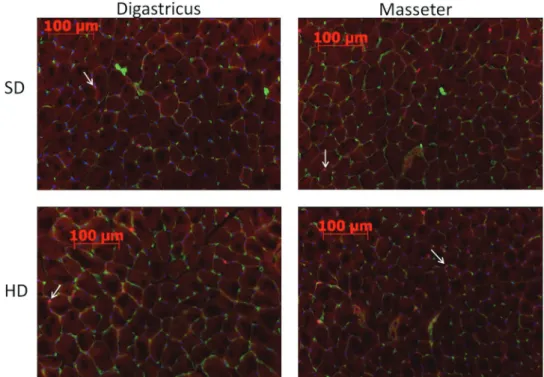
Frozen sections were double-stained for Pax7 and colla-gen type IV (nuclei; Figure 2). The collagen type IV an-tibody stains the basal lamina (green) surrounding every muscle fibre and thus allows the identification of single fibres. The Pax7 antibody stains the nuclei of the SCs (red), but the combination with the blue DAPI yields a purple staining. Multiple cross-sections were used to determine the number of fibres per millimetre square (Figure 3a), the number of SCs per fibre (Figure 3b), and the total fraction of SCs (Figure 3c).
No significant differences between HD and SD in any of the measurements for the m. digastricus were found. There were no significant differences for the m. masseter in any of the measurements except for the number of SCs per fibre. The m. masseter had more SCs per fibre in HD (0.093 ± 0.007) than in SD (0.081 ± 0.008; P = 0.027;
Figure 3c).
When comparing the muscles in each diet group, some differences were found. The m. masseter had significantly more fibres per surface area than the m. digastricus in rats with an SD group (758.1 ± 101.6 and 568.4 ± 85.6; P = 0.047) and a HD group (737.7 ± 32.6 and 592.2 ± 82.2; P = 0.007; Figure 3a). The m. digastricus had more SCs per fibre than the m. masseter in the SD group (0.094 ± 0.01 and 0.081 ± 0.008; P = 0.039; Figure 3c). The differences between m. masseter and m. digastricus in number of nuclei per fibre in the HD and the SD were not statistically
Figure 1 Immunohistochemistry of the m. masseter and m. digastricus. Paraffin sections from the m. digastricus
(1) and superficial m. masseter (2) from the HD group stained for slow MHC fibres. The arrow indicates the local-ized area of slow fibres in the m. masseter.
significant (Figure 3b). In both muscles, the percentage of SCs on total of nuclei was about 6.1 in HD and 5.3 per cent in SD, which was not statistically significant.
Discussion
The rat model for investigating the effects of changes in masticatory function has been used extensively (Kiliaridis, 1986; Katsaros, 2001; Bresin and Kiliaridis, 2002). These studies mainly showed changes in sutures, craniofacial skeletal growth, and muscle structure. To our knowledge, it has never been tested whether changes in diet consistency also affect the SC population in the masticatory muscles. In our study, the m. digastricus was used as a control muscle as in previous studies (Kiliaridis et al., 1988; He et al., 2004). Theoretically, the m. digastricus is not affected by changes in mastication as its function is to antagonize the masseter during opening of the jaw. We confirmed in our study that diet consistency does not affect the fibre density, cell densi-ty, nor the number of SCs of the m. digastricus. There were also no differences in fibre density in the masseter. How-ever, the size of the fibres of the masseter muscle has been reported to increase (Kiliaridis et al., 1988; He et al., 2004). The distribution of slow fibres was also similar in HD and SD in both muscles. This last finding has already been re-ported by Kiliaridis et al. (1988). However, the reduced functional demand on the m. masseter (SD) led to lower numbers of SCs. SCs continuously react to changes in the
environment. They respond to normal changes in function as well as to injury in order to maintain muscles’ integrity. After injury, SCs give rise to myoblasts that fuse with exist-ing damaged fibres or form new fibres (Anderson, 2006; Zammit et al., 2006). The SCs in both skeletal muscles and craniofacial muscles respond to injury and exercise with proliferation. However, SCs in craniofacial muscles prolif-erate more, yet differentiate later than SCs in skeletal mus-cles (Kadi et al., 2005; Harel et al., 2009; Ono et al., 2010). Previous studies on the effect of function on the SC population in skeletal muscles are based either on exercise models or on disuse models with an extra intervention. In the latter studies, disuse was generally achieved by changing the innervation (Gunderson and Bruusgaard, 2008; Legerlotz and Smith, 2008) and is therefore not completely comparable with our disuse model. These studies in skeletal muscles show a reduction of activated SCs (Gunderson and Bruusgaard, 2008; Legerlotz and Smith, 2008). Studies on the effect of function on the SC population using an exercise model mainly focus on changes during exercise itself and on changes after the cessation of exercise. Skeletal muscles adapt to some types of exercise with an increase in fibre size, the number of nuclei per fibre, and the number of SCs (Kadi
et al., 2005; Mackey et al., 2007; Snijders et al., 2009).
However, a reduction in SC number occurs when training is stopped (Mackey et al., 2007; Snijders et al., 2009). This seems comparable to our results as the rats are switched from the standard hard laboratory chow to soft powdered food.
Figure 2 Frozen sections of the m. masseter and m. digastricus with immunohistochemical staining for
col-lagen type IV (green) to show the muscle fibres and for Pax7 (red) to show the SCs and with DAPI staining for the nuclei (blue). SCs are indicated with white arrows.
In aged as well as young individuals, an increase in SC numbers was found after endurance exercises, whereas resistance training only increased SC numbers in young in-dividuals (Snijders et al., 2009). It is not known whether the increase in SCs during training is related to the exer-cise itself or to damage induced by the exerexer-cise (Kadi
et al., 2005). Exercise always increases the number of SCs,
but does not always increase the number of fibres and fibre diameter. Exercise does occasionally increase the number of myonuclei per fibre, but this does not seem to be the stand-ard mechanism (Kadi et al., 2005; Smith and Merry, 2012). To our knowledge, no previous studies on the SC population
in a masseter muscle disuse model have been carried out. The results of this disuse model suggest that the reactions are similar as in skeletal muscle disuse models. However, it needs to be further investigated whether the differences in SC numbers between SD and HD increase in time or remain stable.
In conclusion, our data show that the SC population is smaller in m. masseter with reduced function. Although this seems to show that the SCs in the masseter muscle adapt to changes in function, the exact mechanism remains to be clarified.
Funding
This study was supported by a European Orthodontic Soci-ety Research Grant 2009-2011.
References
Anderson J E 2006 The satellite cell as a companion in skeletal muscle plasticity; currency, conveyance, clue, connector and colander. Journal of Experimental Biology 209: 2276–2292
Bresin A, Kiliaridis S 2002 Dento-skeletal adaptation after bite-raising in growing rats with different masticatory muscle capacities. European Journal of Orthodontics 24: 223–237
Carlson D S, Ellis E 3rd, Dechow P C 1978 Adaptation of the suprahyoid muscle complex to mandibular advancement surgery. American Journal of Orthodontics and Dentofacial Orthopedics 92: 134–143
Carlson D S, Ellis E 3rd, Dechow P C, Nemeth P A 1989 Short-term stabil-ity and muscle adaptation after mandibular advancement surgery with and without suprahyoid myotomy in juvenile Macaca mulatta. Oral Sur-gery Oral Medicine and Oral Pathology 68: 135–149
Charge S B, Rudnicki M A 2004 Cellular and molecular regulation of mus-cle regeneration. Physiological Reviews 84: 209–238
Dwahan J, Rando T A 2005 Stem cells in postnatal myogenesis: molecular mechanisms of satellite cell quiescence, activation and replenishment. Trends in Cell Biology 15: 666–673
Gedrange T et al. 2003 Regional alterations in fiber type distribution, cap-illary density, and blood flow after lower jaw advancement in pig masti-catory muscles. Journal of Dental Research 82: 570–574
Gedrange T, Lupp A, Walter B, Harzer W, Bauer R 2001 Oxidative state and histological changes in muscles of mastication after conditioning training. Experimental and Toxicologic Patholology 53: 89–96 Grefte S, Kuijpers-Jagtman A M, Torensma R, Von den Hoff J W 2007
Skeletal muscle development and regeneration. Stem Cells and Devel-opment 16: 857–868
Grefte S, Kuijpers-Jagtman A M, Torensma R, Von den Hoff J W 2010 A model for muscle regeneration around fibrotic lesions in recurrent strain injuries. Medicine & Science in Sports & Exercise 42: 813–819 Gunderson K, Bruusgaard J C 2008 Nuclear domains during muscle
atro-phy: nuclei lost or paradigm lost? Journal of Physiology 586: 2675–2681 Harel I et al. 2009 Distinct Origins and Genetic programs of head satellite
cells. Developmental Cell 16: 822–832
He T, Olsson S, Daugaard J R, Kiliaridis S 2004 Functional influence of masticatory muscles on the fibre characteristics and capillary distribu-tion in growing ferrets (Mustela putonusfuro)--a histochemical analysis. Archives of Oral Biology 49: 983–989
Kadi F et al. 2005 The behaviour of satellite cells in response to exercise: what have we learned from human studies? European Journal of Physi-ology 451: 319–327
Katsaros C 2001 Masticatory muscle function and transverse dentofacial growth. Swedish Dental Journal Supplement 151: 1–47
Figure 3 Quantification of the immunohistochemistry. Numbers of
fibres, nuclei, and SCs were counted on frozen sections of the m. mas-seter and m. digastricus stained for collagen type IV (green) and Pax7 (red) and stained with DAPI. *P < 0.05. (a) Number of fibres per surface area, (b) number of nuclei per fibre, and (c) number of SCs per fibre for HD and SD for both the m. digastricus and the m. masseter.
Kiliaridis S 1986 Masticatory muscle function and craniofacial morphol-ogy. An experimental study in the growing rat fed a soft diet. Swedish Dental Journal Supplement 36: 1–55
Kiliaridis S, Engström C, Thilander B 1988 Histochemical analysis of masticatory muscle in the growing rat after prolonged alteration in the consistency of the diet. Archives of Oral Biology 33: 187–193 Kitagawa Y, Mitera K, Ogasawara T, Nojyo Y, Miyauchi K, Sano K 2004
Alterations in enzyme histochemical characteristics of the masseter muscle caused by long-term soft diet in growing rabbits. Oral Diseases 10: 271–276
Langenbach G E J, van de Pavert S, Savalle W P M, Korfage J A M, van Eijden T M G J 2003 Influence of food consistency on the rabbit mas-seter muscle. European Journal of Oral Sciences 111: 81–84
Legerlotz K, Smith H K 2008 Role of MyoD in denervated, disused, and exercised muscle. Muscle & Nerve 38: 1087–1100
Mackey A L et al. 2007 Enhanced satellite cell proliferation with resist-ance training in elderly men and women. Scandinavian Journal Medi-cine and Science in Sports 17: 34–42
Mauro A 1961 Satellite cell of skeletal muscle fibres. Journal of Cell Biol-ogy 9: 493–495
McLoon L K, Thorstenson K M, Solomon A, Lewis M P 2007 Myogenic precursor cells in craniofacial muscles. Oral Diseases 13: 134–140 Muir A R, Kanji A H M, Allbrook D 1965 The structure of the satellite
cells in skeletal muscle. Journal of Anatomy 99: 435–444
Miura M, Miura Y, Sonoyama W, Yazama T, Gronthos S, Shi S 2006 Bone marrow-derived mesenchymal stem cells for regenerative medicine in craniofacial region. Oral Diseases 12: 514–522
Negroni E, Butler-Browne G S, Mouly V 2006 Myogenic stem cells: re-generation and cell therapy in human skeletal muscle. Pathologie Biolo-gie 54: 100–108
Ono Y, Boldrin L, Knopp P, Morgan J E, Zammit P S 2010 Muscle satellite cells are a functionally heterogeneous population in both somite-derived and branchiomeric muscles. Developmental Biology 337: 29–41
Proff P et al. 2007 Histological and histomorphometric investigation of the condylar cartilage of juvenile pigs after anterior mandibular displace-ment. Annals of Anatomy 189: 269–275
Renault V, Thornell L E, Eriksson P O, Butler-Browne G, Mouly V 2002 Regenerative potential of skeletal muscle during aging. Aging Cell 1: 132–139
Saito T et al. 2004 Effects of diet consistency on the expression of insu-lin-like growth factors (IGFs), IGF receptors and IGF binding proteins during the development of rat masseter muscle soon after weaning. Ar-chives of Oral Biology 49: 777–782
Smith H K, Merry T L 2012 Voluntary resistance wheel exercise during postnatal growth in rats enhances skeletal muscle satellite cell and myo-nuclear content at adulthood. Acta Physiol 204: 393–402
Snijders T, Verdijk L B, van Loon L J C 2009 The impact of sarcopenia and exercise training on skeletal muscle satellite cells. Ageing Research Reviews 8: 328–338
Taylor A B, Jones K E, Kunwar R, Ravosa M J 2006 Dietary consistency and plasticity of masseter fiber architecture in postweaning rabbits. The Anatomical Record. Part A, Discoveries in Molecular, Cellular, and Evolutionary Biology 288: 1105–1111
Tüz H H, Kisnisci R S, Günhan O 2003 Histomorphometric evaluation of short-term changes in masseter muscle after lengthening the rabbit man-dible by distraction osteogenesis. Journal of Oral Maxillofacial Surgery 61: 615–620
Urushiyama T, Akutsu S, Miyazaki J, Fukui T, Diekwisch T G, Yamane A 2004 Change from a hard to soft diet alters the expression of insulin-like growth factors, their receptors, and binding proteins in association with atrophy in adult mouse masseter muscle. Cell and Tissue Research 315: 97–105
Zammit P S, Partridge T A, Yablonka-Reuveni Z 2006 The skeletal muscle satellite cell: the stem cell that came in from the cold. Journal Histo-chemistry and CytoHisto-chemistry 54: 1177–1191