Human Reproduction vol.13 no.1 pp.220–223, 1998
Second-trimester maternal serum screening for Down’s
syndrome: free
β-human chorionic gonadotrophin
(HCG) and
α-fetoprotein, with or without unconjugated
oestriol, compared with total HCG,
α-fetoprotein and
unconjugated oestriol
Philippe Extermann
1,4, Paul Bischof
1,
Philippe Marguerat
3and Bernadette Mermillod
21Department of Obstetrics and Gynaecology and
2Centre d’Informatique Hospitalier, Geneva University Hospital, Geneva, and3Division of Medical Genetics, Lausanne University Hospital, Lausanne, Switzerland
4To whom correspondence should be addressed at: Department of Obstetrics and Gynaecology, Hoˆpital Cantonal Universitaire, 1211 Geneva 14, Switzerland
The aim of our study was to compare three protocols for second-trimester maternal serum screening for Down’s syndrome in the same serum samples, using two triple tests [total human chorionic gonadotrophin (HCG), α-fetoprotein, unconjugated oestriol; and free β-HCG, α-fetoprotein, unconjugated oestriol] and a double test (freeβ-HCG and α-fetoprotein). The three protocols were compared in a series of 23 serum samples from Down’s syndrome pregnancies and in a cohort of 2516 pregnant women receiving routine antenatal care between June 1992 and June 1993. Among the 23 affected cases, at a cut-off risk of 1:380, the detection rate of Down’s syndrome was comparable with the double test (74%; 17/23) and the triple tests (65%; 15/23) (not significantly different). At the same cut-off risk, in the cohort of 2516 pregnant women screened between 15 and 18 weeks gestation, both protocols using freeβ-HCG achieved a significant reduction of the number of false positive cases (P 5 0.013 and 0.004 for double and triple tests respectively). We conclude that, compared to total HCG, α-fetoprotein and unconjugated oestriol, use of freeβ-HCG and α-fetoprotein represents a better second-trimester screening test for Down’s syn-drome, because it significantly decreases the false positive rate at a lower running cost. The addition of unconjugated oestriol to the double test adds no further advantage.
Key words: Down’s syndrome/α-fetoprotein/free β-HCG/ maternal serum screening/unconjugated oestriol
Introduction
Second-trimester maternal serum screening for Down’s syn-drome (DS) has become common practice in several Western countries. Despite the large number of pregnant women screened to date, the relative merit of total human chorionic gonadotrophin (HCG) versus its freeβ-subunit, as well as the adjunctive role of unconjugated oestriol in such programmes, remain controversial issues. The improved screening perform-ances reported by several investigators when using freeβ-HCG instead of total HCG (Macri et al., 1990b, 1994; Cuckle et al.,
220 © European Society for Human Reproduction and Embryology
1992; Ryall et al., 1992; Spencer et al., 1992, 1993) have not been confirmed by others (Milunski et al., 1993; Stone et al., 1993; Wald et al., 1993). The effectiveness of unconjugated oestriol as a screening variable is equally debated (Wald et al., 1988; Macri et al., 1990a; Spencer et al., 1992; Crossley et al., 1993). These conflicting results have prompted us to perform a direct comparison of three protocols for DS screening in the same serum samples, using two ‘triple tests’ [total HCG, α-fetoprotein, unconjugated oestriol (‘TT’); and freeβ-HCG, α-fetoprotein, unconjugated oestriol (‘TTFB’)] and a ‘double test’ [freeβ-HCG andα-fetoprotein (‘DT’)].
Materials and methods
To achieve this comparison, the three protocols were compared in a series of 23 serum samples from DS pregnancies (18 frozen samples and five samples collected during the prospective study) and in a cohort of 2516 pregnant women with normal pregnancy outcome receiving routine antenatal care in Geneva between June 1992 and June 1993.
Hormone measurements were performed in fresh serum samples in three different laboratories, using the following methods: labora-tories 1 and 2 measuredα-fetoprotein (αFP) and total HCG by IMX (Abbott AG, Cham, Switzerland); laboratory 3 measuredαFP with ES600 (Boehringer, Mannheim, Germany) and total HCG by Stratus (Baxter AG, Zu¨rich, Switzerland). Unconjugated oestriol and free β-HCG were measured by radioimmunoassay in all samples by laboratory 1, using Kodak-Amerlex-M Estriol kit (Polymed SA, Geneva, Switzerland) and FBHCG (CIS-Bio-International, Gif-sur-Yvette, France) respectively.
Median values for weeks 15–18 were previously established in the three laboratories for αFP and total HCG and in laboratory 1 for unconjugated oestriol and freeβ-HCG, using the same sera (50 per week) obtained from pregnant women with a normal pregnancy outcome. Median values were monitored throughout the study and updated as necessary. Concentrations of the serum markers were expressed in multiples of the medians (MOM) for pregnancies of the same gestational age.
Gestational age was determined from last menstrual period (LMP) or ultrasound examination. Almost all our patients had an ultrasound dating scan prior to or at the time of serum screening. When LMP and ultrasound estimates of gestational age were in agreement, LMP estimate was considered. When LMP and ultrasound estimates were divergent, ultrasound estimate was selected if the difference between LMP and ultrasound derived gestational age was.12 days. This was the case in 10% of our patients.
Serum samples were obtained between 15 and 18 weeks gestation. Of the 23 patients with fetal DS (median age 32 years), six (26%) wereù35 years old. Of the 2516 patients screened, 133 (5.3%) were
ù35 years old (median age 36 years); 2383 (94.7%) were ,35 years
(median age 29 years).
Maternal serum screening for Down’s syndrome
Table I. Down’s syndrome (DS) detection rate depending on serum markers
used (total number of DS cases5 23). Cut-off risk 1:380 DS cases detected TT DT TTFB n % n % n % Stored samples (n5 18) 12 67 15 83 14 78 Per-study samples (n5 5) 3 60 2 40 1 20 Total (n5 23) 15 65 17 74 15 65
TT5 triple test [total human chorionic gonadotrophin (HCG), α-fetoprotein, unconjugated oestriol], DT5 double test (freeβ-HCG, α-fetoprotein), TTFB5 triple test (freeβHCG,α-fetoprotein, unconjugated oestriol).
and the trivariate or bivariate Gaussian frequency distribution of the serum markers, using commercially available software programs (ALPHA, Logical Medical Systems Ltd., London, UK; and CIS-Bio-International). Later comparisons were computed with hospital-developed software, using the data from Wald et al. (1988) and Spencer et al. (1992). Cut-off risk indicating further investigation was set at 1:380.
Clinical management was based on the triple test (TT) results. Free β-HCG was concurrently assayed on the serum samples for later evaluation and comparison. This study received ethical approval from the Ethics Committee of our institution. All patients participating in the study gave their informed consent.
Statistical comparisons were done using the McNemar test, or its exact version, when appropriate.
Results
Table I shows the results obtained with the three protocols in the series of 23 serum samples from DS pregnancies. Overall, at a cut-off risk of 1:380, two more DS cases were detected with the protocol using freeβ-HCG andα-fetoprotein [17/23 (74%; 95% confidence interval 52–90%); versus 15/23 (65%; 43–84%)]. This difference in detection rate did not reach statistical significance (P5 0.69). The median MOM values for free β-HCG, total HCG, α-fetoprotein and unconjugated oestriol in the DS cases were 2.68, 1.94, 0.77 and 0.81 respectively. From the five DS cases ascertained during the prospective study, at a cut-off risk of 1:380, three were detected with the triple test using total HCG, two with the double test and one with the triple test using freeβ-HCG (Table I). At a cut-off risk of 1:270, the respective numbers were three, none and one (Table II).
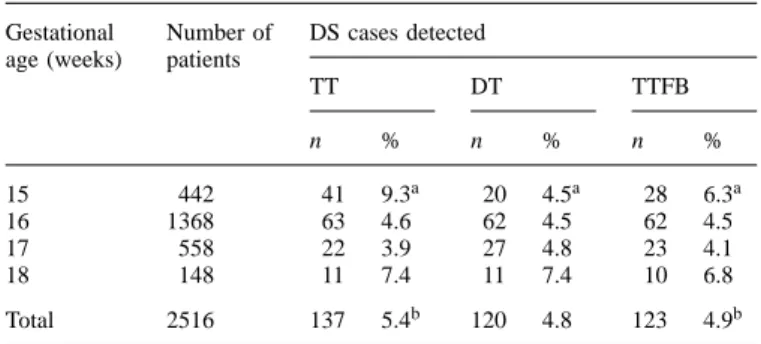
Results of the screening study are presented in Table III. Overall, at a cut-off risk of 1:380, we observed a significant reduction of the number of positive cases with both protocols using free β-HCG instead of total HCG. This reduction of positive rate was only observed at 15 weeks gestation; this effect at 15 weeks persisted at a cut-off of 1:270 with use of freeβ-HCG (Table IV).
Discussion
Our results in DS pregnancies are in agreement with those reported by Spencer et al. (1992) and Macri et al. (1994). The
221 Table II. Down’s syndrome (DS) detection rate depending on serum
markers used (total number of DS cases5 23). Cut-off risk 1:270 DS cases detected TT DT TTFB n % n % n % Stored samples (n5 18) 12 67 14 78 14 78 Per-study samples (n5 5) 3 60 0 0 1 20 Total (n5 23) 15 65 14 61 15 65
TT5 triple test [total human chorionic gonadotrophin (HCG), α-fetoprotein, unconjugated oestriol], DT5 double test (freeβ-HCG, α-fetoprotein), TTFB5 triple test (freeβHCG,α-fetoprotein, unconjugated oestriol).
Table III. Screening study: number of positive cases depending on serum
markers used; cut-off risk 1:380
Gestational Number of DS cases detected age (weeks) patients
TT DT TTFB n % n % n % 15 442 52 11.8a 29 6.6a 36 8.1a 16 1368 95 6.9 90 6.6 82 6.0 17 558 39 7.0 36 6.5 34 6.1 18 148 16 10.8 12 8.1 13 8.8 Total 2516 202 8.0b 167 6.6b 165 6.6b
TT5 triple test [total human chorionic gonadotrophin (HCG), α-fetoprotein, unconjugated oestriol], DT5 double test (freeβ-HCG, α-fetoprotein), TTFB5 triple test (freeβHCG,α-fetoprotein, unconjugated oestriol).
aP-values: DT versus TT: 0.0002; TTFB versus TT: 0.0052; DT versus TTFB: 0.14.
bP-values: DT versus TT: 0.013; TTFB versus TT: 0.0037; DT versus TTFB: 0.92.
Table IV. Screening study: number of positive cases depending on serum
markers used; cut-off risk 1:270
Gestational Number of DS cases detected age (weeks) patients
TT DT TTFB n % n % n % 15 442 41 9.3a 20 4.5a 28 6.3a 16 1368 63 4.6 62 4.5 62 4.5 17 558 22 3.9 27 4.8 23 4.1 18 148 11 7.4 11 7.4 10 6.8 Total 2516 137 5.4b 120 4.8 123 4.9b
TT5 triple test [total human chorionic gonadotrophin (HCG), α-fetoprotein, unconjugated oestriol], DT5 double test (freeβ-HCG, α-fetoprotein), TTFB5 triple test (freeβHCG,α-fetoprotein, unconjugated oestriol).
aP-values: DT versus TT: 0.0008; TTFB versus TT: 0.015; DT versus TTFB: 0.096.
bP-values: DT versus TT: 0.18; TTFB versus TT: 0.22; DT versus TTFB: 0.82.
P.Extermann et al.
higher detection rate of DS pregnancies reported with free β-HCG as compared with total HCG is explained by the wider separation between the median concentrations in affected and unaffected pregnancies [2.07 MOM and 2.64 MOM for total HCG and freeβ-HCG respectively, as reported by Macri et al. (1994)]. For a fixed false positive rate, an 8–10% higher detection rate can be predicted (Cuckle et al., 1992). In our series of 23 affected cases, the median concentrations measured were 1.94 MOM for total HCG and 2.68 MOM for freeβ-HCG.
This improvement of the detection rate was, however, not confirmed in our screening study. Given the small number of affected cases (n 5 5), an estimate of the detection rate is subject to considerable random error, and no valid conclusion can be drawn from this observation. Moreover, the unusually low median concentration of freeβ-HCG (1.77 MOM) in these five affected cases may explain the lower detection rate. Thus, our results are not in conflict with those reported in larger prospective studies using the same serum markers (Spencer
et al., 1993; Macri et al., 1994).
Furthermore, it should be pointed out that among the prospective studies comparing different DS screening protocols published to date, including ours, none has the statistical power to document a real difference in detection rates (to be able to detect a 15% difference (for example 75 versus 60%), with a two-sided test atα5 5% and a power of 80% would require about 290 DS cases). Thus, lack of difference in detection rates cannot be considered as equivalence before a sufficient number of affected cases has been ascertained.
The design of our study allowed us, by comparing several combinations of four screening parameters, to evaluate the relative contribution of a given variable to the final screening results. The relative roles of unconjugated oestriol and free β-HCG can be estimated from the comparisons of DT with TTFB and of TT with TTFB respectively. These comparisons allow us to conclude that use of free β-HCG in a screening protocol is associated with a lower false-positive rate and that the addition of unconjugated oestriol to the double test adds no further advantage (Table III).
Kellner et al. (1995), in a similar study comparing TT with DT in the same serum samples, reached opposite conclusions. The only apparent difference between that study and ours is the lower cut-off risk selected (1:270). However, in our prospective cohort, at the same cut-off risk, the results obtained with all three protocols are comparable and we still observe a significant reduction of the false-positive rate at 15 weeks with use of freeβ-HCG (Table IV). Considering the limited number of affected cases included, it is impossible to reach definitive conclusions concerning the detection rate of fetal DS. Addi-tional comparative studies are needed to clarify these points further.
The significant reduction of the number of false positive cases observed in our prospective study is an important advantage of the screening protocols using freeβ-HCG. Indeed, lowering the false positive rate is a critical issue in patient care, as it means less parental anxiety, fewer unnecessary invasive tests and reduced costs. In practice, by using the double test instead of the triple test with total HCG, we would 222
have reduced our potential number of amniocenteses by 35 (17%) (Table III).
Finally, a further advantage of using freeβ-HCG as a serum marker for DS is that it has its highest detection efficiency between 14 and 16 weeks gestation (Spencer et al., 1993; Macri et al., 1994). This feature is reflected in our prospective study by the significant reduction of the false positive rate observed at 15 weeks. Free β-HCG can thus be used for screening before 15 weeks, and has been advocated to be a promising marker, together with PAPP-A, during the first trimester (MacIntosh et al., 1994).
We thus conclude from the comparison of the triple and double tests for the detection of DS pregnancies that, in our hands, the combined use of freeβ-HCG andα-fetoprotein instead of total HCG, α-fetoprotein and unconjugated oestriol is a better screening test, because it significantly decreases the number of false positive cases at a lower running cost. The addition of unconjugated oestriol to free β-HCG and α-fetoprotein adds no further advantage. Additional comparative studies are still needed to confirm our results and to allow a better estimation of DS detection rates.
Acknowledgements
The authors would like to thank Rene´ Stricker and Dr Claude Rufener for their collaboration during the study.
References
Crossley, J.A., Aitken, D.A. and Connor, J.M. (1993) Second-trimester unconjugated oestriol levels in maternal serum from chromosomally abnormal pregnancies using an optimized assay. Prenat. Diagn., 13, 271–280.
Cuckle, H. and Lilford, R. (1992) Antenatal screening for Down’s syndrome. Br. Med. J., 305, 1017.
Kellner, L.H., Weiner, Z., Weiss, R.R. et al. (1995) Triple marker (alpha-fetoprotein, unconjugated estriol, human chorionic gonadotrophin) versus alpha-fetoprotein plus free-beta-subunit in second-trimester maternal serum screening for fetal Down syndrome: a prospective comparison study. Am. J. Obstet. Gynecol., 173, 1306–1313.
MacIntosh, M.C.M., Iles, R., Teisner, B. et al. (1994) Maternal serum human chorionic gonadotrophin and pregnancy-associated plasma protein A, markers for fetal Down syndrome at 8–14 weeks. Prenat. Diagn., 14, 203–208.
Macri, J.N., Kasturi, R.V., Krantz, D.A. et al. (1990a) Maternal serum Down syndrome screening: unconjugated estriol is not useful. Am. J. Obstet. Gynecol., 162, 672–673.
Macri, J.N., Kasturi, R.V., Krantz, D.A. et al. (1990b) Maternal serum Down syndrome screening: free beta-protein is a more effective marker than human chorionic gonadotropin. Am. J. Obstet. Gynecol., 163, 1248–1253. Macri, J.N., Spencer, K., Garver, K. et al. (1994) Maternal serum free
beta-HCG screening: results of studies including 480 cases of Down’s syndrome. Prenat. Diagn., 14, 97–103.
Milunsky, A., Nebiolo, L.M. and Bellet, D. (1993) Maternal screening for chromosome defects: human chorionic gonadotropin versus its free-beta subunit. Fetal Diagn.Ther., 8, 221–224.
Ryall, R.G., Staples, A.J., Robertson, E.F. et al. (1992) Improved performance in a prenatal screening programme for Down’s syndrome incorporating serum-free HCG subunit analyses. Prenat. Diagn., 12, 251–261.
Spencer, K. and Carpenter, P. (1993) Prospective study of prenatal screening for Down’s syndrome with free beta human chorionic gonadotropin. Br. Med. J., 307, 764–769.
Maternal serum screening for Down’s syndrome
Spencer, K., Coombes, E.J., Mallard, A.S. et al. (1992) Free beta human choriogonadotropin in Down’s syndrome screening: a multicentre study of its role compared with other biochemical markers. Ann. Clin. Biochem.,
29, 506–518.
Stone, S., Henley, R., Reynolds, T. et al. (1993) A comparison of total and free beta-HCG assays in Down’s syndrome screening. Prenat. Diagn., 13, 535–537.
Wald, N.J., Cuckle, H.S., Densem, J.W. et al. (1988) Maternal serum screening for Down’s syndrome in early pregnancy. Br. Med. J., 297, 883–887. Wald, N., Densem, J., Stone, R. et al. (1993) The use of freeβ-HCG in
antenatal screening for Down’s syndrome. Br. J. Obstet. Gynaecol., 100, 550–557.
Received on July 23, 1997; accepted on October 8, 1997