Supplementary Materials for
Structural insights into the EGO-TC–mediated membrane tethering of the
TORC1-regulatory Rag GTPases
Tianlong Zhang, Marie-Pierre Péli-Gulli, Zhen Zhang, Xin Tang, Jie Ye, Claudio De Virgilio*, Jianping Ding* *Corresponding author. Email: [email protected] (C.D.V.); [email protected] (J.D.)
Published 25 September 2019, Sci. Adv. 5, eaax8164 (2019) DOI: 10.1126/sciadv.aax8164
This PDF file includes:
Fig. S1. Sequence alignment of Ego1 from different species as analyzed by ESPript 3.0 (34).
Fig. S2. Deletion of residues 98 to 121 of Ego1 does not affect the vacuolar membrane location
of Ego1 nor the subsequent vacuolar recruitment of the other EGOC components.
Fig. S3. Strains expressing Ego1 and Ego3 mutant variants that are unable to bind the Rag
GTPases are sensitive to rapamycin and have reduced TORC1 activity.
Fig. S4. Overall structure of the two EGOC molecules in the asymmetric unit.
Fig. S5. Representative composite 2Fo-Fc omit maps (contoured at 1σ) of the EGOC (molecule
A).
Fig. S6. Structural comparison of associated and free Gtr1-Gtr2 heterodimers.
Fig. S7. Structural comparison of the EGOC and the Ragulator-Rag complex.
Fig. S8. Analysis of the diffraction data with the Diffraction Anisotropy Server (26).
Table S1. Strains used in this study.
Table S2. Plasmids used in this study.
References (34–38)
Supplementary Materials
Supplementary Figures
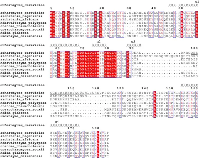
Fig. S1. Sequence alignment of Ego1 from different species as analyzed by ESPript 3.0 (34). The secondary structures (1-4) of Ego1 in the EGOC are schematized above the alignment. Residues 98-121 of Saccharomyces cerevisiae Ego1 which were deleted in the construct are underlined.
A
B
C
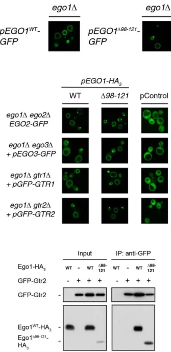
Fig. S2. Deletion of residues 98 to 121 of Ego1 does not affect the vacuolar membrane location of Ego1 nor the subsequent vacuolar recruitment of the other EGOC components. (A) Ego1∆98-121-GFP readily localizes to the vacuolar limiting membrane. Localization of plasmid-expressed Ego1WT-GFP or Ego1∆98-121-GFP was examined in prototrophic ego1∆ cells grown exponentially in synthetic drop out medium. (B) The EGOC components are efficiently recruited to the vacuolar rim by Ego1∆98-121-HA3. Localization of each plasmid-expressed GFP-tagged EGOC component was determined in indicated prototrophic strains co-expressing, or not (pControl), Ego1WT-HA3 or Ego1∆98-121-HA3,andcultured as in A.
(C) Ego1∆98-121 and Gtr2 interact with each other. Anti-GFP immunoprecipitations were performed on lysates from a subset of cells described in panel (B; bottom row) or from ego1∆ cells expressing Ego1WT -HA3.Input and IP fractions were analyzed by western blot and probed with anti-HA and anti-GFP antibodies.
A
B
C
D
Fig. S3. Strains expressing Ego1 and Ego3 mutant variants that are unable to bind the Rag GTPases are sensitive to rapamycin and have reduced TORC1 activity. (A-B) Prototrophic WT and ego1 strains expressing, or not (+ empty vec; -), the indicated plasmid-expressed Ego1-GFP variants were grown exponentially in synthetic defined dropout medium. Serial 10-fold dilutions were then spotted on YPD plates supplemented with vehicle (veh.) or 5 ng ml-1 rapamycin (RAP) and incubated for 4 days at 30°C (A). Alternatively, in vivo TORC1 activity was assessed by examining the phosphorylation status of Thr737 within the TORC1 target Sch9 by immunoblot analysis using anti-Sch9-pThr737 and anti-Sch9 antibodies (B). (C-D) Prototrophic WT and ego3 strains expressing, or not (+ empty vec; -), the indicated
plasmid-expressed Ego3-GFP variants were grown and treated as in (A) and (B), respectively.
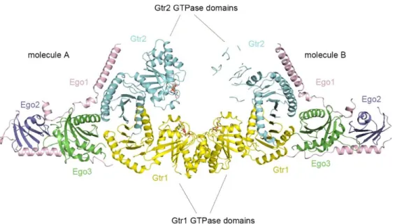
Fig. S4. Overall structure of the two EGOC molecules in the asymmetric unit. The color codes of the components are the same as in Fig. 1B.
A
B
C
D
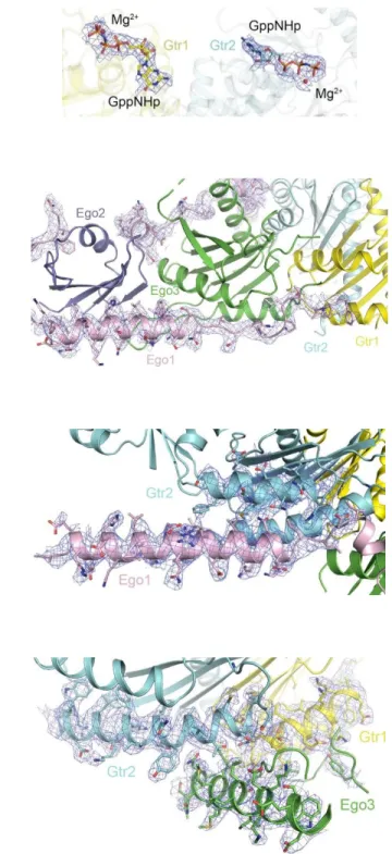
Fig. S5. Representative composite 2Fo-Fc omit maps (contoured at 1σ) of the EGOC (molecule A). (A) The bound GppNHp molecules and Mg2+ ions in Gtr1-Gtr2. The color codes of the components are the same as in Fig. 1B. (B) Ego1 in the interaction interfaces with Ego2, Ego3, and Gtr1. (C) The interaction interface between the 1-helix of Ego1 and Gtr2. (D) The interaction interface between Ego3 and Gtr1-Gtr2.
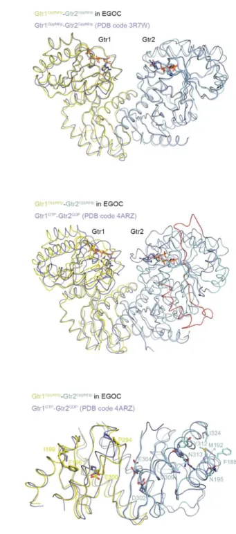
A
B
C
Fig. S6. Structural comparison of associated and free Gtr1-Gtr2 heterodimers. (A) Superposition of the structures of Gtr1GppNHp-Gtr2GppNHp in the EGOC (colored as in Fig. 1B) and free Gtr1GppNHp-Gtr2GppNHp (PDB code 3R7W) (colored in blue). (B) Superposition of the structures of Gtr1GppNHp-Gtr2GppNHp in the EGOC and free Gtr1GTP-Gtr2GDP (PDB code 4ARZ) (colored in blue). The segment composed of residues 28-70 of Gtr2GDP, which exhibits a conformational rearrangement, is colored in red. (C) Superposition of the Roadblock domains of Gtr1GppNHp-Gtr2GppNHp in the EGOC and free Gtr1GTP-Gtr2GDP (PDB code 4ARZ) (colored in blue). The residues involved in the interactions with Ego1 and Ego3 are shown in ball-and-stick models.
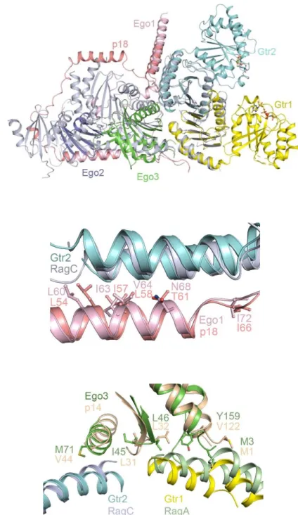
A
B
C
Fig. S7. Structural comparison of the EGOC and the Rag complex. (A) The Ragulator-RagA(CTD)-RagC(CTD) complex (PDB code 6EHR) is superimposed onto the EGOC. The color codes of the EGOC components are the same as in Fig. 1B. For clarity, p18 is colored in red and the other
components in the Ragulator-Rag complex are colored in gray. (B) Comparison of the residues from the
1-helix of Ego1 and p18 in the interacting interfaces with Gtr2/RagC. Ego1, p18, Gtr2, and RagC are colored in pink, red, cyan, and gray, respectively. (C) Comparison of the residues from Ego3 and p14 in the interaction interfaces with Gtr1/RagA and Gtr2/RagC. Ego3, p14, Gtr1, Gtr2, RagA, and RagC are colored in green, wheat, yellow, cyan, palegreen, and gray, respectively.
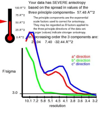
Fig. S8. Analysis of the diffraction data with the Diffraction Anisotropy Server (26). Based on the analysis of the server, the aniostropic native dataset for the EGOC is truncated to 3.5, 4.5 and 3.2 Å resolution along the reciprocal space directions a*, b* and c*, respectively. The figure was generated with the server.
Supplementary Tables
Table S1. Strains used in this study.
Strain Genotype Source Figure
KT1960 MAT; his3, leu2, ura3-52, trp1 (35)
KT1961 MATa; his3, leu2, ura3-52, trp1 (35) S3A-D
MP2649 [KT1960] ego1∆::KanMX4 This study 3A, C, E; S2A, C; S3A, B
MP279-18A [KT1961] ego1∆::KanMX4, gtr2∆::NatMX4 (18) 3B, C; S2B, C MP279-18B [KT1960] ego1∆::KanMX4, gtr1∆::NatMX4 (18) 3D, E; S2B MP2650 [KT1961] ego3∆::KanMX4 (21) 3F, H; S3C, D MP5811 [KT1961] ego3∆::KanMX4, gtr1∆::NatMX4, URA3::GTR1p-GFP-GTR1 This study 3G, H MP5854 [KT1961] ego1∆::NatMX4, ego2∆::KanMX6, HIS3::EGO2p-EGO2-GFP This study S2B MP278-1A [KT1960] ego1∆::KanMX4, ego3∆::KanMX4 This study S2B
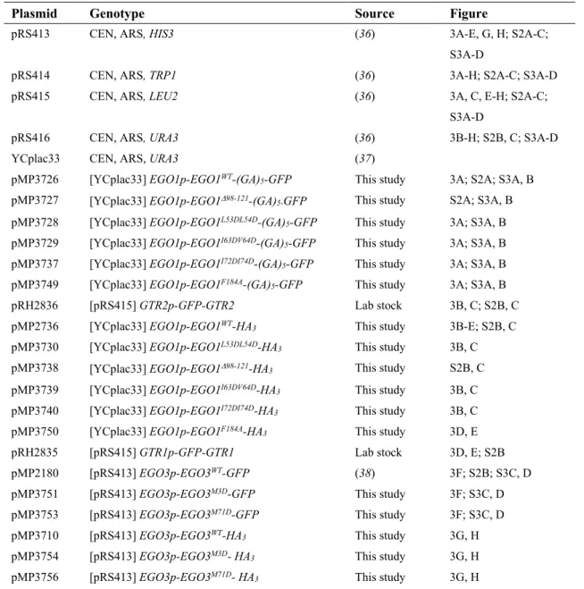
Table S2. Plasmids used in this study.
Plasmid Genotype Source Figure
pRS413 CEN, ARS, HIS3 (36) 3A-E, G, H; S2A-C; S3A-D
pRS414 CEN, ARS, TRP1 (36) 3A-H; S2A-C; S3A-D pRS415 CEN, ARS, LEU2 (36) 3A, C, E-H; S2A-C;
S3A-D
pRS416 CEN, ARS, URA3 (36) 3B-H; S2B, C; S3A-D YCplac33 CEN, ARS, URA3 (37)
pMP3726 [YCplac33] EGO1p-EGO1WT-(GA)5-GFP This study 3A; S2A; S3A, B
pMP3727 [YCplac33] EGO1p-EGO198-121-(GA)
5-GFP This study S2A; S3A, B
pMP3728 [YCplac33] EGO1p-EGO1L53DL54D-(GA)
5-GFP This study 3A; S3A, B
pMP3729 [YCplac33] EGO1p-EGO1I63DV64D-(GA)5-GFP This study 3A; S3A, B
pMP3737 [YCplac33] EGO1p-EGO1I72DI74D-(GA)5-GFP This study 3A; S3A, B
pMP3749 [YCplac33] EGO1p-EGO1F184A-(GA)5-GFP This study 3A; S3A, B
pRH2836 [pRS415] GTR2p-GFP-GTR2 Lab stock 3B, C; S2B, C pMP2736 [YCplac33] EGO1p-EGO1WT-HA3 This study 3B-E; S2B, C
pMP3730 pMP3738
[YCplac33] EGO1p-EGO1L53DL54D-HA3
[YCplac33] EGO1p-EGO198-121-HA3
This study This study
3B, C S2B, C pMP3739 [YCplac33] EGO1p-EGO1I63DV64D-HA3 This study 3B, C
pMP3740 [YCplac33] EGO1p-EGO1I72DI74D-HA3 This study 3B, C
pMP3750 [YCplac33] EGO1p-EGO1F184A-HA
3 This study 3D, E
pRH2835 [pRS415] GTR1p-GFP-GTR1 Lab stock 3D, E; S2B pMP2180 [pRS413] EGO3p-EGO3WT-GFP (38) 3F; S2B; S3C, D
pMP3751 [pRS413] EGO3p-EGO3M3D-GFP This study 3F; S3C, D
pMP3753 [pRS413] EGO3p-EGO3M71D-GFP This study 3F; S3C, D
pMP3710 [pRS413] EGO3p-EGO3WT-HA3 This study 3G, H
pMP3754 [pRS413] EGO3p-EGO3M3D- HA3 This study 3G, H
pMP3756 [pRS413] EGO3p-EGO3M71D- HA
pSIVu integrative, URA3 (38)
pMP3439 [pSIVu] GTR1p-GFP-GTR1 This study 3G, H pSIVh integrative, HIS3 (38)