HAL Id: hal-01455502
https://hal.archives-ouvertes.fr/hal-01455502
Submitted on 3 Feb 2017
HAL is a multi-disciplinary open access
archive for the deposit and dissemination of
sci-entific research documents, whether they are
pub-lished or not. The documents may come from
teaching and research institutions in France or
abroad, or from public or private research centers.
L’archive ouverte pluridisciplinaire HAL, est
destinée au dépôt et à la diffusion de documents
scientifiques de niveau recherche, publiés ou non,
émanant des établissements d’enseignement et de
recherche français ou étrangers, des laboratoires
publics ou privés.
Room temperature inorganic polycondensation of oxide
(Cu2O and ZnO) nanoparticles and thin films
preparation by the dip-coating technique
Guillaume Salek, Christophe Tenailleau, Pascal Dufour, Sophie
Guillemet-Fritsch
To cite this version:
Guillaume Salek, Christophe Tenailleau, Pascal Dufour, Sophie Guillemet-Fritsch. Room
temper-ature inorganic polycondensation of oxide (Cu2O and ZnO) nanoparticles and thin films
prepa-ration by the dip-coating technique.
Thin Solid Films, Elsevier, 2015, vol.
589, pp.
872-876.
Open Archive TOULOUSE Archive Ouverte (OATAO)
OATAO is an open access repository that collects the work of Toulouse researchers and
makes it freely available over the web where possible.
This is an author-deposited version published in :
http://oatao.univ-toulouse.fr/
Eprints ID : 16791
To link to this article : DOI : 10.1016/j.tsf.2015.04.082
URL :
http://dx.doi.org/10.1016/j.tsf.2015.04.082
To cite this version : Salek, Guillaume and Tenailleau, Christophe
and Dufour, Pascal and Guillemet-Fritsch, Sophie Room
temperature inorganic polycondensation of oxide (Cu2O and ZnO)
nanoparticles and thin films preparation by the dip-coating
technique. (2015) Thin Solid Films, vol. 589. pp. 872-876. ISSN
0040-6090
Any correspondence concerning this service should be sent to the repository
administrator:
staff-oatao@listes-diff.inp-toulouse.fr
Room temperature inorganic polycondensation of oxide (Cu
2
O and ZnO)
nanoparticles and thin films preparation by the dip-coating technique
G. Salek, C. Tenailleau
⁎
, P. Dufour, S. Guillemet-Fritsch
Centre Interuniversitaire de Recherche et d'Ingénierie des MATériaux (CIRIMAT), UMR CNRS 5085, Université de Toulouse — UPS, 118 route de Narbonne, 31062 Toulouse Cedex 09, France
a b s t r a c t
Keywords: Cuprous oxide Cuprite Zinc oxide Precipitation method Colloidal suspension Optical properties Thin filmsOxide thin solid films were prepared by dip-coating into colloidal dispersions of oxide nanoparticles stabilized at room temperature without the use of chelating or complex organic dispersing agents. Crystalline oxide nanopar-ticles were obtained by inorganic polycondensation and characterized by X-ray diffraction and field emission gun scanning electron microscopy. Water and ethanol synthesis and solution stabilization of oxide nanoparticle method was optimized to prepare two different structural and compositional materials, namely Cu2O and ZnO.
The influence of hydrodynamic parameters over the particle shape and size is discussed. Spherical and rod shape nanoparticles were formed for Cu2O and ZnO, respectively. Isoelectric point values of 7.5 and 8.2 were
de-termined for cuprous and zinc oxides, respectively, after zeta potential measurements. A shear thinning and thixotropic behavior was observed in both colloidal sols after peptization at pH ~6 with dilute nitric acid. Every colloidal dispersion stabilized in a low cost and environmentally friendly azeotrope solution composed of 96 vol.% of ethanol with water was used for the thin film preparation by the dip-coating technique. Optical properties of the light absorber cuprous oxide and transparent zinc oxide thin solid films were characterized by means of transmittance and reflectance measurements (300–1100 nm).
1. Introduction
Cuprous oxide and zinc oxide are non-toxic, low-cost, chemically stable compounds that contain abundant elements. Cuprous oxide and zinc oxide are interesting materials individually and when combined, and are thus used for various types of applications mainly due to their unique semiconductor and optical properties.
Cuprous oxide (Cu2O) is a p-type conducting oxide with a direct
band gap of 2.2 eV suitable for sunlight absorbance[1]. Cu2O has
attracted much attention for decades, in particular due to its potential uses in solar cells[2], photocatalysis[3], photoelectrochemical water splitting[4], transistors[5], and thin films for gas sensing[6].
Zinc oxide (ZnO) is an n-type conducting material with a large direct band gap of 3.4 eV [7]. The recent development of ZnO based nanomaterials and thin films has boosted research on making it suitable in shape, size and morphology for solar cells[8,9], light emitting diodes [10], microwave absorbers[11], gas sensors[12,13], piezoelectric trans-ducers[14], and varistors[15].
Oxide thin films can be prepared by different techniques such as phys-ical or chemphys-ical vapor deposition[16–18], spray pyrolysis[19], sputtering [20,21], electrodeposition[22,23]and sol–gel[24,25]. The latter technique is a simple and cost efficient engineering process well suited for preparing substrate coatings with controlled physical and chemical properties at
low temperature. The sol–gel method usually requires surfactant or com-plex organic agents for preparing stable colloidal suspensions (or sols) of materials which can then be used for thin layers deposition[26,27]. Inor-ganic polycondensation of oxide nanoparticles is a soft-chemistry method which consists of the salts dissociation in aqueous medium with a precip-itating agent such as lithium hydroxide. This method usually involves the preparation of an organometallic precursor[28]. With optimized condi-tions, such oxide nanoparticles can then be stabilized in a homogeneous suspension when superficial electrical charges are formed at the surface of nanoparticles due to a dispersing agent with appropriate dipolar forces. We here report on a low-cost and complex agent-free synthesis and sol stabilization of Cu2O and ZnO nanoparticles obtained at room
tempera-ture. Cu2O and ZnO thin films were then prepared by the dip-coating
technique and their microstructural and optical properties studied. 2. Experimental details
2.1. Sample preparation
Zn(NO3)2·6H2O (99% purity, purchased from PROLABO) or
CuSO4·5H2O (99%, VWR) dissolved in pure distilled water (0.03 mol,
100 mL) were rapidly poured (5.5 L/s) at room temperature into a dilute buffered solution of LiOH (n = 0.1 mol, V = 1400 mL). After 30 min of stirring, the precipitate was washed with deionized water to remove re-sidual ions (OH−, Li+and SO
4
2−). An additional step consisting of
reduc-ing the Cu(OH)2precipitate was necessary for the formation of cuprous ⁎ Corresponding author. Tel.: +33 561556283.
E-mail address:tenailleau@chimie.ups-tlse.fr(C. Tenailleau).
oxide. Ascorbic acid solution (0.015 mol) was quickly poured into the cuprous hydroxide precipitate diluted with water (500 mL) and was stirred for two hours at a fast stirring rate (240 rpm). The mixture was then centrifuged (4200 rpm for 10 min) and washed with distilled water. This step was repeated three times, removing the supernatant after each stage of centrifugation in order to eliminate the non-reactive and/or excess ions. This simple synthesis method requires only a quick precipitation, a few washings with water and neutraliza-tion (plus a fast reducneutraliza-tion process for Cu2O) in order to form pure and
crystalline oxide powders at room temperature.
For the preparation of colloidal suspensions, a peptization step consisting of charging the nanoparticle surface was performed by the addition of nitric acid (68%) solution for stabilizing colloidal suspensions at pH ~6. A sonication step was applied to the solution at 35 kHz for 10 min to break apart possible aggregates formed in solution. Finally, colloidal suspensions of pure oxide phases were stabilized in an azeotrope solution composed of 96 vol.% absolute ethanol mixed with ultra-pure water after centrifugation of the peptized compounds before dip-coating with a NIMA DC small dip-coater from Lot–Oriel (withdrawal rate of 200 mm/min) for thin film preparation.
2.2. Sample characterization
X-ray diffraction patterns were recorded on a Bruker D4-Endeavor instrument (40 kV, 40 mA) with a CuKα wavelength, from 10 to 100° in 2-Theta, step size of 0.02° and 3.6 s/step scan. Field emission gun scanning electron microscopy (FEG-SEM) images were recorded on a JEOL 6700F instrument.
Film thickness analysis was performed with a Dektak 3030ST system from Veeco (1 mm scan length, low speed, 0.01 mN applied force and Valley profile).
Optical properties characterizations of the films were carried out using a PV300 instrument from Bentham after deposition on 5 mm thick com-mercial pre-cleaned and ready to use glass substrate (Menzel–Gläser pur-chased from Thermo Scientific) in the UV-Visible-NIR regions.
3. Results and discussion
3.1. Precipitation of oxide nanoparticles
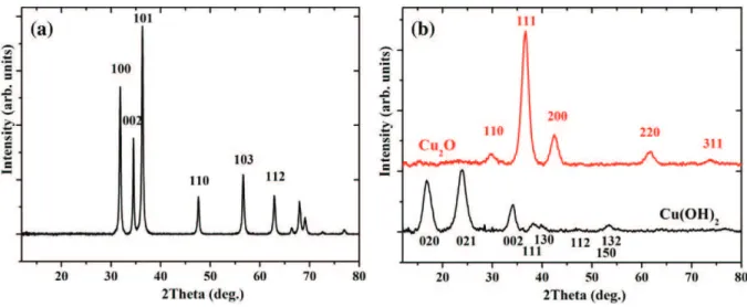
X-ray diffraction analysis showed that pure zinc oxide of wurtzite structural type (with P63mc space group) is formed after precipitation
and the cupric hydroxide phase (Cmc21space group) is observed in
the copper based synthesis (Fig. 1). When raw cupric salt is used for
the sample preparation, the copper ions possess a + II oxidation state. The ascorbic acid (C6H8O6) was then used as a reducing agent in order
to promote the following two reactions and form the cuprous oxide: Cu(OH)2+ C6H8O6= Cu2++ C6H6O62 −+ 2H2O
2 Cu2++ C
6H6O62 −+ H2O = Cu2O + C6H6O6+ 2 H+.
Shape control mechanism of cuprous oxide nanoparticles was recent-ly studied in aqueous solutions with the use of a porecent-lyvinylpyrrolidone surfactant[29]. Size and low-dimensional structures are governed by thermodynamic and kinetic parameters. A supersaturated medium con-taining a large excess of ascorbic acid will drive the reaction to the forma-tion of Cu2O nanoparticles of cubic shape (200 nm average diameter
size). But large excess of ascorbic acid (twice the amount of reactant) can be too reducing and metal copper is usually formed after stirring for half an hour or more. The particle morphology is also sensitive to the stirring rate. A fast stirring rate strongly increases the probability of collisions between particles and can lead to the formation of nano-spheres[29]. Here, the quick addition of the ascorbic acid solution into the cuprous hydroxide precipitate at a fast stirring rate allows the forma-tion of spherical cuprous oxide nanospheres with the cuprite structure (Pn-3m space group) by a successive dissolution/reprecipitation mecha-nism (Figs. 1c and2a). Monodisperse solutions contain particles with an average size of 84 ± 21 nm. Larger spheres of Cu2O can be obtained
when equivalent molar numbers of Cu2+and OH−(n = 0.03 mol) and
volumes of salt and alkaline solutions are used during the precipitate for-mation step (seeFig. 2b).
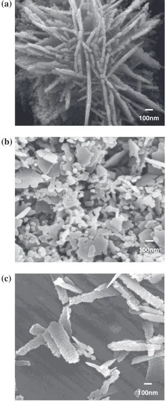
Micrometer flakes or nanorod-like particles of zinc oxide are formed depending on the nature of the solvent used during the synthetic pro-cess (Fig. 3). When the solvent used during precipitation is water crystal plates will assemble together to form flakes (Fig. 3a), that can be even-tually broken apart by sonication (Fig. 3b), while a mixture of 70 vol.% of water and 30 vol.% of alcohol leads to uniquely zinc oxide nanorods (Fig. 3c). The nature of the solvent can indeed influence the mechanism of nucleation[30]. Particle shape obtained during precipitation depends on the number of hydrogen bonds offered by the solvent. Solvent mol-ecules can considerably modify the sample stability and agglomeration phenomenon. Moreover, the germination and growth processes, which are governed by the degree of supersaturation and interfacial energy, can be strongly modified by the change of solvent[31,32]. The oxide particles morphology also strongly depends on the dielectric constant of the aqueous medium. A mixed solution of water and ethanol with
Fig. 1.X-ray diffraction patterns of zinc oxide (left) and copper hydroxide (bottom right) obtained after precipitation. In the latter case, ascorbic acid was used to form crystalline cuprous oxide (top right). Miller indices are given for the first main diffraction peaks.
decreased dielectric constant, influence the growth process along a special crystallographic axis which leads to single rod-like shaped nanoparticles (Fig. 3c).
3.2. Sol stabilization
It is well known that zinc oxide and cuprous oxide are sensitive to dis-solution in aqueous dis-solutions at ambient temperature. This phenomenon closely depends on the chemical species present in solution. Pourbaix di-agrams are often used to view the equilibrium chemistry of a species in terms of redox behavior or solubility alone[33]. Their stabilization in aqueous medium by modification of their hydroxyl groups with inorganic acids is only possible in a small specific pH region. For zinc oxide, at am-bient temperature and in acid medium (pH b6), the dissolution is in prin-ciple due to the direct attack of protons on the particles surface. This promotes the formation of the soluble forms Zn(OH)+[34,35]. In alkaline
media (pH N9), the dissolution phenomenon is generally attributed to the formation of different hydroxyl ions such as Zn(OH)3−and Zn(OH)42−[34,
36]. Similarly, for cuprous oxide under acidic conditions, the solubility is closely related to the chemical species with which copper can form com-plexes[37]. Similarly, following the Pourbaix diagram for copper oxide stabilization, Cu+
is formed in slightly acid solution (pH ~5–6), in a small potential area compared to Cu2+, without any complexing agent. CuII(OH)
2and CuI(OH) are predominant in water under mildly alkaline
conditions (pH N9) and their presence also depends on the oxygen solu-bility in water[33,37].
Zeta potential measurements showed an isoelectric point value of 7.5 and 8.2 for Cu2O and ZnO, respectively (Fig. 4). Zeta potential is
max-imum at a pH value close to 6. Zeta potential decrease observed either at
lower or higher pH could be explained by a partial dissolution of the particles of ZnO and Cu2O. At this range of pH, the increase of ionic
strength by dissolution of particles could induce a compression of the ionic double layer of particles thus changing the size of the agglomerate mobility.
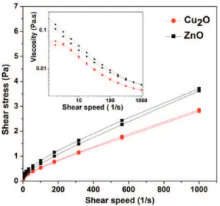
Therefore, a short strong ultrasonication (35 kHz for 5 min) coupled with the addition of an acidic solution of dilute nitric acid (pH ~6) was utilized to disperse and stabilize the particles in solution. After this step of peptization/deagglomeration, an azeotrope mixture containing 96 vol.% of absolute ethanol and 4 vol.% of ultrapure water was used to obtain the final sol, allowing the stabilization of particles and improv-ing the wettability of the sol on a glass substrate. The viscosity variations of this sol as a function of the shear strain is given inFig. 5for each oxide 100nm 1µm 100nm 1µm
(b)
(a)
Fig. 2.Scanning electron microscopy images of Cu2O nanoparticles in the case of excess
(a) and (b) equimolar ratio of (ascorbic acid/solution salt) used to form the precipitate.
(a)
(b)
(c)
100nm 100nm 100nmcolloidal dispersion. Both dispersions of Cu2O and ZnO exhibit a
rheofluidifying (pseudo-plastic) and slightly thixotropic behavior as shown by the non-linearity of the curves and the presence of a small hysteresis. Particle granulometric distributions in colloidal dispersions were measured by dynamic light scattering after ultrasonication of each solution at 35 kHz for 10 min. Cuprous oxide and zinc oxide solu-tions exhibit a narrow distribution of aggregates with an average parti-cle diameter centered around 198 and 255 ± 5 nm, respectively (Fig. 6). Solutions of ZnO were stable for a couple of weeks while colloidal dis-persions of Cu2O were stable for a few months.
3.3. Oxide thin films preparation and their optical properties
Oxide thin films of Cu2O and ZnO were prepared by dip-coating into
the corresponding colloidal dispersions. Contact profilometry measure-ments showed that a thin layer of ~300 nm can be obtained after one dip-coating sequence for both compounds, although particle shapes are different for Cu2O (spheres) and ZnO (rods).
Fig. 7shows room temperature transmittance and reflectance data and insets are FEG-SEM images of a micrometer thick and homogeneous oxide layer obtained after three successive dip-coatings. Optical features are identical for 300 nm thin layers of oxides. Absorbance curves were deduced by summing all three contributions to 100%.
A strong absorption edge is observed for the orange/red Cu2O thin
films, from the UV region to the low wavelengths of the visible region. In the cuprous oxide, the top part of the valence band does not only con-stitute oxygen orbitals, as it is usually observed for transition metal ox-ides, but contains also fully occupied metal d orbitals[38–40]. The Cu+
ions 4 s0orbitals correspond to the conduction band lower energy levels
and charge transfer can occur from the hybridized orbitals forming the valence band to the empty Cu states at the conduction band. The optical direct band gap value of ~ 2.4 eV, which was determined following Tauc's equation[41], is higher than the band gap (~2 eV) measured in Cu2O films grown by electrodeposition technique[42]. The higher
band gap observed in our work can be related to a quantum confine-ment effect due to smaller particle sizes[43]. Indeed, the average crys-tallite size is approximately 10 nm in diameter, as determined by Scherrer's law from the X-ray diffraction patterns, but even smaller par-ticles (below 5 nm) can also be seen in SEM images, supporting the as-sumption of quantum confinement effect.
ZnO thin films are highly transparent over the visible-NIR region. A direct energy band gap of 3.3 eV for ZnO was determined by extrapola-tion of the vertical line calculated following Tauc's equaextrapola-tion[41], in ac-cordance with the literature[7].
These homogeneous thin films of Cu2O and ZnO obtained at room
temperature by the dip-coating method after a strict control of the pa-rameters for the precipitation process and sol dispersion stabilization needed to be sintered to form compact layers. The two materials could then be combined for various applications including photovoltaics. Fi-nally, this water and ethanol synthesis and solution stabilization of oxide nanoparticle method have proven to be efficient for the prepara-tion of thin and crystallized oxide solid films at room temperature, and can thus be extended to a large variety of target structures.
4. Conclusions
Monodisperse distributions of oxide particles were stabilized in col-loidal dispersions using a simple and low cost method that was opti-mized at room temperature using only water and ethanol as solvents. Cu2O nano- or micro-particles are spherical and their size can be
adjust-ed by changing the proportions of metal salts and alkaline solutions. Two types of ZnO particle morphology, flakes or rods, were synthetized by modification of the dielectric constant of the solvent.
After peptization at pH ~6, oxide colloidal suspensions were stabi-lized in an azeotrope ethanol mixed water solution. The small size of hy-drodynamic particles and the high positive zeta potential used to
Fig. 4.Zeta potential measurements vs pH for ZnO (squares) and Cu2O (circles).
Fig. 5.Shear stress variation as a function of the shear strain for a colloidal dispersion of zinc oxide (squares) and cuprous oxide (circles). Inset shows the viscosity variation vs shear speed.
Intensity (arb. units)
Fig. 6.Particles granulometry distributions in colloidal dispersions of zinc oxide (squares) and cuprous oxide (circles).
stabilize our sols allow the formation of stable suspensions which ex-hibit a rheofluidifiant behavior. As-prepared dispersions were then used for the oxide thin films preparation by the dip-coating technique. Homogeneous thin films (~300 nm after one dip-coating sequence) of oxides were formed on glass substrates. The dark orange cuprous oxide films strongly absorb in the UV region and low wavelengths of the visible region while the whitish zinc oxide films are transparent from the UV to the IR regions.
Acknowledgments
This work was supported by the French Ministry of Education and Research. Isabelle Pasquet and Jean-Jacques Demai are thanked for their assistance in recording the SEM images.
References
[1] M.Y. Shen, T. Yokouchi, S. Koyama, T. Goto, Dynamics associated with Bose–Einstein statis-tics of orthoexcitons generated by resonant excitations in cuprous oxide, Phys. Rev. B 56 (1997) 13066.
[2] H.M. Wei, H.B. Gong, L. Chen, M. Zi, B.Q. Cao, Photovoltaic efficiency enhancement of Cu2O
solar cells achieved by controlling homojunction orientation and surface microstructure, J. Phys. Chem. C 116 (2012) 10510.
[3] Y. Zhang, B. Deng, T. Zhang, D. Gao, A.-W. Xu, Shape effects of Cu2O polyhedral
microcrys-tals on photocatalytic activity, J. Phys. Chem. C 114 (2010) 5073.
[4] M. Hara, T. Kondo, M. Komoda, S. Ikeda, J.N. Kondo, K. Domen, M. Hara, K. Shinohara, A. Tanaka, Cu2O as a photocatalyst for overall water splitting under visible light irradiation,
Chem. Commun. 2 (1998) 357.
[5] E. Fortunato, V. Figueiredo, P. Barquiha, E. Elamurugu, P. Barros, G. Gongcalves, S.H.K. Park, C.S. Hwang, R. Martins, Thin-film transistors based on p-type Cu2O thin films produced at
room temperature, Appl. Phys. Lett. 96 (2010) 192102.
[6]P.A. Praveen Janantha, L.N.L. Perera, K.M.D.C. Jayathilaka, J.K.D.S. Jayanetti, D.P. Dissanayaka, W.P. Siripala, Use of Cu2O microcrystalline thin film semiconductors for
gas sensing, Proc. Tech. Sessions 25 (2009) 70.
[7] R. Hauschild, H. Priller, M. Decker, J. Brückner, H. Kalt, C. Klingshirn, The exciton polariton model and the diffusion of excitons in ZnO analyzed by time-dependent photoluminescence spectroscopy, Phys. Status Solidi C 3 (4) (2006) 980.
[8] J. Wu, G. Chen, H. Yang, C. Ku, J. Lai, Effects of dye adsorption on the electron transport properties in ZnO-nanowire dye-sensitized solar cells, Appl. Phys. Lett. 90 (2007) 2131091.
[9] M. Igalson, C. Platzer-Björkman, The influence of buffer layer on the transient behavior of thin film chalcopyrite devices, Sol. Energy Mater. Sol. Cells 84 (2004) 93.
[10] K. Kim, J.S. Horwitz, W.H. Kim, A.J. Makinen, Z.H. Kafafi, D.B. Chrisey, Doped ZnO thin films as anode materials for organic light-emitting diodes, Thin Solid Films 420–421 (2002) 539.
[11] Z.W. Zhou, W.M. Peng, S.Y. Ke, H. Deng, Tetrapod-shaped ZnO whisker and its composites, J. Mater. Process. Technol. 89–90 (1999) 415.
[12] J. Chen, J. Lin, J. Li, G. Xiao, X. Yang, Large-scale syntheses of uniform ZnO nanorods and ethanol gas sensors application, J. Alloys Compd. 509 (2011) 740.
[13] S.J. Pearton, D.P. Norton, K. Ip, Y.W. Heo, Recent progress in processing and properties of ZnO, T. Steiner, Prog. Mater. Sci. 50 (2005) 293.
[14]S.C. Ko, Y.C. Kim, S.S. Lee, S.H. Choi, S.R. Kim, Micromachined piezoelectric membrane acoustic device, Sensors Actuators A Phys. 103 (2003) 130.
[15] L. Saint Macary, M.L. Kahn, C. Estournès, P. Fau, D. Trémouilles, M. Bafleur, P. Renaud, B. Chaudret, Size effect on properties of varistors made from zinc oxide nanoparticles through low temperature spark plasma sintering, Adv. Funct. Mater. 19 (2009) 1775.
[16] J. Kouam, T. Ait-Ahcene, A.G. Plaiasu, M. Abrudeanu, A. Motoc, E. Beche, C. Monty, Charac-terization and properties of ZnO based nanopowders prepared by solar physical vapor de-position (SPVD), Sol. Energy 82 (2008) 226.
[17] X.H. Wang, R.B. Li, D.H. Fan, Control growth of catalyst-free high-quality ZnO nanowire ar-rays on transparent quartz glass substrate by chemical vapor deposition, Appl. Surf. Sci. 257 (2011) 2960.
[18] G. Gugliettaa, T. Wangaa, R. Patib, S. Ehrmanb, R.A. Adomaitisa, Chemical vapor deposition of copper oxide films for photoelectrochemical hydrogen production, in: Frank E. Osterloh (Ed.),Proc. of SPIE, Solar Hydrogen and Nanotechnology IV, 7408 2009, p. 7408071.
[19] R. Ayouchi, D. Leinen, F. Martin, M. Gabas, E. Dalchiele, J.R. Ramos-Barrado, Preparation and characterization of transparent ZnO thin films obtained by spray pyrolysis, Thin Solid Films 426 (2003) 68.
[20]J.R. Ray, M.S. Desai, C.J. Panchal, P.B. Patel, Magnetron sputtered Al–ZnO thin films for photovolataic applications, J. Nano. Electron. Phys. 3 (2011) 755.
[21] S. Izhizuka, S. Kato, T. Maruyama, K. Akimoto, Nitrogen doping into Cu2O thin films
depos-ited by reactive radio–frequency magnetron sputtering, Jpn. J. Appl. Phys. 40 (2001) 2765.
[22]S. Sanchez, D. Aldakov, D. Rouchon, L. Rapenne, A. Delamoreanu, C. Lévy-Clément, V. Ivanova, Sensitization of ZnO nanowire arrays with CuInS2 for extremely thin absorber solar cells, J. Renewable Sustainable Energy 5 (2013) 011207.
[23] K. Mukhopadhyay, A.K. Chakroborty, A.P. Chatterjee, S.K. Lahiri, Galvanostatic deposition and characterization of cuprous oxide thin films, Thin Solid Films 209 (1992) 92.
[24] C.D. Bojorge, H.R. Canepa, U.E. Gilabert, D. Silva, E.A. Dalchiele, R.E. Marotti, Synthesis and optical characterization of ZnO and ZnO:Al nanocrystalline films obtained by the sol–gel dip-coating process, J. Mater. Sci. Mater. Electron. 18 (2007) 1119.
[25] H.Y. Xu, C. Chen, L. Xu, J.K. Dong, Direct growth and shape control of Cu2O film via one-step
chemical bath deposition, Thin Solid Films 527 (2013) 76.
[26] A.K. Kyaw, X.W. Sun, C.Y. Jiang, Efficient charge collection with sol–gel derived colloidal ZnO thin film in photovoltaic devices, J. Sol-Gel Sci. Technol. 52 (2009) 348.
[27] L. Znaidi, Sol–gel-deposited ZnO thin films: a review, Mater. Sci. Eng. B 174 (2010) 18.
[28] L. Spanhel, M.A. Anderson, Semiconductor clusters in the sol–gel process: quantized aggre-gation, gelation, and crystal growth in concentrated zno colloids, J. Am. Chem. Soc. 113 (1991) 2826.
[29] Y. Bai, T. Yang, Q. Gu, G. Cheng, R. Zheng, Shape control mechanism of cuprous oxide nano-particles in aqueous colloidal solutions, Powder Technol. 227 (2012) 35.
[30] A.E. Nielsen, Precipitation, Croat. Chem. Acta 42 (1970) 319.
[31] C. Marcilly, Revue de l'Institut Français du Pétrole, 391984. 189.
[32]G. Charlot, B. Tremillon, Les réactions chimiques dans les solvants et les sels fondus, Gauthier-Villars, Paris, 1963.
[33] M. Pourbaix, Atlas of Electrochemical Equilibria in Aqueous Solutions, Pergamon, New York, 1966.
[34] S. Yamabi, H. Imai, Growth conditions for wurtzite zinc oxide films in aqueous solutions, J. Mater. Chem. 12 (2002) 3773.
[35] A. Degen, M Kosec, Effect of pH and impurities on the surface charge of zinc oxide in aque-ous solution, J. Eur. Ceram. Soc. 20 (2000) 667.
[36] S.W. Bian, I.A. Mudunkotuwa, T. Rupasinghe, V.H. Grassian, Aggregation and dissolution of 4 nm ZnO nanoparticles in aqueous environments: influence of pH, ionic strength, size, and adsorption of humic acid, Langmuir 27 (2011) 6059.
[37] D.A. Palmer, Solubility measurements of crystalline Cu2O in aqueous solution as a function
of temperature and pH, J. Solut. Chem. 40 (2011) 1067.
[38] L. Zhang, L. McMillon, J. McNatt, Gas-dependent bandgap and electrical conductivity of Cu2O thin films, Sol. Energy Mater. Sol. Cells 108 (2013) 230.
[39] Y. Nakano, S. Saeki, T. Morikawa, Optical bandgap widening of p -type Cu2O films by
nitro-gen doping, Appl. Phys. Lett. 94 (2009) 2008.
[40]J.P. Hu, D.J. Payne, R.G. Egdell, On-site interband excitations in resonant inelastic x-ray scattering from Cu2O, Phys. Rev. B 155115 (2008) 1.
[41] J. Tauc, Optical properties and electronic structure of amorphous Ge and Si, Mater. Res. Bull. 3 (1968) 37.
[42] W. Siripala, L.D.R.D. Perera, K.T.L. De Silva, J.K.D.S. Jayanetti, Study of annealing effects of cuprous oxide grown by electrodeposition technique, Sol. Energy Mater. Sol. Cells 44 (1996) 251.
[43] P. He, X. Shen, H. Gao, Size-controlled preparation of Cu2O octahedron nanocrystals and
studies on their optical absorption, J. Colloid Interface Sci. 284 (2005) 510.
400 600 800 1000 0 20 40 60 80 100
Inte
ns
ity
(%
)
Wavelength (nm)
Transmittance Reflectance Absorbance 1 m 400 600 800 1000 0Wavelength (nm)
1 m 20 40 60 80 100Inte
nsity (%
)
Transmittance Absorbance Reflectance µ µFig. 7.Transmittance, reflectance and deduced absorbance spectra of ZnO (left) and Cu2O (right) measured on thin films at room temperature. Insets show side cuts scanning electron