Association between the Rate of CD4
+T Cell Decrease and the Year
of Human Immunodeficiency Virus (HIV) Type 1 Seroconversion
among Persons Enrolled in the Swiss HIV Cohort Study
Philippe Vanhems,1Jean Lambert,3Marta Guerra,3 Bernard Hirschel,2Robert Allard,3and the Swiss HIV Cohort Studya
1Laboratoire d’Epide´miologie et de Sante´ Publique, INSERM Unite´
271, Universite´ Claude Bernard, Lyon, France, for the Coordinating Center of the Swiss HIV Cohort Study, CHUV, Lausanne;2Division
of Infectious Diseases, University Hospital, Geneva, Switzerland;
3Department of Social and Preventive Medicine, University
of Montreal, Montreal, Canada
The aim of this study was to investigate the early CD4+
T cell response among human immunodeficiency virus type 1 (HIV-1) seroconverters in relation to their year of serocon-version. Study participants were enrolled in the Swiss HIV Cohort Study between 1985 and 1995 and had not received antiretroviral treatment. The slope of the CD4+
T cell count within 2 years after seroconversion was significantly associated with the year of seroconversion, by sex and by use of injection drugs, when controlling for initial CD4+
cell count. These results show that the loss of CD4+
cells might be associated with the year of seroconversion, suggesting a change in the pathogenesis of HIV across the years. If these results are confirmed, they could have important implications for the pathogenesis of and therapeutic strategies for HIV-1 infection.
CD41T cell count is associated with human immunodefi-ciency virus type 1 (HIV-1) disease progression and survival [1, 2] among patients infected by HIV from an unknown date and among seroconverters [3, 4]. Recently, the prognostic value of the CD41cell count was confirmed independently of the virus load [5]. Two studies done in the US Navy reported that the initial CD41cell count was !500/mm3in nearly 40% and
!200/mm3in nearly 4% of HIV-1 seroconverters 1 year after
seroconversion [6, 7]. These results suggest that host or viral factors (or both) are associated with a faster loss of CD41cells early in the course of HIV infection. The emergence of more virulent strains has also been suggested as a factor influencing the rate of CD41cell loss. Holmberg et al. [8] did not find that recently infected persons lost their CD41cells faster by year of seroconversion, and this finding was confirmed among patients from the Multicenter AIDS Cohort Study [9]. In addition, no major change, such as a more rapid loss of CD41T cells, seems
Received 17 September 1998; revised 1 June 1999; electronically published 12 November 1999.
Presented in part: 12th World AIDS Conference, Geneva, 28 June–3 July 1998 (abstract 12119).
Financial support: Swiss Federal Office of Public Health (The Swiss HIV Cohort Study, project 168).
a Study group members are listed after the text.
Reprints or correspondence: Dr. Philippe Vanhems, Laboratoire d’Epide´miologie et de Sante´ Publique, Universite´ Claude Bernard Lyon 1, 8, av. Rockefeller, 69373 Lyon Cedex 08, France (philipva@lyon-sud .univ-lyon1.fr).
The Journal of Infectious Diseases 1999;180 :1803–8
q 1999 by the Infectious Diseases Society of America. All rights reserved. 0022-1899/1999/18006-0007$02.00
to have occurred in the natural history of HIV-1 infection in US patients.
In Europe, analysis of data from the Italian Seroconversion Study [10] did not show a secular trend by year of serocon-version for the initial CD41cell measurement obtained within 24 months of seroconversion. Keet et al. [11] suggested a pro-longation of the AIDS-free period for patients in the Amster-dam Cohort Study, a finding in contrast to those from the Tricontinental Seroconverters Study, which showed a more rapid progression to AIDS in homosexual men who serocon-verted between 1982 and 1987 [12]. In addition, Sinicco et al. [13] showed that HIV-1 seroconversion after 1989 was an in-dependent predictor of disease progression.
Additional studies are needed of the natural history of HIV-1 infection for regional public health interventions and com-parisons between countries. The Swiss HIV Cohort Study (SHCS) provides an opportunity for such investigation [14, 15]. We studied the slope of CD41cell decrease among HIV sero-converters enrolled in SHCS between 1985 and 1995, to identify any association between the loss of CD41cells and the year of seroconversion.
Methods
Study population. The SHCS is a prospective study based on voluntary participation of persons infected by HIV-1; persons in-fected by HIV-2 were not included. The rationale of the study, the organization of the study, and the baseline characteristics of the SHCS have been described in detail elsewhere [14, 15]. In brief, this multicenter project was implemented in 7 university hospitals
in Switzerland: Basel, Bern, Geneva, Lausanne, Lugano, St-Gall, and Zurich. Study participants are from both sexes, belong to var-ious groups at risk for HIV infection, and have a physical and blood sample examination every 6 months.
In the first part of the study, we investigated HIV seroconverters with a lag time for seroconversion of!12 months (n = 266). The
lag time for seroconversion was defined as the time elapsed between the last HIV-negative screening test and the first HIV-positive screening test. The date of infection was estimated as the midpoint between these 2 tests, to facilitate comparisons with other inves-tigations [9, 10].
To determine secular trends for the initial CD41 cell
measure-ment by year of seroconversion, we compared the first count among patients after stratification by the year of seroconversion. This ap-proach is similar to that of Galai et al. [10], except that we restricted the lag time of the first CD41 cell determination to 1 year after the date of HIV seroconversion, whereas Galai et al. used 2 years. The lag time for the initial CD41cell measurement was the time
elapsed from the estimated date of infection to the first CD41cell measurement. The mean lag time of the first CD41cell
determi-nation was 5.6 months (median, 5.5 months;SD = 2.9 months). No patient had received antiretroviral therapy before the first CD41 cell count.
In the second part of the study, we estimated secular trends of CD41cell slopes within 2 years after HIV seroconversion. To do this, we restricted the study population to patients with at least 4 CD41 cell determinations within 2 years since the date of HIV seroconversion (n = 69). This inclusion criterion was chosen so that there would be enough reliable data for assessing a slope. To ex-plore the natural history of CD41cell response early after HIV infection, we analyzed data from persons who did not receive any antiretroviral drug during the 2 years after seroconversion or whose treatment began!30 days before the last CD41cell measurement
(1 month of treatment is unlikely to have an effect on the CD4 cell count).
Laboratory methods. HIV seroconversion was assessed by ELISA, and Western blot was used to confirm the presence of HIV-1 antibodies. CD41cell counts were done similarly in each center [16]. All participating centers used cytofluorometry and fluorescein isothiocyanate– or phycoerythrin-labeled antibodies.
Statistical analysis. The baseline characteristics of the patient population were reported by use of means, medians, and SDs for continuous variables and percentages and frequencies for categor-ical variables.
In the first part of the study (n = 266), we performed one-way analyses of variance (ANOVA) to compare age, CD41cell count, lag time for seroconversion, and lag time for the first CD41cell count by year of seroconversion. The x2
test was used to compare sex and risk factors of HIV-infection distributions. The proportions of patients having baseline CD41 cell counts !500/mm3
,
!350/mm3
, and!200/mm3
by year of seroconversion were compared by use of the x2
test for trend [17].
In the second part of this study (n = 69), we analyzed individual repeated measures of CD41cell counts, to identify a secular trend of these values over the years. We have included in the analysis the data of persons with at least 4 determinations within 2 years from seroconversion, with at least 2 measurements in the first year and 2 in the second year. The analysis was done in 2 steps. First,
the slopes of the CD41 cell counts, with respect to time, were
estimated for each patient. In the second step, these slopes were entered as dependent variables in a linear regression model in-cluding years of seroconversion and adjusted for center, age, sex, risk factors, initial CD41cell count, time elapsed between the
se-roconversion and the first CD41cell count, and the following fac-tors in relation to years since seroconversion: center3 year of seroconversion, age3 year of seroconversion, sex 3 year of se-roconversion, and risk factors3 year of seroconversion. We used a backward stepwise method to identify the appropriate model. To check the adequacy of the model and to identify influential cases, studentized deleted residuals, leverages, and Cook distances were computed. The statistical analyses were done by use of SPSS (ver-sion 6.1; SPSS, Chicago) and Epi Info (ver(ver-sion 6.04; Centers for Disease Control and Prevention, Atlanta).P!.05was considered statistically significant.
Results
First part of study. A total of 266 patients were included in the first part of the study: 181 were male (68%), 93 (35%) were exposed to HIV by homosexual contact, 86 (32%) were exposed by heterosexual contact, 42 (16%) were exposed by injection drug use (IDU), and 45 (17%) were exposed by sexual contact and IDU or by another route. The mean age was
years (range, 17–60; median, 28.0). 30.35 8.7
The overall median lag time for seroconversion was 6.0 months (range, 0.2–12.0), and the median lag time for the first CD41cell measurement was 5.5 months (range, 0.1–11.7). The
baseline characteristics did not differ by center (data not shown). The overall median of the first CD41cell count after seroconversion was 546/mm3(range, 4–2100/mm3). The overall
proportions of patients with CD4 cell counts!500/mm3,!350/
mm3, and!200/mm3were 38.6%, 20.1%, and 3.6%, respectively.
Age, median lag times, and characteristics of CD41 cell counts, by calendar year of seroconversion, are shown in table 1. There were no statistically significant differences for age, lag for seroconversion, lag for CD41cell measurement, and CD41 cell counts between years of seroconversion. Similarly, sex and risk factor distributions did not show statistically significant differences among years of seroconversion (data not shown). The x2tests for linear trend of these proportions, by year of
seroconversion, were not statistically significant.
Second part of study. Sixty-nine patients were included in the second part of the study (table 2): 71% were male, 43.5% were exposed to HIV by homosexual contact, 29% were ex-posed by heterosexual contact, 12% were exex-posed by IDU, and 16% were exposed by other routes. The mean age was years (range, 18–60). ANOVA was done on the 31.65 10.4
initial CD41cell counts, to identify possible differences among years of seroconversion; the results were nonsignificant (P = ). In addition, there was no difference in the lag time for .42
Table 1. First CD41cell count of 266 Swiss HIV Cohort Study participants who seroconverted for HIV between 1985 and 1995. Year No. of patients Median age, years
Median lag for seroconversion,
monthsa
Median lag for CD41cell count, monthsb Median no. of CD41 cells/mm3 Mean (SD) no. of CD41 cells/mm3 % with CD41 cells/mm3!500c % with CD41 cells/mm3!350d % with CD41 cells/mm3!200e <1987 11 27.0 5.08 7.11 600 721 (400) 27 18 0 1988 29 28.0 5.50 6.59 540 676 (410) 34 14 7 1989 50 26.5 6.29 5.49 540 617 (350) 36 24 12 1990 40 26.0 4.81 4.85 528 518 (270) 45 32 15 1991 36 28.0 5.13 5.42 519 627 (300) 42 14 3 1992 39 29.5 6.21 5.96 571 615 (370) 28 18 15 1993 21 32.0 6.22 5.40 740 712 (279) 33 5 0 1994 19 35.0 8.95 6.85 560 684 (300) 26 16 0 1995 21 31.0 6.65 5.01 450 435 (230) 62 38 14 Total 266 28.0 6.01 5.49 546 612 (330) 38.6 20.1 3.6
NOTE. Represents first part of current study. HIV, human immunodeficiency virus.
a
Time midpoint between last HIV-negative and first HIV-positive ELISA.
b
Time between estimated date of infection and date of first CD41T cell count.
c
, x2
for linear trend.
P = .42 d
, x2
for linear trend.
P = .83 e
, x2for linear trend.
P = .98
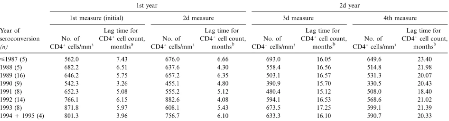
Table 2. Descriptive statistics of the 4 CD41cell determinations performed within 2 years of seroconversion among 69 participants of the Swiss HIV Cohort Study, 1985–1995.
Year of seroconversion
(n)
1st year 2d year
1st measure (initial) 2d measure 3d measure 4th measure
No. of CD41cells/mm3
Lag time for CD41cell count,
monthsa
No. of CD41cells/mm3
Lag time for CD41cell count,
monthsb
No. of CD41cells/mm3
Lag time for CD41cell count,
monthsb
No. of CD41cells/mm3
Lag time for CD41cell count, monthsb <1987 (5) 562.0 7.43 676.0 6.66 693.0 16.05 649.6 23.40 1988 (5) 682.2 6.51 637.6 4.30 558.4 16.56 514.8 21.98 1989 (16) 646.2 5.75 657.2 6.35 503.1 16.57 531.3 20.07 1990 (9) 542.3 3.26 455.1 4.80 390.9 15.70 330.5 20.43 1991 (8) 652.3 5.08 555.2 5.12 480.4 15.12 508.0 18.40 1992 (14) 766.1 6.15 882.6 4.08 594.1 16.53 568.6 21.02 1993 (8) 871.8 5.97 608.1 5.43 673.5 17.25 599.1 21.39 19941 1995 (4) 801.3 3.96 756.7 6.10 633.3 16.10 590.7 20.33
NOTE. Represents second part of current study. Statistical level of significance of differences of CD41cell counts at 6, 12, 18, and 24 months between patients grouped by year of seroconversion areP = .42, .08, .07, and .46, respectively.
a
Time elapsed between estimated date of seroconversion and initial measure of CD41cells.
b
Time elapsed between date of initial measurement of CD41cells and every other count. seroconversion (P = .60) or the lag time for the first CD41cell
measurement by year of seroconversion (P = .14).
The overall mean of the individual CD41cell count slopes was246 cells/mm3per semester. The results of the statistical
analysis relating individual CD41 cell count slopes to prog-nostic factors are shown in table 3. The initial CD41cell count was strongly associated with the slope (P = .001): the higher the initial count, the steeper the negative slope. As an example, an increase of 100 cells/mm3for the initial count was associated
with a supplementary loss of 9.4 cells/mm3per semester.
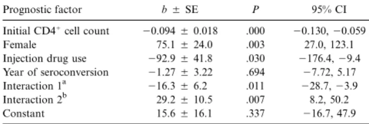
There were 2 significant interactions: year of seroconversion by sex (P = .011) and year of seroconversion by IDU (P =
). Owing to the presence of these interactions in the model, .007
the regression coefficients associated with the main effects in-cluded in these interactions should be interpreted with caution. Our objective was to study the impact of year of seroconversion on the CD41cell count slopes, and this effect can be obtained
for each sex by combining with IDU in the linear regression model shown in table 3. The results are shown in table 4.
As shown in table 4, year of seroconversion was positively associated with the CD41cell count slope for men using in-jection drugs (b = 27.9cells/mm3per semester) and negatively
associated with the CD41cell count slope for women not using injection drugs (b =217.6cells/mm3per semester). These results
mean that, after controlling for the initial CD41cell count, the rate of CD41cell count loss is less important during the recent years for men using injection drugs and more important during the recent years for women not using injection drugs.
Discussion
The objective of this study was to explore the relationship between the CD4 cell response early in the course of HIV
in-Table 3. Multiple linear regression model relating individual CD41 cell count slopes to prognostic factors (n = 68).
Prognostic factor b5 SE P 95% CI
Initial CD41cell count 20.094 5 0.018 .000 20.130, 20.059
Female 75.15 24.0 .003 27.0, 123.1
Injection drug use 292.9 5 41.8 .030 2176.4, 29.4 Year of seroconversion 21.27 5 3.22 .694 27.72, 5.17
Interaction 1a 216.3 5 6.2 .011 228.7, 23.9
Interaction 2b 29.25 10.5 .007 8.2, 50.2
Constant 15.65 16.1 .337 216.7, 47.9
NOTE. CI, confidence interval.
a
Year of seroconversion in relation to sex.
b
Year of seconversion in relation to injection drug use.
Table 4. Effect of year of human immunodeficiency virus type 1 seroconversion on individual CD41cell count slopes with respect to each sex in combination with injection drug use (IDU).
b5 SE 95% CI No IDU Men 21.27 5 3.22 27.72, 5.17 Women 217.6 5 5.5 228.6, 26.6 IDU Men 27.95 10.1 7.7, 48.1 Women 11.65 11.5 211.4, 34.6
NOTE. CI, confidence interval. fection and in the year of seroconversion among SHCS
par-ticipants who were free of antiretroviral treatment.
Linear analysis of the CD41cell count within 2 years after
seroconversion showed interactions between the year of sero-conversion and sex and IDU. This finding differed from those of other studies [9, 18]. Our results are in the same direction as those of Sinicco et al. [13] but are not completely similar. Sinicco et al. [13] reported that the probability of the CD41 cell count falling to!500,!400, and!2003 106cells/L and of
AIDS progression was higher for patients who seroconverted after December 1989. Nevertheless, in our study, the significant effect of the year of seroconversion was observed only through the interaction terms, which do not seem to have been explored in the Sinicco study. In our study, the year of seroconversion had an effect among patients followed for 2 years with a similar censoring strategy [19]. The main questions were the following: (1) did we observe the emergence of HIV strains with various levels of virulence in recent years; (2) is there any interaction between the virus strain pathogenicity and the use of injection drugs; and (3) is there any interaction between the virus strain pathogenicity and the sex of the host? Fardzadegan et al. [20] showed that women with the same virus load as men had 1.6-fold higher risk of AIDS [20]. These results, combined with ours, suggest that HIV pathogenicity might differ for males and females and by route of HIV infection.
We do not have any information on the phenotype or ge-notype of the virus strains, and little is known on the distri-bution of those strains in different populations at risk of HIV infection [21]. The phenotype defined by syncytium-inducing strains is associated with a faster progression of disease [22], but, until now, no particular genotype seemed to be associated with a faster decrease in CD41cell counts. For example,
pri-mary HIV infection by a strain with a mutation at codon 215, which indicates acquired resistance to zidovudine, was not as-sociated with more-severe clinical features [23, 24] or with a faster loss of CD41cells 1 year after HIV-1 infection [24]. There is also little evidence that the viral subtype determines the rate of disease progression among US patients [25], but HIV-1 sub-types may differ in rates of progression to AIDS among female sex workers in Senegal [26]. The host’s genetic status regarding
the CCR-5 chemokine receptor has been associated with var-ious rates of progression [27, 28]. In particular, HIV-1–positive patients who are heterozygous for the CCR-5 D32 deletion seem to have a slower early HIV-1 progression [27, 28]. We did not have this information for the SHCS participants; however, a trend toward change of the genotype over time would be sur-prising. This mutation alone does not explain the various rates of HIV-1 disease progression [29], which can involve many fac-tors [30–34].
Our study did have some limitations. Even with standardized criteria of enrollment in the cohort, we cannot totally eliminate recruitment bias toward inclusion of more severely ill patients in recent years. Because the proportion of practitioners with good experience in HIV medicine increased with time, they could have referred to the university hospitals only the patients with severe clinical features at seroconversion. Thus, since the severity of acute HIV infection has been shown to be associated with faster disease progression [35], our results could be biased by a greater proportion of symptomatic seroconverters detected in the recent years. Unfortunately, valid information on the acute illness is not available in the Swiss cohort, especially for patients who seroconverterted before 1990. Moreover, the pa-tients who received antiretroviral treatment at primary HIV infection and who were most likely to be recent seroconverters were not analyzed, so that the study could be restricted to the natural course of HIV infection.
The use of a standardized inclusion protocol and the absence of statistical differences of major baseline characteristics be-tween centers may not have prevented some factors, such as genetic markers (i.e., the human major histocompatibility com-plex genes HLA groups) associated with disease progression [36], from being unequally distributed in Switzerland. The pro-portion of patients who received an antiretroviral treatment could have affected the results, but that proportion is expected to be higher in the recent seroconverters. Thus, we should have observed the opposite relationship between the year of sero-conversion and the CD41cell decrease. The new antiretroviral therapy guidelines proposing to treat patients as early as pos-sible after infection could definitively mask the natural history of the CD41cell response early in the course of infection among patients with access to health care in the future [37].
In summary, we found that the loss of CD41cells was
as-sociated with the year of seroconversion by sex and IDU, sug-gesting that the pathogenetic effect of HIV could have changed since 1985 and differed by sex and route of HIV infection. Physicians should be aware of this finding when estimating drug efficacy. Moreover, if changes of HIV pathogenetic mechanisms are confirmed, they can occur on targets other than CD41cells and need to be investigated.
Members of the Swiss HIV Cohort Study (SHCS) The following are members of the SHCS: M. Bateguay (Co-chair of the Scientific Board); E. Bernasconi, Ph. Bu¨rgisser, M. Egger, and P. Erb (Chairs of the Group “Laboratories”); W. Fierz and M. Flepp (Chairs of the Group “Clinics”); P. Fran-cioli (President of the SHCS, Centre Hospitalier Universitaire Vaudois, Lausanne); H. J. Furrer, P. Grob, and B. Hirschel (Chairs of the Scientific Board); and L. Kaiser, B. Ledergerber, R. Lu¨thy, R. Malinverni, L. Matter, M. Opravil, F. Paccaud, G. Pantaleo, L. Perrin, W. Pichler, J-C. Piffaretti, M. Ricken-bach, P. Sudre, J. SchupRicken-bach, A. Telenti, and P. Vernazza.
Acknowledgments
We thank R. Gaudet (University of Montreal) for his statistical as-sistance and L. Ayzac and J. Este`ve (Claude Bernard University, Lyon) for their comments.
References
1. Phillips AN, Pezzotti P, Cozzi Lepri A, et al. CD4 lymphocyte count as a determinant of the time from seroconversion to AIDS and death from AIDS: evidence from the Italian seroconversion study. AIDS 1994; 8: 1299–305.
2. Stein DS, Korvick JA, Vermund SH. CD41lymphocyte cell enumeration for prediction of clinical course of human immunodeficiency virus disease: a review. J Infect Dis 1992; 165:352–63.
3. Mellors JW, Rinaldo CR Jr, Gupta P, White RM, Todd JA, Kingsley LA. Prognosis of HIV-1 infection predicted by the quantity of virus in plasma. Science 1996; 272:1167–70.
4. Mellors J, Kingsley LA, Rinaldo CR Jr, et al. Quantification of HIV-1 RNA in plasma predicts outcome after seroconversion. Ann Intern Med 1995; 122:573–9.
5. Vlahov D, Graham N, Hoover D, et al. Prognostic indicators for AIDS and infectious disease death in HIV-infected injection drug users. JAMA 1998; 279:35–40.
6. Weiss PJ, Brodine SK, Goforth RR, et al. Initial CD4 lymphocyte counts in recent human immunodeficiency virus infection and lack of association with identified coinfections. J Infect Dis 1992; 166:1149–53.
7. Gorham ED, Garland FC, Mayers DL, et al. CD4 lymphocyte counts within 24 months of human immunodeficiency virus seroconversion. Arch Intern Med 1993; 153:869–76.
8. Holmberg SD, Conley LJ, Luby SP, Cohn S, Wong LC, Vlahov D. Recent infection with human immunodeficiency virus and possible rapid loss of
CD4 T lymphocytes. J Acquir Immune Defic Syndr Hum Retrovirol 1995; 9:291–6.
9. O’Brien TR, Hoover DR, Rosenberg P, et al. Evaluation of secular trends in CD41lymphocyte loss among human immunodeficiency virus type 1 (HIV-1)–infected men with known dates of seroconversion. Am J Epi-demiol 1995; 142:636–42.
10. Galai N, Lepri AC, Vlahov D, et al. Temporal trends of initial CD4 cell counts following human immunodeficiency virus seroconversion in Italy, 1985–1992. Am J Epidemiol 1996; 143:278–82.
11. Keet IPM, Veugelers PJ, Koot M, et al. Temporal trends of the natural history of HIV-1 infection following seroconversion between 1984 and 1993. AIDS 1996; 10:1601–2.
12. Veugelers PJ, Page KA, Tindall B, et al. Determinants of HIV disease pro-gression among homosexual men registered in the Tricontinental Sero-converters Study. Am J Epidemiol 1994; 140:747–58.
13. Sinicco A, Fora R, Raiteri R, et al. Is the clinical course of HIV-1 changing? Cohort study. BMJ 1997; 314:1232–7.
14. Ledergerber B, von Overbeck J, Egger M, Luthy R. The Swiss HIV cohort study: rationale, organization and selected baseline characteristics. Soz Praventivmed 1994; 39:387–94.
15. Egger M, Hirschel B, Francioli, et al. Impact of antiretroviral combination therapies in HIV infected patients in Switzerland: prospective multicentre study. BMJ 1997; 315:1194–9.
16. Ledergerber B, Kno¨pfli D, Lu¨thy R. Laboratory studies. In: Documentation of the Swiss HIV Cohort Study. Lausanne, Switzerland: Swiss HIV Cohort Study Coordinating Center, 1991:41–2.
17. Fleiss JL. Statistical methods for rates and proportions. 2nd ed. New York: John Wiley, 1981:143–6.
18. Carre N, Prins M, Meyer L, et al. Has the rate of progression to AIDS changed in recent years? AIDS 1997; 11:1611–8.
19. Cozzi Lepri A, Phillips AN, Pezzotti P, Rezza G. Study’s censoring strategy may be source of bias. BMJ 1997; 315:1237.
20. Farzadegan H, Hoover DR, Astemborski J, et al. Sex differences in HIV-1 viral load and progression to AIDS. Lancet 1998; 352:1510–4. 21. Lukashov VV, Goudsmit J. HIV heterogeneity and disease progression in
AIDS: a model of continuous virus adaptation. AIDS 1998; 12:S43–52. 22. Nielsen C, Pedersen C, Lundgren JD, Gerstoft J. Biological properties of
HIV isolates in primary HIV infection: consequences for the subsequent course of infection. AIDS 1993; 7:1035–40.
23. Vanhems P, Allard R, Cooper DA, et al. Acute human virus type 1 disease as a mononucleosis-like illness: is the diagnosis too restrictive? Clin Infect Dis 1997; 24:965–70.
24. Imrie A, Carr A, Duncombe C, Finlayson R, et al. Primary infection with zidovudine-resistant human immunodeficiency virus type 1 does not ad-versely affect outcome at 1 year. J Infect Dis 1996; 174:195–8. 25. Operskalski EA, Busch MP, Mosley JW, Stram DO. Comparative rates of
disease progression among persons infected with the same or different HIV-1 strains. J Acquir Immune Defic Syndr Hum Retrovirol 1997; 15: 145–50.
26. Kanki PJ, Hamel DJ, Sankale´ JL, et al. Human immunodeficiency virus type 1 subtypes differ in disease progression. J Infect Dis 1999; 179:68–73. 27. Martin PM, Dean M, Smith MW, et al. Genetic acceleration of AIDS
pro-gression by a promoter variant of CCR5. Science 1998; 282:1907–11. 28. Meyer L, Magierowska M, Hubert JB, et al. Early protective effect of
CCR-5 D32 heterozygosity on HIV-1 disease progression: relationship with viral load. AIDS 1997; 11:F73–F84.
29. Cohen OJ, Vaccarezza M, Lam GK, et al. Heterozygosity for a defective gene for CC chemokine receptor 5 is not the sole determinant for the immunologic and virologic phenotype of HIV-infected long-term non-progressors. J Clin Invest 1997; 100:1581–9.
30. Kirchhoff F, Greenough TC, Brettler D, Sullivan JL, Desrosier RC. Absence of intact nef sequences in a long-term survivor with nonprogressive HIV-1 infection. N Engl J Med HIV-1995; 332:228–32.
Cytotoxic-T-cell responses, viral load, and disease progression in early human im-munodeficiency virus type 1 infection. N Engl J Med 1997; 337:1267–4. 32. Desrosier RC, Sullivan JL. Declining CD4 T-cell counts in a person infected
with nef-deleted HIV-1. N Engl J Med 1999; 340:236–7.
33. Darby SC, Ewart DW, Giangrande PLF, et al. Importance of age at infection with HIV-1 for survival and development of AIDS in UK haemophilia population. Lancet 1996; 347:1573–9.
34. Schechter MT, Hogg RS, Aylward B, et al. Higher socioeconomic status is associated with slower progression of HIV infection independent of access
to health care. J Clin Epidemiol 1994; 47:59–67.
35. Vanhems P, Lambert J, Cooper DA, et al. Severity and prognosis of acute human immunodeficiency virus type 1 illness: a dose-response relationship. Clin Infect Dis 1998; 26:314–22.
36. Kaslow RA, Carrington M, Apple R, et al. Influence of combinations of human major histocompatibility complex genes on the course of HIV-1 infection. Nat Med 1996; 2:405–11.
37. Carpenter CCJ, Fischel MA, Hammer SM, et al. Antiretroviral therapy for HIV infection in 1996. JAMA 1996; 276:146–54.