Reduced Vagal Activity in Salt-Sensitive
Subjects During Mental Challenge
Konrad Buchholz, Hartmut Scha¨chinger, Miriam Wagner,
Arya M. Sharma, and Hans Christian Deter
Background: Salt-sensitive normotensive men exhibit
an enhanced pressor and heart rate (HR) response to mental stress. Stress-induced HR acceleration may result from sympathetic activation or vagal withdrawal. We stud-ied the importance of vagal withdrawal for the increased stress responsiveness of salt-sensitive subjects.
Methods: We studied cardiovascular reactivity to
men-tal challenge in 17 salt-sensitive healthy white male stu-dents and 56 salt-resistant control subjects who were comparable with respect to age, body mass index, and physical fitness. Salt sensitivity was determined by a 2-week dietary protocol (20 mmol v 240 mmol sodium/day). Mental stress was induced by a computerized informa-tion-processing task (manometer test). Electrocardio-gram and finger blood pressure (BP; Finapres, Ohmeda, Louisville, CO) were registered continuously to deter-mine HR and interbeat-interval length. Time and fre-quency domain (spectral power) based measures of respiratory-related heart rate variability (HRV) were calculated to estimate vagal cardiac control; diastolic
BP reactivity was assessed to estimate peripheral sym-pathetic effects.
Results: Stress-induced increase in HR was higher in
salt-sensitive than in salt-resistant subjects. Salt-sensitive subjects, in comparison to salt-resistant subjects, showed significantly reduced respiratory-related HRV during baseline and mental stress conditions (P ⬍ .01). The increase in diastolic BP during mental challenge was sig-nificantly greater in salt-sensitive subjects (P⬍ .05).
Conclusions: Our findings suggest reduced vagal and
increased sympathetic tone during mental challenge in salt-sensitive subjects. Altered autonomic nervous system function may contribute to later development of hyperten-sion in salt-sensitive individuals. Am J Hypertens 2003; 16:531–536 © 2003 American Journal of Hypertension, Ltd.
Key Words: Heart rate variability, vagal activity,
men-tal stress, sodium-dependent hypertension, blood pressure.
E
nhanced cardiovascular responsiveness to mental challenge was found in subjects at future risk for the development of arterial hypertension, such as borderline hypertensives and healthy offspring of hyper-tensive parents.1Increased pressor responsiveness has also been described as a consequence of salt loading in a proportion of young normotensive individuals. Several lines of evidence suggest that these salt-sensitive individ-uals may be genetically prone to the development of hypertension.2– 4 We have previously shown that young normotensive salt-sensitive subjects display an elevated heart rate (HR) response to mental challenge,5suggestingthat autonomic nervous system (ANS) factors may
con-tribute to the development of hypertension in these sub-jects.
In accordance, enhanced anxiety and irritability was found in salt-sensitive subjects,6compatible with a
con-tribution of higher central nervous system factors. How-ever, HR changes may result from parasympathetic or sympathetic nervous system changes and it remains un-clear whether vagal withdrawal or sympathetic activation is responsible for the enhanced cardiovascular responsive-ness to mental challenge described in salt-sensitive sub-jects.
Several investigators have shown that the high fre-quency component of heart rate variability (HRV)
primar-Received January 23, 2003. First decision February 14, 2003. Accepted March 11, 2003.
From the Division of Psychosomatic Medicine (KB), Benjamin Franklin Medical Center, Free University of Berlin, Berlin, Germany; Department of Medicine (HS), Division of Psychosomatic Medicine, University Hospital Basel, Basel, Switzerland; Division of Psychoso-matic Medicine (MW, HCD), Benjamin Franklin Medical Center, Free University of Berlin, Berlin, Germany; and Franz-Volhard Klinik
(AMS), Medical Faculty of the Charite´, Humboldt University of Berlin and Max Delbru¨ck Center for Molecular Medicine, Berlin, Germany.
The study was supported by the German Research Council (DFG De 224/6-1).
Address correspondence and reprint requests to Dr. Konrad Buch-holz, Department of Nephrology, Hannover Medical School, Carl-Neu-berg-Str. 1, D-30625 Hannover, Germany; e-mail: konrad.buchholz@ gmx.de
0895-7061/03/$30.00 © 2003 by the American Journal of Hypertension, Ltd.
doi:10.1016/S0895-7061(03)00905-1 Published by Elsevier Inc.
ily reflects vagal tone.7–10 Diastolic blood pressure (BP) responses have been used as estimates of sympathetic nervous system excitement.
The aim of the present study was to examine ANS changes in sensitive subjects, as compared to salt-resistant subjects, during rest and mental stress conditions.
Methods
Study Population
Seventy-three healthy white men (age range: 20 to 30 years), who responded to an advertisement posted on the university campus, volunteered for the study. All but four students of other faculties (two salt-sensitive and two salt-resistant subjects) were enrolled in medical school. Before entering the study, all subjects underwent routine physical and laboratory evaluation to ensure that none had hypertension, diabetes mellitus, hepatic, pulmonary, or renal disease, or abnormal weight (body mass index,⬍19 or ⬎26 kg/m2). All subjects had a resting diastolic BP ⬍85 mm Hg and a systolic BP ⬍135 mm Hg, defined by the mean of 60 oscillometric measurements (DINAMAP, Criticon, Tampa, FL) in the recumbent position. Subjects were excluded if they reported a neurologic or psychiatric disease, drug abuse (including nicotine and alcohol), vas-cular damage, or current medication.
All subjects underwent phenotypic characterization re-garding their BP response to a dietary salt-loading/restric-tion protocol as previously described.11,12 In brief,
subjects were given a standardized low-salt diet containing 20 mmol sodium, 20 mmol chloride, 60 mmol potassium, and 20 mmol calcium per day for 14 days. To this diet, a daily supplement of 22 tablets of Slow Sodium (10 mmol NaCl per tablet; CIBA-GEIGY, Horsham, United King-dom) and placebo were each given in a randomized single-blind crossover fashion for 7 days. Dietary compliance was assessed by daily measurement of electrolyte excre-tion. At the end of each week, after a 30-min resting period, BP was measured for assessment of salt sensitivity in the recumbent subject during 1 h at 1-min intervals with an automatic oscillometric device (DINAMAP 1846 SX, Critikon). As in previous studies,11,12 salt sensitivity was
defined as a significant decrease in mean arterial pressure ⬎3 mm Hg under the low-salt diet calculated as the difference between the average of the 60 readings under the high salt and low salt periods. Following the supine measurements, subjects were asked to stand upright, and BP and HR was measured during 15 min at 1-min inter-vals. To assess nonspecific sympathetic reactivity under low and high salt intake, BP and HR readings of the 15 min in the orthostatic position were averaged and com-pared to the average of the 60 readings in the supine position.
Physical fitness was estimated by subjects reports about their exercise habits and was classified as 1 point for⬍1
h, 2 points for 1 to 2 h, and 3 points for⬎2 h of physical exercise per week.
Study Protocol
All psychophysiologic tests were conducted between 8 and 12 AM, with the subjects seated comfortably in a high-backed chair in a quiet room with an ambient tem-perature of 20°C to 24°C. Electrodes and transducers for detection of physiologic parameters were fastened to the chest, nondominant arm, and hand. Blood pressure was continuously registered by the Finapres device (Ohmeda, Louisville, CO), attached to the middle digit of the non-dominant hand. Heart rate was continuously registered by electrocardiography. Respiratory rate was continuously registered with a piezo-electric transducer that was fas-tened to the chest with an elastic belt.
The mental challenge test (manometer test) was per-formed 1 to 3 days before the salt loading/restriction protocol began. Subjects were instructed to abstain from coffee and black tea for at least 12 h before testing. Before the manometer test, subjects remained on their normal salt intake with a comparable sodium excretion between salt-sensitive and salt-resistant subjects (184.3⫾ 61.5 v 181.6 ⫾ 91.2 mmol within 24 h before the test). After the first instructions, a venous cannula was placed in the dominant forearm. Thereafter, subjects rested for 30 min to get accustomed to the environment. The duration of the ma-nometer test was 8 min, and it was preceded by a resting phase of 5 min. Blood samples for catecholamine levels (measured by HPLC) and plasma renin activity (measured by radioimmunoassay) were drawn immediately before the resting phase and immediately after the manometer test.
The manometer test13consisted of a standardized com-puter-based information-processing task, performed under time pressure. All tests and instructions were presented on a computer monitor, and data entry was performed by manipulating a mouse with the dominant hand. Subjects were presented with an increasing number of 3 to 11 manometer clocks, and had to rapidly decide whether one or more hands of the clocks deviated by more than 90 degrees from the direction of an arrow shown on the top of the screen. Wrong answers were indicated by a loud acoustic signal. During the course of the test, the time allowed for the decision was subsequently shortened by 30% until the participant made a mistake. After a mistake, the time allowed for the decision was extended by 30%. This algorithm maintained an individually adapted work-load. In previous studies the manometer test was judged as emotionally distressing by most participants. The test was shown to elicit reproducible BP and HR responses that are within the expected range of comparable mental challenge tests (eg, mental arithmetic).14,15
The protocol of the study, including the process for obtaining informed consent, was approved by the ethics committee of our hospital.
Data Analysis
All physiologic signals were continuously recorded at 1000 Hz with DOS-Software (Turbolab, Stemmer, Puch-heim, FRG). The electrocardiographic and finger BP curves were analyzed off-line under visual artifact control by ALYS software (version 1.18, T. Sudhop and H. Scha¨chinger, Basel, Switzerland). Blood pressure and HR responses to the mental task were determined by subtract-ing the average of the readsubtract-ings dursubtract-ing the baseline period from the average of the readings during the manometer test.
Power spectral density of HR and BP was analyzed by Fourier transformation according to published standards.16
The very low, low, and high bands were defined as 0.02 to 0.06 Hz, 0.07 to 0.14 Hz, and 0.15 to 0.4 Hz, respectively. Respiratory frequency is an important determinator of HRV17and was assessed by calculating the modal value of
respiratory frequency tracings during each test period. The root mean square of successive differences (RMSSD) of inter-beat interval (IBI) length was calculated as a time domain-based measure of respiratory-related HRV.18,19
Variability measures were log-transformed to achieve nor-mally distributed parameters.
Statistical Analysis
Repeated measures analysis of variance was performed by SAS GLM-Procedure (version 8.1; SAS Inc., Cary, NC) to determine group (salt-sensitive versus salt-resistant) and stress effects (resting baseline versus mental stress) and potential interactions. A statistically significant interaction term reflects different effects of stress between groups. All data are presented as means⫾ standard deviation. A P ⬍ .05 was considered statistically significant.
Results
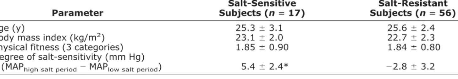
Baseline characteristics of salt-sensitive and salt-resistant subjects are shown in Table 1. Subjects were comparable with respect to age, body mass index, and physical fitness. By definition, the difference in mean arterial BP between the high salt and low salt periods was greater in the salt-sensitive than in the salt-resistant group.
Baseline levels of HR (69.1⫾ 9.9 beats/min v 67.0 ⫾ 10.2 beats/min), systolic BP (109.05 ⫾ 13.09 mm Hg v 107.31⫾ 14.83 mm Hg), and diastolic BP (64.50 ⫾ 7.16 mm Hg v 63.73⫾ 11.20 mm Hg) before the mental stress test were comparable between salt-sensitive and salt-resis-tant subjects, respectively. During the manometer test, systolic and diastolic BP increased in all subjects, but salt-sensitive subjects displayed greater heart rate (10.7⫾ 9.0 beats/min v 5.9⫾ 6.1 beats/min; P ⬍ .01) and diastolic BP responses (11.3⫾ 3.7 mm Hg v 9.2 ⫾ 3.7 mm Hg; P ⬍ .05) than salt-resistant subjects, and tended to display greater systolic BP responses (19.2⫾ 8.5 mm Hg v 16.4 ⫾ 8.5 mm Hg; P ⬍ .10).
Salt-sensitive subjects tended to display lower respira-tory rates during baseline (0.233⫾ 0.046 Hz) as compared to salt-resistant subjects (0.252 ⫾ 0.039 Hz; P ⫽ .05). During mental stress, respiratory rate increased signifi-cantly in both study groups (P⬍ .0001) but did not differ between salt-sensitive (0.306⫾ 0.038 Hz) and salt-resis-tant subjects (0.324⫾ 0.039 Hz).
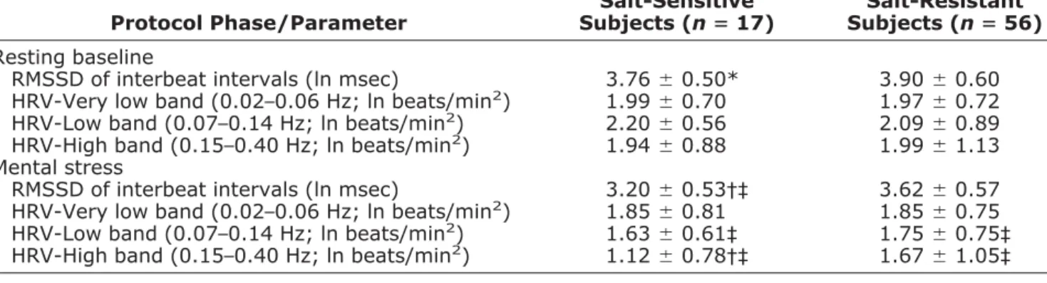
Time and frequency domain based measures of HRV are presented in Fig. 1 and Table 2. The time domain-based variability estimate RMSSD of IBI length revealed significant stress (time) [F(1,70)⫽ 57.0; P ⬍ .0001] and
Table 1. Demographic parameters of salt-sensitive and salt-resistant subjects
Parameter Salt-Sensitive Subjects (nⴝ 17) Salt-Resistant Subjects (nⴝ 56) Age (y) 25.3⫾ 3.1 25.6⫾ 2.4 Body mass index (kg/m2) 23.1⫾ 2.0 22.7⫾ 2.3
Physical fitness (3 categories) 1.85⫾ 0.90 1.84⫾ 0.80 Degree of salt-sensitivity (mm Hg)
(MAPhigh salt period⫺ MAPlow salt period) 5.4⫾ 2.4* ⫺2.8 ⫾ 3.2
MAP⫽ mean arterial pressure. Values are means⫾ SD.
* P⬍ .001 v salt-resistant subjects.
FIG. 1. Time domain-based heart rate variability (RMSSD) during
baseline and mental stress in salt-sensitive and salt-resistant sub-jects. RMSSD⫽ root mean square of successive differences of inter-beat intervals.
group effects [F(1,70) ⫽ 3.7; P ⬍ .05] and a significant group ⫻ stress interaction [F(1,70) ⫽ 6.7; P ⬍ .01], indicating reduced HRV in salt-sensitive subjects during baseline and mental stress.
The analysis of spectral power-based measures showed a significant stress effect [F(1,71)⫽ 45.1; P ⬍ .0001] and a significant group⫻ stress interaction [F(1,71) ⫽ 9.1; P ⬍ .003] in the high band of HRV (0.15 to 0.40 Hz), indicating reduced HRV in salt-sensitive subjects during mental stress. No group effects and no significant interac-tions in the very low and low bands of HRV and in the respective bands of BP variability (BPV) could be de-tected, although there were significant stress effects in the low bands of HRV [F(1,71)⫽ 21.9; P ⬍ .0001] and BPV [F(1,71)⫽ 10.9; P ⬍ .001; data not shown].
Analysis of BP and HR response to upright standing after 90 min in the supine position revealed no different increases of mean, systolic, and diastolic BP or HR
be-tween salt-sensitive and salt-resistant subjects (Table 3). With respect to absolute values, only diastolic BP was slightly higher in salt-resistant subjects during low salt intake, both in supine and standing positions (P⬍ .05). In addition, we analyzed neurohumoral measures of the adrenergic tone. Plasma catecholamine levels neither differed between salt-sensitive and salt-resistant subjects before mental challenge (norepinephrine: 178.4⫾ 86.6 v 217.8⫾ 85.4 pg/mL, epinephrine: 40.6 ⫾ 17.5 v 38.3 ⫾ 20.3 pg/mL) nor after mental challenge (norepinephrine: 184.9⫾ 99.1 v 229.3 ⫾ 87.7 pg/mL, epinephrine: 38.4 ⫾ 17.7 v 46.3⫾ 20.8 pg/mL). Renin activity was lower in salt-sensitive than in salt-resistant subjects before men-tal challenge (1.09 ⫾ 0.74 v 1.47 ⫾ 0.83 g Ang I/L/min; P ⬍ .05), and was comparable after mental challenge (1.35⫾ 0.78 v 1.67 ⫾ 0.90g Ang I/L/min). Only in salt-resistant subjects, renin activity was sig-nificantly higher after mental challenge than before
Table 2. Heart rate variability during baseline and mental stress in salt-sensitive and salt-resistant subjects
Protocol Phase/Parameter Salt-Sensitive Subjects (nⴝ 17) Salt-Resistant Subjects (nⴝ 56) Resting baseline
RMSSD of interbeat intervals (ln msec) 3.76⫾ 0.50* 3.90⫾ 0.60 HRV-Very low band (0.02–0.06 Hz; ln beats/min2) 1.99⫾ 0.70 1.97⫾ 0.72
HRV-Low band (0.07–0.14 Hz; ln beats/min2) 2.20⫾ 0.56 2.09⫾ 0.89
HRV-High band (0.15–0.40 Hz; ln beats/min2) 1.94⫾ 0.88 1.99⫾ 1.13
Mental stress
RMSSD of interbeat intervals (ln msec) 3.20⫾ 0.53†‡ 3.62⫾ 0.57 HRV-Very low band (0.02–0.06 Hz; ln beats/min2) 1.85⫾ 0.81 1.85⫾ 0.75
HRV-Low band (0.07–0.14 Hz; ln beats/min2) 1.63⫾ 0.61‡ 1.75⫾ 0.75‡
HRV-High band (0.15–0.40 Hz; ln beats/min2) 1.12⫾ 0.78†‡ 1.67⫾ 1.05‡
RMSSD⫽ root mean square of successive differences; HRV ⫽ heart rate variability.
Values are means⫾ SD. Between-group differences: * P ⬍ .05, † P ⬍ .01; intragroup differences v baseline: ‡ P ⬍ .001.
Table 3. Blood pressure and heart rate in supine and standing position in salt-sensitive and salt-resistant subjects Protocol Phase/Parameter Salt-Sensitive Subjects* (nⴝ 17) Salt-Resistant Subjects (nⴝ 56) Low Salt Period High Salt Period Low Salt Period High Salt Period Supine position Systolic BP (mm Hg) 109.7⫾ 8.3 113.9⫾ 7.5 111.7 ⫾ 10.9 110.5⫾ 9.5 Diastolic BP (mm Hg) 54.7⫾ 4.8† 58.3⫾ 5.3 58.8⫾ 5.7 57.9⫾ 5.6 MAP (mm Hg) 75.7⫾ 4.9 80.0⫾ 6.3 78.6⫾ 8.3 78.3⫾ 6.2 Heart rate (beats/min) 56.2⫾ 7.1 55.0⫾ 6.4 59.1⫾ 9.4 56.0⫾ 8.9 Standing position
Systolic BP (mm Hg) 110.1⫾ 12.3 115.7⫾ 10.9 114.4 ⫾ 11.2‡ 113.8⫾ 9.8‡ Diastolic BP (mm Hg) 61.0⫾ 5.1†§ 63.3⫾ 7.3‡ 65.4⫾ 6.4§ 65.7⫾ 7.2§ MAP (mm Hg) 83.4⫾ 6.9§ 87.3⫾ 8.2§ 87.1⫾ 7.3§ 87.0⫾ 7.8§ Heart rate (beats/min) 79.7⫾ 9.1§ 75.8⫾ 9.4§ 81.0⫾ 10.5§ 76.5⫾ 11.2§
BP⫽ blood pressure; MAP ⫽ mean arterial pressure. Values are means⫾ SD.
* In salt-sensitive subjects, all BP values were higher during high salt periods, all P⬍ .05. Between-group differences, within same dietary regimen: † P⬍ .01; intragroup differences v supine position: ‡ P ⬍ .01, § P ⬍ .001.
mental challenge (P⬍ .05). Aldosterone and hematocrit levels at baseline were not different between study groups (data not shown).
Discussion
In a previous study, we have shown that salt-sensitive normotensive subjects display an increased pressor and HR response to mental stress.6 In the present study, we extend these findings by demonstrating that salt-sensitive subjects, compared to salt-resistant subjects, also display a reduced vagal activity during baseline and mental stress, as demonstrated by the finding of a significant reduction of respiratory related HRV in the time (RMSSD) and fre-quency domains (high band HRV).
The finding of a reduced vagal tone under mental stress is well in line with previous reports on stress-related HRV in healthy volunteers and postinfarct patients.20,21 De-creased vagal tone has been shown in established hyper-tension,22 as well as borderline hypertension.23 The reduction of vagal tone in salt-sensitive subjects in the present study may partly explain their higher HR and BP responses under mental challenge. Withdrawal of vagal tone as the cause of enhanced cardiovascular reactivity is well in line with the hypothesis that the low HR of humans under resting condition predominantly results from a strong parasympathetic cardiac input.24
It may be criticized that the slightly lower respiratory rate of salt-sensitive subjects during the resting baseline could have biased the results. However, the lower respi-ratory rate observed in the salt-sensitive group would have favored increased RMSSD of IBI—just the opposite of what was actually detected.
Diastolic BP reactivity during mental stress was en-hanced in salt-sensitive subjects, suggesting enen-hanced pe-ripheral sympathetic effects. Enhanced peripheral sympathetic reactivity in normotensive salt-sensitive sub-jects was also demonstrated in former studies that showed an enhanced pressor response to noradrenaline infu-sion.25,26Some investigators suggest low frequency (LF) HRV to reflect sympathetic cardiac activity.18Contrasting to the increase of diastolic BP, in the present study there was a significant decrease of LF HRV that tended to be greater in salt-sensitive subjects (⫺26%) than in salt-resistant subjects (⫺16%). However, literature on stress-induced changes of low band HRV is inconsistent. With respect to the LF band, published data are not conclusive, and it remains unclear whether a decrease or increase of LF HRV during mental challenge is to be expected. Con-troversial findings have been reported. Spectral analysis techniques revealing non-normalized power results (such as the technique used by our study group) indicated sig-nificantly decreased LF HRV during mental stress27,28or nonsignificant decreases.29In case of normalized spectral power units, results are also inconsistent. Some found normalized LF HRV to remain unchanged during mental stress,30 others found increases in normalized LF HRV
during mental stress.31 The latter finding may be poten-tially explained by altered breathing patterns induced by mental stress tests using verbal activities.32In the present study, there was no significant difference between salt-sensitive and salt-resistant subjects in the reduction of the LF band during the manometer test. Thus we avoid spec-ulating on potential differences in cardiac sympathetic tone. Yet, another possible explanation for conflicting results with respect to sympathetic cardiac activity in salt-sensitive subjects could be that their defect is located in the adrenoreceptor sensitivity rather than in central sympathetic output, as proposed by Skrabal and co-work-ers.2On the other hand, an increase of sympathetic muscle nerve activity under reduced sodium intake was found in hypertensive and normotensive subjects, which was attrib-uted to an impaired baroreflex control.33,34Unfortunately, the relation of sympathetic reactivity to sodium sensitivity is not reported in these studies; therefore, comparisons to our results are difficult.
Thus, our study shows that the withdrawal of vagal activity under mental stress is more pronounced in salt-sensitive compared to salt-resistant subjects. As the sub-jects in the present study were normotensive volunteers, this implies that the tendency to markedly withdraw vagal tone precedes the hypertensive state, and may thus play a causal role in the later development of hypertension in salt-sensitive subjects. Decreased vagal tone has been shown in established hypertension,14as well as borderline hypertension.15
We may speculate that the withdrawal of vagal tone in salt-sensitive subjects is one of the mediators through which enhanced negative affect (eg, elicited by mental challenge) can lead to enhanced cardiovascular reactions. In a recent publication,35 we reported an association be-tween enhanced cardiovascular responsiveness and en-hanced affective startle modulation in salt-sensitive subjects, suggesting an enhanced neurovegetative output from the amygdala. Perhaps the output of the amygdala is not only mediated by sympathetic neural efferences, as suggested by Holand et al,36 but also by modulation of vagal efferences.
As possible limitations of our study, it should be noted that reports on reproducibility of salt sensitivity classifi-cation as well as reproducibility of cardiovascular reac-tions to mental stress are inconsistent. Nevertheless, in our own laboratory a good reliabilitiy of classification of salt sensitivity,37as well as a good reliabilitiy of cardiovascu-lar reactivity to mental stress13–15 were demonstrated. Furthermore, it should be said that the interpretation of reduced HRV as a reflection of vagal withdrawal is some-what speculative and was not confirmed by experiments with vagolytic drugs in this pilot study.
In summary, in this study we provide evidence for reduced respiratory sinus arrhythmia during baseline and mental challenge in salt-sensitive subjects. The reduction of high band HRV suggests diminished vagal activity. Diminished vagal activity under mental challenge,
to-gether with enhanced sympathetic reactivity, may contrib-ute to the later development of hypertension in salt-sensitive individuals.
References
1. Fredrikson M, Matthews K: Cardiovascular responses to behavioral stress and hypertension: A meta-analytic review. Ann Behav Med 1990;12:30 –39.
2. Skrabal F, Hamberger L, Ledochowski M: Inherited salt sensitivity in normotensive humans as a cause of essential hypertension: a new concept. J Cardiovasc Pharmacol 1984;6(Suppl 1):S215–S223. 3. Sharma AM: Salt sensitivity as a phenotype for genetic studies of
human hypertension ( Editorial). Nephrol Dial Transplant 1996;11: 927–929.
4. Weinberger MH, Fineberg NS: Sodium and volume sensitivity of blood pressure. Age and pressure change over time. Hypertension 1991;18:67–71.
5. Deter HC, Buchholz K, Schorr U, Schachinger H, Turan S, Sharma AM: Psychophysiological reactivity of salt-sensitive normotensive subjects. J Hypertens 1997;15:839 –844.
6. Buchholz K, Schorr U, Turan S, Sharma AM, Deter HC: Emotional irritability and anxiety in salt-sensitive persons at risk for essential hypertension. Psychother Psychosom Med Psychol 1999;49:284 – 289.
7. Akselrod S, Gordon D, Ubel FA, Shannon DC, Barger AC, Cohen RJ: Power spectrum analysis of heart rate fluctuation: a quantitative probe of beat-to-beat cardiovascular control. Science 1981;213: 220 –223.
8. Pomeranz B, Macaulay RJB, Caudill MA, Kutz I, Adam D, Gordon D, Kilborn KM, Barger AC, Shannon DC, Cohen RJ, Benson H: Assessment of autonomic function in humans by heart rate spectral analysis. Am J Physiol 1985;248:H151–H153.
9. Malliani A, Pagani M, Lombardi F, Cerutti S: Cardiovascular neural regulation explored in the frequency domain. Circulation 1991;84: 482–492.
10. Task Force of the European Society of Cardiology and the North American Society of Pacing and Electrophysiology: Heart rate vari-ability: standards of measurement, physiological interpretation, and clinical use. Circulation 1996;93:1043–1065.
11. Sharma AM, Cetto C, Schorr U, Spies K-P, Distler A: Renal acid– base excretion in normotensive salt-sensitive humans. Hyper-tension 1993;22:884 –890.
12. Sharma AM, Schorr U, Distler A: Insulin resistance in young salt-sensitive normotensive subjects. Hypertension 1993;21:273– 279.
13. Johannes B: A complex experimental assessment for objective de-scription of hierarchical psychophysiological behavior as human regulatory phenotype. J Gravitational Physiol 1994;1:P73–P74. 14. Seiffert, K: Psychophysiologische Reaktionsmuster unter
standard-isierter Stressbelastung bei Patienten mit atopischer Dermatitis. Medical Dissertation, Faculty of Medicine, Free University of Ber-lin, BerBer-lin, 1998.
15. Luck H: Psychophysiologische Reaktionsmuster von Angstpati-enten vor und nach einer stationa¨ren Psychotherapie. Medical Dis-sertation, Faculty of Medicine, Free University of Berlin, Berlin, 1998.
16. Schachinger H, Weinbacher M, Kiss A, Ritz R, Langewitz W: Cardiovascular indices of peripheral and central sympathetic acti-vation. Psychosom Med 2001;63:788 –796.
17. Bernardi L, Wdowczyk-Szulc J, Valenti C, Castoldi S, Passino C, Spadacini G, Sleight P: Effects of controlled breathing, mental activity and mental stress with or without verbalization on heart rate variability. JACC 2000;35:1462–1469.
18. Berntson GG, Bigger JT Jr, Eckberg DL, Grossman P, Kaufmann PG, Malik M, Nagaraja HN, Porges SW, Saul JP, Stone PH, van der Molen MW: Heart rate variability: origins, methods, and interpre-tative caveats. Psychophysiology 1997;34:623–648.
19. Sloan RP, Bagiella E, Shapiro PA, Kuhl JP, Chernikhova D, Berg J, Myers MM: Hostility, gender, and cardiac autonomic control. Psy-chosom Med 2001;63:434 –440.
20. McCraty R, Atkinson M, Tiller WA, Rein G, Watkins AD: The effects of emotions on short-term power spectrum analysis of heart rate variability. Am J Cardiol 1995;76:1089 –1093.
21. Tuininga YS, Crijns HJ, Brouwer J, van den Berg MP, Man in’t Veld AJ, Mulder G: Evaluation of importance of central effects of atenolol and metoprolol measured by heart rate variability during mental performance tasks, physical exercise, and daily life in stable postinfarct patients. Circulation 1995;92:3415–3423.
22. Guzzetti S, Piccaluga E, Casati R, Cerutti S, Lombardi F, Pagani M, Malliani A: Sympathetic predominance in essential hypertension: a study employing spectral analysis of heart rate variability. J Hyper-tens 1988;6:711–717.
23. Grossman P, Brinkman A, de Vries J: Cardiac autonomic mecha-nisms associated with borderline hypertension under varying behav-ioral demands: evidence for attenuated parasympathetic tone but not for enhanced beta-adrenergic activity. Psychophysiology 1992;29: 698 –711.
24. Persson PB, Kirchheim HR: Baroreceptor Reflexes. Berlin, Heidel-berg, New York, Tokyo, Springer, 1991.
25. Skrabal F, Herholz H, Neumayr M, Hamberger L, Ledochowski M, Sporer H, Ho¨rtnagl H, Schwarz S, Scho¨nitzer D: Salt sensitivity in humans is linked to enhanced sympathetic responsiveness and to enhanced proximal tubular reabsorption. Hypertension 1984;6:152– 158.
26. Sharma AM, Schattenfroh S, Thiede H-M, Oelkers W, Distler A: Effects of sodium salts on pressor reactivity in salt-sensitive men. Hypertension 1992;19:541–548.
27. Langewitz W, Ru¨ddel H, Scha¨chinger H: Reduced parasympathetic cardiac control in patients with hypertension at rest and under mental stress. Am Heart J 1994;127:122–128.
28. Madden K, Savard GK: Effects of mental state on heart rate and blood pressure variability in men and women. Clin Physiol 1995; 15:557–569.
29. Hoshikawa Y, Yamamoto Y: Effects of stroop color–word conflict test on the autonomic nervous system responses. Am J Physiol 1997;272:H1113–H1121.
30. Sloan RP, Shapiro PA, Bagiella E, Boni SM, Paik M, Bigger JT, Steinman RC, Gorman JM: Effect of mental stress throughout the day on cardiac autonomic control. Biol Psychol 1994;37:89 –99. 31. Pagani M, Mazzuero G, Ferrari A, Liberati D, Cerutti S, Vaitl D,
Tavazzi L, Malliani A: Sympathovagal interaction during mental stress. A study using spectral analysis of heart rate variability in healthy control subjects and patients with prior myocardial infarc-tion. Circulation 1991;83(Suppl 4):II43–II51.
32. Bernarndi L, Wdowczyk-Szulc J, Valenti C, Castoldi S, Passino C, Spadacini G, Sleight P: Effects of controlled breathing, mental activity and mental stress with and without verbalization on heart rate variability. J Am Coll Cardiol 2000;35:1462–1469.
33. Anderson EA, Sinkey CA, Lawton WJ, Mark AL: Elevated sym-pathetic nerve activity in borderline hypertensive humans. Evidence from direct intraneural recordings. Hypertension 1989;14:177–183. 34. Grassi G, Dell’Oro R, Seravalle G, Foglia G, Trevano FQ, Mancia G: Short- and long-term neuroadrenergic effects of moderate dietary sodium restriction in essential hypertension. Circulation 2002;106: 1957–1961.
35. Buchholz K, Schachinger H, Wagner M, Schorr U, Sharma AM, Deter HC: Enhanced affective startle modulation in salt-sensitive subjects. Hypertension 2001;38:1325–1329.
36. Holand S, Girard A, Laude D, Meyer-Bisch C, Elghozi JL: Effects of an auditory startle stimulus on blood pressure and heart rate in humans. J Hypertens 1999;17:1893–1897.
37. Sharma AM, Schattenfroh S, Kribben A, Distler A: Reliability of salt-sensitivity testing in normotensive subjects. Klin Wochenschr 1989;67:632–634.