Review
The causes of Charcot-Marie-Tooth disease
P. Young
a, band U. Suter
b,*
a
Department of Neurology, University of Münster (Germany)
b
Institute of Cell Biology, Department of Biology, Swiss Federal Institute of Technology, ETH Hönggerberg, Zürich
(Switzerland), Fax: +41 1 633 1190, e-mail: usuter@cell.biol.ethz.ch
Received 5 April 2003; received after revision 20 May 2003; accepted 23 May 2003
Abstract. Charcot-Marie-Tooth (CMT) disease serves as
the summary term for the most frequent forms of
inher-ited peripheral neuropathies that affect motor and sensory
nerves. In the last 12 years, 14 genes have been identified
that cause different CMT subforms. The genes found
ini-tially are predominantly responsible for demyelinating
DOI 10.1007/s00018-003-3133-5© Birkhäuser Verlag, Basel, 2003
and dysmyelinating neuropathies. Genes affected in
ax-onal and rare forms of CMT have only recently been
iden-tified. In this review, we will focus on the currently
known genes that are associated with CMT syndromes
with regards to their genetics and function.
Key words. Myelin; peripheral nervous system; Schwann cell; neurodegeneration; Charcot-Marie-Tooth disease;
hereditary neuropathy; axon degeneration.
Introduction
Charcot-Marie-Tooth (CMT) disease is a major genetic
disease in clinical neurology with a prevalence of
ap-proximately 1 in 2500 [1]. Since the first descriptions by
Charcot, Marie and Tooth in 1886, the main clinical
fea-tures of this syndrome were defined as distal peroneal
weakness accompanied by muscular atrophy [2, 3]. More
than 100 years later, the classification of CMT
syn-dromes today has been revised and extended based on
clinical features, electrophysiological, histopathological
and genetic findings [4]. This development was mainly
guided by progress in clinical electrophysiology and later
by the advances in molecular genetics. In this review, we
will predominantly concentrate on the genetic and
mole-cular understanding of CMT. But first, we shall provide a
short overview of the manifestation and biological basis
of the disease.
*
Corresponding author.Classical clinical classification of CMT
The typical CMT patient is affected by slowly progressive
distal muscle weakness and atrophy that primarily affects
the small foot muscles, peroneal muscles and, often later,
those of the hands and forearms. Foot deformities, mostly
pes cavus and claw toes are common, leading to gait
im-pairments. Although the disease is usually progressive, it
rarely causes wheelchair dependence but considerably
af-fects the quality of life.
CMT is subdivided into demyelinating (CMT1) and
ax-onal (CMT2) forms according to clinical,
electrophysio-logical and histopathoelectrophysio-logical features. CMT1 is
charac-terized by disease onset in the 1 or 2 decade of life, nerve
conduction velocities (NCVs) less than 38 m/s, and
seg-mental demyelination, remyelination and onion-bulb
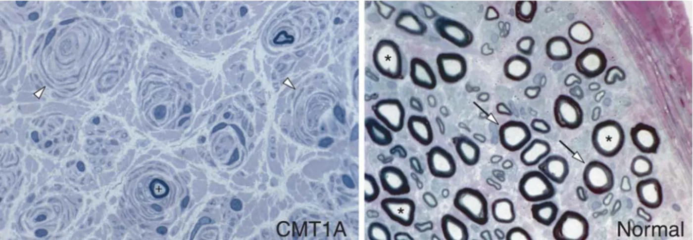
for-mations in nerve biopsies (fig. 1). CMT2 is associated
with normal or near-normal NCVs, and nerve biopsies
show loss of myelinated axons [5]. A neuropathy is
called ‘axonal’ if the axon (or neuron) is affected by the
primary injury. If the primary insult occurs in the
myeli-nating Schwann cell, this is considered a ‘demyelimyeli-nating
neuropathy’. This distinction is based on clinical and
pathological evidence. Progress in identifying the genes
responsible for CMT has now revealed, however, that
some of these disease genes are expressed by both cell
types, neurons and Schwann cells, making the
deter-mination of the primary defect often uncertain.
Fur-thermore, axon and Schwann cells interact intensively,
leading to secondary effects that might be difficult to
separate from cell-autonomous events [6]. Recent
exper-iments on myelinated axons in the CNS suggest that the
myelinating cell type may even appear morphologically
completely normal and still affect the associated axon
dramatically [7].
In the late sixties, Dyck and Lambert [8] presented an
ini-tial classification based on electrophysiological changes
in the peripheral nerves of CMT patients. Differentiating
hallmarks were NCVs, compound motor action potentials
(CMAPs), compound sensory action potentials (SNAPs),
and the patterns of inheritance. Furthermore, the term
‘hereditary motor and sensory neuropathy’ (HMSN) was
created as a synonym for CMT disease. Over time,
diag-nostic parameters for CMT were more and more
influ-enced by the increasing possibilities of molecular
genet-ics [4]. The first description of a defined genetic basis for
a given CMT subtype was discovered in 1991, when a
1.5-megabase-long, intrachromosomal duplication on the
short arm of chromosome 17 (17p11.2) was found to be
associated with the most common subtype CMT type 1A
(CMT1A) [9, 10]. Due to the fortuitous help of the
analy-sis of two spontaneous animal models for CMT, Trembler
(Tr; ([11] and Tr-J; [12]), the peripheral myelin protein 22
(PMP22) was identified as an excellent dosage-sensitive
candidate gene located on the CMT1A duplication
[13 – 16]. The final proof that PMP22 was indeed the
re-sponsible disease gene was provided a few years later
us-ing transgenic rodents [17, 18]. This initial findus-ing of a
myelin gene as the CMT1A disease gene sparked a flurry
of further investigations that led to the identification of
two other genes encoding myelin components, protein
zero (P0; MPZ) and connexin32 (Cx32; GJB1) as major
loci for demyelinating forms of CMT, collectively termed
CMT1 [19]. In the meantime, 14 genes have been found
that cause different forms of CMT. Based on these recent
genetic findings, a reevaluation of the existing
classifica-tion may be warranted. The clinical and pathological
vari-ability of CMT, however, even when the same gene is
in-volved, and the fact that mutations in different genes can
manifest as similar phenotypes, might argue differently.
A novel genetics-based categorization will be an
impor-tant additional help for the clinician, but it is not likely to
fully replace the traditional classification. Valuable
fur-ther information for a more integrated approach to this
problem will come from the detailed understanding of the
pathobiological basis of CMT syndromes. The key
ques-tion that needs to be answered is: How is the sequence of
events defined that leads from a particular genetic
alter-ation to biological effects that are deleterious to the
proper function of peripheral nerves (for reviews: [6,
19 – 22])? If we are able to find the answers to this
ques-tion, we will not only understand CMT disease but also
learn important lessons about the basic molecular cell
bi-ology of the development and maintenance of peripheral
motor and sensory nerves.
Genes mutated in CMT
For a complete and updated list of CMT genes, CMT loci
and mutations, consult: http://molgen-www.uia.ac.be/
CMTMutations/
Figure 1. Histopathological comparison of a normal human sural nerve and a nerve from a CMT1A patient. Arrows indicate normally myelinated fibers; asterisks denote axons on a nerve cross-section. Open arrowheads point to onion-bulb formations consisting of super-numerary Schwann cells that surround concentrically a demyelinated axon in the diseased state. A ‘+’ sign indicates a partially remyeli-nated axon (the pictures are a kind gift from Dr Steven S. Scherer, University of Pennsylvania).
Demyelinating forms of CMT
Peripheral myelin protein 22 (PMP22)
PMP22 was the first gene to be identified in the
patho-genesis of the development of CMT [11 – 16]. Nelis and
co-workers showed that the heterozygous duplication of
PMP22 is by far the most frequent mutation in CMT [23].
The reciprocal event to the duplication, the deletion of the
same fragment on chromosome 17p11.2 containing
PMP22, causes a neuropathy which is characterized by an
increase in vulnerability to pressure trauma resulting in
temporary nerve palsies [24]. This neuropathy, called
hereditary neuropathy with liability to pressure palsies
(HNPP), is associated with focal hypermyelination
(tomacula). In general the clinical phenotype of HNPP is
not progressive, although CMT-like symptoms may
occa-sionally develop with age [25 – 27]. Some point- and
frame-shifting mutations in PMP22, most likely causing
functional null alleles, have also been identified in HNPP
pedigrees [28, 29].
Most patients carrying the heterozygous CMT1A
dupli-cation show the classical demyelinating phenotype with
reduced NCV below 38 m/s, associated with rather
ho-mogeneous myelin defects [30, 31]. Histopathologically,
the typical changes in myelin fibers are demyelination,
thin myelin sheaths and onion-bulb formation [32]
(fig. 1). Axonal damage was thought to be rare and
pre-dominantly secondary to long-lasting deymelination.
Ex-tended electrophysiological studies revealed, however,
that the clinical degree of handicap is correlated with
ax-onal atrophy and loss, as indicated by the reduction of
CMAP and SNAP, but not with reduced NCV [33 – 35].
Homozygous CMT1A duplications cause very severe
dysmyelinating phenotypes [9, 36, 37].
Besides gene duplications and deletions, a number of
point mutations in PMP22 have been described [38].
Some of them appear to be heterozygous null alleles and
lead to HNPP, as described above. Very few mutations are
recessively inherited [39, 40]. The vast majority, however,
are associated with membrane-associated domains of the
PMP22 protein and are dominantly inherited. In general,
this last group of mutations causes a more severe CMT1
phenotype than duplication, and some have been
classi-fied as Déjérine-Sottas syndrome (DSS) [41, 42].
How does this pletora of mutations affect PMP22 and
cause the different phenotypes? The answer to this
intrigu-ing question is far from clear, but appropriate animal
mod-els mimicking the different genetic situations have been
generated. They will help to shed light on this interesting
problem [43]. This includes Pmp22 null mice [44–46],
mice and rats carrying additional copies of the PMP22
gene [17, 18, 47–49], mice with an internal Pmp22
dele-tion [50] and Pmp22 point mutadele-tions [11, 12, 51, 52].
Clearly, the key to understanding the disease phenotypes
is knowledge of the detailed function of the PMP22
pro-tein. This polypeptide comprises 160 amino acids and is
membrane associated [53]. It is most abundant in
peri-pheral nerves, and mainly expressed by myelinating
Schwann cells [54 – 56]. PMP22 plays a crucial role in the
development and maintenance of compact myelin, as is
well established by the disease phenotypes of CMT1A
and HNPP patients as well as animal models [38].
Addi-tional in vitro data suggest that PMP22 may regulate cell
proliferation [57 – 59], cell death [59 – 61], cell
differenti-ation [61 – 64] and membrane traffic [65]. Furthermore, a
function of PMP22 as a constituent of intercellular
junc-tions in epithelia has been suggested [66]. The
signifi-cance of the latter finding in the etiology of CMT,
al-though intriguing, remains to be elucidated. Such a
func-tion might rather be related to the fact that PMP22 is also
expressed outside of the nervous system [55], shows
dis-tant similarities to the tight junction components of the
claudin protein family [67] and belongs to the closely
re-lated PMP22/EMP gene family [68].
How do altered levels of PMP22 produce their
pheno-types in HNPP and CMT1A? It has been suggested that
PMP22 is part of a stoichiometric complex with P0, the
major adhesion protein of peripheral myelin and evidence
for direct interaction of the two proteins has been
pre-sented [69] but is also debated [70]. However, the
possi-bility that a reduced PMP22/P0 ratio (in HNPP) or the
op-posite situation (in CMT1A) may affect myelin stability
remains an interesting hypothesis. Alternatively, or in
ad-dition, Schwann cell proliferation and differentiation
might be affected [71, 72]. Importantly, from the point of
developing future treatment, the effects of PMP22
over-expression in Schwann cells in a CMT1A animal model
appear to be reversible, at least with regard to myelination
[73]. Whether neuronal deficiencies that correlate with
the disease-associated handicaps can also be reversed
re-mains open [6, 33 – 35, 74, 75].
How do dominant PMP22 point mutations cause the
dis-ease? At least some of them appear to produce a
gain-of-abnormal function, since most PMP22 point mutations in
human and animal models have more severe phenotypes
compared with HNPP and heterozygous PMP22
knock-out mice. One disesease mechanism has been elucidated
in that some PMP22 mutant proteins are retained
intra-cellularly, in the endoplasmic reticulum (ER) and/or the
intermediate compartment [38, 61, 64, 69, 76 – 80]. In
ad-dition, the mutant proteins encoded by the Tr (G150D)
and Tr-J alleles (L16P) aggregate abnormally in
trans-fected fibroblasts [70], although such aggregates have
been suggested to be even protective in PMP22 point
mu-tation-based peripheral neuropathies [52]. Since PMP22
forms dimers and multimers, mutant PMP22 retained in
the ER and/or intermediate compartment may prevent the
efficient transport of wild-type PMP22 to the cell
mem-brane in the form of a classical dominant-negative effect
[38, 70, 80]. However, genetic evidence shows that at
least the Tr mutation causes also a toxic gain of function
in the absence of wild-type PMP22. Heterozygous Tr
mice have a more pronounced phenotype than
heterozy-gous PMP22 knockout mice (and HNPP patients with a
heterozygous PMP22 deletion). In addition, mice
carry-ing a PMP22 null allele and the Tr allele as a compound
heterozygote display a much worse neuropathy than
het-erozygous or homozygous PMP22 knockout animals
[46].
How could a toxic gain of function be generated by
im-paired PMP22 trafficking? In analogy to the involvement
of the unfolded protein response in modulation of disease
severity in Pelizaeus-Merzbacher disease due to
prote-olipid protein mutations [81], accumulation of mutant
PMP22 may trigger a similar effect. However, there is no
experimental evidence to support such a mechanism.
An-other ER chaperone, calnexin, may be involved by getting
sequestered away by mutant PMP22 and contributing to
the resulting neuropathy [77]. Furthermore, inefficient
proteasome function could be toxic to the cell and may
add to the potential disease mechanism [82, 83]. Finally,
PMP22 accumulates in lysosomes of (de)myelinating
Schwann cells of Tr-J mice, potentially related to a role of
the endosomal/lysosomal degradation pathway in the
pathogenesis of demyelination [79, 82, 83]. This is of
spe-cial interest considering that a lyosomal protein of
un-known function, LITAF/SIMPLE, was recently identified
as being mutated in the dominant demyelinating CMT1C
disease [84].
P0, MPZ
In 1981, the first genetic linkage data suggested a gene
located on chromosome 1 to be associated with an
auto-somal dominant demyelinating CMT syndrome [85].
This was classified later as CMT1B [5], and the
muta-tions responsible were found in MPZ [86 – 89]. P0/MPZ is
a major PNS myelin component that belongs to the
im-munoglobulin superfamily [90] and plays a major role in
compaction of the myelin sheath as a homophilic
adhe-sion protein [91 – 95]. Structural analysis of the P0
extra-cellular domain suggests that four molecules form a
tetramer in cis that interacts homophilically in trans with
tetramers in the opposing membrane [96, 97]. Similar to
mutations in PMP22, the clinical, electrophysiological
and histopathological findings turned out to be
heteroge-neous for MPZ mutations [89]. These range from
autoso-mal dominant CMT1B to severe forms classified as DSS.
Furthermore, the same mutation can cause different
de-grees of disease severity in different patients [98]. Close
to a hundred MPZ mutations have been described up to
now. The main phenotype is a demyelinating neuropathy
that is clinically and by electrophysiological means
indis-tinguishable from CMT1A. Less frequently, patients are
very severely affected [5, 89]. As expected from the
func-tion of P0, transfecfunc-tion studies in cultured cells revealed
that some CMT1B mutations in the P0 extracellular
do-main [99] and cytoplasmic mutants of a protein kinase C
(PKC) phosphorylation site [100] reduced adhesion. The
latter finding is particularly interesting since it may imply
that loss of P0 signaling function (which is largely
un-known) is part of the disease mechanism.
A second set of dominant mutations in MPZ is associated
with CMT2 [98, 101 – 106]. The mechanism of how these
mutations in a gene that is exclusively expressed by
Schwann cells but not neurons are causing axonal
neu-ropathy is unclear. The answer to this question will
re-quire the construction of precise mouse models
mimick-ing these mutations, and their detailed analysis. Such
ef-forts have begun, and they have revealed potential
mutation-specific effects [94, 107, 108]. However,
fur-ther refinements are needed before more definitive
state-ments can be made.
Lipopolysaccharide-induced tumor necrosis factor
a
a
factor LITAF/SIMPLE
Mutations in LITAF/SIMPLE cause the dominant
inher-ited demyelinating CMT1C disease [84]. In the three
families described so far, the phenotype cannot be
distin-guished from other forms of CMT1. Little is known about
the biological function of LITAF/SIMPLE. The
corre-sponding messenger RNA (mRNA) is found in the sciatic
nerve, but in contrast to the other genes causing CMT1,
its expression level is not altered after nerve injury.
Orig-inally described as a transcription factor involved in
tu-mor necrosis factor
a (TNF-a) gene regulation (hence the
name; [109]), further analysis indicates that
LITAF/SIM-PLE encodes a small integral membrane protein of
lyso-somes/late endosomes [110]. It has been speculated that
altered lysosomal function and protein degradation may
have an impact on myelin development and maintenance
based on upregulation of this pathway in Tr-J [79]. If
cor-rect, mutated LITAF/SIMPLE may exert its effect in a
similar way [84].
Early growth response gene (EGR2/Krox20)
Based on theoretical considerations, transcriptional
regu-lators of the myelin-associated CMT genes are likely
can-didates to cause CMT1-like phenotypes [111]. Indeed,
mutations in SOX10, a key regulator of myelin genes,
lead to syndromes including peripheral neuropathies
[112 – 114]. Similarly, several dominant mutations in the
zinc-finger transcription factor EGR2/Krox20 have been
found in patients suffering from severe forms of
demyeli-nating CMT (CMT1D and DSS) [115, 116]. An
addi-tional mutation results in the syndrome congenital
hypomyelination (CH; [116]), which shows absence of
peripheral nervous system (PNS) myelin from birth.
EGR2/Krox20 is a zinc-finger transcription factor with a
crucial role in the regulation of PNS myelination, since
Krox20-deficient mice show a complete lack of myelin
(amyelination) [117]. Overexpression of EGR2/Krox20
in Schwann cells strongly increased the expression of
MPZ, PMP22, GJB1/Cx32, periaxin (PRX) and other
myelin-related genes [118]. Furthermore, EGR2/Krox20
can activate the MPZ promoter in cotransfected Schwann
cells [119]. EGR2/Krox20 mutations are likely to act
through gain of function, since heterozygous Krox20-null
mice are not affected. Some of the mutant proteins show
reduced DNA binding and transactivation in in vitro
as-says [120], and a dominant-negative effect on myelin
gene expression has been suggested [118]. The
pathogen-esis seems to consist of a combination of (partial)
loss-of-function and gain-of-abnormal-loss-of-function mechanisms that
require further clarification.
One recessive mutation in the R1 domain of EGR2/
Krox20 preventing interactions with the NAB
corepres-sor has been classified as CMT4E [116]. It probably
re-sults in the deregulation of EGR2 by increasing its
activ-ity on myelin genes. The recessive nature of this mutation
may be due to a threshold effect in which increased levels
of expression of a target gene have to be reached for
caus-ing a phenotype [120]. This hypothesis appears plausible
in the context of the dosage sensitivity of the PMP22 [17,
18] and P0 [107] genes.
Gap junction protein beta 1 (GJB1/Cx32)
Mutations affecting GJB1 cause CMT1X. Linked to the
X-chromosome and thus inherited without male-to-male
transmission, this is the second most common form of
de-myelinating CMT, accounting for 10 – 15 % of all cases.
CMT1X is considered to be an X-linked dominant trait
because it affects female carriers with variable clinical
in-volvement due to random X-chromosome inactivation
[121]. Males are uniformly affected, and the clinical
man-ifestations are indistinguishable from those in patients
with CMT1A or CMT1B. In comparison to CMT1A and
CMT1B biopsies, less demyelination and remyelination
and more axonal degeneration/regeneration are observed
in CMT1X [122 – 124].
More than 200 mutations have been found in GJB1 since
the original CMT1X gene was identified [125]. Despite
this genetic heterogeneity, the disease severity caused by
GJB1 mutations is similar in affected men [122, 126,
127]. Notably in contrast to the other CMT1 genes, some
particular GJB1 mutations show signs of central nervous
system (CNS) involvement [128 – 131].
GJB1/Cx32 is a four-transmembrane protein of the
con-nexin family, components of gap junctions that permit the
exchange of small signaling molecules across the
mem-branes of cells. In PNS nerves, Cx32 is found in incisures
and paranodal loops of myelinating Schwann cells [132].
It has been suggested that Cx32 may form gap junctions
between adjacent layers of the myelin sheath to establish
a short radial pathway, and convincing experimental
evi-dence has been obtained [133]. However, the pathway and
rate of diffusion of 5,6-carboxyfluorescein in Gjb1
knockout mice were not altered compared with wild-type
mice. Since other connexins such as Cx29 (human
homo-logue, Cx31.3) are also present in inscisures [134, 135],
they may partially substitute for the lack of Cx32. One
would then hypothesize that unknown peculiar features of
Cx32-containing channels are the basis for more specific
disruptions of the radial pathway by GJB1/Gjb1
muta-tions that cause demyelination in humans and mice
[121, 136].
The consequences of the molecular alterations of
CMT1X-associated Cx32 mutants have been analyzed by
expression studies in heterologous cells in culture and
Xenopus oocytes [137]. These experiments revealed that
many mutants are not able to form functional channels.
Others build up channels but with altered biophysical
characteristics. As also observed for PMP22 mutant
pro-teins, a number of altered Cx32 proteins show defects in
intracellular trafficking [138]. Some are completely
blocked in the ER. Others reach the cell membrane but
with an increased accumulation in the Golgi apparatus
when compared with wild-type Cx32. The detailed
func-tional consequences of these findings remain to be
deter-mined and appear to be complex [139, 140]. In particular,
the situation has to be assessed in myelinating Schwann
cells, since it is likely to differ considerably from the
set-tings in cultured nonmyelinating cells [141].
Some Cx32 mutants may have additional toxic
gain-of-function effects, in particular when associated with
un-usually severe phenotypes. Since connexins assemble
into hexamers, dominant-negative affects appear likely.
Cx32 mutants, however, cannot interact with themselves
since GJB1 is subject to X-chromosome inactivation
[121]. They may interact, however, with other connexins
expressed by myelinating Schwann cells (or if CNS
ab-normalities have been observed, with connexins
ex-pressed by cells in the CNS).
Ganglioside-induced differentiation-associated
protein-1 (GDAP1)
Mutations in GDAP1 are associated with a severe
reces-sive form of CMT termed CMT4A [142 – 146]. Patients
have strongly reduced NCV with a clinical onset early in
childhood, often progressing to strong clinical
impair-ment. On nerve biopsies, severe hypomyelination, basal
lamina onion bulbs (possibly indicating Schwann cell
death) and loss of large myelinated axons are observed. In
contrast, a second set of GDAP1 mutations shows a
re-cessive CMT2-like phenotype with normal NCV and loss
of myelinated fibers without signs of demyelination.
Vo-cal cord paresis was noted as additional feature [146].
Re-cent data suggest that patients carrying mutations in
GDAP1 can exhibit a definitive demyelinating
neuropa-thy, an axonal form or an intermediate peripheral
neu-ropathy with both features [147, 148].
GDAP1 encodes a protein with two predicted
transmem-brane domains and a glutathione S-transferase domain.
The gene was originally identified due to its high
expres-sion in differentiated Neuro2a cells induced by GD3
syn-thase overexpression and in retinoic acid-induced
neural-differentiated mouse embryonic carcinoma P19 cells
[149]. GDAP1 is expressed in the brain and in peripheral
nerve [146], but no data are yet available at cellular
reso-lution. One might only speculate at this time whether
cell-automomous or non-cell-autonomous mechanisms
re-lated to specific GDAP1 mutations are responsible for the
observed peculiar Schwann cell and/or axonal
pheno-types. The glutathione S-transferase domain indicates
some function of GDAP1 in detoxification and protection
against reactive oxygen species. Such processes are
im-plicated in motor neuron death in amyotrophic lateral
sclerosis and other neurodegenerative diseases [150].
Similar mechanisms are also likely to be involved in
Schwann cell death in diabetic neuropathies. Thus, the
function of GDAP1 might be a key element in defining
potential common disease mechanisms of various
neu-ropathies.
Myotubularin-related protein-2 (MTMR2)
Mutations affecting MTMR2 are the cause of a severe
au-tosomal recessive, demyelinating form of CMT that has
been named CMT4B1 [151, 152]. CMT4B1 has its
clini-cal onset in early childhood and leads progressively to
wheelchair dependence [153]. NCV is strongly reduced.
Nerve biopsies show a peculiar feature with the presence
of focally infolded and redundant loops of myelin sheets.
These characteristics suggest a primary insult to the
myelinating Schwann cell. MTMR2 is, however, also
ex-pressed by peripheral neurons [154, 155], and the
contri-bution of the different cell types remains to be determined.
MTMR2 belongs to a family of myotubularin-related
pro-teins that is named after the founding member
myotubu-larin (MTM), the mutated culprit gene in X-linked
my-otubular myopathy [156]. MTMR2 contains a pleckstrin
homology-GRAM (glucosyltransferase, Rab-like GTPase
activator and myotubularin) domain, a phosphatase
do-main, a coiled-coil domain and a PDZ-binding motif
[156]. The membrane phospholipids
phosphatidylinosi-tol-3-phosphate [PI(3)P] and
phosphatidylinositol-3,5-phosphate [PI(3,5)P2] are dephosphorylated by MTM,
MTMR2, MTMR3 and MTMR6 [155, 157 – 160]. Since
phosphoinositides regulate intracellular membrane
traf-ficking [161], the demyelinating neuropathy CMT4B1
might be triggered by the malfunction of neural
mem-brane recycling, and/or endocytic or exocytotic processes,
and/or disturbed membrane-mediated transport pathways.
This may include autophagy, the process that targets
cy-tosolic proteins and organelles to lysosomes for
hydro-lase-mediated degradation [162]. If this pathway is indeed
disturbed, other proteins involved in the same process
might be good candidates for other forms of CMT. Altered
lysosomal function can cause demyelinating peripheral
neuropathies as revealed by mice deficient in the
lysoso-mal membrane protein LIMP-2/LGP85 [163]. In addition,
the disease mechanisms in CMT1C (LITAF/SIMPLE)
and possibly of some PMP22 point mutations [79] might
be related. Furthermore, recent observations that
overex-pression of PMP22 affects membrane trafficking and the
finding of vesicles resembling autophagic vacuoles in
PMP22 overexpressing cells might hint toward some kind
of functional connection [65, 77].
The known MTMR2 mutations show a complex pattern
[152, 154, 164], but loss of phosphatase activity appears
to be frequent [155].
Myotubularin-related protein-13/Set binding factor 2
(MTMR13/SBF2)
Based on the finding of mutations in MTMR2 in
CMT4B1, related genes that show similar pathological
features have been analyzed. As a result, mutations in
MTMR13/SBF2 have been identified in this recessive
de-myelinating CMT subtype [165, 166]. In one family, the
disease was also associated with glaucoma [166].
MTMR13/SBF2 encodes a homologue of MTMR2 but
without a functional phosphatase domain. Loss of
phos-phatase activity was previously shown to be associated
with disease-causing mutations of MTMR2 [155]. A
po-tential disease mechanism that would reconcile these
findings may be that the two MTMRs that cause CMT4B,
MTMR13/SBF2 as phospatase-inactive adaptor and
MTMR2 as catalytic subunit interact as a complex
to-gether to regulate the levels of PI(3), PI(5) and PI(3,5)
phosphatidylinositol and/or the subcellular localization
of the complex. Such a role for MTMR-like
pseudophos-phatases was proposed earlier [167]. Furthermore, at
least some monomeric myotubularins appear to be
cat-alytically inactive, and binding of substrate
phospho-inositides and the allosteric activator PI(5) triggers
oligomerization and activation [157]. How
pseudophos-phatases may affect this regulatory circuit is an exciting
open question.
Crude expression analysis of MTMR13/SBF2 revealed
mRNA in multiple tissues, especially in brain, spinal cord
and sciatic nerve. Cellular resolution combined with
functional experiments in the protein-expressing cell
types will be required to elucidate the tantalizing cell
bi-ology of the CMT4B diseases. Such analyses will also
help to understand why the disease is affecting only
pe-ripheral nerves or, in the case of the glaucoma-associated
mutation, also other cell types [166].
N-myc-downstream regulated gene 1 (NDRG1)
Mutations in NDRG1 are responsible for CMT4D (also
called HMSN-Lom; [168, 169]). This is a rare recessive
demyelinating syndrome that also includes hearing loss
and dysmorphic features caused by a protein-truncating
nonsense mutation. Onset of disease is early in life with
fast progression leading to severe handicaps. NDRG1 is
widely expressed, but its function is unknown [170, 171].
Recent computer-aided modeling suggests that NDRG1
belongs in the
a/b hydrolase superfamily, but it is
pre-dicted not to be enzymatically active [172]. If it
associ-ates with other hydrolases, this may hint toward a
func-tion in degradafunc-tion processes that have been implicated in
other types of demyelinating CMT. Alternatively,
NDRG1 might be involved in the cellular stress response
and the regulation of cell growth [173].
PRX
PRX was identified as a potential candidate for CMT
based on the phenotype of Prx-deficient mice [174].
These animals showed PNS myelin outfoldings
(tomac-ula, focal hypermyelination), followed by demyelination
similar to Pmp22-deficient mice, but with a delayed
on-set [45]. In addition, Prx-deficient mice displayed the
unique feature among myelin mutants of neuropathic
pain [174]. Prominent sensory impairments were also
ob-served in CMT4F patients carrying PRX mutations
[175 – 177]. Recessive mutations in PRX were initially
identified in patients presenting with a severe form of
de-myelinating DSS. However, PRX mutations can cause a
broad spectrum of demyelinating neuropathies [178].
PRX is a PDZ domain-containing protein that is
exclu-sively expressed by myelinating Schwann cells [179, 180].
In the adult myelinated fibers, PRX is connected to the
dy-stroglycan complex by dystrophin related protein-2
(DRP-2) linking the basal lamina to the cytoskeleton of the
Schwann cell, thus allowing potential signal transduction
[181]. During development, PRX is found in the adaxonal
membrane of the myelinating Schwann cell and may have
some additional function [180]. Furthermore, an isoform
of PRX is targeted to the nucleus of embryonic Schwann
cells, suggesting that this protein can shuttle between the
nucleus and cortical signaling/adherence complexes [182].
Axonal forms of CMT
Kinesin1B (KIF1B)
A single mutation in the kinesin superfamily motor
pro-tein KIF1B has been identified up to now in dominant
CMT2A [183]. The mutant allele leads to loss of function
in the motor domain and indicates that defects in axonal
transport due to a mutated motor protein can be
responsi-ble for axonal peripheral neuropathy. Heterozygous
Kif1B null mice develop a peripheral neuropathy similar
to humans, supporting haploinsufficiency as the
underly-ing genetic mechanism [183]. It is currently debated,
however, which of the two isoforms generated by
differ-ential splicing of KIF1B, KIF1B
a and/or KIF1Bb, is
de-fective in CMT2A [183, 184].
The kinesin superfamily (KIF) motors are responsible for
microtubule-dependent transport of a variety of
or-ganelles and vesicles [185]. KIF1B
a mediates the
trans-port of mitochondria [186]. KIF1B
b associates with
synaptic vesicles containing synaptophysin,
synaptotag-min, and SV2 [183]. If, indeed, the transport of
mito-chondria is affected, the resulting phenotype may be
caused by a similar mechanism as suggested for
neurofil-ament mutations associated with CMT4E (see below;
[187]). In any case, motor and sensory neurons possess
very long axons and might be particularly sensitive to
al-tered transport of various loads. Such disturbed transport
mechanisms were suggested earlier to be a potential
com-mon denominator in several peripheral neuropathies,
since axonal impairment, also in demyelinting forms of
CMT, appears to start almost always distally and is
asso-ciated with defects in the cytoskeleton [21].
Small GTP-ase late endosomal protein gene 7 (RAB7)
CMT2B is characterized by marked distal muscle
weak-ness and wasting, high frequency of foot ulcers, and
am-putations of the toes due to recurrent infections [188 –
190]. Two missense mutations have been found in RAB7
as the underlying genetic defect [191].
RAB7 encodes a member of the Rab family of ras-related
GTPases that are involved in intracellular membrane
traf-ficking [191]. RAB7 is widely expressed, but its function
in the nervous system remains to be determined. In
ticular, it will be an important task to elucidate why
par-ticular RAB7 mutations manifest themselves exclusively
in the peripheral nervous system.
Lamin A/C (LMNA)
Recessive mutations affecting LMNA are the cause of the
axonal neuropathy CMT2B1 [192, 193]. The onset of this
CMT disease form is usually in the 2 decade with rapid
progression involving upper limbs and proximal muscles,
leading to severe handicaps. Motor NCVs are normal or
slightly slowed, and biopsies reveal a reduction of
myeli-nated axons and clusters of regenerated axons. Similar
pathological features have been observed in sciatic
nerves of Lmna-deficient mice [193]. Particular LMNA
mutations are associated with a number of other inherited
diseases, including limb-girdle muscular dystrophy type
1B, autosomal dominant Emery-Dreifuss muscular
dys-trophy, dilated cardiomyopathy type 1A and autosomal
dominant partial lipodystrophy. This suggests the
exis-tence of distinct functional domains in lamin A/C that are
essential for different cell types.
LMNA encodes the nuclear components lamins A/C.
These proteins are thought to be the evolutionary
progen-itors of intermediate filament proteins of the cytoskeleton
and may have dual functions as building blocks as well as
transcriptional regulators [194]. Knowledge of the
struc-ture-function relationship of lamins A/C will
undoubt-edly provide the key to understanding how specific
muta-tions in LMNA lead to the observed multitude of genetic
diseases.
Neurofilament light chain (NEFL)
NEFL encodes an intermediate filament protein that is a
major cytoskeleton component of neurons. The first
pa-tients identified as carriers of dominant NEFL mutations
showed the clinical picture of axonal CMT with
addi-tional hyperkeratosis, classified as CMT2E with early
on-set and slightly reduced NCVs [195, 196]. Later,
addi-tional mutations were reported with significant slowing
of NCVs [197], some with the more severe DSS
pheno-type [198]. The nerve biopsy of one patient showed
dys-myelination, loss of myelinated fibers, onion bulbs and
clusters of regenerating axons. Thus, the phenotype of
NFEL mutations appears to be heterogeneous, with
dif-ferent degrees of severity of axonal neuropathy and/or
oc-casional features of severe demyelinating CMT [198].
NEFL is mainly expressed by neurons, although some
ex-pression has also been found in Schwann cells deprived
of axonal contact [199]. Nefl null mice do not develop a
CMT2-like neuropathy [200], but a point mutation that
disrupts the assembly of neurofilaments causes severe
peripheral neuropathy and massive motor neuron death in
transgenic mice [201]. Since CMT2E is autosomal
dom-inantly inherited, these data suggest that NEFL mutations
associated with CMT2E act by gain of function rather
than haploinsufficiency. This hypothesis is further
sup-ported by recent findings that some CMT2E-NEFL
pro-teins disrupt assembly and axonal transport of
neurofila-ments as well as mitochondria localization in various
transfected cells, including sensory neurons [187, 202,
203].
Closing remarks
Recent progress in the genetics of CMT has been
re-markable, and 14 genes have been described so far in the
pathogenetics of the different forms of the disease. For
some of the disease-causing proteins, we have a rather
clear picture about their function. For others, we are just
at the beginning of determining their role in myelinated
nerves. With regard to disease mechanisms, the crucial
interplay between neurons (axons) and myelinating
Schwann cells has turned into a prominent factor [6]. This
has become particularly obvious with the finding that the
disability in most if not all inherited neuropathies, axonal
and demyelinating forms, is correlated with axonal loss
[33, 35, 74, 204]. Myelin deficiency also leads to a
de-crease in axonal caliber, axonal transport, and affects
neurofilaments and microtubules [205]. However, the
signals that mediate this cell-to-cell communication are
largely unknown. Loss of trophic support by damaged
Schwann cells may contribute, and recent data suggest
that inflammation is involved [206].
Further work will focus on the identification of other
genes that can cause CMT. Modern molecular methods
are expected to lead to fast progress on this issue by
high-throughput transcriptomics [118, 207] and proteomics in
conjunction with simplified chromosomal mapping,
made possible due to the advances in the Human Genome
Project. Deciphering the exact role of the different
mu-tated CMT proteins in neurons and/or Schwann cells and
elucidating the underlying disease mechanisms will
con-tinue to be a formidable task. This will include
elucida-tion of the specific contribuelucida-tions of the affected proteins
in Schwann cells and/or neurons to the disease phenotype
(primary and secondary insults). On the cellular level,
aberrant regulation of intracellular transport of proteins
and lipids (membranes) in conjunction with altered
intra-cellular degradation mechanisms, together with impaired
axonal transport, have emerged as novel potentially
im-portant contributors to the various CMT phenotypes.
Clinical neurologists, pathologists, geneticists, cell
biolo-gists and molecular biolobiolo-gists will be required to work
closely together to meet the challenge of understanding
CMT. This endeavor should not only bring us closer to the
development of potential treatment strategies, but we will
also learn important lessons about the biology of the
pe-ripheral nerve.
Acknowledgments. We thank Dr Steven S. Scherer for providing
the pictures shown in figure 1. This work was supported by a re-search fellowship to P.Y. from the Deutsche Forschungsgemein-schaft, Bonn (YO 48/1-1), a grant of the commission ‘Innovative Medizinische Forschung’ (IMF, YO 1 2 01 11), the Swiss National Science Foundation, the Swiss Muscle Disease Foundation, the Na-tional Center of Competence in Research ‘Neural Plasticity and Re-pair’, and the Swiss Bundesamt for Science related to the Commis-sion of the European Communities, specific RTD programme ‘Quality of Life and Management of Living Resources’, QLK6-CT-2000-00179.
1 Skre H. (1974) Genetic and clinical aspects of Charcot-Marie-Tooth’s disease. Clin. Genet. 6: 98 – 118
2 Charcot J.-M. and Marie P. (1886) Sur une forme particuliére d`atrophie musculaire progressive, souvent familiale,
debu-tant par les pieds et les jambes et atteignant plus tard les mains. Rev. Méd. 6: 97 – 138
3 Tooth H. H. (1886) The Peroneal Type of Progressive Muscu-lar Atrophy, H. K. Lewis, London
4 Kuhlenbaumer G., Young P., Hunermund G., Ringelstein B. and Stogbauer F. (2002) Clinical features and molecular ge-netics of hereditary peripheral neuropathies. J. Neurol. 249: 1629 - 1650
5 Dyck P. J., Chance P., Lebo R. and Carney J. A. (1993) Hered-itary motor And sensory neuropathy. In: Peripheral Neuropa-thy 3, pp. 1094 – 1136, Dyck P. J. and Thomas P. K. (eds), W. B. Saunders, Philadelphia
6 Maier M., Berger P. and Suter U. (2002) Understanding Schwann cell-neurone interactions: the key to Charcot-Marie-Tooth disease? J. Anat. 200: 357 – 366
7 Lappe-Siefke C., Goebbels S., Gravel M., Nicksch E., Lee J., Braun P. E. et al. (2003) Disruption of Cnp1 uncouples oligo-dendroglial functions in axonal support and myelination. Nat. Genet. 33: 366 – 374
8 Dyck P. J. and Lambert E. H. (1968) Lower motor and primary sensory neuron diseases with peroneal muscular atrophy I. Neurologic, genetic and electrophysiologic findings in hered-itary polyneuropathies. Arch. Neurol. 18: 603 – 618
9 Lupski J., de Oca-Luna R., Slaugenhaupt S., Pentao L., Guzzetta V., Trask B. et al. (1991) DNA duplication associated with Charcot-Marie-Tooth disease type 1A. Cell 66: 219 – 232 10 Raeymaekers P., Timmerman V., Nelis E., De Jonghe P., Hoogendijk J. E., Baas F. et al. (1991) Duplication in chromo-some 17p11.2 in Charcot-Marie-Tooth neuropathy type 1a (CMT 1a). The HMSN Collaborative Research Group. Neu-romuscul. Disord. 1: 93 – 97
11 Suter U., Welcher A. A., Ozcelik T., Snipes G. J., Kosaras B., Francke U. et al. (1992) Trembler mouse carries a point muta-tion in a myelin gene. Nature 356: 241 – 244
12 Suter U., Moskow J. J., Welcher A. A., Snipes G. J., Kosaras B., Sidman R. L. et al. (1992) A leucine-to-proline mutation in the putative first transmembrane domain of the 22-kDa pe-ripheral myelin protein in the trembler-J mouse. Proc. Natl. Acad. Sci. USA 89: 4382 – 4386
13 Matsunami N., Smith B., Ballard L., Lensch M., Robertson M., Albertsen H. et al. (1992) Peripheral myelin protein-22 gene maps in the duplication in chromosome 17p11.2 associ-ated with Charcot-Marie-Tooth 1A. Nat. Genet. 1: 176 – 179 14 Valentijn L., Bolhuis P., Zorn I., Hoogendijk J., van den Bosch
N., Hensels G. et al. (1992) The peripheral myelin gene PMP-22/GAS-3 is duplicated in Charcot-Marie-Tooth disease type 1A. Nat. Genet. 1: 166 – 170
15 Timmerman V., Nelis E., Van Hul W., Nieuwenhuijsen B., Chen K., Wang S. et al. (1992) The peripheral myelin protein gene PMP-22 is contained within the Charcot-Marie-Tooth disease type 1A duplication. Nat. Genet. 1: 171 – 175 16 Patel P., Roa B., Welcher A., Schoener-Scott R., Trask B.,
Pen-tao L. et al. (1992) The gene for the peripheral myelin protein PMP-22 is a candidate for Charcot-Marie-Tooth disease type 1A. Nat. Genet. 1: 159 – 165
17 Magyar J., Martini R., Ruelicke T., Aguzzi A., Adlkofer K., Dembic Z. et al. (1996) Impaired differentiation of Schwann cells in transgenic mice with increased PMP22 gene dosage. J. Neurosci. 16: 5351 – 5360
18 Sereda M., Griffiths I., Puhlhofer A., Stewart H., Rossner M. J., Zimmerman F. et al. (1996) A transgenic rat model of Char-cot-Marie-Tooth disease. Neuron 16: 1049 – 1060
19 Suter U. and Snipes G. (1995) Biology and Genetics of hered-itary motor and sensory Neuropathies. Annu. Rev. Neurosci.
18: 45 – 75
20 Martini R. (2001) The effect of myelinating Schwann cells on axons. Muscle Nerve 24: 456 – 466
21 Berger P., Young P. and Suter U. (2002) Molecular cell biology of Charcot-Marie-Tooth disease. Neurogenetics 4: 1 – 15
22 Young P. and Suter U. (2001) Disease mechanisms and poten-tial therapeutic strategies in Charcot-Marie-Tooth disease. Brain Res. Brain Res. Rev. 36: 213 – 221
23 Nelis E., Van Broeckhoven C., De J. P., Lofgren A., Vanden-berghe A., Latour P. et al. (1996) Estimation of the mutation frequencies in Charcot-Marie-Tooth disease type 1 and hered-itary neuropathy with liability to pressure palsies: a European collaborative study. Eur. J. Hum. Genet. 4: 25 – 33
24 Chance P., Alderson M., Leppig K., Lensch M., Matsunami N., Smith B. et al. (1993) DNA deletion associated with hered-itary neuropathy with liability to pressure palsies. Cell 72: 143 – 151
25 Stögbauer F., Young P., Kuhlenbäumer G., De Jonghe P. and Timmerman V. (2000) Hereditary recurrent focal neu-ropathies: clinical and molecular features. Neurology 54: 546 – 551
26 Stögbauer F. and Van Broeckhoven C. (2000) Hereditary re-current focal neuropathies (6th Workshop of the European Charcot-Marie-Tooth Disease Consortium). Neuromuscular Disorders 10: 518 – 524
27 Windebank A. (1993) Inherited recurrent focal neuropathies. In: Peripheral Neuropathy, 3rd edn, pp. 1137 – 1148, Dyck P., Thomas P. and Griffin J. (eds), W. B. Saunders, Philadelphia 28 Nicholson G., Valentijn L., Cherryson A., Kennerson M.,
Bragg T., DeKroon R. et al. (1994) A frame shift mutation in the PMP22 gene in hereditary neuropathy with liability to pressure palsies. Nat. Genet. 6: 263 – 266
29 Young P., Wiebusch H., Stögbauer F., Ringelstein B., Assmann G. and Funke H. (1997) A novel frameshift mutation in PMP22 accounts for hereditary neuropathy with liability to pressure palsies. Neurology 48: 450 – 452
30 Gabreels-Festen A. and Wetering R. V. (1999) Human nerve pathology caused by different mutational mechanisms of the PMP22 gene. Ann. N. Y. Acad. Sci. 883: 336 – 343
31 Gabreels-Festen A. and Gabreels F. (1993) Hereditary de-myelinating motor and sensory neuropathy. Brain Pathol. 3: 135 – 146
32 Thomas P. K., King R. H., Small J. R. and Robertson A. M. (1996) The pathology of Charcot-Marie-Tooth disease and re-lated disorders. Neuropathol. Appl. Neurobiol. 22: 269 – 284 33 Berciano J., Garcia A., Calleja J. and Combarros O. (2000)
Clinico-electrophysiological correlation of extensor digito-rum brevis muscle atrophy in children with charcot-marie-tooth disease 1A duplication. Neuromuscul. Disord. 10: 419 – 424
34 Krajewski K., Turansky C., Lewis R., Garbern J., Hinderer S., Kamholz J. et al. (1999) Correlation between weakness and axonal loss in patients with CMT1A. Ann. N. Y. Acad. Sci.
883: 490 – 492
35 Krajewski K. M., Lewis R. A., Fuerst D. R., Turansky C., Hin-derer S. R., Garbern J. et al. (2000) Neurological dysfunction and axonal degeneration in Charcot-Marie-Tooth disease type 1A. Brain 123: 1516 – 1527
36 LeGuern E., Gouider R., Mabin D., Tardieu S., Birouk N., Par-ent P. et al. (1997) PatiPar-ents homozygous for the 17p11.2 du-plication in Charcot-Marie-Tooth type 1A disease. Ann. Neu-rol. 41: 104 – 108
37 Lupski J., Wise C., Kuwano A., Pentao L., Parke J., Glaze D. et al. (1992) Gene dosage is a mechanism for Charcot-Marie-Tooth disease type 1A. Nat. Genet. 1: 29 – 33
38 Naef R. and Suter U. (1999) Impaired intracellular trafficking is a common disease mechanism of PMP22 point mutations in peripheral neuropathies. Neurobiol. Dis. 6: 1 – 14
39 Lupski J. R. (2000) Recessive Charcot-Marie-Tooth disease. Ann. Neurol. 47: 6 – 8
40 Parman Y., Plante-Bordeneuve V., Guiochon-Mantel A., Erak-soy M. and Said G. (1999) Recessive inheritance of a new point mutation of the PMP22 gene in Dejerine-Sottas disease. Ann. Neurol. 45: 518 – 522
41 Roa B. B., Dyck P. J., Marks H. G., Chance P. F. and Lupski J. R. (1993) Dejerine-Sottas syndrome associated with point mutation in the peripheral myelin protein 22 (PMP22) gene. Nat. Genet. 5: 269 – 273
42 Valentijn L. J., Ouvrier R. A., van den Bosch N. H., Bolhuis P. A., Baas F. and Nicholson G. A. (1995) Dejerine-Sottas neu-ropathy is associated with a de novo PMP22 mutation. Hum. Mutat. 5: 76 – 80
43 Suter U. and Nave K. A. (1999) Transgenic mouse models of CMT1A and HNPP. Ann. N. Y. Acad. Sci. 883: 247 – 253 44 Adlkofer K., Frei R., Neuberg D. H., Zielasek J., Toyka K. V.
and Suter U. (1997) Heterozygous peripheral myelin protein 22-deficient mice are affected by a progressive demyelinating tomaculous neuropathy. J. Neurosci. 17: 4662 – 4671 45 Adlkofer K., Martini R., Aguzzi A., Zielasek J., Toyka K. V.
and Suter U. (1995) Hypermyelination and demyelinating pe-ripheral neuropathy in Pmp22-deficient mice. Nat. Genet. 11: 274 – 280
46 Adlkofer K., Naef R. and Suter U. (1997) Analysis of com-pound heterozygous mice reveals that the Trembler mutation can behave as a gain-of-function allele. J. Neurosci. Res. 49: 671 – 680
47 Huxley C., Passage E., Manson A., Putzu G., Figarella-Branger D., Pellissier J. F. et al. (1996) Construction of a mouse model of Charcot-Marie-Tooth disease type 1A by pronuclear injection of human YAC DNA. Hum. Mol. Genet.
5: 563 – 569
48 Niemann S., Sereda M. W., Rossner M., Stewart H., Suter U., Meinck H. M. et al. (1999) The ‘CMT rat’: peripheral neu-ropathy and dysmyelination caused by transgenic overexpres-sion of PMP22. Ann. N. Y. Acad. Sci. 883: 254 – 261 49 Robertson A. M., Perea J., McGuigan A., King R. H., Muddle
J. R., Gabreels-Festen A. A. et al. (2002) Comparison of a new pmp22 transgenic mouse line with other mouse models and human patients with CMT1A. J. Anat. 200: 377 – 390 50 Suh J. G., Ichihara N., Saigoh K., Nakabayashi O., Yamanishi
T., Tanaka K. et al. (1997) An in-frame deletion in peripheral myelin protein-22 gene causes hypomyelination and cell death of the Schwann cells in the new Trembler mutant mice. Neu-roscience 79: 735 – 744
51 Isaacs A. M., Davies K. E., Hunter A. J., Nolan P. M., Vizor L., Peters J. et al. (2000) Identification of two new Pmp22 mouse mutants using large-scale mutagenesis and a novel rapid map-ping strategy. Hum. Mol. Genet. 9: 1865 – 1871
52 Isaacs A. M., Jeans A., Oliver P. L., Vizor L., Brown S. D., Hunter A. J. et al. (2002) Identification of a new Pmp22 mouse mutant and trafficking analysis of a Pmp22 allelic series sug-gesting that protein aggregates may be protective in Pmp22-associated peripheral neuropathy. Mol. Cell. Neurosci. 21: 114 – 125
53 Taylor V., Zgraggen C., Naef R. and Suter U. (2000) Mem-brane topology of peripheral myelin protein 22. J. Neurosci. Res 62: 15 – 27
54 Spreyer P., Kuhn G., Hanemann C. O., Gillen C., Schaal H., Kuhn R. et al. (1991) Axon-regulated expression of a Schwann cell transcript that is homologous to a ‘growth ar-rest-specific’ gene. EMBO J. 10: 3661 – 3668
55 Welcher A. A., Suter U., De Leon M., Snipes G. J. and Shooter E. M. (1991) A myelin protein is encoded by the homologue of a growth arrest-specific gene. Proc. Natl. Acad. Sci. USA
88: 7195 – 7199
56 Snipes G. J., Suter U., Welcher A. A. and Shooter E. M. (1992) Characterization of a novel peripheral nervous system myelin protein (PMP-22/SR13). J. Cell Biol. 117: 225 – 238 57 D’Urso D., Schmalenbach C., Zoidl G., Prior R. and Muller H.
W. (1997) Studies on the effects of altered PMP22 expression during myelination in vitro. J. Neurosci. Res. 48: 31 – 42 58 Hanemann C. O., Rosenbaum C., Kupfer S., Wosch S.,
Stoeg-bauer F. and Müller H. W. (1998) Improved culture methods to
expand Schwann cells with altered growth behaviour from CMT1A patients. Glia 22: 89 – 98
59 Zoidl G., Blass-Kampmann S., D’Urso D., Schmalenbach C. and Muller H. (1995) Retroviral-mediated gene transfer of the peripheral myelin protein PMP22 in Schwann cells modula-tion of cell growth. EMBO J. 14: 1122 – 1128
60 Fabbretti E., Edomi P., Brancolini C. and Schneider C. (1995) Apoptotic phenotype induced by overexpression of wild-type gas3/PMP22: its relation to the demyelinating peripheral neu-ropathy CMT1A. Genes Dev. 9: 1846 – 1856
61 Brancolini C., Marzinotto S., Edomi P., Agostoni E., Fioren-tini C., Muller H. W. et al. (1999) Rho-dependent regulation of cell spreading by the tetraspan membrane protein Gas3/PMP22. Mol. Biol. Cell 10: 2441 – 2459
62 Hanemann C. O., Gabreels-Fasten A. A., Muller H. W. and Stoll G. (1996) Low affinity NGF receptor expression in CMT1A nerve biopsies of different disease stages. Brain 119: 1461 – 1469
63 Hanemann C. O., Gabreels-Festen A. A., Stoll G. and Müller H. W. (1997) Schwann cell differentiation in Charcot-Marie-Tooth disease type 1A (CMT1A): normal number of myeli-nating Schwann cells in young CMT1A patients and neural cell adhesion molecule expression in onion bulbs. Acta Neu-ropathol. 94: 310 – 315
64 Brancolini C., Edomi P., Marzinotto S. and Schneider C. (2000) Exposure at the cell surface is required for gas3/PMP22 to regulate both cell death and cell spreading: implication for the Charcot-Marie-Tooth type 1A and De-jerine-Sottas diseases. Mol. Biol. Cell 11: 2901 – 2914 65 Chies R., Nobbio L., Edomi P., Schenone A., Schneider C. and
Brancolini C. (2003) Alterations in the Arf6-regulated plasma membrane endosomal recycling pathway in cells overexpressing the tetraspan protein Gas3/PMP22. J. Cell Sci. 116: 987– 999 66 Notterpek L., Roux K. J., Amici S. A., Yazdanpour A., Rahner
C. and Fletcher B. S. (2001) Peripheral myelin protein 22 is a constituent of intercellular junctions in epithelia. Proc. Natl. Acad. Sci. USA 98: 14404 – 14409
67 Takeda Y., Notsu T., Kitamura K. and Uyemura K. (2001) Functional analysis for peripheral myelin protein PASII/ PMP22: is it a member of claudin superfamily? Neurochem. Res. 26: 599 – 607
68 Jetten A. M. and Suter U. (2000) The peripheral myelin pro-tein 22 and epithelial membrane propro-tein family. Prog. Nucleic Acid Res. Mol. Biol. 64: 97 – 129
69 D’Urso D., Ehrhardt P. and Muller H. W. (1999) Peripheral myelin protein 22 and protein zero: a novel association in pe-ripheral nervous system myelin. J. Neurosci. 19: 3396 – 3403 70 Tobler A. R., Liu N., Mueller L. and Shooter E. M. (2002)
Dif-ferential aggregation of the Trembler and Trembler J mutants of peripheral myelin protein 22. Proc. Natl. Acad. Sci. USA
99: 483 – 488
71 Hanemann C. O. and Müller H. W. (1998) Pathogenesis of Charcot-Marie-Tooth 1A (CMT1A) neuropathy. Trends Neu-rosci. 21: 282 – 286
72 Sancho S., Young P. and Suter U. (2001) Regulation of Schwann cell proliferation and apoptosis in PMP22-deficient mice and mouse models of Charcot-Marie-Tooth disease type 1A. Brain 124: 2177 – 2187
73 Perea J., Robertson A., Tolmachova T., Muddle J., King R. H., Ponsford S. et al. (2001) Induced myelination and demyelina-tion in a condidemyelina-tional mouse model of Charcot-Marie-Tooth disease type 1A. Hum. Mol. Genet. 10: 1007 – 1018 74 Sancho S., Magyar J. P., Aguzzi A. and Suter U. (1999) Distal
axonopathy in peripheral nerves of PMP22-mutant mice. Brain 122: 1563 – 1577
75 Norreel J. C., Vinay L., Fontes M. and Clarac F. (2003) Close relationship between motor impairments and loss of func-tional motoneurons in a Charcot-Marie-Tooth type 1A model. Neuroscience 116: 695 – 703
76 Colby J., Nicholson R., Dickson K. M., Orfali W., Naef R., Suter U. et al. (2000) PMP22 carrying the trembler or trem-bler-J mutation is intracellularly retained in myelinating Schwann cells. Neurobiol. Dis. 7: 561 – 573
77 Dickson K. M., Bergeron J. J., Shames I., Colby J., Nguyen D. T., Chevet E. et al. (2002) Association of calnexin with mutant peripheral myelin protein-22 ex vivo: a basis for ‘gain-of-func-tion’ ER diseases. Proc. Natl. Acad. Sci. USA 99: 9852– 9857 78 Naef R., Adlkofer K., Lescher B. and Suter U. (1997) Aberrant protein trafficking in Trembler suggests a disease mechanism for hereditary human peripheral neuropathies. Mol. Cell. Neurosci. 9: 13 – 25
79 Notterpek L., Shooter E. M. and Snipes G. J. (1997) Upregu-lation of the endosomal-lysosomal pathway in the trembler-J neuropathy. J. Neurosci. 17: 4190 – 4200
80 Tobler A. R., Notterpek L., Naef R., Taylor V., Suter U. and Shooter E. M. (1999) Transport of Trembler-J mutant periph-eral myelin protein 22 is blocked in the intermediate compart-ment and affects the transport of the wild-type protein by di-rect interaction. J. Neurosci. 19: 2027 – 2036
81 Southwood C. M., Garbern J., Jiang W. and Gow A. (2002) The unfolded protein response modulates disease severity in Pelizaeus-Merzbacher disease. Neuron 36: 585 – 596 82 Ryan M. C., Shooter E. M. and Notterpek L. (2002)
Aggre-some formation in neuropathy models based on peripheral myelin protein 22 mutations. Neurobiol. Dis. 10: 109 – 118 83 Notterpek L., Ryan M. C., Tobler A. R. and Shooter E. M.
(1999) PMP22 accumulation in aggresomes: implications for CMT1A pathology. Neurobiol. Dis. 6: 450 – 460
84 Street V. A., Bennett C. L., Goldy J. D., Shirk A. J., Kleopa K. A., Tempel B. L. et al. (2003) Mutation of a putative protein degradation gene LITAF/SIMPLE in Charcot-Marie-Tooth disease 1C. Neurology 60: 22 – 26
85 Bird T., Ott J. and Giblett E. (1982) Evidence for linkage of Charcot-Marie-Tooth neuropathy to the Duffy locus on chro-mosome 1. Am. J. Hum. Genet. 34: 388 – 394
86 Hayasaka K., Himoro M., Sawaishi Y., Nanao K., Takahashi T., Takada G. et al. (1993) De novo mutation of the myelin Po gene in Dejerine-Sottas disease (hereditary motor and sensory neuropathy type III). Nat. Genet. 5: 266 – 268
87 Hayasaka K., Himoro M., Takada G., Takahashi E., Mi-noshima S. and Shimizu N. (1993) Structure and localization of the gene encoding human peripheral myelin protein 2 (PMP2). Genomics 18: 244 – 248
88 Hayasaka K., Ohnishi A., Takada G., Fukushima Y. and Murai Y. (1993) Mutation of the myelin P0 gene in Charcot-Marie-Tooth neuropathy type 1. Biochem. Biophys. Res. Commun.
194: 1317 – 1322
89 Warner L. E., Hilz M. J., Appel S. H., Killian J. M., Kolodny E. H., Karpati G. et al. (1996) Clinical phenotypes of different MPZ (P0) mutations may include Charcot-Marie-Tooth Type
1B, Dejerine-Sottas and congenital hypomyelination. Neuron
17: 451 – 460
90 Lemke G. and Axel R. (1985) Isolation and sequence of a cDNA encoding the major structural protein of peripheral myelin. Cell 40: 501 – 508
91 D’Urso D., Brophy P. J., Staugaitis S. M., Gillespie C. S., Frey A. B., Stempak J. G. et al. (1990) Protein zero of peripheral nerve myelin: biosynthesis, membrane insertion and evidence for homotypic interaction. Neuron 4: 449 – 460
92 Filbin M. T., Walsh F. S., Trapp B. D., Pizzey J. A. and Ten-nekoon G. I. (1990) Role of myelin P0 protein as a homophilic adhesion molecule. Nature 344: 871 – 872
93 Giese K. P., Martini R., Lemke G., Soriano P. and Schachner M. (1992) Mouse P0 gene disruption leads to hypomyelina-tion, abnormal expression of recognition molecules, and de-generation of myelin and axons. Cell 71: 565 – 576
94 Martini R., Zielasek J., Toyka K., Giese P. and Schachner M. (1995) Protein zero (P0)-deficient mice show myelin
degen-eration in peripheral nerves characteristic of inherited human neuropathies. Nat. Genet. 11: 281 – 286
95 Schneider-Schaulies J., von Brunn A. and Schachner M. (1990) Recombinant peripheral myelin protein P0 confers both adhesion and neurite outgrowth-promoting properties. J. Neurosci. Res. 27: 286 – 297
96 Shapiro L., Doyle J. P., Hensley P., Colman D. R. and Hen-drickson W. A. (1996) Crystal structure of the extracellular domain from P0, the major structural protein of peripheral nerve myelin. Neuron 17: 435 – 449
97 Suter U. (1997) Myelin: keeping nerves well wrapped up. Curr. Biol. 7: R21 – 23
98 Young P., Grote K., Kuhlenbäumer G., Debus O., Kurlemann H., Halfter H. et al. (2001) Mutation analysis in Chariot-Marie Tooth disease type 1: point mutations in the MPZ gene and the GJB1 gene cause comparable phenotypic heterogeneity. J. Neurol. 248: 410 – 415
99 Matsuyama W., Nakagawa M., Takashima H. and Osame M. (2002) Altered trafficking and adhesion function of MPZ mu-tations and phenotypes of Charcot-Marie-Tooth disease 1B. Acta Neuropathol. 103: 501 – 508
100 Xu W., Shy M., Kamholz J., Elferink L., Xu G., Lilien J. et al. (2001) Mutations in the cytoplasmic domain of P0 reveal a role for PKC-mediated phosphorylation in adhesion and myelination. J. Cell Biol. 155: 439 – 446
101 Marrosu M. G., Vaccargiu S., Marrosu G., Vannelli A., Cianchetti C. and Muntoni F. (1998) Charcot-Marie-Tooth disease type 2 associated with mutation of the myelin protein zero gene. Neurology 50: 1397 – 1401
102 Chapon F., Latour P., Diraison P., Schaeffer S. and Vanden-berghe A. (1999) Axonal phenotype of Charcot-Marie-Tooth disease associated with a mutation in the myelin protein zero gene. J. Neurol. Neurosurg. Psychiatry 66: 779 – 782 103 De Jonghe P., Timmerman V., Ceuterick C., Nelis E., De
Vriendt E., Lofgren A. et al. (1999) The Thr124Met mutation in the peripheral myelin protein zero (MPZ) gene is associated with a clinically distinct Charcot-Marie-Tooth phenotype. Brain 122: 281 – 290
104 Senderek J., Hermanns B., Lehmann U., Bergmann C., Marx G., Kabus C. et al. (2000) Charcot-Marie-Tooth neuropathy type 2 and P0 point mutations: two novel amino acid substitu-tions (Asp61Gly; Tyr119Cys) and a possible hotspot on Thr124Met. Brain Pathol. 10: 235 – 248
105 Hanemann C. O., Gabreels-Festen A. A. and De Jonghe P. (2001) Axon damage in CMT due to mutation in myelin pro-tein P0. Neuromuscul. Disord. 11: 753 – 756
106 Misu K., Yoshihara T., Shikama Y., Awaki E., Yamamoto M., Hattori N. et al. (2000) An axonal form of Charcot-Marie-Tooth disease showing distinctive features in association with mutations in the peripheral myelin protein zero gene (Thr124Met or Asp75Val). J. Neurol. Neurosurg. Psychiatry
69: 806 – 811
107 Wrabetz L., Feltri M. L., Quattrini A., Imperiale D., Previtali S., D’Antonio M. et al. (2000) P(0) glycoprotein overexpres-sion causes congenital hypomyelination of peripheral nerves. J. Cell Biol. 148: 1021 – 1034
108 Previtali S. C., Quattrini A., Fasolini M., Panzeri M. C., Villa A., Filbin M. T. et al. (2000) Epitope-tagged P(0) glycoprotein causes Charcot-Marie-Tooth-like neuropathy in transgenic mice. J. Cell Biol. 151: 1035 – 1046
109 Myokai F., Takashiba S., Lebo R. and Amar S. (1999) A novel lipopolysaccharide-induced transcription factor regulating tu-mor necrosis factor alpha gene expression: molecular cloning, sequencing, characterization and chromosomal assignment. Proc. Natl. Acad. Sci. USA 96: 4518 – 4523
110 Moriwaki Y., Begum N. A., Kobayashi M., Matsumoto M., Toyoshima K. and Seya T. (2001) Mycobacterium bovis Bacil-lus Calmette-Guerin and its cell wall complex induce a novel lysosomal membrane protein, SIMPLE, that bridges the