p53 and the CNS
Tumors and Developmental Abnormalities
Giulia Fulci and Erwin G. Van Meir*
Laboratory of Molecular Neuro-Oncology, Neurosurgery Department and Winship Cancer Center,
Emory University, 1365B Clifton Road, Building B-5100, Atlanta, GA 30322; and Laboratory of
Tumor Biology and Genetics, University Hospital, 1011 Lausanne, Switzerland
Abstract
This article reviews the recent molecular and clinical studies that characterize the role of p53 in pathologies of the central nervous system, p53 has many important biological functions, notably, maintenance of DNA stability and regulation of apoptosis. These features are essential to avoid cellular transformation and ensure normal brain development. Lack of p53 function in the brain results in tumor formation in the astrocytic and lymphoid lineages and in severe neurodevelop- mental diseases, such as exencephaly.
Index Entries: Brain development; central nervous system; tumor suppressor gene; glioma; anencephaly.
Introduction
p53 is a protein with a very large spectrum of biological and physiological functions including safeguard of genomic stability, cell cycle regulation, cell differentiation, apoptosis, and angiogenesis (1,2).
p53 primarily acts as a transcription factor, but can also interact with other proteins mak- ing it an important player in different cellular pathways, p53 is involved in regulating the cell cycle and maintaining the g e n o m e ' s integrity. Consequently, p53 malfunction has
b e e n related to tumor formation, p53 is a tumor suppressor, as its loss of activity is asso- ciated w i t h more than 50% of h u m a n tumors, including primary brain tumors of astrocytic and l y m p h o i d origin. Nevertheless, tumors are not the only type of brain pathologies in w h i c h p53 is involved. Recent data have s h o w n neuronal d e v e l o p m e n t a l defects in p53 null mice, such as exencephaly.
In this article w e present the structure of the
TP53 gene and describe in detail the biochemi- cal aspects and biological functions of its gene product. Maintenance of genomic stability, cell * Author to whom all correspondence should be addressed.
62 Fulci and Van Meir
cycle regulation, control over differentiation, and apoptosis of central nervous system (CNS) cells are all determinant in avoiding glioma formation and CNS malformations.
p53 Gene and Protein
TP53
Gene Structure
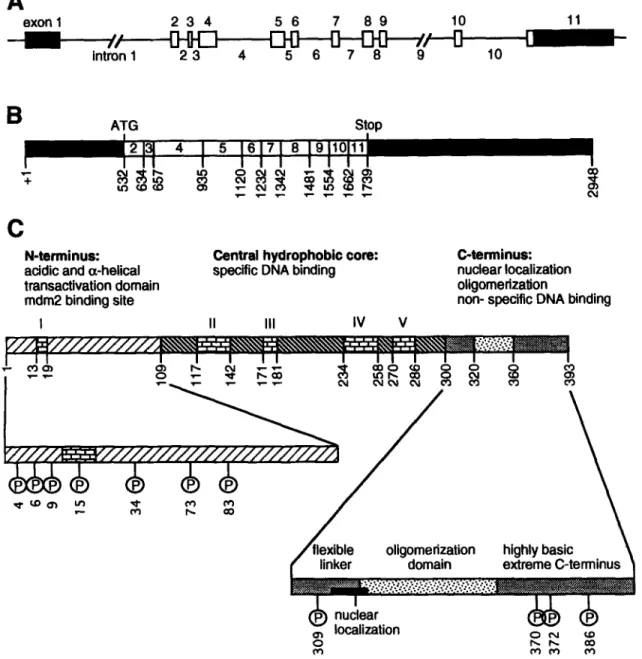
The TP53 gene consists of 11 exons and 10 introns (Fig. 1A) spanning 20 kb of genomic DNA on the short arm of chromosome 17, at 17p13.1 (3). TP53 encodes a 2.8 kb mRNA, which is translated into a 53-kDa nuclear phosphoprotein that acts as a transcription factor (4,5) and functions as a tumor suppres- sor (6).
Regulation
ofTP53
Gene Expression
Different regulatory sites have been mapped in the 5' upstream region of the TP53 gene, in exon 1 and in intron I (7). Analysis of the TP53
promoter region evidenced the lack of TATA and CAAT sequences and the presence of differ- ent recognition sites for oncogenic transcription factors such as NF1, jun (8), and Myc/Max (9).
p53 may have autoregulatory functions: responsive elements are present on the TP53
promoter and reporter constructs under the control of these elements are downregulated by p53 (10). Nevertheless, there is still no evi- dence for p53 binding to its own promoter.
Recently, a PAX-5 binding site was identified within the untranslated first exon of TP53 (11).
PAX genes encode nuclear transcription factors implicated in the control of mammalian embryogenesis. In particular, PAX-2, 5, and -8 are highly expressed during the development of the CNS. It has been hypothesized that PAX- 5 may have a physiological downregulatory function on TP53 expression in order to allow proliferation of stem cells that migrate from the ventricular zone to the intermediate zone of the neural tube. Stuart et al. (11) reported that PAX-5 is often overexpressed in high- grade gliomas, suggesting that constitutive
expression of PAX-5 may allow tumors to bypass the need for p53 mutation to abrogate p53-mediated processes. However, subsequent work failed to show a correlation between PAX5 expression and p53 status in a series of patients showing recurrence from low-grade astrocytoma to glioblastoma (E.T. Stuart and E. G. Van Meir, unpublished data). In addition, whereas PAX5 overexpression was confined to late stage glioblastoma, p53 mutations are known to occur early in the progression of astrocytoma.
Other proteins identified as potential induc- ers of TP53 transcription are ETS1 and ETS2 factors (12) and the C7 poxviral transcription factor (13).
p53 Protein
Functional p53 is a nonglobular tetramer of a single peptidic chain that comprises three functionally characterized segments (Fig. 1C): the N-terminal transcription activation region, a central core responsible for specific DNA binding and a C-terminal oligomerization sequence.
Exon 11 of p53 mRNA codes for the very C- terminal region of the protein that is a negative regulator of the protein's activity (14). This ter- minal segment of p53 seems to interact some- how with the core of the protein and prevents p53-DNA binding. This allosteric model for negative regulation of p53 activity by the C- terminus is confirmed by different experimen- tal data showing that disruption of this interaction, by both physiological and artificial events, induces a conformational change that results in p53 activation (14,15).
Moreover, five highly conserved and inde- pendently folding domains (I-IV) can be dis- tinguished in the p53 protein (Fig. 1C).
Crystallography shows that the central core of p53 is a sandwich of two antiparallel sheets formed of four and five ~ strands and a loop sheet helix motif packed against the sandwich (16). The overall structure of the sandwich is stabilized by two large loops held together by a Zn atom.
I intron 1 2 3 4 5 6 7 8
/,"
9Et
10B
ATG Stop GO O4C
N-terminus: acidic and (x-helical transacUvation domain mdm2 binding siteI
Central hydrophobic core: specific DNA binding
II III IV V
C-terminus: nuclear localization oUgomerization
non- specific DNA binding
I I I I I I I I I I I I I
Fig. 1. Schematic representation of the p53 gene, mRNA and protein structures. (A) TP53 gene structure: boxes stand for exons (white parts are translated, black ones are not) and segments for introns. Numbers of exons and introns are indicated. (B) mRNA structure: nucleotide positions at splicing junctions of exons 1-11 are indicated. Start (ATG) and stop codons are also shown. The predicted protein size from mRNA is 43.5 kDa. (C) Protein structure: amino acid localization of the five evolutionarily conserved domains (I-V) are shown. Enlargements of the N- and C-termini show the phosphorylation sites in the mouse (human phosphorylation sites are similar) and the distinct functional domains.
Although the 13 sandwich is an uncommon structure for transcription factors, the loop sheet helix and the two large loops are typical motifs of proteins capable of complexing with DNA. In fact, crystallographic studies of the
p53/DNA complex demonstrated that the loop sheet helix motif binds to the DNA's major groove. One of the loops binds to the minor groove, and the other has an important role in stabilizing the overall structure of the complex.
64
Fulci and Van Meir
60 5O (n p. .o.,., 40 E 3O tO Q . ,I0 E lO c- O conserved domainsloop sheet helix motif involved in contact with DNA major groove loop 3 for stabilization of the protein folding
loop 2 involved in contact with ~ DNA minor groove
175 I I I I 0 3 03 0 3 0 3 U3 (D I ~ 00 248
L J j ,
273llJ=._ _1
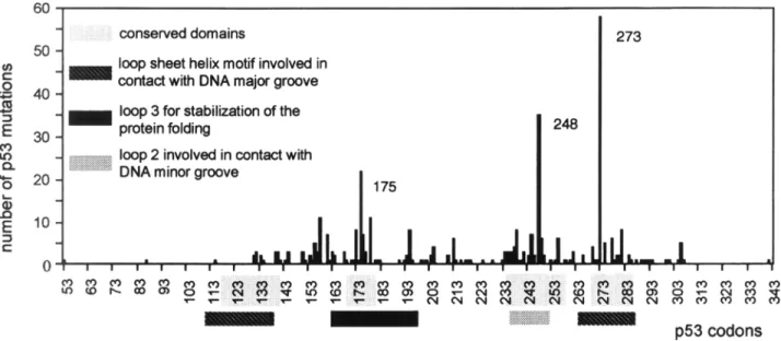
9 - - d ~ I I I I I I I I I I I I I I I I I I l I I I I p53 codonsFig. 2. Frequencies of p53 exon mutations found in brain tumors reported by the IARC database: http://www.iarc.fr. Regions implicated in DNA interaction and protein stability as well as the conserved domains of the protein are indicated.
Moreover, it has been demonstrated that these motifs are the sites of the most frequently mutated p53 codons found in tumors and cor- respond to the highly conserved domains of the protein, suggesting that these regions have a biological importance (Fig. 2).
p53 accomplishes its biological roles either by interaction with other proteins or acting as a transcription factor. It enhances transcription of specific genes by binding to promoters contain- ing four copies of the p53 pentameric consensus sequence PuPuPuC(A/T)-(T/A)GPyPyPy with a stoichiometry of one core domain for one pen- tamer (17).
Three factors have been shown to act as p53 coactivators and potentiate its transcriptional activity: the nuclear tyrosine kinase
c-abl (18)
and the two homologous transcription factors p300 and CBP, all of which are implicated in cell proliferation and differentiation. The syn- ergistic transcription activity of p300/CBP and p53 is permitted by physical interaction between the proteins
(19-21).
More recent data indicate that acetylation of the p53 C-terminal domain by its coactivator p300 dramaticallyincreases sequence-specific DNA binding of p53
(22)
(Fig. 3).p53 is also able to repress transcription of genes containing TATA boxes by binding, through its oligomerization domain, to the TATA binding protein (TBP) and preventing its interaction with DNA
(23).
Although it has been shown that genes that have neither TATA boxes nor p53 consensus sequences are not submitted to direct p53 regulation(24),
more recent data indicate that p53 inhibits the tran- scription of genes containing an AP1 site by sequestering the transcription factor p300 (19).Regulation of p53 Activity
Responses to Physiological Stress
p53 is a sensor of a variety of physiological stress conditions to the cell such as hypoxia, heat and starvation
(25,26).
The response to such insults is either growth arrest or apopto- sis (Fig. 3). Graeber et al.(25)
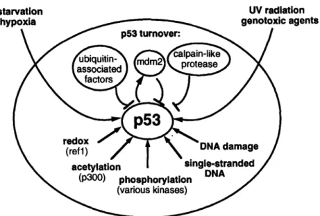
compared the mechanisms and the effects of p53 induction by different types of stress. Their experimentsstarvation UV radiation
hypoxia genotoxic agents
Fig. 3. Schematic representation of factors regulating p53 activity.
Arrows,
induction; T-bars indicate repression.showed that heat induces a mutant conforma- tion on p53 allowing it to complex with heat shock protein hsc70 and to increase its half-life. This is accompanied by cytoplasmic instead of nuclear p53 accumulation during high temper- ature shifts. On the other hand, p53 accumu- lates in the nucleus during hypoxia, and an increased p53 transactivational activity can be observed. Recent data also suggest that p53 might be stabilized by an interaction with hypoxia inducible factor (HIF-1)
(27).
Sensing of DNA Damage
DNA damaging agents such as ionizing radiation
(28)
and genotoxic and antimicro- tubule agents(29,30)
induce p53 expression (Fig. 3). The precise sequence of events that link DNA damage to p53 induction is unknown, but involves the ATM gene product(31)
and the DNA repair machinery. Within this context, it is interesting to mention the capacity of p53 to bind repair proteins XPD and XPB(32).
Moreover, in vitro experiments have shown that single stranded DNA, within the size range generated during excision repair, stimu- lates p53-DNA binding
(33-35).
When increased, p53 may function as a tumor suppressor by inducing tumor cell apoptosis. This in turn may lead to clonal selection in vivo of cells lacking wild-type (wt) p53
(36).
As a consequence, anticancer treat- ments inducing necrosis and DNA damage, such as radio- and chemotherapy, could also favor the expansion of p53 mutated cells and induce malignant progression(37).
Phosphorylation
p53 has multiple phosphorylation sites both in its N- and C-termini (Fig. 1C). The N-ter- minal phosphorylation seems to be preferen- tially accomplished by cyclin-dependent kinases, double-stranded DNA activated pro- tein kinase, mitogen-activated protein kinase, Jun N-terminal kinase and Raf kinase; whereas the C-terminal domain is phosphorylated by cyclin-dependent kinases, casein kinase II, pro- tein kinase C (PKC), and the CDK7-cyclinH- p36 complex of TFIIH
(38-40).
Even though there is considerable evidence that phosphorylation regulates p53 activity in vivo
(41),
it is unclear how the cited kinases affect p53. Different single p53 phosphoryla-66
Fulci and Van Meir
tion mutants were tested for their transcription activity and for their capacity to suppress cell proliferation, but no difference was found in comparison with wt p53
(42).
By contrast, in some cases, altering two or more phosphoryla- tion sites at once could significantly lessen p53 transactivation potential or DNA binding, or both(40).
Furthermore, the conformation of p53 is determinant for its availability as a substrate for different kinases and for the phosphoryla- tion pattern generated by the same kinase
(43).
This information is coherent with the observa- tion that the phosphorylation status of human p53 at serines 15 and 392 was found to be dif- ferent between the wt and a conformational mutant p53 in glioblastoma cells. In particular, phosphorylation of serine 15 was reduced in the mutant p53 compared with wt, while phosphorylation of ser 392 was increased
(44).
Taken together, these data indicate that it is not possible to consider phosphorylation as a simple on/off switch for p53 functions, but it is rather a complex regulatory system that could also be cell type dependent. Recent data indi- cate that phosphorylation of N-terminal ser- ines 15 and 37 by DNA-PK is induced after DNA damage and inhibits p53 interaction with MDM2, resulting in p53 activation
(45).
Phos- phorylation of the C-terminus of p53 by casein kinase II was also shown to activate p53-DNA binding in vitro(14).
Moreover, p53 hyper- phosphorylation by oncogene activation of the MAPK pathway may be the molecular basis for the ability of p53 to sense oncogene trans- formation in the cell and exert its antionco- genic effect(46).
Redox State of the Protein
Another effector of p53 activity is the redox state of the protein (Fig. 3). It is now appreciated that oxydization renders p53 unable to bind DNA, whereas reduction enhances this capac- ity. According to this idea, the redox repair pro- tein Ref 1, which was shown to activate AP-1 by reducing a conserved cystein in its DNA bind- ing domain, has been recently identified as a potent activator of p53 both in a redox-depen- dent and an -independent manner
(47,48).
MDM2 Binding
The activity of p53 is downregulated in an autoregulatory feedback loop: p53 induces the transcription of the
MDM2
gene and the accumulation of the oncoprotein mdm2 represses p53 (Fig. 3). The delay between p53 induction andMDM2
activation defines the time during which p53 can exert its activity(49,50).
This mechanism is used by certain tumors, including osteosarcomas(51)
and about 10% of astrocytomas(52)
to inactivate p53 byMDM2
gene amplification. A novel player in p53 regulation was identified recently: p14 ARF, the product of a gene com- monly deleted in human cancer, was found to inhibit mdm2 function. The absence of p14 ARF increases the availability of mdm2 for p53 downregulation, which should favor cellular transformation(53).
p53 Turnover
The most apparent and important regulatory mechanism of the activity displayed by p53 is its rapid turnover (half-life of about 20-30 min), which limits the quantity of wt p53 in the nucleus (Fig. 3).
One of the mechanisms that regulate p53 turnover is the ubiquitin-dependent pathway (Fig. 3). In human papillomavirus-infected cells a tripartite complex forms between the viral protein E6, p53, and the cellular protein E6-AP and targets p53 for ubiquitin-dependent proteolysis (54). Accordingly, p53 was shown to accumulate in cell lines lacking ubiquitin- dependent mechanisms
(55).
Nevertheless, the precise molecular mechanisms of this pathway and its general applicability are unclear.Recent experiments suggest that p53 turnover may also be accomplished by an ubiquitin degradation pathway independent of E6-AP. It was shown that E6-AP has no effect on p53 lev- els in nonvirus-infected cells
(56)
and that mdm2 is able to induce p53 degradation by a proteasome complex(57,58).
These data are confirmed by recent results showing that p53 mutants that are unable to bind mdm2 are more stable than other p53 mutants or the wt protein. Moreover, treatment of tumor cells containingAngiogenesis
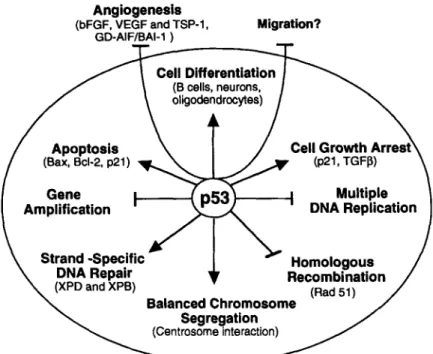
(bFGF, VEGF and TSP-1,
GD-AIF/BAI-1
)
Migration?~~~'C el~ Dc
'sff: rnentr
ioat:~
Fig. 4. Schematic representation of p53 functions. Arrows, induction; %bars indicate inhibition.
wt p53 with agents that disrupt the p53-mdm2 interaction induces p53 accumulation (59).
Finally, a completely different mechanism for p53 degradation has been recently observed. In this case, p53 turnover is regulated by a cal- pain-like protease
(60)
(Fig. 3).p53: "Guardian" of the Genome
One of the most important functions of p53 in cells is maintenance of genomic stability. This term encompasses all the mechanisms that ensure transmission of an intact genome from parental to descendent cells during mitosis. DNA replication, repair, and chromosome seg- regation should occur in an orderly fashion, and biochemical signal transduction pathways must ensure completion of each step before entering subsequent phases. Some of these pathways, known as cell cycle checkpoints, involve p53, either acting as a transcription factor or by its interaction with other proteins, p53 can thus be a direct participant in maintaining DNA integrity by acting in the mechanisms of homol-
ogous and illegitimate recombination
(61-63),
DNA repair
(32),
DNA replication(64-66),
gene amplification(67),
and chromosome segrega- tion(68-70).
p53 can also indirectly maintain DNA integrity by sensing abnormal cell cycle progression and maintaining cell populations with intact genomes through cell cycle arrest (allowing for DNA repair) or by eliminating damaged cells (inducing senescence or by stim- ulating apoptosis) (Fig. 4).Loss of p53 Function in Cell Growth
Arrest, Differentiation,
or Apoptosis: Abnormal CNS
Development and Neoplastic
Transformation
p53 plays a central role in determining whether a cell must undergo differentiation, senescence, or apoptosis.
p53-dependent cell cycle arrest and apopto- sis can be seen as indirect mechanisms by
68
Fulci and Van Meir
which p53 accomplishes maintenance of genomic stability. Both pathways are induced by DNA damage, the ultimate result of which is the repair or elimination of DNA damaged cells. Therefore, they are two important mecha- nisms avoiding tumor initiation or progres- sion, or both. p53-dependent apoptosis and cell differentiation are two crucial pathways of CNS development. Lack of p53 can sometimes result in severe developmental abnormalities leading to exencephaly.
p53 and Cell Cycle Arrest
p53 is able to block the cell cycle both at the G1 checkpoint and at the G2/M transition
(71-73). The
loss of cell cycle control functions normally assumed by p53 is believed to con- tribute to tumor development in the CNS. Upon p53 restoration in glioma cells cell cycle arrest is observed, which can be either a reversible arrest or an irreversible senescent-like event(74,75).
At the molecular level, p53-dependent growth arrest is induced by p53 transactivating differ- ent cell cycle regulators, such as p21 and TGF-~(76-79).
p21 arrests the cell cycle by inhibiting the activity of cyclin-dependent kinase com- plexes(78)
and is the most important p53- induced cell cycle regulator. Indeed, human glioblastoma cells lacking p53 genes (LN-Z308) have low levels of p21, and transfection of wt p53 in such cells activates p21 expression and inhibits cell growth(80,81).
Direct transfer ofCDKN1,
the gene encoding p21 in different glioma cell lines (U373MG, U87MG, GB-1), also induces growth arrest(82).
Moreover, p21 over- expression is accompanied by a diminished malignant phenotype, as demonstrated in vivo using peripheral and intracerebral xenograft models(82).
Accordingly, the introduction of theCDKN1
gene in a rat glioma cell line was shown to induce growth arrest, cell susceptibil- ity to radiation, and tumor necrosis(83).
Never- theless, the absence ofCDKN1
gene mutations in human gliomas suggests that it cannot be considered a tumor suppressor gene(84).
In other tumor models, it was shown that overexpression of one of the growth arrest spe-
cific genes (gas1) blocks cell proliferation in a p53-dependent manner, although p53 transac- tivating function was dispensable in this case
(85).
This may be related to the fact that p53 is also responsible for the transcriptional repres- sion of different cell cycle inducer genes through its interaction with the TATA box binding protein(23),
but these factors have not yet been studied in brain tumors.p53 and Apoptosis
It is now common knowledge that p53 is able to trigger apoptosis in different human cells including undifferentiated neurons, oligo- dendrocytes and glioma cell lines
(86-89).
This mechanism is essential for the development of the central nervous system as well as in tumor prevention and treatment. Different stimuli like DNA damage, myc and adenovirus EIA expression and withdrawal of growth factors can trigger p53-dependent apoptosis (1).Studies made on the molecular mechanisms of p53-dependent apoptosis result in unclear and contradictory data. Both the transactivating function of p53 and its capacity to repress gene transcription seem to be important to accom- plish apoptosis
(90,91).
Indeed, p53 induces the expression of Bax and represses Bcl-2(92,93)
and, in a human glioblastoma, it was shown that p21 is a downstream mediator in p53- dependent apoptosis
(94).
Recentl~ it was pro- posed that p53 transactivating function can trigger apoptosis through induction of redox- controlling genes, which in turn increases reac- tive oxygen species (ROS), causing oxidative damage, which produces apoptosis(95).
Other data indicate that p53 transactivation function is dispensable for induction of apop- tosis
(96).
Deletion of the N-terminal proline- rich domain of p53 abolishes p53-induced apoptosis, although the protein retains its transactivation capacity. This p53 domain could probably be determinant in inducing apoptosis by interacting with other proteins through its Pro-X-X-Pro motifs(97,98).
p53-dependent apoptosis plays an important role in the development of the CNS, when mat-
uration of the CNS involves massive scale death of neurons.
In vitro experiments show that apoptotic death of undifferentiated neurons and of oligo- dendrocytes is p53-dependent
(86-88,99,100).
High levels of Bax and reduced levels of Bcl-2 are found in some neurons before ischemic death; therefore, it is likely that a change in bal- ance between these two molecules is a key event in p53-mediated neuronal death
(87).
In vivo, a significant fraction of p53 knock- out mice were found to die before birth. Analy- sis of these embryos showed that about 20% present a failure in closure of the neuronal tube, which results in exencephaly followed by anencephaly
(101,102).
The normal develop- ment of surviving p53 knockout mice shows that, in brain development, the missing p53 function is probably substituted by other pro- teins, perhaps one of the recently cloned p53 family members(103,104).
Because p53 is associated with neuronal damage and is involved in apoptotic death of oligodendrocytes
(86),
clarifying the role of p53 in the processes underlying neuronal and oligodendrocytic death should provide novel information in the understanding of CNS development, but also of certain CNS patholo- gies/injuries. Degenerative disorders, includ- ing Alzheimer's disease and brain trauma, involve neuronal cell death(87,
and references therein), and multiple sclerosis is characterized by oligodendrocytic death (105). Accordingly, p53-dependent apoptosis seems to be the major cause of adrenalectomy-induced degen- eration of hippocampal granule cells(106).
p53 in Differentiation
Clues as to the involvement of p53 in the dif- ferentiation processes are given by the obser- vation of induction of several differentiation markers after p53 overexpression. For exam- ple, immunoglobulin chains ~t and K are induced in pre-B cells after p53 induction
(107).
Within the context of the CNS p53 acts as a regulatory protein for the differentiation of
neurons and oligodendrocytes in vitro. Subcel- lular localization of p53 from cytoplasm to nucleus occurs in oligodendrocyte progenitors at 24 h after the addition of differentiating medium. Subsequently, p53 nuclear staining decreases to basal levels in fully differentiated cells
(87).
These results were confirmed by observation of a block in neurite extension (marker of oligodendrocyte differentiation) after adding a dominant negative p53 protein to the cells(87). The
physiological mechanism(s) by which differentiation signals may mediate nuclear translocation are unknown. Once in the nucleus, p53 may control differentiation by transcriptionally activating a specific subset of differentiation-related genes.To examine when and where p53 might be important in development, p53 expression and transcriptional activity was followed during mice embryonic development using transgenic mice harboring a lacZ gene under the control of a p53-regulated promoter. The resulting data indicate that p53 is highly expressed in the early developing CNS, in undifferentiated cells, in neuroblasts, and in neurons
(99,100,108).
These results support a role for p53 in certain stages of CNS development, although the precise topog- raphy and dynamics of p53 expression in differ- ent brain areas have not yet been defined.p53 Mutations in Tumors of the CNS
Analysis of
TP53
gene status and overexpres- sion of its protein product have been well docu- mented in primary CNS tumors (Fig. 2). Although the entireTP53
gene is a good target for all different types of mutations, such as dele- tions, insertions, transitions, and transversions, a differential distribution of these classes of mutations can be observed along the gene. Although alterations truncating the protein, such as insertions and deletions, were found along the whole gene, point mutations that alter p53 function seem to be situated only in the hydrophobic core of the protein (87% in exons 5-8), where single base substitutions can com- promise the protein conformation or function,70
Fulci and Van Meir
or both. The lack of point mutations outside thecore of the protein is probably also the result of the fact that most investigators have focused their analyses on exons 5-8
(109).
Additionally, of 250 potential sites for mutations present inthe TP53
gene, 25% of all mutations found in human tumors cluster at codons 175, 245, 248, 249, and 273. The preferential alteration of par- ticular sites of a gene can have different reasons: (1) they may occur for structural and chemical reasons (repetitive sequences or CpG dinu- cleotides and susceptibility to carcinogenic agents); (2) they may be due to biochemical problems related to the transcription and repair machinery; (3) they may be biologically moti- vated; for example, some mutants may give a growth advantage to the cells.Crystallographic studies distinguished two types of mutation sites. Some are directly involved in DNA binding and include the hot spot codons 248 and 273, whereas others are required for the stable folding of the protein such as codon 175. Moreover, this analysis evidenced that the frequency of TP53 point mutations decreases at increased distances from the biologically important structures of the gene product
(16).
Statistical analysis of
TP53
gene mutations found in tumors of the CNS has shown thatTP53
mutations are mostly restricted to tumors of astrocytic origin (33%)(110).
Recent data obtained with a more sensitive protocol to detect p53 mutations show even higher fre- quencies: 67% in anaplastic astrocytoma and 41% in glioblastoma multiforme(111).
Lower mutation frequencies are found in glioblas- toma, because p53 mutation appears to occur preferentially only in some subtypes(112,113).
TP53
gene mutations also occur frequently in primary CNS lymphomas (30%)(114)
but are quite rare in oligodendroglioma (13%) and in medulloblastoma (11%) and are apparently absent in other tumors of the CNS.Most
TP53
gene alterations are spontaneous GC-AT transversions arising by deamination of 5' methylcytosine at CpG sites. There are no brain tumor-specific mutations; the three most frequent alterations are at codons 175, 248, and273. The frequency of mutations differs some- what since mutation at codon 273 is predomi- nant, whereas in other human tumors the most frequently mutated codons are 248, 249, and 175
(115).
Nevertheless, there are still no data indicating a specific role of these mutants in the genesis of astrocytic tumors.More contradictory data were obtained in an effort to understand whether p53 mutation is an initial, early or late event in glioma tumori- genesis. Evidence for p53 mutation as an initia- tion event in glioblastoma derives from the finding of brain tumors in patients with germline p53 mutations, such as occurs in patients with multifocal glioma
(116)
and in families with Li-Fraumeni syndrome. These groups present a high incidence of tumors of the CNS (13%), most of which are astrocy- tomas (73%)(117).
Additionally, the pattern of mutations in sporadic and inherited brain tumors is similar. The in vitro transformation of spontaneously immortalized cortical astro- cytes of p53 knockout mice(118,119)
gives additional strength to the theory that p53 mutation is an early initiation event in the for- mation and progression of astrocytomas. As a result of the early death of p53 knockout mice attributable to lymphomas and sarcomas, the causality of loss of p53 wt function in CNS tumors could not be demonstrated.Genetic analysis of cell lines derived from gliomas induced in rats by N-ethyl-N-nitro- sureas (ENU) showed high frequency of p53 mutations
(120,121)
in domains II-V(120,121).
Immortalization and transformation of astroglial cells can also occur independent of p53 muta- tion(120,121).
Either cell immortalization is induced by genes other thanTP53,
or p53 mutation is associated with later stages of tumorigenesis, or both.Conclusions
During the past 10 years, the importance of p53 in tumor research has increased dramati- cally and p53 has become involved in a wide range of functions: safeguard of genome stabil-
ity, cell cycle arrest, differentiation, apoptosis, angiogenesis, and t u m o r cell invasion. Addi- tionally, both biological and clinical analysis of p53 in tumors has demonstrated its particular importance in the most malignant type of pri- mary brain tumor: astrocytoma. By contrast, for example, to carcinomas in which p53 muta- tion is a late event in the formation of the tumor, in astrocytoma there is considerable evidence that p53 alteration occurs early in the progression of the disease. Moreover, although the primary characterized function of p53 is as an oncosuppressor, in the brain p53 m a y also function as a regulator of CNS development. Lack of p53 may cause exencephaly, although in this disease, p53 absence can, in some cases, be compensated by other proteins. In both brain tumors and exencephaly, the capacity of p53 to induce apoptosis m a y be determinant in causing the disease. In the first case, as a way to eliminate genetically d a m a g e d cells; while in the second case, as a normal physiological process essential to the orderly d e v e l o p m e n t of the CNS. Therefore, the comprehension of p53 structure and regulation, as well as the molec- ular pathways of p53-dependent apoptosis, cell cycle arrest, and differentiation should bring us closer to gaining molecular insights into these severe diseases of the CNS.
Acknowledgments
The authors w o u l d like to thank Larry R. Paul and Laura H. Matthews for expert editor- ial assistance.
References
1. Ko, L. J. and Prives, C. (1996) p53--Puzzle and paradigm. Genes Dev. 10, 1054-1072.
2. Levine, A. J. (1997) p53, the cellular gatekeeper for growth and division. Cell 88, 323-331. 3. Lamb, P. and Crawford, L. (1986) Characteri-
zation of the human p53 gene. Mol. Cell. Biol. 6,
1379-1385.
4. Farmer, G., Friedlander, P., Colgan, J., Manley, J. L., and Prives, C. (1996) Transcriptional repres-
sion by p53 involves molecular interactions dis- tinct from those with the TATA box binding protein. Nucleic Acids Res. 24, 4281-4288. 5. Fields, S. and Jang, S. K. (1990) Presence of a
potent transcription activating sequence in the p53 protein. Science 249, 1046-1049.
6. Raycroft, L., Wu, H. Y., and Lozano, G. (1990) Transcriptional activation by wild-type but not transforming mutants of the p53 anti-onco- gene. Science 249, 1049-1051.
7. Reisman, D., Greenberg, M., and Rotter, V. (1988) Human p53 oncogene contains one pro- moter upstream of exon 1 and a second, stronger promoter within intron 1. Proc. Natl.
Acad. Sci. USA 85, 5146-5150.
8. Ginsberg, D., Oren, M., Yaniv, M., and Piette, J. (1990) Protein-binding elements in the pro- moter region of the mouse p53 gene. Oncogene
5, 1285-1290.
9. Reisman, D., Elkind, N. B., Roy, B., Beamon, J., and Rotter, V. (1993) c-Myc transactivates the p53 promoter through a required downstream CACGTG motif. Cell Growth Diff. 4, 57-65. 10. Deffie, A., Wu, H., Reinke, V., and Lozano, G.
(1993) The tumor suppressor p53 regulates its own transcription. MoI. Cell. Biol. 13, 3415--3423. 11. Stuart, E. T., Haffner, R., Oren, M., and Gruss, P. (1995) Loss of p53 function through PAX- mediated transcriptional repression. EMBO J. 14, 5638-5645.
12. Venanzoni, M. C., Robinson, L. R., Hodge, D. R., Kola, I., and Seth, A. (1996) ETS1 and ETS2 in p53 regulation: Spatial separation of ETS binding sites (EBS) modulate protein: DNA interaction. Oncogene 12, 1199-1204.
13. Wali, A. and Strayer, D. S. (1996) Regulation of p53 gene expression by a poxviral transcrip- tion factor. Virology 224, 63-72.
14. Hupp, T. R., Meek, D. W., Midgley, C. A., and Lane, D. P. (1992) Regulation of the specific DNA binding function of p53. Cell 71, 875-886. 15. Selivanova, G., Iotsova, V., Okan, I., Fritsche, M., Strom, M., Groner, B., Grafstrom, R. C., and Wiman, K. G. (1997) Restoration of the growth suppression function of mutant p53 by a synthetic peptide derived from the p53 C- terminal domain. Nature Med. 3, 632-638. 16. Cho, Y., Gorina, S., Jeffrey, P.D., and Pavletich,
N. P. (1994) Crystal structure of a p53 tumor suppressor-DNA complex: Understanding tumorigenic mutations. Science 265, 346-355. 17. E1-Deiry, W. S., Kern, S. E., Pietenpol, J. A.,
Kinzler, K. W., and Vogelstein, B. (1992) Defin-
72 Fulci and Van Meir
ition of a consensus binding site for p53.
Nature Genet. 1, 45-49.
18. Goga, A., Liu X., Hambuch, T. M., Senechal, K., Major, E., Berk, A. J., Witte, O. N., and Sawyers, C. L. (1995) p53 dependent growth suppression by the c-Abl nuclear tyrosine kinase. Oncogene 11, 791-799.
19. Avantaggiati, M. L., Ogryzko, V., Gardner, K., Giordano, A., Levine, A. S., and Kelly, K. (1997) Recruitment of p300/CBP in p53-dependent signal pathways. Cell 89, 1175-1184.
20. Gu, W., Shi, X. L., and Roeder, R. G. (1997) Synergistic activation of transcription by CBP and p53. Nature 387, 819-823.
21. Lill, N. L., Grossman, S. R., Ginsberg, D., Decaprio, J., and Livingston, D. M. (1997) Binding and modulation of p53 by p300/cbp coactivators. Nature 387, 823-827.
22. Gu, W. and Roeder, R. G. (1997) Activation of p53 sequence-specific DNA binding by acety- lation of the p53 C-terminal domain. Celt 90,
595-606.
23. Seto, E., Usheva, A., Zambetti, G. P., Momand, J., Horikoshi, N., Weinmann, R., Levine, A. J., and Shenk, T. (1992) Wild-type p53 binds to the TATA-binding protein and represses tran- scription. Proc Natl Acad Sci USA 89, 12,028-12,032.
24. Mack, D. H., Vartikar, J., Pipas, J. M., and Laimins, L. A. (1993) Specific repression of TATA-mediated but not initiator-mediated transcription by wild-type p53. Nature 363, 281-283.
25. Graeber, T. G., Peterson, J. F., Tsai, M., Monica, K., Fornace, A. J., and Giaccia, A. J. (1994) Hypoxia induces accumulation of p53 protein, but activation of a Gl-phase checkpoint by low-oxygen conditions is independent of p53 status. Mol. Cell. Biol. 14, 6264-6277.
26. Ohnishi, T., Wang, X., Ohnishi, K., Matsumoto, H., and Takahashi, A. (1996) p53-dependent induction of WAF1 by heat treatment in human glioblastoma cells. J. Biol. Chem. 271,
14,510-14,103.
27. An, W. G., Kanekal, M., Simon, M. C., Maltepe, E., Blagosklonny, M. V., and Neckers, L. M. (1998) Stabilization of wild-type p53 by hypoxia-inducible factor 1-alpha. Nature 392, 405-408.
28. Maltzman, W. and Czyzyk, L. (1984) UV irra- diation stimulates levels of p53 cellular tumor antigen in nontransformed mouse cells. Mol.
Cell. Biol. 4, 1689-1694.
29. Kastan, M. B., Onyekwere, O., Sidransky, D., Vogelstein, B., and Craig, R. W. (1991) Partici- pation of p53 protein in the cellular response to DNA damage. Cancer Res. 51, 6304-6311. 30. Yuan, J. N., Liu, B. H., Lee, H., Shaw, Y. T.,
Chiou, S. T., Chang, W. C., and Lai, M. D. (1993) Release of the p53-induced repression on thymidine kinase promoter by single p53- binding sequence. Biochem. Biophys. Res. Com-
mun. 191, 662-668.
31. Kastan, M. B., Zhan, Q., el Deiry, W. S., Carrier, E, Jacks, T., Walsh, W. V., Plunkett, B. S., Vogel- stein, B., and Fornace, A. J. (1992) A mam- malian cell cycle checkpoint pathway utilizing p53 and GADD45 is defective in ataxia-telan- glectasia. Cell 71, 587-597.
32. Wang, X. W., Yeh, H., Schaeffer, L., Roy, R., Moncollin, V., Egly, J. M., Wang, Z., Freidberg, E. C., Evans, M. K., Taffe, B. G., et al. (1995) p53 modulation of TFIIH-associated nucleotide excision repair activity. Nature Genet. 10, 188-195.
33. Bakalkin, G., Yakovleva, T., Selivanova, G., Magnusson, K. P., Szekely, L., Kiseleva, E., Klein, G., Terenius, L., and Wiman, K. G. (1994) p53 binds single-stranded DNA ends and catalyzes DNA renaturation and strand transfer. Proc. Natl. Acad. Sci. USA 91, 413-417.
34. Jayaraman, J. and Prives, C. (1995) Activation of p53 sequence-specific DNA binding by short single strands of DNA requires the p53 C-terminus. Cell 81, 1021-1029.
35. Selivanova, G., Iotsova, V., Kiseleva, E., Strom, M., Bakalkin, G., Grafstrom, R. C., and Wiman, K. G. (1996) The single-stranded DNA end binding site of p53 coincides with the C-termi- nal regulatory region. Nucleic Acids Res. 24, 3560-3567.
36. Graeber, T. G., Osmanian, C., Jacks, T., Hous- man, D. E., Koch, C. J., Lowe, S. W., and Giac- cia, A. J. (1996) Hypoxia-mediated selection of cells with diminished apoptotic potential in solid tumours. Nature 379, 88-91.
37. Van Meir, E. G. (1996) Hypoxia-mediated selection of cells with diminished apoptotic potential in solid tumours. Neurosurgery 39, 878-879.
38. Lu, H., Fisher, R. P., Bailey, P., and Levine, A. J. (1997) The CDK7-cycH-p36 complex of tran- scription factor IIH phosphorylates p53, enhancing its sequence-specific DNA binding activity in vitro. Mol. Cell. Biol. 17, 5923-5934.
39. Meek, D. W., Simon, S., Kikkawa, U., and Eck- hart, W. (1990) The p53 tumour suppressor protein is phosphorylated at serine 389 by casein kinase II. EMBO J. 9, 3253-3260.
40. Milczarek, G. J., Martinez, J., and Bowden, G. T. (1997) p53 phosphorylation-biochemical and functional consequences. Life Sci. 60, 1-11. 41. Ullrich, S. J., Mercer, W. E., and Appella, E. (1992) Human wild-type p53 adopts a unique conformational and phosphorylation state in vivo during growth arrest of glioblastoma cells. Oncogene 7, 1635-1643.
42. Fuchs, B., O'Connor, D., Fallis, L., Scheidt- mann, K. H., and Lu, X. (1995) p53 phosphory- lation mutants retain transcription activity.
Oncogene 10, 789-793.
43. Adler, V., Pincus, M. R., Minamoto, T., Fuchs, S. Y., Bluth, M. J., Brandtrauf, P. W., Friedman, F. K., Robinson, R. C., Chen, J. M., Wang, X. W., Harris, C. C., and Ronai, Z. (1997) Conforma- tion-dependent phosphorylation of p53. Proc.
Natl. Acad. Sci. USA 94, 1686-1691.
44. Ullrich, S. J., Sakaguchi, K., Lees, M. S., Fis- cella, M., Mercer, W. E., Anderson, C. W., and Appella, E. (1993) Phosphorylation at Ser-15 and Ser-392 in mutant p53 molecules from human tumors is altered compared to wild- type p53. Proc. Natl. Acad. Sci. USA 90, 5954-5958.
45. Shieh, S. Y., Ikeda, M., Taya, Y., and Prives, C. (1997) Dna damage-induced phosphorylation of p53 alleviates inhibition by mdm2. Cell 91, 325-334.
46. Fukasawa, K. and Vande, W. G. (1997) Synergy between the Mos/mitogen-activated protein kinase pathway and loss of p53 function in transformation and chromosome instability.
Mol. Cell. Biol. 17, 506-518.
47. Hainaut, P. and Milner, J. (1993) Red0x modu- lation of p53 conformation and sequence-spe- cific DNA binding in vitro. Cancer Res. 53, 4469-4473.
48. Jayaraman, L., Murthy, K., Zhu, C., Curran, T., Xanthoudakis, S. and Prives, C. (1997) Identifi- cation of redox/repair protein ref-1 as a potent activator of p53. Genes Dev. 11, 558-570. 49. Montes de Oca Luna, R, Wagner, D. S., and
Lozano, G. (1995) Rescue of early embryonic lethality in mdm2-deficient mice by deletion of p53. Nature 378, 203-206.
50. Picksley, S. M. and Lane, D. P. (1993) The p53- mdm2 autoregulatory feedback loop: A para-
digm for the regulation of growth control by p53?. BioEssays 15, 689-690.
51. Lonardo, F., Ueda, T., Huvos, A. G., Healey, J., and Ladanyi, M. (1997) p53 and MDM2 alter- ations in osteosarcomas: Correlation with clin- icopathologic features and proliferative rate.
Cancer 79, 1541-1547.
52. Reifenberger, G., Liu, L., Ichimura, K., Schmidt, E. E., and Collins, V. P. (1993) Ampli- fication and overexpression of the MDM2 gene in a subset of human malignant gliomas with- out p53 mutations. Cancer Res. 53, 2736-2739. 53. Zhang, Y. P., Xiong, Y., and Yarbrough, W. G.
(1998) Arf promotes mdm2 degradation and stabilizes p53--arf-ink4a locus deletion impairs both the Rb and p53 tumor suppres- sion pathways. Cell 92, 725-734.
54. Scheffner, M., Huibregtse, J. M., Vierstra, R. D., and Howley, P. M. (1993) The HPV-16 E6 and E6-AP complex functions as a ubiquitin-pro- tein ligase in the ubiquitination of p53. Cell 75, 495-505.
55. Chowdary, D. R., Dermody, J. J., Jha, K. K., and Ozer, H. U (1994) Accumulation of p53 in a mutant cell line defective in the ubiquitin pathway. Mol. Cell. Biol. 14, 1997-2003.
56. Beer-Romero, P., Glass, S., and Rolfe, M. (1997) Antisense targeting of E6-AP elevates p53 in hpv-infected cells but not in normal cells.
Oncogene 14, 595-602.
57. Haupt, Y., Maya, R., Kazaz, A., and Oren, M. (1997) Mdm2 promotes the rapid degradation of p53. Nature 387, 296-299.
58. Kubbutat, M., Jones, S. N., and Vousden, K. H. (1997) Regulation of p53 stability by mdm2.
Nature 387, 299-303.
59. Midgley, C. A. and Lane, D. P. (1997) p53 pro- tein stability in tumour cells is not determined by mutation but is dependent on Mdm2 bind- ing. Oncogene 15, 1179-1189.
60. Zhang, W. L., Lu, Q., Xie, Z. J., and Mellgren, R. L. (1997) Inhibition of the growth of wi-38 fibroblasts by benzyloxycarbonyMeu-leu-tyr diazomethyl ketone Evidence that cleavage of p53 by a calpain-like protease is necessary for g(1) to S-phase transition. Oncogene 14, 255-263.
61. Bertrand, P., Rouillard, D., Boulet, A., Levalois, C., Soussi, T., and Lopez, B. S. (1997) Increase of spontaneous intrachromosomal homolo- gous recombination in mammalian cells expressing a mutant p53 protein. Oncogene 14, 1117-1122.
74 Fulci and Van Meir
62. Mekeel, K. L., Tang, W., Kachnic, L. A., Luo, C. M., Defrank, J. S., and Powell, S. N. (1997) Inactivation of p53 results in high rates of homologous recombination. Oncogene 14, 1847-1857.
63. Sturzbecher, H. W., Donzelmann, B., Henning, W., Knippschild, U., and Buchhop, S. (1996) p53 is linked directly to homologous recombi- nation processes via RAD51/RecA protein interaction. EMBO J. 15, 1992-2002.
64. Albertoni, M., Daub, D. M., Arden, K. C., Viars, C. S., Powell, C., and Van Meir, E. G. (1998) Genetic instability leads to loss of both p53 alleles in a human glioblastoma. Oncogene
16, 321-326.
65. Di Leonardo, A., Khan, S. H., Linke, S. P., Greco, V., Seidita, G., and Wahl, G. M. (1997) DNA rereplication in the presence of mitotic spindle inhibitors in human and mouse fibrob- lasts lacking either p53 or pRb function. Cancer
Res. 57, 1013-1019.
66. van Meyel, D. J., Ramsay, D. A., Casson, A. G., Keeney, M., Chambers, A. F., and Cairncross, J. G. (1994) p53 mutation, expression, and DNA ploidy in evolving gliomas: Evidence for two pathways of progression. ]. Natl. Cancer Inst.
86, 1011-1017.
67. Livingstone, L. R., White, A., Sprouse, J., Livanos, E., Jacks, T., and Tlsty, T. D. (1992) Altered cell cycle arrest and gene amplifica- tion potential accompany loss of wild-type p53. Cell 70, 923-935.
68. Brown, C. R., Doxsey, S. J., White, E., and Welch, W. J. (1994) Both viral (adenovirus E1B) and cellular (hsp 70, p53) components interact with centrosomes. J. Cell Physiol. 160, 47-60.
69. Cross, S. M., Sanchez, C. A., Morgan, C. A., Schimke, M. K., Ramel, S., Idzerda, R. L., Raskind, W. H., and Reid, B. J. (1995) A p53- dependent mouse spindle checkpoint. Science
267, 1353-1356.
70. Fukasawa, K., Choi, T., Kuriyama, R., Rulong, S., and Vande, W. G. (1996) Abnormal centro- some amplification in the absence of p53. Sci-
ence 271, 1744-1747.
71. Agarwal, M. L., Agarwal, A., Taylor, W. R., and Stark, G. R. (1995) p53 controls both the G2/M and the G1 cell cycle checkpoints and mediates reversible growth arrest in human fibroblasts. Proc. Natl. Acad. Sci. USA 92, 8493-8497.
72. Lin, D., Shields, M. T., Ullrich, S. J., Appella, E., and Mercer, W. E. (1992) Growth arrest induced by wild-type p53 protein blocks cells prior to or near the restriction point in late G1 phase. Proc. Natl. Acad. Sci. USA 89, 9210-9214. 73. Stewart, N., Hicks, G. G., Paraskevas, F., and Mowat, M. (1995) Evidence for a second cell cycle block at G2/M by p53. Oncogene 10, 109-115.
74. Mercer, W. E., Shields, M. T., Amin, M., Sauve, G. J., Appella, E., Romano, J. W., and Ullrich, S. J. (1990) Negative growth regulation in a glioblastoma tumor cell line that conditionally expresses human wild-type p53. Proc. Natl.
Acad. Sci. USA 87, 6166-6170.
75. Van Meir, E. G., Roemer, K., Diserens, A.-C., Kikuchi, T., Rempel, S. A., Haas, M., Huang, H.-J. S., Friedmann, T., de Tribolet, N., and Cavenee, W. K. (1995) Single cell monitoring of growth arrest and morphological changes induced by transfer of wild-type p53 alleles to glioblastoma cells. Proc. Natl. Acad. Sci. USA
92, 1008-1012.
76. E1-Deiry, W. S., Tokino, T., Velculescu, V. E., Levy, D. B., Parsons, R., Trent, J. M., Lin, D., Mercer, W. E., Kinzler, K. W., and Vogelstein, B. (1993) WAF1, a potential mediator of p53 suppression. Cell 75, 817-825.
77. Fujiwara, T., Mukhopadhyay, T., Cai, D. W., Morris, D. K., Roth, J. A., and Grimm, E. A. (1994) Retroviral-mediated transduction of p53 gene increases TGF-beta expression in a human glioblastoma cell line. Int. J. Cancer 56, 834-839.
78. Harper, J. W., Adami, G. R., Wei, N., Key- omarsi, K., and Elledge, S. J. (1993) The p21 Cdk-interacting protein is a potent inhibitor of G1 cyclin-dependent kinases. Cell 75, 805-816.
79. Mercer, W. E., Shields, M. T., Lin, D., Appella, E., and Ullrich, S. J. (1991) Growth suppression induced by wild-type p53 protein is accompa- nied by selective down-regulation of prolifer- ating-cell nuclear antigen expression, Proc.
Natl. Acad. Sci. USA 88, 1958-1962.
80. Jung, J. M., Li, H., Kobayashi, T., Kyritsis, A. P., Langford, L. A., Bruner, J. M., Levin, V. A., and Zhang, W. (1995) Inhibition of human glioblas- toma cell growth by WAF1/Cip1 can be atten- uated by mutant p53, Cell Growth Diff. 6, 909-913.
81. Van Meir, E. G., Kikuchi, T., Tada, M., Li, H., Diserens, A.-C., Wojcik, B. E., Huang, H.-J. S.,
Friedmann, T., de Tribolet, N., and Cavenee, W. K. (1994) Analysis of the p53 gene and its expression in human glioblastoma cells. Can-
cer Res. 54, 649-652.
82. Chen, J., Willingham, T., Shuford, M., Bruce, D., Rushing, E., Smith, Y., and Nisen, P. D. (1996) Effects of ectopic overexpression of p21(WAF1/CIP1) on aneuploidy and the malignant phenotype of human brain tumor cells. Oncogene 13, 1395-1403.
83. Hsiao, M., Tse, V., Carmel, J., Costanzi, E., Strauss, B., Haas, M., and Silverberg, G. D. (1997) Functional expression of human p21(WAF1/CIP1) gene in rat glioma cells sup- presses tumor growth in vivo and induces radiosensitivity. Biochem. Biophys. Res. Com-
mun. 233, 329-335.
84. Tenan, M., Carrara, F., Di Donato, S., and Finocchiaro, G. (1995) Absence of mutations and identification of two polymorphisms in the SSCP and sequence analysis of p21CKI gene in malignant gliomas. Int. J. Cancer 62, 115-117.
85. Del Sal, G., Ruaro, E. M., Utrera, R., Cole, C. N., Levine, A. J., and Schneider, C. (1995) Gasl-induced growth suppression requires a transactivation-independent p53 function.
Mol. Cell. Biol. 15, 7152-7160.
86. Eizenberg, O., Faber, E. A., Gottlieb, E., Oren, M., Rotter, V., and Schwartz, M. (1995) Direct involvement of p53 in programmed cell death of oligodendrocytes. EMBO J. 14, 1136-1144.
87. Eizenberg, O., Faber, E. A., Gottlieb, E., Oren, M., Rotter, V., and Schwartz, M. (1996) p53 plays a regulatory role in differentiation and apoptosis of central nervous system-associ- ated cells. Mol. Cell. Biol. 16, 5178-5185.
88. Eves, E. M., Boise, L. H., Thompson, C. B., Wagner, A. J., Hay, N. and Rosner, M. R. (1996) Apoptosis induced by differentiation or serum deprivation in an immortalized central ner- vous system neuronal cell line. J. Neurochem.
67, 1908-1920.
89. Li, H. W., Lochmuller, H., Yong, V. W., Karpati, G., and Nalbantoglu, J. (1997) Aden- ovirus-mediated wild-type p53 gene transfer and overexpression induces apoptosis of human glioma cells independent of endoge- nous p53 status. J. Neuropathol. Exp. Neurol.
56, 872-878.
90. Sabbatini, P., Chiou, S. K., Rao, L., and White, E. (1995) Modulation of p53-mediated tran-
scriptional repression and apoptosis by the adenovirus EIB 19K protein. Mol. Cell. Biol. 15, 1060-1070.
91. Shen, Y. and Shenk, T. (1994) Relief of p53- mediated transcriptional repression by the adenovirus EIB 19-kDa protein or the cellular Bcl-2 protein. Proc. Natl. Acad. Sci. USA 91, 8940-8944.
92. Miyashita, T., Krajewski, S., Krajewska, M., Wang, H. G., Lin, H. K., Liebermann, D. A., Hoffman, B., and Reed, J. C. (1994) Tumor sup- pressor p53 is a regulator of bcl-2 and bax gene expression in vitro and in vivo. Oncogene 9, 1799-805.
93. Miyashita, T. and Reed, J. C. (1995) Tumor sup- pressor p53 is a direct transcriptional activator of the human bax gene. Cell 80, 293-299. 94. Kondo, S., Barna, B. P., Kondo, Y., Tanaka, Y.,
Casey, G., Liu, J., Morimura, T., Kaakaji, R., Peterson, J. W., Werbel, B., and Barnett, G. H. (1996) WAF1/CIP1 increases the susceptibility of p53 non-functional malignant glioma cells to cisplatin-induced apoptosis. Oncogene 13, 1279-1285.
95. Polyak, K., Xia, Y., Zweier, J. L., Kinzler, K. W., and Vogelstein, B. (1997) A model for p53- induced apoptosis. Nature 389, 300-305. 96. Caelles, C., Helmberg, A., and Karin, M. (1994)
p53-dependent apoptosis in the absence of transcriptional activation of p53-target genes.
Nature 370, 220-223.
97. Sakamuro, D., Sabbatini P., White, E., and Prendergast, G. C. (1997) The polyproline region of p53 is required to activate apopto- sis but not growth arrest. Oncogene 15, 887-898.
98. Walker, K. K. and Levine, A. J. (1996) Identifi- cation of a novel p53 functional domain that is necessary for efficient growth suppression.
Proc. Natl. Acad. Sci. USA 93, 15,335-15,340.
99. Gottlieb, E., Haffner, R., King, A., Asher, G., Gruss, P., Lonai, P., and Oren, M. (1997) Trans- genic mouse model for studying the transcrip- tional activity of the p53 protein: Age- and tissue-dependent changes in radiation-induced activation during embryogenesis. EMBO J. 16, 1381-1390.
100. Komarova, E. A., Chernov, M. V., Franks, R., Wang, K., Armin, G., Zelnick, C. R., Chin, D. M., Bacus, S. S., Stark, G. R., and Gudkov, A. V. (1997) Transgenic mice with p53-responsive lacZ: p53 activity varies dramatically during
76 Fulci and Van Meir
normal development and determines radia- tion and drug sensitivity in vivo. EMBO J. 16, 1391-1400.
101. Armstrong, J. F., Kaufman, M. H., Harrison, D. J., and Clarke, A. R. (1995) High-frequency developmental abnormalities in p53-deficient mice. Curr. Biol. 5, 931-936.
102. Sah, V. P., Attardi, L. D., Mulligan, G. J., Williams, B. O., Bronson, R. T., and Jacks, T. (1995) A subset of p53-deficient embryos exhibit exencephaly. Nature Genet. 10, 175-180. 103. Bian, J. and Sun, Y. (1997) p53CP, a putative
p53 competing protein that specifically binds to the consensus p53 DNA binding sites: A third member of the p53 family? Proc. Natl.
Acad. Sci. USA 94, 14,753-14,758.
104. Kaghad, M., Bonnet, H., Yang, A., Creancier, L., Biscan, J. C., Valent, A., Minty, A., Chalon, P., Lelias, J. M., Dumont, X., Ferrara, P., McK- eon, E, and Caput, D. (1997) Monoallelically expressed gene related to p53 at lp36, a region frequently deleted in neuroblastoma and other human cancers. Cell 90, 809-819.
105. Rodriguez, M., Scheithauer, B. W., Forbes, G., and Kelly, P. J. (1993) Oligodendrocyte injury is an early event in lesions of multiple sclero- sis. Mayo Clin Proc. 68, 627-636.
106. Schreiber, S. S., Sakhi, S., Dugich, D. M. and Nichols, N. R. (1994) Tumor suppressor p53 induction and DNA damage in hippocampal granule cells after adrenalectomy. Exp. Neurol. 130, 368-376.
107. Aloni, G. R., Schwartz, D., and Rotter, V. (1995) Accumulation of wild-type p53 protein upon gamma-irradiation induces a G2 arrest-depen- dent immunoglobulin kappa light chain gene expression. EMBO J. 14, 1392-1401.
108. MacCallum, D. E., Hupp, T. R., Midgley, C. A., Stuart, D., Campbell, S. J., Harper, A., Walsh, F. S., Wright, E. G., Balmain, A., Lane, D. P. and Hall, P. A. (1996) The p53 response to ionising radiation in adult and developing murine tis- sues. Oncogene 13, 2575--2587.
109. Greenblatt, M. S., Bennett, W. P., Hollstein, M., and Harris, C. C. (1994) Mutations in the p53 tumor suppressor gene: Clues to cancer etiol- ogy and molecular pathogenesis. Cancer Res.
54, 4855-4878.
110. Kleihues, P., Ohgaki, H., Eibl, R. H., Reichel, M. B., Mariani, L., Gehring, M., Petersen, I., Holl, T., von Deimling, A., Wiestler, O. D., et al. (1994) Type and frequency of p53 mutations in
tumors of the nervous system and its cover- ings. Recent Results Cancer Res. 135, 25-31. 111. Tada, M., Iggo, R. D., Waridel, F., Nozaki, M.,
Matsumoto, R., Sawamura, Y., Shinohe, Y., Ikeda, J., and Abe, H. (1997) Reappraisal of p53 mutations in human malignant astrocytic neo- plasms by p53 functional assay: comparison with conventional structural analyses. Mol.
Carcinogen. 18, 171-176.
112. von Deimling, A., Eibl, R. H., Ohgaki, H., Louis, D. N., von Ammon, K., Petersen, I., Kleihues, P., Chung, R. Y., Wiestler, O. D., and Seizinger, B. R. (1992) p53 mutations are associated with 17p allelic loss in grade II and grade III astrocytoma.
Cancer Res. 52, 2987-2990.
113. Watanabe, K., Tachibana, O., Sata, K., Yonekawa, Y., Kleihues, P., and Ohgaki, H. (1996) Overexpression of the EGF receptor and p53 mutations are mutually exclusive in the evolution of primary and secondary glioblas- tomas. Brain Pathol. 6, 217-223.
114. Nozaki, M., Tada, M., Matsumoto, R., Sawa- mura, Y., Abe, H., and Iggo, R. D. (1998) Rare occurrence of inactivating p53 gene mutations in primary non-astrocytic tumors of the central nervous system--Reappraisal by yeast func- tional assay. Acta Neuropathol. 95, 291-296. 115. B6gler O., Huang H. J., Kleihues P., and Cave-
nee W. K. (1995) The p53 gene and its role in human brain tumors. Glia 15, 308-327.
116. Kyritsis, A. P., Yung, W. K., Leeds, N. E., Bruner, J., Gleason, M. J., and Levin, V. A. (1992) Multifocal cerebral gliomas associated with secondary malignancies Lancet 339, 1229-1230.
117. Kleihues, P., Schauble, B., zur H a u s e n A., Esteve, J. and Ohgaki, H. (1997) Tumors associ- ated with p53 germline mutations: A synopsis of 91 families. Am. J. Pathol 150, 1-13.
118. B6gler, O., Huang, H. J., and Cavenee, W. K. (1995) Loss of wild-type p53 bestows a growth advantage on primary cortical astrocytes and facilitates their in vitro transformation. Cancer
Res. 55, 2746-2751.
119. Yahanda, A. M., Bruner, J. M., Donehower, L. A., and Morrison, R. S. (1995) Astrocytes derived from p53-deficient mice provide a multistep in vitro model for development of malignant gliomas. Mol. Cell. Biol. 15,
4249-4259.
120. Asai, A., Miyagi, Y., Sugiyama, A., Gamanuma, M., Hong, S. H., Takamoto, S., Nomura, K.,
Matsutani, M., Takakura, K., and Kuchino, Y. (1994) Negative effects of wild-type p53 and s-
Myc on cellular growth and tumorigenicity of
glioma cells. Implication of the tumor suppres- sor genes for gene therapy. J. Neuro-oncol. 19,
259-268.
121. Hiraga, S., Arita, N., Ohnishi, T., Izumoto, S., Taki, T., Higuchi, M., Iwaisako, K., Sakoda, S., Yamamoto, Y., and Hayakawa, T. (1996) Estab- lishment of spontaneously immortalized rat type 1 astroglial cell lines: the role of p53 in astroglial carcinogenesis. Glia 18, 185-199.