HAL Id: inserm-00276332
https://www.hal.inserm.fr/inserm-00276332
Submitted on 29 Apr 2008HAL is a multi-disciplinary open access archive for the deposit and dissemination of sci-entific research documents, whether they are pub-lished or not. The documents may come from teaching and research institutions in France or abroad, or from public or private research centers.
L’archive ouverte pluridisciplinaire HAL, est destinée au dépôt et à la diffusion de documents scientifiques de niveau recherche, publiés ou non, émanant des établissements d’enseignement et de recherche français ou étrangers, des laboratoires publics ou privés.
receptor antagonist, [3H]SR 142948A, in the rat brain.
Catalina Betancur, Maryse Canton, Alain Burgos, Bernard Labeeuw, Danielle
Gully, William Rostène, Didier Pélaprat
To cite this version:
Catalina Betancur, Maryse Canton, Alain Burgos, Bernard Labeeuw, Danielle Gully, et al.. Charac-terization of binding sites of a new neurotensin receptor antagonist, [3H]SR 142948A, in the rat brain.. European Journal of Pharmacology, Elsevier, 1998, 343 (1), pp.67-77. �inserm-00276332�
Characterization of binding sites of a new neurotensin
receptor antagonist, [
3H]SR 142948A, in the rat brain
Catalina Betancur
a,*, Maryse Canton
b, Alain Burgos
b, Bernard Labeeuw
b,
Danielle Gully
b, William Rostène
aand Didier Pélaprat
aa INSERM U. 339, Hôpital Saint-Antoine, 184 rue du Faubourg Saint-Antoine,
75571 Paris Cedex 12, France
b Sanofi Recherche, 195 route d'Espagne, 31036 Toulouse Cedex, France
*Corresponding author. Tel.: +33-1-49284688; fax: +33-1-43408270; e-mail: betancur@adr.st-antoine.inserm.fr
Abstract
The present study describes the characterization of the binding properties and autoradiographic distribution of a new nonpeptide antagonist of neurotensin receptors, [3H]SR 142948A
(2-{[5-(2,6-dimethoxyphenyl)-1-(4-(N-(3-dimethylaminopropyl)-N- methylcarbamoyl)-2-isopropylphenyl)-1H-pyrazole-3-carbonyl]-amino}-adamantane-2-carboxylic acid, hydrochloride), in the rat brain. The binding of [3H]SR 142948A in brain
membrane homogenates was specific, time-dependent, reversible and saturable. [3H]SR
142948A bound to an apparently homogeneous population of sites, with a Kd of 3.5 nM and
a Bmax value of 508 fmol/mg of protein, which was 80% higher than that observed in
saturation experiments with [3H]neurotensin. [3H]SR 142948A binding was inhibited by SR
142948A, the related nonpeptide receptor antagonist, SR 48692 (2-{[1-(7-chloroquinolin-4-yl)-5-(2,6-dimethoxyphenyl)-1H-pyrazole-3-carbonyl]amino}-adamantane-2-carboxylic acid) and neurotensin. Saturation and competition studies in the presence or absence of the histamine H1 receptor antagonist, levocabastine, revealed that [3H]SR 142948A bound with
similar affinities to both the levocabastine-insensitive neurotensin NT1 receptors (20% of the
total binding population) and the recently cloned levocabastine-sensitive neurotensin NT2
receptors (80% of the receptors) (Kd=6.8 and 4.8 nM, respectively). The regional distribution
of [3H]SR 142948A binding in the rat brain closely matched the distribution of
[125I]neurotensin binding. In conclusion, these findings indicate that [3H]SR 142948A is a new
potent antagonist radioligand which recognizes with high affinity both neurotensin NT1 and
NT2 receptors and represents thus an excellent tool to study neurotensin receptors in the rat
brain.
Keywords: [3H]SR 142948A; Neurotensin receptor; Nonpeptide receptor antagonist; Levocabastine; Receptor autoradiography; Brain
1. Introduction
Neurotensin is a 13-amino acid neuropeptide found in the central nervous system and peripheral tissues of numerous mammalian species (Emson et al., 1982; Mai et al., 1987). Neurotensin acts as a neurotransmitter–neuromodulator in a variety of physiological processes. In particular, neurotensin has been shown to play an important role in the modulation of midbrain dopamine transmission (Kasckow and Nemeroff, 1991); neurotensin is also involved in nociception, hypothermia and control of anterior pituitary hormone secretion (Rostène and Alexander, 1997). In the adult rat and mouse brain, neurotensin can bind to two different binding sites which can be distinguished by their affinity for neurotensin (Mazella et al., 1983), as well as by their sensitivity to levocabastine, a histamine H1 receptor antagonist (Schotte et al., 1986). In other species, including humans, rabbits and guinea pigs, only levocabastine-insensitive sites have been detected in the brain (Schotte et al., 1986). Until recently, it was believed that the physiological effects of neurotensin were mediated through a single class of G protein-coupled receptors, corresponding to the levocabastine-insensitive binding sites, cloned from rat brain (Tanaka et al., 1990) and human adenocarcinoma HT-29 cell line (Vita et al., 1993). In contrast, it was assumed that levocabastine-sensitive neurotensin binding sites lacked signalling activity and were thus considered as acceptor sites, devoid of function. Very recently, however, a novel neurotensin receptor (called NT2)
sensitive to levocabastine was cloned in the rat hypothalamus (Chalon et al., 1996) and mouse brain (Mazella et al., 1996). It also belongs to the family of G protein-coupled receptors and has about 40% homology with the previously cloned rat and human neurotensin receptors (NT1). The biological function of the neurotensin NT2 receptor remains to be determined.
A major advancement in the field of neurotensin research was provided by the discovery of the first highly potent and selective nonpeptide neurotensin receptor antagonist, SR 48692 (Gully et al., 1993). SR 48692 is orally active, crosses the blood brain barrier and has a long-lasting action; it shows higher affinity for neurotensin NT1 than for NT2 receptors. This
antagonist can counteract the effects of neurotensin in numerous in vitro and in vivo assays (Gully et al., 1993); however, SR 48692 is unable to inhibit neurotensin-induced hypothermia and analgesia in rats and mice (Dubuc et al., 1994). This compound also fails to reverse dopamine release in the nucleus accumbens evoked by neurotensin injection in the ventral tegmental area (Steinberg et al., 1994), as well as the hypolocomotion induced by intracerebroventricular administration of the peptide (Pugsley et al., 1995). These findings suggest that these effects of neurotensin could be mediated through a neurotensin receptor subtype which is insensitive to SR 48692 (Le et al., 1996).
Although SR 48692 has proved an important pharmacological tool for studying neurotensin receptors and for exploring the existence of possible neurotensin receptor subtypes, it has certain properties that could limit its usefulness. In particular, SR 48692 has very low aqueous solubility and 100 times lower affinity for the rat brain when compared to the guinea pig brain (Gully et al., 1993). Thus, the tritiated ligand derived from this antagonist, [3H]SR 48692, bound with high affinity to the guinea pig brain (K
d=2 nM) (Betancur et al.,
1995) and to cells transfected with the rat neurotensin NT1 receptor (Kd=3 nM) (Labbé-Jullié
et al., 1995), but exhibited high levels of nonspecific binding when tested in adult rat brain homogenates or sections (unpublished observation).
Recently, Sanofi developed a second nonpeptide antagonist of neurotensin receptors, SR 142948A, which is chemically related to SR 48692 but has better solubility and increased affinity in the rat brain (Gully et al., 1997). SR 142948A recognizes with similar affinity (in the nanomolar range) both neurotensin NT1 and NT2 receptors (Gully et al., 1997).
Interestingly, in contrast to SR 48692, SR 142948A blocked the hypothermia and analgesia induced by central injection of neurotensin, revealing a wider spectrum of action, probably through inhibition of different neurotensin receptor subtypes (Gully et al., 1997).
In the present study, we examined the binding properties of a newly developed tritiated form of SR 142948A ([3H]SR 142948A) in adult rat brain membrane homogenates.
Furthermore, we studied the autoradiographic distribution of [3H]SR 142948A binding sites in
the rat brain and compared it with that of [125I]neurotensin binding. [3H]SR 142948A binding
was studied in the presence or absence of levocabastine, in order to determine its binding properties to neurotensin NT1 and NT2 receptors.
2. Materials and methods
2.1. Chemicals
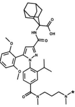
SR 142948A (2-{[5-(2,6-dimethoxyphenyl)-1-(4-(N-(3-dimethylaminopropyl)-N- methylcarbamoyl)-2-isopropylphenyl)-1H-pyrazole-3-carbonyl]-amino}-adamantane-2-carboxylic acid, hydrochloride) (Fig. 1) and SR 48692 (2-{[1-(7-chloroquinolin-4-yl)-5-(2,6-dimethoxyphenyl)-1H-pyrazole-3-carbonyl]amino}-adamantane-2-carboxylic acid) were synthesized at Sanofi Recherche (Montpellier, France). Both compounds were dissolved in dimethylsulfoxide and stored in aliquots at -20°C until the day of the experiment. [3H]SR
142948A (83 Ci/mmol) was tritiated at Sanofi Recherche (Alnwick, Great Britain). [3H]Neurotensin (104 Ci/mmol) was purchased from New England Nuclear (Les Ulis, France)
and monoiodo-[125I-Tyr3]neurotensin (2000 Ci/mmol) was iodinated and purified as described
previously (Sadoul et al., 1984). Unlabeled neurotensin was purchased from Neosystem Laboratories (Strasbourg, France). Levocabastine was kindly provided by Janssen Pharmaceutica (Beerse, Belgium) and was solubilized in ethanol.
2.2. Preparation of brain membrane homogenates
Male Sprague–Dawley rats (180–220 g, Charles River, Saint Aubain-les-Elboeuf, France) were killed by cervical dislocation. The whole brain (minus the cerebellum) was removed rapidly and homogenized in 10 volumes (original wet weight/volume) of 50 mM Tris–HCl ice-cold buffer (pH 7.4) for 30 s by using a polytron (setting 5). After 20 min of centrifugation at 50 000 g, the pellet was washed and again centrifuged as above. The final pellet was resuspended in binding assay buffer containing 50 mM Tris–HCl (pH 7.4), 1 mM EDTA,
0.1% bovine serum albumin, 1 mM 1,10 orthophenanthroline (Sigma, Saint Louis, MO), 5 mM dithiothreitol and 40 mg/l bacitracin and stored as aliquots in liquid nitrogen until used.
2.3. Binding assays in brain homogenates
Aliquots of brain membranes (300 µg/assay) were incubated in 0.5 ml (final volume) of binding assay buffer containing the appropriate concentrations of [3H]SR 142948A and
unlabeled drugs. After incubation at 20°C for 60 min, the assay medium was diluted with 4 ml of ice-cold 50 mM Tris–HCl buffer (pH 7.4) and filtered rapidly under reduced pressure through Whatman glass-fiber GF/B filters pretreated with 0.1% polyethylenimine. The filters were washed 3 times under the same conditions and transferred to vials containing 4 ml of scintillation cocktail. The bound radioactivity was determined by liquid scintillation counting. Nonspecific binding was determined in the presence of 1 µM unlabeled SR 142948A.
Saturation experiments were carried out with increasing concentrations of [3H]SR
142948A (0.05 to 16 nM). In association kinetic experiments, rat brain homogenates were incubated with [3H]SR 142948A (2 nM) for various time periods. For dissociation studies,
[3H]SR 142948A was incubated for 30 min with brain membranes, unlabeled SR 142948A
was then added at a final concentration of 1 µM and incubations were stopped at the indicated times. Competition studies were conducted with a single concentration of [3H]SR 142948A (2
nM) and at least 10 concentrations of unlabeled ligands. In addition, saturation and competition studies with [3H]SR 142948A were performed in the presence of a constant
concentration of levocabastine (10 µM), which selectively inhibits binding to neurotensin NT2
receptors. The effect of guanyl nucleotides on [3H]SR 142948A binding was examined by
adding increasing concentrations of the nonhydrolyzable GTP analog 5 -guanylylimidodiphosphate (Gpp(NH)p) to the binding assay. [3H]neurotensin binding to rat
brain membranes was performed as described previously (Goedert et al., 1984), in the presence or absence of levocabastine (10 µM). Nonspecific binding was determined by incubation with 1 µM neurotensin. All experiments were performed 2 or 3 times in triplicate. Data from association, saturation and competition studies were analyzed by using a nonlinear regression program, LIGAND (Munson and Rodbard, 1980). Ki values were calculated
according to the Cheng and Prusoff (1973) equation.
2.4. Autoradiography of [3H]SR 142948A binding sites
Adult male rats were killed by decapitation, the brain was removed rapidly, frozen on dry ice and stored at -80°C. Coronal sections (20 µm thick) were cut on a cryostat at -16°C, mounted on slides (Superfrost plus, Menzel-Glaser, Madison, WI) and stored at -20°C until used. [3H]SR 142948A binding was performed by incubating the sections for 60 min at room
temperature with 300 µl of 2 nM [3H]SR 142948A in 50 mM Tris–HCl buffer (pH 7.4),
containing 1 mM EDTA, 0.1% bovine serum albumin, 40 mg/ml bacitracin and 0.5 mM 1,10 orthophenanthroline, in the presence or absence of 1 µM levocabastine. Additional sections were incubated with 10 µM SR 142948A for the determination of nonspecific binding. After
incubation, the sections were washed 3 times for 10 min each at 4°C in 50 mM Tris–HCl buffer (pH 7.4), containing 1 mM EDTA and 0.1% bovine serum albumin. Slides were then dipped briefly in distilled water and dried under a stream of air. Sections were placed in X-ray cassettes, apposed to Hyperfilm-3H (Amersham, France) for 3 weeks and developed by
standard photographic procedures.
2.5. Autoradiography of [125I]neurotensin binding sites
[125I]Neurotensin binding was performed as described previously (Moyse et al., 1987).
Briefly, slide-mounted brain sections were incubated for 60 min at 4°C with 300 µl of 0.3 nM [125I]neurotensin in 50 mM Tris–HCl buffer (pH 7.4), containing 5 mM MgCl2, 0.2% bovine
serum albumin and 0.5 mM 1,10 orthophenanthroline, in the presence or absence of 1 µM levocabastine. Nonspecific binding was determined in the presence of 10 µM unlabeled neurotensin. The sections were washed 4 times for 2 min each at 4°C in 40 mM Tris–HCl buffer (pH 7.4), dipped briefly in distilled water and dried. Radiolabeled sections were exposed to Hyperfilm-βmax (Amersham) for 1 week.
2.6. Densitometric analysis
Quantitative optical density measurements of film autoradiographs were carried out with a computer-based image analysis system (HISTO-RAG, Biocom, Les Ulis, France). Optical densities of nonspecific binding were subtracted from total binding to obtain specific binding. Measurements were performed bilaterally on 4 brain sections per level. Values were expressed as nCi/mg tissue by using autoradiographic 3H and 125I micro-scales (Amersham) that were
exposed together with the tissue sections. Brain structures were identified according to the atlas of Paxinos and Watson (1986).
3. Results
3.1. Biochemical profile of [3H]SR 142948A binding to rat brain membrane
homogenates
3.1.1. Tissue concentration linearity
Fig. 2 shows the total, nonspecific and specific binding of [3H]SR 142948A to rat brain
homogenates as a function of membrane protein concentration. The specific binding of [3H]SR
142948A measured at 2 nM was linear with increasing protein concentration until 400 µg of protein/tube.
3.1.2. Saturation studies
Fig. 3 shows a saturation isotherm of the binding of [3H]SR 142948A to rat brain membranes.
Analysis of the saturation curves by computer-assisted nonlinear regression or by Scatchard analysis (Fig. 3, inset) revealed a single class of high-affinity binding sites (Kd=3.48±0.22 nM;
mean±S.E.M., n=3) for radioligand concentrations ranging from 0.05 to 16 nM, with a maximal binding capacity (Bmax) of 508.4±45.0 fmol/mg of protein.
3.1.3. Kinetic studies
The specific binding of [3H]SR 142948A to rat brain membranes was time dependent, reaching a steady state in about 60–80 min at 20°C (Fig. 4A). Fig. 4B illustrates the rate of dissociation, measured at various time intervals after addition of 1 µM unlabeled SR 142948A at equilibrium binding. The dissociation kinetics revealed a first-order process with a dissociation rate constant (k-1) of 0.021 min-1 (mean, n=2). The observed association rate
constant (kobs) was 0.033 min-1 and the kinetic association constant (k1), calculated from the
equation k1=(kobs-k1)/([3H]SR 142948A), was 0.006 109 M-1 min-1. The dissociation
constant (Kd) calculated from the ratio k-1/k1 was 3.5 nM, similar to the dissociation constant
determined in saturation studies.
3.1.4. Competition studies
Fig. 5 shows the inhibition of [3H]SR 142948A binding by increasing concentrations of
unlabeled SR 142948A, SR 48692 and neurotensin in rat brain homogenates. The Ki value
obtained for unlabeled SR 142948A, 5.0±0.4 nM, was close to the values determined in kinetic studies (Hill coefficient, nH=0.98±0.01). The binding of [3H]SR 142948A to rat brain
membranes was fully displaced by the natural ligand neurotensin, as well as by the previously described neurotensin receptor antagonist SR 48692, a nonpeptide molecule chemically related to SR 142948A. These competition curves gave Ki values of 32.8±5.9 nM (nH=0.99±0.10) for
neurotensin and 123.6±15.7 nM (nH=0.95±0.06) for SR 48692.
3.1.5. Effect of levocabastine on [3H]SR 142948A and [3H]neurotensin binding
The addition of increasing concentrations of levocabastine to the [3H]SR 142948A binding
assay resulted in progressive blockade of neurotensin NT2 receptors (Fig. 6). Concentrations
greater than 1 µM levocabastine completely inhibited the binding of [3H]SR 142948A to NT 2
receptors, which constituted 80% of the whole population of sites. The presence of 10 µM levocabastine in the [3H]SR 142948A binding assay revealed neurotensin NT
1 receptors,
which were recognized by SR 142948A with an IC50 value of 4.0±0.3 nM, in the same
nanomolar range as for experiments without levocabastine (IC50=7.0±1.5 nM).
Saturation experiments were also performed in the presence or absence of 10 µM levocabastine with a fixed concentration of [3H]SR 142948A and increasing concentrations of
unlabeled SR 142948A. Scatchard analysis of [3H]SR 142948A binding data in the absence of
levocabastine yielded a linear plot (Fig. 7A), indicating the presence of a homogeneous population of binding sites (Kd=6.1±1.6 nM, Bmax=527±22 fmol/mg protein; n=3). However,
the parallel leftward shift of this straight line obtained in the presence of 10 µM levocabastine indicated binding to neurotensin NT1 receptors (Kd=3.4±0.5 nM, Bmax=104±8 fmol/mg
protein) and, by difference, revealed neurotensin NT2 receptors (Kd=8.5±4.1 nM,
Bmax=422±58 fmol/mg protein). These data confirmed that [3H]SR 142948A exhibits similar
affinities for the two subtypes of neurotensin receptors. Similar experiments performed with [3H]neurotensin (Fig. 7B) revealed K
d values comparable to those of [3H] SR 142948A (6.8
and 4.8 nM for NT1 and NT2 receptors, respectively). However, [3H]neurotensin bound to a
lower number of binding sites than [3H]SR 142948A (B
max values of 32 and 265 fmol/mg for
NT1 and NT2 receptors, respectively).
3.1.6. Effect of guanyl nucleotides on [3H]SR 142948A binding
In order to determine whether the binding of [3H]SR 142948A was sensitive to guanyl
nucleotides, we examined the affinity of the radioligand in the presence of Gpp(NH)p, a nonhydrolyzable analog of GTP. The addition of Gpp(NH)p in concentrations up to 100 µM did not modify the specific binding of [3H]SR 142948A to rat brain membranes (data not
shown).
3.2. Autoradiographic localization of [3H]SR 142948A binding sites in the rat brain:
Comparison with [125I]neurotensin binding
Preliminary experiments showed that the characteristics of [3H]SR 142948A binding to rat
midbrain sections were similar to those observed for brain membrane homogenates. [3H]SR
142948A binding was saturable and reached a steady state by 60 min. Specific [3H]SR
142948A binding was approximately 90% of the total binding, as determined with 2 nM radioligand. Analysis of competition studies performed on rat midbrain sections showed that SR 142948A, SR 48692 and neurotensin induced a dose-dependent and complete inhibition of [3H]SR 142948A binding (data not shown).
Fig. 8 shows the regional distribution of [3H]SR 142948A and [125I]neurotensin binding
sites in the rat brain. The highest density of [3H]SR 142948A and [125I]neurotensin binding
was present in the midbrain, in the ventral tegmental area and substantia nigra pars compacta. Intense labeling was also observed in the perirhinal area as well as in the dorsal peduncular, anterior cingulate and agranular insular cortices, posteromedial cortical amygdaloid nucleus, medial habenula and ventral dentate gyrus. The endopiriform nucleus, septohippocampal nucleus, central amygdaloid nucleus and zona incerta exhibited moderate levels of binding. Finally, a low density of binding sites was observed in the frontal, parietal, temporal and retrosplenial granular cortices, caudate putamen, nucleus accumbens, lateral septum, hypothalamus, hippocampus, substantia nigra pars reticulata and in the superficial gray layer of the superior colliculus.
Table 1 shows the comparative distribution of [3H]SR 142948A and [125I]neurotensin
binding sites in the rat brain, in the presence or absence of levocabastine. An excellent correlation between the regional distribution of the two ligands was observed, in agreement with the autoradiographic localization of neurotensin receptors in rat brain described previously (Moyse et al., 1987). The presence of levocabastine in the incubation buffer inhibited binding to neurotensin NT2 receptors and decreased the amount of labeling with
[3H]SR 142948A and [125I]neurotensin in all brain regions studied, indicating that both ligands
labeled neurotensin NT1 and NT2 receptors. This result is in agreement with the ubiquitous
distribution of NT2 receptors in the rat brain (Schotte et al., 1986).
4. Discussion
The binding of the nonpeptide neurotensin receptor antagonist [3H]SR 142948A to rat brain
membranes was rapid, tissue concentration and time dependent, saturable and reversible. Scatchard analyses of saturation experiments indicated that [3H]SR 142948A binds with high
affinity (Kd=3.5 nM) and apparently recognizes a single class of binding sites. The number of
sites labeled by [3H]SR 142948A (B
max=508 fmol/mg protein) was 80% greater than the Bmax
value determined with [3H]neurotensin under the same experimental conditions (297 fmol/mg
protein). Competition experiments with unlabeled SR 142948A yielded a Hill coefficient near unity, further suggesting that the antagonist bound to an apparently homogeneous population of binding sites.
The potencies of the neurotensin receptor antagonists SR 142948A and SR 48692 in inhibiting specific [3H]SR 142948A binding (K
i=5 and 123.6 nM, respectively) were similar
to their previously reported potencies in displacing [125I]neurotensin binding in adult rat brain
homogenates (IC50=3.96 and 82 nM for SR 142948A and SR 48692, respectively) (Gully et
al., 1997). In contrast, the potency of the natural peptide agonist neurotensin for inhibiting [3H]SR 142948A binding (K
i=32.8 nM) was 10-fold lower than its potency in displacing
[125I]neurotensin binding (IC
50=3.2 nM). This is consistent with results obtained with other
receptor systems, demonstrating that estimates of agonist affinity are lower when an antagonist rather than an agonist radioligand is displaced. Indeed, a reduced potency for agonists to compete against radiolabeled antagonist ligands has been observed previously for neurotensin receptors in the guinea pig brain (Betancur et al., 1995), as well as for muscarinic cholinergic receptors (Waelbroeck et al., 1982), cholecystokinin CCKA (Chang et al., 1986; Talkad et al., 1994) and CCKB receptors (Chang et al., 1989), tachykinin NK1 receptors
(McLean et al., 1991) and 5-HT2 receptors (Teitler et al., 1990). These findings have been
interpreted as indicating the existence of different conformational states of the same receptor, with different affinities for agonist and antagonist ligands (Schwartz et al., 1995).
The higher number of receptors detected with [3H]SR 142948A when compared with the
number detected with the agonist radioligand, [3H]neurotensin, also supports this hypothesis.
The ability of radiolabeled nonpeptide antagonists to recognize a larger number of receptors, characterized by low affinity for the agonist, than agonist-derived radioligands appears to be a common phenomenon. For instance, we showed previously that in the guinea pig brain the number of binding sites labeled by the antagonist [3H]SR 48692 exceeded by 20-fold the
number of receptors labeled with the agonist [125I]neurotensin (Betancur et al., 1995). The
binding sites detected by [3H]SR 48692 were characterized by a low affinity for neurotensin
and were insensitive to GTP, suggesting that they represent the uncoupled form of the neurotensin receptor. These data, together with current hypotheses on the molecular
interactions of ligands with their receptors, suggest that agonists bind with high affinity to the active receptor conformation, whereas antagonists bind with higher affinity to the inactive conformation (Schwartz et al., 1995). Thus, peptide agonists and nonpeptide antagonists act as allosteric competitive ligands by binding in a mutually exclusive fashion to sites occurring in different receptor conformations. In support of this model, recent site-directed mutational studies of the rat neurotensin NT1 receptor showed that mutations in the N-terminal part
eliminate neurotensin binding without affecting the binding of [3H]SR 48692 (Labbé-Jullié et
al., 1995). This finding suggests the existence of distinct agonist and antagonist binding domains on the neurotensin NT1 receptor, similarly to what has been reported for several
other neuropeptide receptors (for review, see Betancur et al., 1997).
Levocabastine is a nonpeptide antagonist of histamine H1 receptors that is structurally unrelated to neurotensin (Stockbroekx et al., 1986), but binds also to neurotensin NT2
receptors (Schotte et al., 1986). The recent cloning of the mouse NT2 receptor and its
expression in Xenopus oocytes indicated that levocabastine acts as an agonist in this system, since it triggers a Cl- inward current, as does neurotensin (Mazella et al., 1996). Saturation and
competition experiments performed with or without levocabastine indicated that [3H]SR
142948A bound with high affinity to neurotensin NT1 and NT2 receptors (Kd=3.4 and 8.5
nM, respectively). Indeed, the addition of levocabastine to the [3H]SR 142948A binding assay
caused a parallel leftward shift of the Scatchard plot, indicating the displacement of [3H]SR
142948A binding from levocabastine-sensitive NT2 receptors. The similar affinity of [3H]SR
142948A for both subtypes of neurotensin receptors explains the apparently homogeneous population of binding sites detected with this ligand in the absence of levocabastine. Moreover, our results indicate that neurotensin NT2 receptors constitute 80% of the whole
population of sites labeled by [3H]SR 142948A (B
max=104 and 422 fmol/mg protein for NT1
and NT2 receptors, respectively) on rat brain membranes.
Guanyl nucleotides differentially affect agonist and antagonist binding in several neurotransmitter receptor systems. Accordingly, guanyl nucleotides have been reported to significantly reduce [125I]neurotensin binding to NT
1 receptors, by interfering with the
formation of the high-affinity agonist-receptor-G protein ternary complex (Hermans et al., 1996). In contrast, the present study showed that addition of Gpp(NH)p had no effect on specific [3H]SR 142948A binding. This finding is consistent with previous data showing that
antagonist ligands bind with high affinity to the G protein-uncoupled state of receptors (Teitler et al., 1990; Rosenkilde et al., 1994; Betancur et al., 1995). It should be noted, however, that the levocabastine-sensitive neurotensin binding site is insensitive to GTP (Vincent, 1995), although recent data indicate that the cloned mouse neurotensin NT2 receptor
is coupled functionally to phospholipase C when expressed in oocytes (Mazella et al., 1996). Other studies have shown that an absence of GTP-sensitive binding does not necessarily indicate failure to activate a second messenger cascade (Chung et al., 1988; Maeda et al., 1990; Hermans et al., 1996).
The different sensitivity to Gpp(NH)p of neurotensin NT1 and NT2 receptors is
particularly interesting in view of the fact that the lowest homology between the two
neurotensin receptors is in their third cytoplasmic loop and C-terminal domain (Chalon et al., 1996; Mazella et al., 1996), two regions implicated in the coupling to G proteins (Yamada et al., 1994; Hermans et al., 1996). Furthermore, it has been proposed that the extremely high number of Ser/Thr residues in the third intracytoplasmic loop and the C-terminal domain of the neurotensin NT2 receptor protein could be associated with a basal phosphorylated state
resulting in desensitization of the receptor (Mazella et al., 1996). This could result in a higher proportion of receptors in the uncoupled form expressed in the membrane and might explain the insensitivity of the NT2 receptor to GTP analogs. The use of recently developed
radiolabeled agonist ligands specific for the G protein-coupled state of neurotensin receptors (Gaudriault et al., 1996), in the presence or absence of levocabastine, could help to determine the different states of coupling of neurotensin NT1 and NT2 receptors.
The autoradiographic distribution of [3H]SR 142948A binding in sections of rat brain was
consistent with its selective binding to neurotensin receptors. The heterogeneous pattern of specific [3H]SR 142948A binding closely matched the localization of [125I]neurotensin binding
observed in adjacent sections. The addition of levocabastine to the incubation buffer resulted in a small and diffuse reduction of [3H]SR 142948A and [125I]neurotensin binding in all brain
structures studied, in agreement with previous studies indicating that neurotensin NT2
receptors are distributed throughout the rat central nervous system (Schotte et al., 1986; Kitabgi et al., 1987) and are predominantly associated with glial cells (Schotte et al., 1988). The ubiquitous distribution of neurotensin NT2 receptors contrasts with the highly regional
localization of NT1 receptors in the brain. Indeed, NT1 receptors are particularly abundant in
brain regions rich in dopamine neurons, such as the substantia nigra and the ventral tegmental area, as well as in certain cortical areas. These results are in agreement with the previously described distribution of [125I]neurotensin binding (Moyse et al., 1987) and neurotensin NT
1
receptor mRNA (Nicot et al., 1994) observed in the rat brain.
The functional characterization of the actions of SR 142948A in the central nervous system showed that this compound, like the first-generation neurotensin receptor antagonist SR 48692, antagonizes the turning behavior induced by intrastriatal injection of neurotensin in mice as well as acetylcholine release evoked by neurotensin in the striatum (Gully et al., 1993 and Gully et al., 1997). Neither compound modified dopamine release in the nucleus accumbens after injection of neurotensin into the ventral tegmental area (Steinberg et al., 1994; Gully et al., 1997). However, unlike SR 48692, SR 142948A blocked the hypothermia and analgesia induced by central injection of neurotensin in rodents (Gully et al., 1997). These results suggest that SR 142948A may interact with a neurotensin receptor subtype which is not blocked by SR 48692. It would be interesting to assess the potential involvement of the recently identified neurotensin NT2 receptor in the mediation of the hypothermia and
anti-nociceptive effects induced by neurotensin.
In conclusion, [3H]SR 142948A represents a new potent nonpeptide antagonist
radioligand specific for neurotensin receptors. The compound binds to rat neurotensin NT1
and NT2 receptors with nanomolar affinity, close to that of the natural ligand neurotensin. The
ligand previously described, [3H]SR 48692, exhibits a higher affinity for NT
1 than for NT2
receptors and has a low ratio of total binding to nonspecific binding in rat brain membranes and tissue sections, which limits its utility. Consequently, the availability of [3H]SR 142948A
provides a valuable tool for the study of neurotensin receptors in the central nervous system.
Acknowledgements
We thank A.M. Lhiaubet for labeling [125I]neurotensin. C.B. is supported by a post-doctoral
fellowship from INSERM (Institut National de la Santé et de la Recherche Médicale), France.
References
Betancur, C., Canton, M., Gully, D., Vela, G., Pélaprat, D. and Rostène, W. 1995. Characterization and distribution of binding sites for a new neurotensin receptor antagonist ligand, [3H]SR 48692, in the guinea pig brain. J. Pharmacol. Exp. Ther. 273, pp. 1450–1458 Betancur, C., Azzi, M. and Rostène, W. 1997. Nonpeptide antagonists of neuropeptide receptors:
Tools for research and therapy. Trends Pharmacol. Sci. 18, pp. 372–386
Chalon, P., Vita, N., Kaghad, M., Guillemot, M., Bonnin, J., Delpech, B., Le Fur, G., Ferrara, P. and Caput, D. 1996. Molecular cloning of a levocabastine-sensitive neurotensin binding site. FEBS Lett. 386, pp. 91–94
Chang, R.S.L., Lotti, V.J., Chen, T.B. and Kunkel, K.A. 1986. Characterization of the binding of [3H]-(±)-L-364,718: A new potent, nonpeptide cholecystokinin antagonist radioligand selective for peripheral receptors. Mol. Pharmacol. 30, pp. 212–217
Chang, R.S.L., Chen, T.B., Bock, M.G., Freidinger, R.M., Chen, R., Rosegay, A. and Lotti, V.J. 1989. Characterization of the binding of [3H]L-365,260: A new potent and selective brain cholecystokinin (CCK-B) and gastrin receptor antagonist radioligand. Mol. Pharmacol. 35, pp. 803–808
Cheng, Y. and Prusoff, W. 1973. Relationship between the inhibition constant (Ki) and the
concentration of inhibitor which causes 50% inhibition (IC50) of an enzymatic reaction.
Biochem. Pharmacol. 22, pp. 3099–3108
Chung, F.Z., Wang, C.D., Potter, P.C., Venter, J.C. and Fraser, C.M. 1988. Site-directed mutagenesis and continuous expression of human β-adrenergic receptors. J. Biol. Chem. 263, pp. 4052–4055
Dubuc, I., Costentin, J., Terranova, J.P., Barnouin, M.C., Soubrié, P., Le Fur, G., Rostène, W. and Kitabgi, P. 1994. The nonpeptide neurotensin antagonist, SR 48692, used as a tool to reveal putative neurotensin receptor subtypes. Br. J. Pharmacol. 112, pp. 352–354
Emson, P.C., Goedert, M., Horsfield, P.M., Rioux, F. and St. Pierre, S. 1982. The regional distribution and chromatographic characterization of neurotensin-like immunoreactivity in the rat central nervous system. J. Neurochem. 38, pp. 992–999
Gaudriault, G., Zsürger, N. and Vincent, J.P. 1996. Radiolabeled ligands specific for the G protein-coupled state of neurotensin receptors. J. Neurochem. 67, pp. 2590–2598
Goedert, M., Pittaway, K., Williams, B.J. and Emson, P.C. 1984. Specific binding of tritiated neurotensin to rat brain membranes: Characterization and regional distribution. Brain Res. 304, pp. 71–81
Gully, D., Canton, M., Boigegrain, R., Jeanjean, F., Molimard, J.C., Poncelet, M., Gueudet, C., Heaulme, M., Leyris, R., Brouard, A., Pélaprat, D., Labbé-Jullié, C., Mazella, J., Soubrié, P., Maffrand, J.P., Rostène, W., Kitabgi, P. and Le Fur, G. 1993. Biochemical and pharmacological profile of a potent and selective nonpeptide antagonist of neurotensin receptor. Proc. Natl. Acad. Sci. USA 90, pp. 65–69
Gully, D., Labeeuw, B., Boigegrain, R., Oury-Donat, F., Bachy, A., Poncelet, M., Steinberg, R., Suaud-Chagny, M.F., Santucci, V., Vita, N., Pecceu, F., Labbé-Jullié, C., Kitabgi, P., Soubrié, P., Le Fur, G. and Maffrand, J.P. 1997. Biochemical and pharmacological activities of SR 142948A, a new potent neurotensin receptor antagonist. J. Pharmacol. Exp. Ther. 280, pp. 802–812
Hermans, E., Octave, J.N. and Maloteaux, J.M. 1996. Interaction of the COOH-terminal domain of the neurotensin receptor with a G protein does not control the phospholipase C activation but is involved in the agonist-induced internalization. Mol. Pharmacol. 49, pp. 365–372 Kasckow, J. and Nemeroff, C.B. 1991. The neurobiology of neurotensin: Focus on neurotensin–
dopamine interactions. Regul. Pept. 36, pp. 153–164
Kitabgi, P., Rostène, W., Dussaillant, M., Schotte, A., Laduron, P.M. and Vincent, J.P. 1987. Two populations of neurotensin binding sites in murine brain: Discrimination by the antihistamine levocabastine reveals markedly different radioautographic distribution. Eur. J. Pharmacol. 140, pp. 285–293
Labbé-Jullié, C., Botto, J.M., Mas, M.V., Chabry, J., Mazella, J., Vincent, J.P., Gully, D., Maffrand, J.P. and Kitabgi, P. 1995. [3H]SR 48692, the first nonpeptide neurotensin
antagonist radioligand: Characterization of binding properties and evidence for distinct agonist and antagonist binding domains on the rat neurotensin receptor. Mol. Pharmacol. 47, pp. 1050–1056
Le, F., Cusak, B. and Richelson, E. 1996. The neurotensin receptor: Is there more than one subtype?. Trends Pharmacol. Sci. 17, pp. 1–3
Maeda, S., Lameh, J., Mallet, W.G., Philip, M., Ramachandran, J. and Sadée, W. 1990. Internalization of the Hm1 muscarinic cholinergic receptor involves the third cytoplasmic loop. FEBS Lett. 269, pp. 386–388
Mai, J., Triepel, K.J. and Metz, J. 1987. Neurotensin in the human brain. Neuroscience 22, pp. 499–524
Mazella, J., Poustis, C., Labbé, C., Checler, F., Kitabgi, P., Granier, C., Van Rietschoten, J. and Vincent, J.P. 1983. Monoiodo Trp11-neurotensin, a highly radioactive ligand of neurotensin receptors: Preparation, biological activity, and binding properties to rat brain synaptic membranes. J. Biol. Chem. 258, pp. 3476–3481
Mazella, J., Botto, J.M., Guillemare, E., Coppola, T., Sarret, P. and Vincent, J.P. 1996. Structure, functional expression, and cerebral localization of the levocabastine-sensitive neurotensin/neuromedin N receptor from mouse brain. J. Neurosci. 16, pp. 5613–5620 McLean, S., Ganong, A.H., Seeger, T.F., Bryce, D.K., Pratt, K.G., Reynolds, L.S., Siok, C.J., Lowe
III, J.A. and Heym, J. 1991. Activity and distribution of binding sites in brain of a nonpeptide substance P (NK1) receptor antagonist. Science 251, pp. 437–439
Moyse, E., Rostène, W., Vial, M., Leonard, K., Mazella, J., Kitabgi, P., Vincent, J.P. and Beaudet, A. 1987. Distribution of neurotensin binding sites in rat brain: A light microscopic radioautographic study using monoiodo [125I]Tyr3-neurotensin. Neuroscience 22, pp. 525–
536
Munson, P.J. and Rodbard, D. 1980. LIGAND: A versatile computerized approach for characterization of ligand-binding systems. Anal. Biochem. 107, pp. 220–239
Nicot, A., Rostène, W. and Bérod, A. 1994. Neurotensin receptor expression in the rat forebrain and midbrain: A combined analysis by in situ hybridization and receptor autoradiography. J. Comp. Neurol. 341, pp. 407–419
Paxinos, G., Watson, C. 1986. The Rat Brain in Stereotaxic Coordinates, 2nd ed. Academic Press, Sydney.
Pugsley, T.A., Akunne, H.C., Whetzel, S.Z., Demattos, S., Corbin, A.E., Wiley, J.N., Wustrow, D.J., Wise, L.D. and Heffner, T.G. 1995. Differential effects of the nonpeptide neurotensin
antagonist, SR 48692, on the pharmacological effects of neurotensin agonists. Peptides 16, pp. 37–44
Rosenkilde, M.M., Cahir, M., Gether, U., Hjorth, S.A. and Schwartz, T.W. 1994. Mutations along transmembrane segment II of the NK-1 receptor affect substance P competition with non-peptide antagonists but not substance P binding. J. Biol. Chem. 269, pp. 28160–28164
Rostène, W.H. and Alexander, M.J. 1997. Neurotensin and neuroendocrine regulation. Front. Neuroendocrinol. 18, p. 115
Sadoul, J.L., Mazella, J., Amar, S., Kitabgi, P. and Vincent, J.P. 1984. Preparation of neurotensin selectively iodinated on the tyrosine 3 residue. Biological activity and binding properties on mammalian neurotensin receptors. Biochem. Biophys. Res. Commun. 120, pp. 812–819 Schotte, A., Leysen, J.E. and Laduron, P.M. 1986. Evidence for a displaceable non-specific
[3H]neurotensin binding site in rat brain. Naunyn Schmiedebergs Arch. Pharmacol. 333, pp.
400–405
Schotte, A., Rostène, W. and Laduron, P.M. 1988. Different subcellular localization of neurotensin receptor and neurotensin acceptor sites in the rat brain dopaminergic system. J. Neurochem. 50, pp. 1026–1031
Schwartz, T.W., Gether, U., Schambye, H.T. and Hjorth, S.A. 1995. Molecular mechanism of action of nonpeptide ligands for peptide receptors. Curr. Pharm. Design 1, pp. 355–372 Steinberg, R., Brun, P., Fournier, M., Souilhac, J., Rodier, D., Mons, G., Terranova, J.P., Le Fur,
G. and Soubrié, P. 1994. SR 48692, a non-peptide neurotensin receptor antagonist differentially affects neurotensin-induced behaviour and changes in dopaminergic transmission. Neuroscience 59, pp. 921–929
Stockbroekx, R.A., Luyckx, M.G., Willems, J.M., Janssen, M.A.C., Bracke, J.O.M.M., Joosen, B.L.P. and Van Wauwe, J.P. 1986. Levocabastine (R 50 547), the prototype of a chemical series of compounds with specific H1-antihistamine activity. Drug Dev. Res. 8, pp. 87–93 Talkad, V.D., Fortune, K.P., Pollo, D.A., Shah, G.N., Wank, S.A. and Gardner, J.D. 1994. Direct
demonstration of three different states of the pancreatic cholecystokinin receptor. Proc. Natl. Acad. Sci. USA 91, pp. 1868–1872
Tanaka, K., Masu, M. and Nakanishi, S. 1990. Structure and functional expression of the cloned rat neurotensin receptor. Neuron 4, pp. 847–854
Teitler, M., Leonhardt, S., Weisberg, E.L. and Hoffman, B.J. 1990. 4-[125
I]iodo-(2,5-dimethoxy)phenylisopropylamine and [3H]ketanserin labeling of 5-hydroxytryptamine2
(5HT2) receptors in mammalian cells transfected with a rat 5HT2 cDNA: Evidence for multiple states and not multiple 5HT2 receptor subtypes. Mol. Pharmacol. 38, pp. 594–598 Vincent, J.P. 1995. Neurotensin receptors: Binding properties, transduction pathways and
structure. Cell. Mol. Neurobiol. 15, pp. 501–512
Vita, N., Laurent, P., Leport, S., Chalon, P., Dumont, X., Kaghad, M., Gully, D., Le Fur, G., Ferrara, P. and Caput, D. 1993. Cloning and expression of a complementary DNA encoding a high affinity human neurotensin receptor. FEBS Lett. 317, pp. 139–142
Waelbroeck, M., Robberecht, P., Chatelain, P. and Christophe, J. 1982. Rat cardiac muscarinic receptors. I. Effects of guanine nucleotides on high- and low-affinity binding sites. Mol. Pharmacol. 21, pp. 581–588
Yamada, M., Yamada, M., Watson, M.A. and Richelson, E. 1994. Deletion of the putative third intracellular loop of the rat neurotensin receptor abolishes phosphoinositide hydrolysis but not cyclic AMP formation in CHO-K1 cells. Mol. Pharmacol. 46, pp. 470–476
Table 1. Regional distribution of [3H]SR 142948A and [125I]neurotensin binding sites in the rat brain,
in the presence or absence of levocabastine
Specific Binding
[3H]SR 142948A [125I]neurotensin
Brain area – levo + levo – levo + levo
Cerebral cortex
Frontal cortex 1.91 ± 0.11 1.12 ± 0.02 0.39 ± 0.03 0.29 ± 0.01 Anterior cingulate cortex 6.61 ± 0.27 6.41 ± 0.14 2.01 ± 0.16 1.79 ± 0.14 Dorsal peduncular cortex 9.44 ± 0.18 7.67 ± 0.27 2.47 ± 0.39 1.80 ± 0.09 Agranular insular cortex 7.58 ± 0.21 5.66 ± 0.51 1.62 ± 0.19 1.05 ± 0.09 Dorsal endopiriform nucleus 5.06 ± 0.12 4.59 ± 0.09 1.82 ± 0.13 1.15 ± 0.07 Parietal cortex 1.84 ± 0.05 0.82 ± 0.05 0.40 ± 0.02 0.27 ± 0.01 Perirhinal area 10.54 ± 0.29 8.98 ± 0.22 2.51 ± 0.32 2.29 ± 0.05 Retrosplenial granular cortex 3.11 ± 0.09 1.98 ± 0.12 1.07 ± 0.03 0.84 ± 0.06 Temporal cortex 2.22 ± 0.05 1.01 ± 0.06 0.47 ± 0.01 0.33 ± 0.01 Forebrain
Septohippocampal nucleus 5.90 ± 0.27 5.50 ± 0.29 2.09 ± 0.47 2.01 ± 0.17 Lateral septal nucleus, dorsal part 5.99 ± 0.25 4.65 ± 0.10 1.13 ± 0.02 1.02 ± 0.05 Basal ganglia
Caudate putamen (striatum) 2.57 ± 0.13 2.12 ± 0.06 0.58 ± 0.02 0.51 ± 0.02 Accumbens nucleus, shell 2.62 ± 0.08 1.66 ± 0.07 0.52 ± 0.02 0.35 ± 0.01 Accumbens nucleus, core 2.39 ± 0.09 1.55 ± 0.06 0.55 ± 0.03 0.37 ± 0.01 Amygdala
Central amygdaloid nucleus 5.00 ± 0.33 3.10 ± 0.09 1.29 ± 0.09 0.93 ± 0.02 Posteromedial cortical amygdaloid nucleus 9.17 ± 0.38 8.06 ± 0.26 1.83 ± 0.20 1.60 ± 0.12 Diencephalon
Zona incerta 7.84 ± 0.14 5.67 ± 0.46 1.61 ± 0.06 1.32 ± 0.03 Lateral habenular nucleus 5.41 ± 0.20 4.01 ± 0.11 1.20 ± 0.06 0.80 ± 0.04 Medial habenular nucleus 10.68 ± 0.48 9.01 ± 0.50 2.63 ± 0.14 1.92 ± 0.07 Hippocampal formation
Dentate gyrus, ventral part 8.41 ± 0.44 7.64 ± 0.31 1.19 ± 0.08 0.98 ± 0.14 Midbrain
Substantia nigra, pars compacta 14.25 ± 1.08 11.34 ± 1.08 2.38 ± 0.10 2.05 ± 0.26 Substantia nigra, pars reticulata 3.21 ± 0.13 2.79 ± 0.14 0.99 ± 0.05 0.83 ± 0.07 Ventral tegmental area 15.10 ± 1.08 9.77 ± 0.97 2.18 ± 0.17 1.71 ± 0.19 Superficial gray layer of the superior colliculus 4.10 ± 0.13 2.28 ± 0.08 0.82 ± 0.03 0.64 ± 0.03
[3H]SR 142948A and [125I]neurotensin specific binding to neurotensin NT
1 and NT2 receptors was
determined on rat brain sections by autoradiography, in the presence (+ levo) or absence (– levo) of levocabastine (1 µM). The occlusion of neurotensin NT2 receptors by levocabastine allowed detection
of NT1 receptors. The difference between binding in the absence and in the presence of levocabastine
represents binding to NT2 receptors. Optical densities were measured bilaterally on 4 brain sections per
level and expressed as mean ± S.E.M. nCi/mg tissue.
Fig. 1. Chemical structure of SR 142948A. *: location of the 3 tritium atoms.
Fig. 2. [3H]SR 142948A binding as a function of increasing protein concentrations. Various concentrations of rat brain membranes were incubated with 2 nM [3H]SR 142948A for 60 min at 20°C. Nonspecific binding was determined with 1 µM unlabeled SR 142948A. Specific binding was defined as the difference between total and nonspecific binding. The data represent the mean of triplicate determinations.
Fig. 3. Saturation analysis and Scatchard plot (inset) of [3H]SR 142948A binding to rat brain membranes. Membranes were incubated for 60 min at 20°C over a concentration range of [3H]SR 142948A (0.05 to 16 nM); nonspecific binding was defined with 1 µM unlabeled SR 142948A. The data shown are from one of 3 experiments performed in triplicate.
Fig. 4. Association (A) and dissociation (B) kinetics of specific [3H]SR 142948A binding to rat brain membranes. (A) The association of 2 nM [3H]SR 142948A binding was determined at various time intervals. (B) Time course of dissociation of [3H]SR 142948A binding, initiated with 1 µM unlabeled SR 142948A. The points shown are means of triplicate determinations from a representative experiment.
Fig. 5. Inhibition of specific [3H]SR 142948A binding to rat brain membranes by increasing
concentrations of SR 142948A, SR 48692 and neurotensin. Results represent the means±S.E.M. of 3 independent experiments performed in triplicate.
Fig. 6. Inhibition of specific [3H]SR 142948A binding to rat brain membranes by SR 142948A, levocabastine and SR 142948A in the presence of a constant concentration of levocabastine (10
µM). Specific binding of [3H]SR 142948A without levocabastine represents 100%. In the absence of levocabastine, SR 142948A (●) inhibited [3H]SR 142948A binding to neurotensin NT
1 and
NT2 receptors. Levocabastine (♦) inhibited binding of [3H]SR 142948A to NT2 receptors (80%
of the receptors). In the presence of 10 µM levocabastine, SR 142948A (□) inhibited [3H]SR
142948A binding to NT1 receptors (20% of the receptors). The data shown are from a single
experiment performed in triplicate. The experiment was repeated 3 times with similar results.
Fig. 7. Scatchard analysis of saturation of [3H]SR 142948A (A) and [3H]neurotensin (B) binding
to rat brain membranes performed with or without 10 µM levocabastine. [3H]SR 142948A and
[3H]neurotensin bound to neurotensin NT
1 and NT2 receptors; the addition of 10 µM
levocabastine to the binding assay blocked levocabastine-sensitive NT2 receptors and revealed
NT1 receptors. The NT2 receptor data were calculated as the difference between the results
obtained in the absence and in the presence of levocabastine. The values are from typical experiments and represent the means of triplicate determinations.
Fig. 8. Autoradiographic distribution of [3H]SR 142948A (left) and [125I]neurotensin (right) binding sites in coronal sections of the rat brain. Photographs show [3H]SR 142948A and
[125I]neurotensin binding in the absence of levocabastine. Nonspecific binding was determined in the presence of 10 µM unlabeled SR 142948A or neurotensin, respectively, and was indistinguishable from background. Abbreviations: CPu=caudate putamen; DG=ventral dentate gyrus; En=endopirifom nucleus; Hb=habenula; Hi=hippocampus; LS=lateral septum; Prh=perirhinal area; RSG=retrosplenial granular cortex; SNC=substantia nigra, pars compacta; SNR=substantia nigra, pars reticulata; VTA=ventral tegmental area; ZI=zona incerta.
![Table 1. Regional distribution of [ 3 H]SR 142948A and [ 125 I]neurotensin binding sites in the rat brain, in the presence or absence of levocabastine](https://thumb-eu.123doks.com/thumbv2/123doknet/14652603.551870/15.892.90.803.167.917/table-regional-distribution-neurotensin-binding-presence-absence-levocabastine.webp)
![Fig. 3. Saturation analysis and Scatchard plot (inset) of [ 3 H]SR 142948A binding to rat brain membranes](https://thumb-eu.123doks.com/thumbv2/123doknet/14652603.551870/17.892.221.668.125.562/fig-saturation-analysis-scatchard-inset-binding-brain-membranes.webp)
![Fig. 4. Association (A) and dissociation (B) kinetics of specific [ 3 H]SR 142948A binding to rat brain membranes](https://thumb-eu.123doks.com/thumbv2/123doknet/14652603.551870/18.892.242.647.112.925/fig-association-dissociation-kinetics-specific-binding-brain-membranes.webp)
![Fig. 5. Inhibition of specific [ 3 H]SR 142948A binding to rat brain membranes by increasing concentrations of SR 142948A, SR 48692 and neurotensin](https://thumb-eu.123doks.com/thumbv2/123doknet/14652603.551870/19.892.227.669.98.536/inhibition-specific-binding-brain-membranes-increasing-concentrations-neurotensin.webp)
![Fig. 6. Inhibition of specific [ 3 H]SR 142948A binding to rat brain membranes by SR 142948A, levocabastine and SR 142948A in the presence of a constant concentration of levocabastine (10 µ M)](https://thumb-eu.123doks.com/thumbv2/123doknet/14652603.551870/20.892.220.669.126.562/inhibition-specific-membranes-levocabastine-presence-constant-concentration-levocabastine.webp)
![Fig. 7. Scatchard analysis of saturation of [ 3 H]SR 142948A (A) and [ 3 H]neurotensin (B) binding to rat brain membranes performed with or without 10 µ M levocabastine](https://thumb-eu.123doks.com/thumbv2/123doknet/14652603.551870/21.892.228.665.111.966/scatchard-analysis-saturation-neurotensin-binding-membranes-performed-levocabastine.webp)
![Fig. 8. Autoradiographic distribution of [ 3 H]SR 142948A (left) and [ 125 I]neurotensin (right) binding sites in coronal sections of the rat brain](https://thumb-eu.123doks.com/thumbv2/123doknet/14652603.551870/22.892.111.786.128.825/autoradiographic-distribution-neurotensin-right-binding-sites-coronal-sections.webp)
