HAL Id: hal-02866507
https://hal.archives-ouvertes.fr/hal-02866507
Submitted on 12 Jun 2020HAL is a multi-disciplinary open access archive for the deposit and dissemination of sci-entific research documents, whether they are pub-lished or not. The documents may come from teaching and research institutions in France or abroad, or from public or private research centers.
L’archive ouverte pluridisciplinaire HAL, est destinée au dépôt et à la diffusion de documents scientifiques de niveau recherche, publiés ou non, émanant des établissements d’enseignement et de recherche français ou étrangers, des laboratoires publics ou privés.
technology, experimental and clinical applications
Serge Mordon, Elise Thecua, Laurine Ziane, Fabienne Lecomte, Pascal
Deleporte, Grégory Baert, Anne-Sophie Dewalle-Vignion
To cite this version:
Serge Mordon, Elise Thecua, Laurine Ziane, Fabienne Lecomte, Pascal Deleporte, et al.. Light emitting fabrics for Photodynamic Therapy: technology, experimental and clinical applications. translational biophotonics, Jürgen Popp, Carsten Philipp, Ronald Sroka, 2020, �10.1002/tbio.202000005�. �hal-02866507�
1
Light emitting fabrics for Photodynamic Therapy: technology, experimental
and clinical applications.
Serge Mordon, Elise Thécua, Laurine Ziane, Fabienne Lecomte, Pascal Deleporte, Grégory Baert, Anne-Sophie Vignion-Dewalle.
1
Univ. Lille, Inserm, CHU Lille, U1189 – OncoThAI –Laser Assisted Therapies and Immunotherapies for Oncology, F-59000 Lille, France
Correspondance : Pr. Serge Mordon OncoThAI 1, avenue Oscar Lambret, 59037 LILLE cedex, France
Email : [email protected]
Abstract
A homogeneous and reproducible fluence rate delivery during clinical PDT (PhotoDynamic Therapy) plays a determinant role in preventing under- or overtreatment. The development of a flexible light source able to generate uniform light on all its surface would considerably improve the homogeneity of light delivery. The integration of plastic optical fibers into textile structures offers an interesting alternative. This article aims to describe briefly the technology used to develop Light Emitting Fabrics (LEF) and their use in vitro (CELL-LEF), in vivo (VIVO-LEF) for experimental evaluation of PDT. At last, the use of LEF for clinical applications is given by 3 examples. For in-vitro applications, the CELL-LEF device allows the illumination of several 96-well cell culture plates. For the VIVO-LEF, the system developed for PDT can treat 3 mice simultaneously with a homogeneous and high irradiance The medical LEF systems developed for PDT in Dermatology for the treatment of actinic keratosis demonstrate their superiority thanks to a uniform light distribution due the flexibility of LEF. Interestingly, the technology used for manufacturing LEF is very well known by the textile industry, leading to very competitive production costs. The fact that optical fibers can transmit light from 400 nm to 1200 nm allows the connection of LEF to different laser sources covering the light spectrum of all photosensitizers used for medical applications. New developments should allow to use the LEF inside cavities such as the pleural or the peritoneal cavities.
2 Principle of LEF
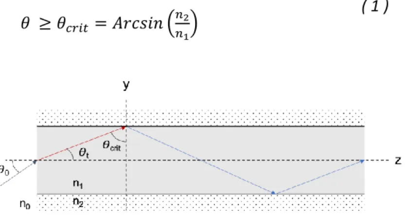
The different technologies based on optical fibers for large area were described by Mordon et al [1]. The technology developed in Lille used optical fibers. Briefly, optical fibers are optical structures, which allow incident light, usually from an optical source, to be guided by a series of internal total reflections that occurs under angular conditions, with minimum losses [1]. Standard step-index optical fiber is composed of a core and a surrounding cladding of cylindrical shapes, and with respective refractive indices and . Geometrical optics defines a particular angle called as the smallest angle of incidence of a ray at
which no refraction occurs at the boundary of two media when . General angular condition of total reflection of an incident light ray of angle is given in (1).
( 1 )
Figure 1: principle of light propagation in an optical fiber
In the optical fiber, total internal reflection occurs for light rays of smaller angles than the angle of acceptation , defined as the minimum angle of incidence to obtain a refracted light ray of angle that satisfies the general angular condition of total reflection ( 1 ). Otherwise, the portion of the incident light rays reflected and/or refracted is described by the Fresnel equations.
( 2 )
Local microscopic variations of core medium density from manufacturing process (variation in density, orientation or molecular composition of the material) lead to local variations in the refractive index, and generate losses by scattering of the light rays. Linear attenuation corresponds to the sum of all absorption and scattering losses that occur in the optical fiber, and is defined by the attenuation coefficient [2].
3 ( 3 )
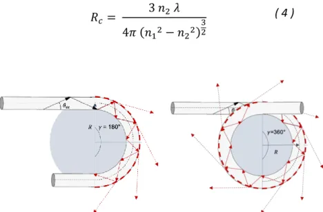
Additional bendings can increase the optical fiber attenuation coefficient, by inducing light leakage through the core. Macrobending is defined as a mechanical stress, which can be punctual or repeated [3], and is characterized by the critical radius of curvature which represent the bending radius from which macrobending losses become significant [4]. When an optical fiber is bent with a bending radius smaller than the critical bending radius
, the angle of incidence of the ray may become smaller than and the ray refracts
within the cladding and part of the ray may be refracted outside the optical fiber [2] (figure 2). The bending radius is associated with the angle of curvature which gives information on the length of the bent section[5][6] .
( 4 )
Figure 2: principle of light emission be optical fiber due to bending
Bending light losses within optical fibers are typically characterized with the objective of minimizing them as much as possible, especially for telecommunication or power transmission applications. In some cases, they are quantified to measure deformations within materials using fiber optic sensors, and maximized when homogeneous light emitting surfaces are desired [7]
By integration of plastic optical fibers within knitted or woven structure, light emission can be obtained over flexible textile surfaces. Homogeneity of spatial light distribution can be obtained under the condition of controlling the density of the fibers and the angles and radii of curvature [8].
Optical fibers are generally woven as conventional yarn according to various satin weave structures along the fabrics length to control light emission [9-11]. As knitting involves bending radii that are too severe to be supported without risk of breakage if the optical fiber
4
is knitted, optical fibers are mainly laid in a partial weft in a warp or weft knitted structures [12] in a straight line or in special patterns [13].
Plastic optical fibers can be gathered and glued within a metallic bundle in order to be coupled to any LASER source by the mean of 2 beam expanders (figures 3 & 4). The injection of light at each end of the textile, allows to balance the bending losses providing a more uniform and intense lateral emission [14].
Figure 3: LEF is illuminated by injecting light at each end of the fibers gathered in a bundle
Figure 4: Light Emitting Fabrics obtained by a knitting process connected to a red laser source (635nm)
LEF is composed of plastic optical fibers. Consequently, it can be connected to lasers of any wavelength from 400 nm to 1200 nm (figure 5).
5 Figure 5: LEF can emit several wavelengths from violet to infrared
In vitro use of LEF : CELL-LEF
Published PDT preclinical studies described various kinds of light sources. In many cases, the light source is handmade with optical fibers connected to laser or with LED panels [15] [16]. However, OF can deliver light only on small areas, while LED panels provide incoherent light with broad spectral width. Although easy to use and quite inexpensive, optical fibers and LED panels do not allow an effective homogenous illumination.
In vitro PDT studies often require illumination of several cells plates either all at once or all within a short period of time. In this context, a cells illuminator, CELL-LEF, able to illuminate several 96-well cell culture plate simultaneously with a homogeneous light has been designed (Figure 6).
CELL-LEF embeds two large LEF (total illumination surface: 750 cm2) sandwiched and kept in place between two rigid, transparent plastic sheets. These sheets also allow protection of the LEF and easy disinfection of CELL-LEF before and after use. Before being sandwiched, the two LEF are jointly sewn on a white textile in order to reflect the light emitted by the bottom face and therefore increase the quantity of light on the top face. A template is placed on the top plastic sheet in order to indicate the emplacement of the 6 multi-well plates (resulting illumination surface: 657 cm2). Finally, a lightproof cover can be used to protect cells from stray light, which could lead to undesired activation of the PS.
6 Figure 6: Schematic view of CELL-LEF, which is made of several layers, from top to down: first rigid transparent plastic sheet, light emitting fabrics (LEF), reflecting white textile, second rigid transparent
plastic sheet, and support feet.
Figure 7: Photograph of one CELL-LEF device. http://www.oncothai.fr/about-the-research-unit/technologies/361-preclinical-illumination-device-in-vitro
For all measurements, CELL-LEF was connected to a 635 nm laser (ONCO THAI, Lille, France) set to achieve a target mean irradiance of 1 mW/cm2. Different tests were performed in order to evaluate the homogeneity of irradiance, temperature evaluation of cell during illumination of 96-well cell culture plates. The measurement methodology has been already described [17]
7 With the CELL-LEF illuminator, irradiance values range from 0.81 to 1.18 mW/cm2 (mean: 0.98 mW/cm2; standard deviation: 0.11 mW/cm2). To obtain these values, a laser output power of 2.6 W was required. Homogeneity was determined using an automatic measurement system specifically developed for this purpose. A homogeneity of 90.9% was recorded. CELL-LEF was classified exempt risk group for all hazard groups according to the IEC 62471 standard, and does not exceed Accessible Emission Limits of class 1 defined by IEC 60825 standard
Figure 8: 3D representation of irradiance distribution over the light emitting fabric surface
At last, temperature elevation measurements inside 96 well plate gave the following results 45 minutes of CW illumination: for a well with cells with 5-ALA, an increase of +1.14 °C was measured but it was only +0.88 °C inside a well with cells without 5-ALA [18] .
CELL-LEF was already used in experimental studies to evaluate a new folate receptor-targeted photosensitizer on peritoneal ovarian cancer cells [19] and in four pancreatic adenocarcinoma (ADKP) i cell lines: Capan-1, Capan-2, MiapaCa-2, and Panc-1. [20]
.
LEF for in vivo experimental evaluation of PDT (VIVO-LEF)
In the framework of the development of an original humanized SCID mouse model of ovarian peritoneal carcinomatosis, a specific device dedicated to mice illumination. A mice box, called VIVO-LEF was developed to illuminate three mice simultaneously with a homogeneous light (Figure 9). VIVO-LEF consists of two separated white 3D-printed plastic bases, on which two light emitting fabrics (LEF) are fixed. The bases are designed to form three cavities, in which mice can be placed in prone position (Figure 10). The materials used make VIVO-LEF strong and lightweight. The total surface of illumination of 250 cm2 allows to
8
cover the whole body of the three mice. For the in vivo experiments performed on the SCID mouse model of ovarian peritoneal carcinomatosis, an irradiance of 11.08 0.58 mW/cm2 is delivered. Since, LEF are secondary light source, VIVO-LEF does not emit heat. Thanks to these performances, VIVO-LEF is far superior to OLED which are limited by their low irradiance and important temperature increase [21].
Figure 9: 3D illustration of the VIVO_LEF device.
Figure 10: VIVO-LEF can illuminate 3 mice simultaneously with homogeneous light
Clinical study #1: Evaluating illumination of actinic keratosis with a flexible LEF compared to the conventional photodynamic therapy with a LED panel: NCT03076918
In dermatology, PDT is used to treat actinic keratosis. Actinic keratosis are common pre-invasive cancerous lesions in sun-exposed skin which negatively affect the quality of life in patients and may progress to invasive squamous cell carcinoma. Actinic keratosis usually
9
develop on areas that are frequently exposed to the sun (e.g., face, ears, scalp, neck, forearms, and back of the hands). Studies have shown that if actinic keratosis are untreated, actinic keratosis may regress, or alternatively, may progress to squamous cell carcinoma, with significant morbidity and possible lethal outcome. Predicting which actinic keratosis may progress to squamous cell carcinoma is not possible, nor is the conversion rate for an actinic keratosis to squamous cell carcinoma clear: the transformation rate from an actinic keratosis lesion to squamous cell carcinoma within one year has been reported to be <1:1000. The malignant potential and the fact that it is impossible to predict which actinic keratosis will evolve into squamous cell carcinoma, have led to the common consensus that actinic keratosis have to be treated. Because of the high prevalence of actinic keratosis, their treatment represents a substantial workload, and must therefore be efficacious and easy to perform. Moreover, for patients an ideal treatment should be well tolerated and result in good cosmesis. The most commonly used treatments for actinic keratosis are cryotherapy, topical chemotherapy and, more recently, photodynamic therapy (PDT) [22] However, for this application, PDT is carried out with a wide variety of light sources delivering a broad range of more or less adapted light doses. Due to the complexities of the human anatomy, these light sources do not in fact deliver a uniform light distribution to the skin. For example, in the case of the LED system used usually in Dermatology, Moseley et al demonstrated that the irradiance may be as low as 38% of the central area at a distance of only 2 cm [23].
The device consists of 3 flexible light-emitting fabrics (size 21.5 cm × 5 cm each) for a total area of 3 × 21.5 cm × 5 cm = 322.5 cm2). Each one is illuminated sequentially with a 635 nm laser at low fluence rate (12.3 mW/cm2) for one minute, such as a fractionated irradiation (1 minute light, 2 minutes dark) is achieved (figure 11). An irradiation time of two and a half hours enables to deliver a total light dose of about 37 J/cm2 anywhere in the treated area (12.3 mW/cm2 × 9000 s × 1 minute light / (1 minute light + 2 minutes dark))[24].
The protocol involved a 30-minute incubation with MAL followed by a 2.5 h irradiation with a light-emitting fabric-based device (FLEXI-PDT). Due to the short incubation time, this device aimed to provide a nearly pain-free, all year round alternative to conventional PDT (C-PDT) performed with a LED panel, 3 hours after MAL application with 75 mW/cm² for 10 minutes[25]. Moreover, the high flexibility of the light-emitting fabric-based device ensured an optimal conformation of the device to the area to be treated, offering clear advantages over other protocols.
10 Figure 11: Each LEF sequentially emits 635 nm red light for one minute resulting in a fractionated
irradiation (1 minute light, 2 minutes dark).
FLEXI-PDT C-PDT Superiority p value for comparison between
randomized group Number of lesions 156 154
Complete lesion response rate (%) at 3 months
66.0 59.1 p=0.070
Complete lesion response rate (%) at 6 months
84.0 76.8 p=0.086
Pain experienced during the 1st treatment
0.4 ± 0.6 5.0 ± 2.6 p < 0.0001
Pain experienced during the 2nd treatment
0.2 ± 0.5 5.0 ± 2.2 p < 0.0001
Table 1: Complete response rate (lesion-level) achieved with FLEXI-PDT and C-PDT at 3 and 6 months [7].
For this clinical protocol, 27 patients were included in the study. Two patients dropped out for personal reasons before treatment. 25 patients with a total of 310 actinic keratosis were treated and examined at three months after the treatment. Due to remaining actinic keratosis, a second treatment session was required for 20 patients with a total of 252 actinic keratosis. Between three and six months following the first treatment session, one patient dropped out due to a serious adverse event not related to the treatment and one patient did not return for the 6-month visit for personal reason. 23 patients with 286 actinic keratosis therefore completed the study at 6 months.). Most of them had phototype II (76.0%). A total of 156 actinic keratosis, the majority of which were in grade I (42.3%) and II (56.4%),
11
received FLEXI-PDT. 154 actinic keratosis (grade I: 42.2%; grade II: 56.5%; grade III: 1.3%) received C-PDT[26].
At 3 month follow up, with 91 actinic keratosis in complete response and 63 actinic keratosis in incomplete response. C-PDT achieved a lesion complete response rate at three months of 59.1% vs 66.0% with FLEXI-PDT. At six months following treatment, the lesion complete response rate achieve 84.0% with FLEXI-PDT vs 76.8% with C-PDT. The response rate at six months for FLEXI-PDT (respectively, C-PDT) was around 1.3 (respectively, 1.3) times higher than that at three months. Similar local side effects, such as erythema and oedema, were observed with both FLEXI-PDT and C-PDT. Usual in dermatological PDT, these effects did not require any special care.
Evaluating illumination of actinic keratosis with an helmet incorporating a LEF compared to the Conventional Photodynamic Therapy: NCT03076918
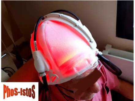
The second clinical evaluation of the LEF technology was carried out with an improved version of the previous device. The clinical protocol was similar to the one used except that the irradiance has been reduced from 12.3 mW/cm2 to 1.3 mW/cm2 and the light dose from 37 J/cm2 to 12 J/cm2. Furthermore, the device has been redesigned so as to be more ergonomic and compact (figure 12). A 21 cm × 18 cm surface (378 cm2) LEF was integrated inside an ergonomic helmet. This device was classified as exempt risk group according to IEC 60601-2-57/2012. [24]
Figure 12: image of the helmet delivering an irradiance of 1.3mW/cm² for 150 minutes for the treatment of Actinic Keratosis
12
For this clinical protocol, 47 patients were included in the study. (C-PDT) was performed as usual with a LED panel, 3 hours after MAL application with 75 mW/cm² for 10 minutes. The final analysis of this study gave the following results: One patient withdrew consent and did not receive treatment. Forty-six patients for a total of 560 actinic keratosis were treated in a split-face manner with C-PDT (285 actinic keratosis) and P-PDT (285 actinic keratosis), and evaluated at 3 months of follow-up. Due to at least one remaining actinic keratosis, 19 patients were required to undergo a second PDT session. Of these, one dropped out after the 3-month visit for fear of a pain as intense as that experienced with C-PDT during the first PDT session. As a result, only 18 patients (for a total of 105 remaining actinic keratosis of the 204 initial actinic keratosis at the first treatment session) were retreated. Forty-five patients completed the study at 6 months. All patients were men, aged 49-89 years (mean age 72.4 years). Most patients had Fitzpatrick skin types II (63.8%). Of the 285 actinic keratosis randomized to receive C-PDT (respectively, P-PDT), 45.6% (respectively, 44.9%) were in grade I and 54.4% (respectively, 55.1%) were in grade II [27].
At 3 month follow up, P-PDT was non-inferior to that obtained with C-PDT (79.3% vs. 80.7%, respectively. Six months following the first treatment session (after one PDT session for 27 patients and two PDT sessions for 18 patients). Whatever the protocol, almost all patients reported adverse effects throughout the study (100% with C-PDT vs. 97.8% with P-PDT). However, the incidence of adverse effects was lower with P-PDT than with C-PDT (161 vs. 264).
The more important observation was the quasi-absence of pain with P-PDT. With all the pain scores ranging from 0 to 2.7, P-PDT was reported to be almost pain-free. Regarding the first PDT session (46 patients), the treatment-related pain at the end of irradiation is significantly lower with P-PDT compared to C-PDT (0.3 ± 0.6 vs. 7.4 ± 2.3, p<0.0001). The same finding was also observed for the second PDT session (18 patients) (Figure 4) (0.2 ± 0.4 for P-PDT vs. 7.7 ± 1.8, p<0.0001 for C-PDT).
P-PDT C-PDT Superiority p value for comparison between
randomised group Number of lesions 280 280
Complete lesion response rate (%) at 3 months
80.7 79.3 p=0.34
Complete lesion response rate (%) at 6 months
94.9 94.2 p=0.66
Pain experienced during the 1st treatment
0.3 ± 0.6 7.4 ± 2.3 p < 0.0001
Pain experienced during the 2nd treatment
13 Table 2: Complete response rate (lesion-level) achieved with P-PDT and C-PDT at 3 and 6 months [8].
PAGETEX
Primary Extramammary Paget’s disease (EMPD) of the vulva is a rare skin cancer that mainly affects the genital region of elderly female population. Patients develop red eczematous and pruriginous plaques with a chronic evolution. Common dermatological symptoms and the lack of knowledge of the Paget’s disease often lead to late diagnosis. To control disease progression and symptoms usually experienced by patients, surgical excision is the mainstay of treatment. The excision can be a total vulvectomy, or be delimitated to the lesions with common margins of 2 cm in width and 0.5 cm in depth. However, recurrences are common (up to 58% within 15 months to 14 years) [28] [28], and recurrent patients suffer from severe functional and sexual alterations. Alternative treatments are studied like topical chemotherapy, laser ablation or radiotherapy but the adverse effects are numerous and the results are not enough superior to surgical excision. To date, none of these treatments can be considered as a solid alternative [29]. PDT is also studied [30-33] but unfortunately, the benefits of using photodynamic therapy for vulvar EMPD remains a challenge to demonstrate, because of the inhomogeneous illumination of vulvar and perianal areas, and the extreme pain that patients usually experienced during the illumination procedure that may lead to premature end of treatment [34, 35]. Inspired from the PHOS-ISTOS® study light parameters [36], the PAGETEX protocol (NCT03713203) involves the application of MAL cream for 30 minutes followed by 2.5 hours of illumination, without removing MAL-cream, such that a total light dose of approximately 12 J/cm² is delivered
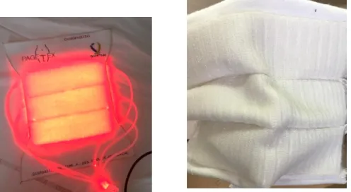
The PAGETEX® device was developed to fit the body shapes and provides a homogeneous light at the entry of the vagina, under the lips and on perianal region safely, [7, 14]. During the PDT treatment, the PAGETEX® device is placed over the vulva and maintained by pants (figures 13, 14). Patients can even slightly move during the illumination session, and also be accompanied while keeping intimacy.
14
Figure 13: PAGETEX® device connected with 635 nm LASER source – flat device (Left), folded device (Right)
Transparent occlusive panties keep the PAGETEX device completely isolated from the patient’s skin and thus to be reusable after specific cleaning.
Figure 14: PAGETEX® device connected with 635 nm LASER source during treatment of a patient
15 Conclusion
The different applications of Light Emitting Fabrics for photodynamic treatment show that this technology is well suited for homogeneous illumination of large areas. The technology used for manufacturing this LEF is very well known by the textile industry, leading to very competitive production costs. The fact that optical fibers can transmit light from 400 nm to 1200 nm allows the connection of LEF to different laser sources covering the light spectrum of all photosensitizers used for medical applications. New developments should allow to use the LEF inside cavities such as the pleural or the peritoneal cavities. At last, other applications such as baby jaundice treatment are already forecast.
Conflicts of interest
The LEF technology is now commercialized by the company, MDB Texinov in France. However, no author of this article has financial interest in the development of the LEF device with this company and consequently the authors have no conflicts to declare.
Acknowledgements
The authors acknowledge the French National Research Agency (ANR) (Projet-ANR-12-EMMA-0018), the European Commission for funding the Phosistos project under the Competitiveness and Innovation Framework Programme (CIP 621103) & the Region Hauts-de-France (European FEDER funding 2017_03029)
References
[1] Jenkins FA, White HE: Fundamentals of Optics 4th Edition. Mc Graw Hill. [2] Agrawal G: Fiber-Optic Communication Systems: Fourth Edition, 2012.
[3] Jay JA: An Overview of Macrobending and Microbending of Optical Fibers. Corning, 2011. [4] Dutton HJR: Understanding Optical Communications: International Technical Support
Organization - IBM, 1998.
[5] Endruweit A, Long AC, Johnson MS: Textile composites with integrated optical fibres:
quantification of the influence of single and multiple fibre bends on the light transmission using a Monte Carlo ray-tracing method. Smart Materials and Structures 2007, 17:015004.
[6] Liu H, Yager P: Modeling of optical bending losses in multimode waveguides by ray
tracing. Photonics West '95. Edited by Katzir JAHDMHA. San Jose, CA, United States.: SPIE, 1995.
[7] Thecua E, Ziane L, Baert G, Deleporte P, Leroux. B, Kumar A, Baydoun M, Morales O,
Delhem N, Mordon S: Light emitting fabric for photodynamic treatment of actinic keratosis. SPIE BIOS. Edited by SPIEDigitalLibrary. San Francisco, 2017.
16 [8] Oguz Y, Cochrane C, Koncar V, Mordon SR: Doehlert experimental design applied to
optimization of light emitting textile structures. Optical Fiber Technology 2016, 30:38-47.
[9] Quandt BM, Pfister MS, Lubben JF, Spano F, Rossi RM, Bona GL, Boesel LF: POF-yarn
weaves: controlling the light out-coupling of wearable phototherapy devices. Biomed Opt Express 2017, 8:4316-30.
[10] Shen J, Chui C, Tao X: Luminous fabric devices for wearable low-level light therapy.
Biomed Opt Express 2013, 4:2925-37.
[11] Cochrane C, Mordon SR, Lesage JC, Koncar V: New design of textile light diffusers for
photodynamic therapy. Mater Sci Eng C Mater Biol Appl 2013, 33:1170-5.
[12] Martens Y, Wehlage D, Ehrmann A: Knitting with optical fibers, 2016.
[13] Gong Z, Ziyang X, OuYang X, Zhang J, Lau N, Zhou J, Chan C: Wearable Fiber Optic
Technology Based on Smart Textile: A Review. Materials 2019, 12:3311.
[14] Mordon S, Cochrane C, Tylcz JB, Betrouni N, Mortier L, Koncar V: Light emitting fabric
technologies for photodynamic therapy. Photodiagnosis Photodyn Ther 2015, 12:1-8.
[15] Doix B, Bastien E, Rambaud A, Pinto A, Louis C, Gregoire V, Riant O, Feron O: Preclinical
Evaluation of White Led-Activated Non-porphyrinic Photosensitizer OR141 in 3D Tumor Spheroids and Mouse Skin Lesions. Front Oncol 2018, 8:393.
[16] Shi L, Buchner A, Pohla H, Pongratz T, Ruhm A, Zimmermann W, Gederaas OA, Zhang L,
Wang X, Stepp H, Sroka R: Methadone enhances the effectiveness of 5-aminolevulinic acid-based photodynamic therapy for squamous cell carcinoma and glioblastoma in vitro. J Biophotonics 2019, 12:e201800468.
[17] Thecua E, Ziane L, Baert G, Deleporte P, Leroux B, Kumar A, Baydoun M, Morales O,
Delhem N, Mordon S: Devices based on Light Emitting Fabrics dedicated to PDT preclinical studies. 17th International Photodynamic Association World Congress, 2019, Cambridge, Massachusetts, United States. Edited by Hasan T. Boston, MA, USA: SPIE, 2019.
[18] Ziane L, Thecua E, Deleporte P, Baert G, Vignion-Dewalle AS, Mordon S: Illumination
device based on light emitting fabrics for in-vitro photodynamic therapy preclinical studies. Photochemical & Photobiological Sciences in press.
[19] Baydoun M, Morales O, Frochot C, Ludovic C, Leroux B, Thecua E, Ziane L, Grabarz A,
Kumar A, de Schutter C, Collinet P, Azais H, Mordon S, Delhem N: Photodynamic Therapy Using a New Folate Receptor-Targeted Photosensitizer on Peritoneal Ovarian Cancer Cells Induces the Release of Extracellular Vesicles with Immunoactivating Properties. J Clin Med 2020, 9.
[20] Quilbe A, Morales O, Baydoun M, Kumar A, Mustapha R, Murakami T, Leroux B, de
Schutter C, Thecua E, Ziane L, Colombeau L, Frochot C, Mordon S, Delhem N: An Efficient Photodynamic Therapy Treatment for Human Pancreatic Adenocarcinoma. J Clin Med 2020, 9.
[21] Guo HW, Lin LT, Chen PH, Ho MH, Huang WT, Lee YJ, Chiou SH, Hsieh YS, Dong CY, Wang
HW: Low-fluence rate, long duration photodynamic therapy in glioma mouse model using organic light emitting diode (OLED). Photodiagnosis Photodyn Ther 2015, 12:504-10.
[22] Peris K, Fargnoli MC: Conventional treatment of actinic keratosis: an overview. Curr
17 [23] Moseley H: Light distribution and calibration of commercial PDT LED arrays. Photochem
Photobiol Sci 2005, 4:911-4.
[24] Vignion-Dewalle AS, Abi Rached H, Thecua E, Lecomte F, Deleporte P, Behal H, Hommel
T, Duhamel A, Szeimies RM, Mortier L, Mordon S: A New Light-Emitting, Fabric-Based Device for Photodynamic Therapy of Actinic Keratosis: Protocol for a Randomized, Controlled, Multicenter, Intra-Individual, Phase II Noninferiority Study (the Phosistos Study). JMIR Res Protoc 2019, 8:e12990.
[25] Lecomte F, Vignion-Dewalle AS, Vicentini C, Thecua E, Deleporte P, Duhamel A, Mordon
S, Mortier L: Evaluating the Noninferiority of a New Photodynamic Therapy (Flexitheralight) Compared With Conventional Treatment for Actinic Keratosis: Protocol for a Phase 2 Study. JMIR Res Protoc 2019, 8:e11530.
[26] Vicentini C, Vignion-Dewalle AS, Thecua E, Lecomte F, Maire C, Deleporte P, Behal H,
Kerob D, Duhamel A, Mordon S, Mortier L: Photodynamic therapy for actinic keratosis of the forehead and scalp: a randomized, controlled, phase II clinical study evaluating the noninferiority of a new protocol involving irradiation with a light-emitting, fabric-based device (the Flexitheralight protocol) compared with the conventional protocol involving irradiation with the Aktilite CL 128 lamp. Br J Dermatol 2019, 180:765-73.
[27] Mordon S, Vignion-Dewalle AS, Abi-Rached H, Thecua E, Lecomte F, Vicentini C,
Deleporte P, Behal H, Kerob D, Hommel T, Duhamel A, Szeimies RM, Mortier L: The conventional protocol vs. a protocol including illumination with a fabric-based biophotonic device (the Phosistos protocol) in photodynamic therapy for actinic keratosis: a randomized, controlled, noninferiority clinical study. Br J Dermatol 2020, 182:76-84.
[28] Parker LP, Parker JR, Bodurka-Bevers D, Deavers M, Bevers MW, Shen-Gunther J,
Gershenson DM: Paget's disease of the vulva: pathology, pattern of involvement, and prognosis. Gynecologic oncology 2000, 77:183-9.
[29] Raspagliesi F, Fontanelli R, Rossi G, Ditto A, Solima E, Hanozet F, Kusamura S:
Photodynamic therapy using a methyl ester of 5-aminolevulinic acid in recurrent Paget's disease of the vulva: a pilot study. Gynecologic oncology 2006, 103:581-6.
[30] Clement E, Sparsa A, Doffoel-Hantz V, Durox H, Prey S, Bonnetblanc JM, Caly H, Aubard
Y, Bedane C: [Photodynamic therapy for the treatment of extramammary Paget's disease]. Ann Dermatol Venereol 2012, 139:103-8.
[31] Fontanelli R, Papadia A, Martinelli F, Lorusso D, Grijuela B, Merola M, Solima E, Ditto A,
Raspagliesi F: Photodynamic therapy with M-ALA as non surgical treatment option in patients with primary extramammary Paget's disease. Gynecologic oncology 2013, 130:90-4.
[32] Kim SJ, Thompson AK, Zubair AS, Otley CC, Arpey CJ, Baum CL, Roenigk RK, Lohse CM,
Brewer JD: Surgical Treatment and Outcomes of Patients With Extramammary Paget Disease: A Cohort Study. Dermatol Surg 2017, 43:708-14.
[33] Louis-Sylvestre C, Haddad B, Paniel BJ: Paget's disease of the vulva: results of different
conservative treatments. European journal of obstetrics, gynecology, and reproductive biology 2001, 99:253-5.
[34] Gao Y, Zhang XC, Wang WS, Yang Y, Wang HL, Lu YG, Fan DL: Efficacy and safety of
18 [35] Rioli DI, Samimi M, Beneton N, Hainaut E, Martin L, Misery L, Quereux G: Efficacy and
tolerance of photodynamic therapy for vulvar Paget's disease: a multicentric retrospective study. Eur J Dermatol 2018, 28:351-5.
[36] Mordon S, Vignion-Dewalle AS, Abi-Rached H, Thecua E, Lecomte F, Vicentini C,
Deleporte P, Behal H, Kerob D, Hommel T, Duhamel A, Szeimies RM, Mortier L: The conventional protocol versus a protocol including illumination with a fabric-based biophotonic device (the Phosistos protocol) in photodynamic therapy for actinic keratosis: a randomized, controlled, non-inferiority clinical study. Br J Dermatol 2019.
19
Light emitting fabrics for Photodynamic Therapy: technology, experimental and clinical Applications" (Manuscript ID : tbio. 202000005. R2)
Graphical abstract :
By integration of plastic optical fibers within knitted or woven structure, light emission can be obtained over large and flexible textile fabrics. Homogeneity of spatial light distribution is obtained by controlling the density of the fibers and the angles and radii of curvature. These Light Emitting fabrics (LEF) can be connected to any light source form 400nm to 1200 nm. LEF are now used for preclinical and clinical applications of phtotodynamic therapy. Several examples are given into this article.


![Table 1: Complete response rate (lesion-level) achieved with FLEXI-PDT and C-PDT at 3 and 6 months [7]](https://thumb-eu.123doks.com/thumbv2/123doknet/14691367.561504/11.892.112.789.105.425/table-complete-response-lesion-level-achieved-flexi-months.webp)