HAL Id: hal-02906377
https://hal.archives-ouvertes.fr/hal-02906377
Submitted on 24 Jul 2020
HAL is a multi-disciplinary open access
archive for the deposit and dissemination of
sci-entific research documents, whether they are
pub-lished or not. The documents may come from
teaching and research institutions in France or
abroad, or from public or private research centers.
L’archive ouverte pluridisciplinaire HAL, est
destinée au dépôt et à la diffusion de documents
scientifiques de niveau recherche, publiés ou non,
émanant des établissements d’enseignement et de
recherche français ou étrangers, des laboratoires
publics ou privés.
the Polymeric Encapsulation of a Ru(II) Polypyridine
Complex with Pluronic F-127 for Photodynamic
Therapy Applications
Johannes Karges, Hui Chao, Gilles Gasser
To cite this version:
Johannes Karges, Hui Chao, Gilles Gasser. Synthesis, Characterisation and Biological Evaluation
of the Polymeric Encapsulation of a Ru(II) Polypyridine Complex with Pluronic F-127 for
Photody-namic Therapy Applications. European Journal of Inorganic Chemistry, Wiley-VCH Verlag, 2020,
�10.1002/ejic.202000545�. �hal-02906377�
Synthesis, Characterisation and Biological Evaluation of the
Polymeric Encapsulation of a Ru(II) Polypyridine Complex with
Pluronic F-127 for Photodynamic Therapy Applications
Johannes Karges,
[a]Hui Chao,*
[b]and Gilles Gasser*
[a]Abstract: The therapy of cancer remains a major challenge for
modern medicine. Complementary to classical treatments, the use of
photodynamic therapy (PDT) has received increasing attention over
the last decades. Among the classes of PDT photosensitizers (PSs)
investigated, Ru(II) polypyridine complexes are currently considered
as a valuable option. To improve the water solubility of these lipophilic
compounds and generate a drug delivery system, these complexes
can be encapsulated into polymers. Herein, the physical
encapsulation of the photoactive
[Ru((E,E’)-4,4´-bis[p-methoxystyryl]-2,2´-bipyridine)
3][PF
6]
2complex with the polymer Pluronic F-127 is
presented. The resulting spherical particles were found to have a toxic
effect in the micromolar range in human cervical carcinoma cells upon
light irradiation (500 nm).
Introduction
The use of metal complexes in medicine has started to develop
during the last century after the discovery of Salvarsan (mixture
of 3-amino-4-hydroxyphenyl-arsenic(III) compounds) by Ehrlich et
al. in 1912
[1]and cisplatin (cis-diamminodichloroplatinum(II)) by
Rosenberg et al. in the 1960’s.
[2]These compounds, which
had/have a tremendous impact on human health, have promoted
the development of other metal-based compounds.
[3]To date,
metal complexes have received special attention in the treatment
of cancer, one of the deadliest diseases worldwide. For example,
the Pt(II)-containing chemotherapeutic drug cisplatin and its
derivatives are very commonly used to treat this disease. Despite
their undeniable success, these drugs are associated with severe
side effects (e.g., nerve and kidney damage, nausea, vomiting,
and bone marrow suppression) as well as an increasing number
of resistance problems, limiting their clinical applications.
[4]As a complementary technique to the traditional medical
treatments against cancer (i.e., surgery, chemotherapy,
radiotherapy), photodynamic therapy (PDT) has received
increasing attention over the last decades. It relies on the
combination of a photosensitiser (PS), light and oxygen. During a
PDT treatment, the PS is selectively activated upon light
irradiation to generate reactive oxygen species (ROS). These
species can cause significant cell damage and ultimately trigger
cell death.
[5]Metal complexes,
[6]and especially Ru(II) polypyridine
complexes,
[7]are currently being investigated as PSs for PDT due
to their ideal photophysical properties. One of such compounds,
namely TLD-1433 from the group of McFarland, is currently in
phase II clinical trial against bladder cancer.
[8]However, the
majority of Ru(II) polypyridine complexes lack significant
absorption in the phototherapeutic window (600-900 nm), which
limits their application for PDT purposes.
[9]For this reason,
research effort are devoted towards the development of
compounds with absorption at these wavelengths.
[10]One
possibility to reach this aim is to expend the aromatic system of
the ligands bound to the Ru(II) centre. However, such synthetic
modifications usually engenders a decrease of the water solubility
of the generated compounds, limiting their applications.
[11]An
additional limitation of the currently investigated and clinically
applied PDT agents is their poor cancer cell selectivity, resulting
in the use of high concentrations for a desired therapeutic
outcome. To overcome this drawback, there is a need for the
development of suitable drug delivery systems. To date, a variety
of different delivery carriers for Ru(II) polypyridine complexes
have
been
reported,
including
polymeric/physical
encapsulations,
[12]loading into nanoparticles
[13], conjugation to
carbon nanotubes,
[14]receptor-targeting moieties/peptides,
[15]antibodies,
[16]or metal-organic frameworks.
[17]However, the
majority of these drug delivery systems are associated with either
a tedious preparation, high price, poor water solubility or reduced
therapeutic effect. There is therefore a need for the development
of a cheap, easy-to-prepare and selective drug carrier. Among
these techniques, the physical encapsulation of a compound into
polymeric particles is known to be synthetically not challenging
and to highly improve water solubility. It also allows to specifically
target tissue thanks to the enhanced permeability and retention
(EPR) effect,
[18]although this concept is currently controversially
discussed.
[19]Among others, poloxamers are investigated as a class of
polymers carriers, which are not or only slowly biodegradable but
biocompatible. These polymeric materials consist of a non-ionic
triblock ABA structure with block A as hydrophilic poly(ethylene
oxide) and block B as hydrophobic poly(propylene oxide). Based
on this difference in water solubility, the polymeric material is able
to self-assemble into spherical micelles, which can be loaded with
hydrophobic compounds.
[20]Recently, Sadler et al. successfully
encapsulated
the
[Ru(p-cymene)(1,2-dicarba-closo-dodecarborane-1,2-dithiolato)] complex for boron neutron capture
therapy with the Pluronic P-123 polymer. The generated micelles
were shown to have a decreased toxicity profile while having an
increased selectivity towards cancer cells in comparison to
non-[a] Dr. Johannes Karges, Dr. Gilles GasserChimie ParisTech, PSL University, CNRS, Institute of Chemistry for Life and Health Sciences, Laboratory for Inorganic Chemical Biology, 75005 Paris, France.
E-mail: gilles.gasser@chimieparistech.psl.eu Homepage: www.gassergroup.com [b] Prof. Hui Chao
MOE Key Laboratory of Bioinorganic and Synthetic Chemistry, School of Chemistry, Sun Yat-Sen University, 510275 Guangzhou, People’s Republic of China.
E-mail: ceschh@mail.sysu.edu.cn
Supporting information for this article is given via a link at the end of the document.
cancerous cells.
[21]The group of Howard et al. has reported the
encapsulation of [Ru(4,7-diphenyl-1,10-phenanthroline)
3]
2+with
Poloxamer-407/Pluronic F-127 as a two-photon excited oxygen
imaging agent in aqueous solution.
[22]Lemercier et al.
encapsulated the PDT PS [Ru(1,10-phenanthroline)
3]
2+derivatives into poly(D,L-lactide-co-glycolide) and Poloxamer
188/Pluronic F-68. They could show that their PS was slowly
released from the particles and had the ability to generate a
phototoxic effect in cancerous cells upon irradiation.
[23]Encouraged by the promising results obtained by our group on
the encapsulation of Ru(II)-based PDT PSs
[24], we report, in this
article, the encapsulation of the lipophilic Ru(II) polypyridine
complex
[Ru((E,E’)-4,4´-Bis[p-methoxystyryl]-2,2´-bipyridine)
3][PF
6]
2(Figure 1a), which we have recently reported
as an effective PS
[10b]with the polymer Poloxamer-407/Pluronic
F-127 (Figure 1b). The generated particles were found to have a
spherical shape as characterised by dynamic light scattering and
transmission electron microscopy. While stable in a biological
environment, the particles were able to trigger cell death in human
cervical carcinoma (HeLa) cells in the micromolar range upon
irradiation at 500 nm. Interestingly, the cytotoxic effect of the
particles could be correlated with their different loading ratios and
particle sizes.
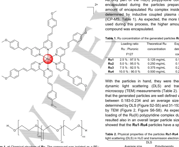
2+ N N N N N N Ru O O O O O Oa)
b)
H O O O O H 100 100 65Figure 1. a) Chemical structure of Ru. The compound was isolated as a PF6
-salt. b) Chemical structure of the block-polymer Pluronic F-127/Poloxamer-407.
Results and Discussion
Synthesis
The
Ru(II)
polypyridine
complex
[Ru((E,E’)-4,4´-Bis[p-methoxystyryl]-2,2´-bipyridine)
3][PF
6]
2(Ru, Figure 1a) was
synthesised as previously reported.
[10b]The purity of the
compound was verified by HPLC (Figure S1) and elemental
analysis. Due to the insolubility of Ru in H
2O, the compound was
encapsulated using the polymer poly(ethylene
glycol)-block-poly(propylene glycol)-block-poly(ethylene glycol), which is also
known as Pluronic F-127/Poloxamer-407 (M
n~ 10000 – 12000,
Figure 1b). In this study, the effect of different loadings (2.5 : 97.5
– Ru1 ; 5 : 95 – Ru2; 7.5 : 92.5 – Ru3; 10 : 90 – Ru4, wt% Ru :
Pluronic F-127) of the Ru complex with the polymer was
investigated. The encapsulation process was performed by
mixing a DCM phase of the Ru(II) polypyridine complex with a
H
2O phase of Pluronic F-127 in the presence of ultrasonic pulses,
as previously published for the encapsulation of metal
complexes.
[25]Following this, the DCM was evaporated and large
aggregates were removed by size exclusion chromatography,
resulting in a clear red aqueous solution.
Particle Characterisation
As only part of the Ru(II) polypyridine complex Ru could be
encapsulated during the particles preparation process, the
amount of encapsulated Ru complex inside the particles was
determined by inductive coupled plasma mass spectrometry
(ICP-MS, Table 1). As expected, the more Ru(II) complex was
used during this process, the higher amount of this lipophilic
compound was encapsulated.
Table 1. Ru concentration of the generated particles Ru1-Ru4. Loading ratio Ru : Pluronic F127 Theoretical Ru concentration Experimentally determined Ru concentration Yield Ru1 2.5 % : 97.5 % 0.125 mg/mL 0.108 mg/mL 86 % Ru2 5.0 % : 95.0 % 0.250 mg/mL 0.179 mg/mL 72 % Ru3 7.5 % : 92.5 % 0.375 mg/mL 0.237 mg/mL 63 % Ru4 10.0 % : 90.0 % 0.500 mg/mL 0.246 mg/mL 49 %
With the particles in hand, they were then characterised by
dynamic light scattering (DLS) and transmission electron
microscopy (TEM) measurements (Table 2). The results indicate
that the generated particles are well defined with a polydispersity
between 0.183-0.234 and an average size of 53-162 nm, as
determined by DLS (Figure S2-S5) and 31-153 nm, as determined
by TEM (Figure 2, Figure S6-S8). As expected, increasing the
loading of the Ru(II) polypyridine complex during their synthesis
resulted also in an overall larger particle size. The TEM images
showed that the Ru1-Ru4 particles have a spherical shape.
Table 2. Physical properties of the particles Ru1-Ru4 determined by dynamic light scattering (DLS) in H2O and transmission electron microscopy (TEM).
DLS TEM
Average size Polydispersity Average size Ru1 53 nm 0.183 31 ± 3 nm Ru2 58 nm 0.217 46 ± 5 nm Ru3 65 nm 0.196 56 ± 6 nm Ru4 162 nm 0.234 153 ± 8 nm
Figure 2. Representative TEM images of Ru3.
Photophysical evaluation
In order to verify that the photophysical properties of the complex
are not significantly influenced by the encapsulation due to, for
example, quenching effects, which would prevent the desired
PDT outcome, the photophysical properties (Table S1) of
Ru1-Ru4 were investigated and compared to the complex alone (Ru).
The absorption and emission spectra upon irradiation at 450 nm
show no significant differences. Interestingly, the particles
(Ru1-Ru4) show an increased luminescence quantum yield. This
finding can be explained: the encapsulated Ru(II) polypyridyl
complexes are surrounded by a lesser number of water molecules,
which could potentially quench their luminescence. The ability to
produce singlet oxygen (
1O
2
) was then investigated to confirm that
oxygen is able to reach the compound and to interact with the
excited state of the complex. For this purpose, the change in
absorbance of the
1O
2
scavenger 1,3-diphenyl-isobenzofuran was
time dependently monitored upon irradiation at 500 nm.
[26]The
results (Table S1) confirm that the particles (Ru1-Ru4) are able
to generate
1O
2
in a similar manner than the compound itself.
Overall, the photophysical evaluation indicated that the
encapsulation of the Ru(II) polypyridine complex did not influence
its excited state behaviour. This finding is in agreement with that
recently observed for the encapsulation of
[Ru(4,7-diphenyl-1,10-phenanthroline)
3]
2+with the same polymer
[22]as well as the
encapsulation
[Ru(2,2´-bipyridine)
2(dipyrido[3,2-a:2′,3′-c]phenazine-7-hydroxymethyl)]
2+with a polylactide polymer.
[24]Stability
As an important factor for biological applications, the stability of
the polymeric particles under biological conditions was
investigated as previous works have shown that this could be
problematic.
[23, 27]For this purpose, the particles were incubated
in H
2O and the cell medium DMEM and their absorption spectra
as well as their size distribution were monitored in various time
intervals (0, 2, 8, 12, 24, 48 h). As no significant changed were
observed within 48 h (Figure S9-S20), the stability of the particles
is confirmed. These findings are in agreement with stability
studies of particles formed by the encapsulation of
[Ru(4,7-diphenyl-1,10-phenanthroline)
3]
2+with the same polymer
carrier.
[22]Biological evaluation
After confirmation of the stability of the particles (Ru1-Ru4) in a
biological environment, the effect these particles have on cancer
cells was investigated. For this purpose, the compounds were
incubated in human cervical carcinoma (HeLa) cells in the dark
as well as exposed to a light irradiation at 500 nm (16.7 min, 10
J/cm
2)
and their cell viability measured (Table 3). All particle
formulations were found to have no cytotoxic effect in the dark up
to high micromolar concentrations (IC
50> 500 μM), as requested
for a PDT agent. Upon light exposure, the compounds were found
to be able to generate highly cytotoxic
1O
2
and therefore trigger
cell death in the micromolar range (IC
50= 93-261 μM).
Interestingly, the particle formulations Ru1-Ru3 were found to
have a stronger phototoxic effect than Ru4. Of note, Ru was
found to have a drastically stronger cytotoxic effect than its
corresponding nanoformulations.
Table 3. IC50 values (μM) in the dark and upon irradiation at 500 nm (16.7 min,
10 J/cm2) for the particles Ru1-Ru4 in comparison to Ru and cisplatin in human
cervical carcinoma (HeLa) cells. Average of three independent measurements. IC50 in the dark IC50 upon irradiation PI
Ru >50 13.6 ± 0.9 >3.7 Ru1 >500 93 ± 8 >5.4 Ru2 >500 104 ± 11 >4.8 Ru3 >500 117 ± 12 >4.3 Ru4 >500 261 ± 23 >1.9 cisplatin 11.2 ± 1.1 - -
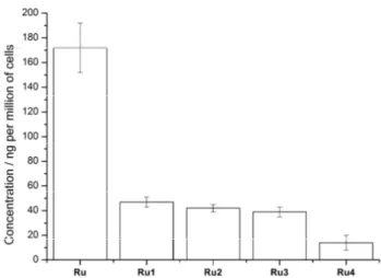
To understand these results, the cellular uptake of the particles
was investigated by incubating the compounds in HeLa cells and
determining the amount of Ru inside the cells by ICP-MS.
Interestingly, Ru has a much higher cellular uptake than the
particle formulations (Ru1-Ru4). In addition, the particles with a
smaller size were found to have a significantly higher cellular
uptake than the larger ones. Overall, these experiments explains
the superior phototoxic effect observed for Ru and the small
particles.
Figure 3. Comparison of the cellular uptake of Ru (50 μM) and its particle formulations Ru1-Ru4 after a 12 h incubation in HeLa cells.
Conclusions
In summary, we have synthesised and characterised polymeric
particles containing the photoactive Ru(II) polypyridine complex
[Ru((E,E’)-4,4´-bis[p-methoxystyryl]-2,2´-bipyridine)
3][PF
6]
2with
different loading ratios of the compound within the polymer carrier
Pluronic F-127. By increasing the amount of the Ru(II)
polypyridine complex during the encapsulation, we observed that
the size of the generated particles is increased. Photophysical
measurements showed that the particles are able to generate
highly cytotoxic singlet oxygen. The particles were found to have
a high stability in water and cell medium. While being non-toxic in
the dark up to high concentrations, the particles had a phototoxic
effect upon light irradiation at 500 nm in cancerous human
cervical carcinoma cells in the micromolar range. Interestingly,
thanks to ICP-MS measurements, the particles with a smaller size
were found to have a significant higher cellular uptake, explaining
their superior phototoxicity.
Experimental Section
MaterialsPoly(ethylene glycol)-block-poly(propylene glycol)-block-poly(ethylene glycol) with the tradename Pluronic F127 (Mn ~ 10000 - 12000) was
commercially obtained from Energy Chemical. Dulbecco's modified eagle medium (DMEM), fetal bovine serum (FBS) and penicillin were purchased from Invitrogen.
Instrumentation and methods
1H and 13C NMR spectra were recorded on a Bruker 500 MHz NMR
spectrometer. Chemical shifts (δ) are reported in parts per million (ppm) referenced to tetramethylsilane (δ 0.00) ppm using the residual proton solvent peaks as internal standards. Coupling constants (J) are reported in Hertz (Hz) and the multiplicity is abbreviated as follows: s (singulet), d (doublet). Elemental microanalyses were performed on a Thermo Flash 2000 elemental analyser. For analytic HPLC the following system has been used: 2 x Agilent G1361 1260 Prep Pump system with Agilent G7115A 1260 DAD WR Detector equipped with an Agilent Pursuit XRs 5C18 (100Å, C18 5 μm 250 x 4.6 mm) column and an Agilent G1364B 1260-FC fraction collector. The solvents (HPLC grade) were millipore water (0.1% TFA, solvent A) and acetonitrile (0.1% TFA, solvent B). The solvents (HPLC grade) were millipore water (0.1% TFA, solvent A) and acetonitrile (solvent B). Method: 0-3 minutes: isocratic 50% A (50% B); 3-17 minutes: linear gradient from 50% A (50% B) to 0% A (100% B). The flow rate was 1 mL/min and the chromatogram was detected at 250 nm. Inductive coupled plasma mass spectrometry (ICP-MS) experiments were carried out on an iCAP RQ ICP-MS instrument (Thermo Fisher). Synthesis
[Ru((E,E’)-4,4´-Bis[p-methoxystyryl]-2,2´-bipyridine)3][PF6]2 (Ru):
The compound was synthesised as previously reported.[10b] Purity was
assessed by HPLC and elemental analysis. Elemental analysis calcd. for C84H72F12N6O6P2Ru (%): C 61.05, H 4.39, N 5.09; found: C 61.17, H 4.44,
N 5.21.
Particle preparation
A solution of [Ru((E,E’)-4,4´-Bis[p-methoxystyryl]-2,2´-bipyridine)3][PF6]2
(Ru) in 0.5 mL DCM was added to a solution of Pluronic F-127 in 19.5 mL H2O in different weight ratios (2.5 : 97.5 – Ru1 ; 5 : 95 – Ru2; 7.5 : 92.5 –
Ru3; 10 : 90 – Ru4). The two phases were vigorously mixed to generate an emulsion. The solution was further treated with ultrasonic pulses with a Scientz – II D ultrasonic homogenizer using a 10 min method (tsonication = 2
s, Power = 15%, tbreak = 1 s) while keeping the sample constantly at 25 °C.
This method has been repeated a second time after a 5 min break. The DCM was removed by evaporation at 50 °C. Large aggregated were removed by size exclusion chromatography. After that a clear transparent solution in H2O was obtained.
Particle characterisation
The amount of encapsulated complex was determined using ICP-MS. Each sample was digested using a 60% HNO3 solution for three days,
followed by a 30% H2O2 solution for an additional day. After that time, the
sample was diluted to a solution of 2% HNO3 in water. The Ru content was
determined using an ICP-MS apparatus and comparing the results with the Ru references. The size and polydispersity of the particles was determined using dynamic light scattering (DLS) measurements with an Omni EliteSizer (Brookhaven). The size and morphology of the particles was determined by transmission electron microscopy (TEM) with a JEM-1400 Plus electron microscope (Jeol).
Spectroscopic measurements
The absorption of a sample was recorded on a Lambda 850 UV/VIS spectrometer (PerkinElmer) at room temperature. Emission spectra were recorded on a LS 55 fluorescence spectrometer (PerkinElmer) at room temperature. For the determination of the luminescence quantum yield, the samples were prepared with an absorbance of 0.2 at 450 nm in H2O. This
solution was irradiated at 450 nm and the emission signal measured. The luminescence quantum yields were determined by comparison with the reference [Ru(bipy)3]Cl2 in acetonitrile (Φem=5.9%)[28] applying the
following formula:
Φem,S = Φem,R * (FR / FS) * (IS / IR) * (nS / nR)2
F = 1 – 10-A
Φem = luminescence quantum yield, F = fraction of light absorbed, I =
integrated emission intensities, n = refractive index, A = absorbance of the sample at irradiation wavelength, S = sample, R = reference.
Singlet oxygen measurements
The singlet oxygen production (Φ(1O2)) was measured my monitoring the
change of the absorbance of the 1O2 scavenger
1,3-Diphenylisobenzofuran. The samples were prepared in a H2O solution
containing the complex with an absorbance of 0.2 at 500 nm and DPBF (30 μM). The samples were aerated and irradiated at 500 nm using different time intervals. The absorbance of the samples at 411 nm was measured during these time intervals with a Lambda 850 UV/VIS spectrometer (PerkinElmer). The difference in absorbance (A0-A) was
calculated and plotted against the irradiation times. From the plot the slope of the linear regression was calculated as well as the absorbance correction factor determined. As reference for the measurement rose bengal (Φ(1O2)rose bengal =76%)[29] in methanol was used. The singlet
oxygen quantum yields were calculated applying the following formula: Φ(1O
2)S = Φ(1O2)R * (BS / BR) * (IR / IS)
I = I0 * (1 – 10-A)
Φ(1O
2) = singlet oxygen quantum yield, B = slope of the linear regression
of the plot of the areas of the singlet oxygen luminescence peaks against the irradiation intensity, I = absorbance correction factor, I0 = light intensity
of the irradiation source, A = absorbance of the sample at irradiation wavelength, S = sample, R = reference.
Stability measurements by UV/Vis spectroscopy
The stability of a sample was investigated upon incubation in water and the cell medium DMEM. In various time intervals (0, 2, 8, 12, 24, 48 h) the absorption of the samples was recorded with a Lambda 850 UV/VIS spectrometer (PerkinElmer).
Stability measurements by DLS
The stability of the sample was investigated upon incubation in water and the cell medium DMEM. In various time intervals (0, 2, 8, 12, 24, 48 h) the size distribution of the particles was recorded with an Omni EliteSizer (Brookhaven) apparatus.
Cell culture
Human cervical carcinoma (HeLa) cells were obtained from the American Type Culture Collection (Manassas, VA). The cells were cultured using DMEM media with addition of 10% FBS and 1% penstrep. The cells were
cultivated and maintained in a cell culture incubator at 37 °C with 5% CO2
atmosphere. Before an experiment, the cells were passaged three times. (Photo-)cytotoxicity
The cytotoxicity of a sample was accessed using a 3-(4,5-dimethylthiazol-2-yl)-2,5-diphenyltetrazolium bromide (MTT) assay. Cells were seeded in 96 well plates (1000 cells per well in 100 μL of media) and allowed to adhere upon incubation for 24 h. The cells were treated with increasing concentrations of the compounds diluted in cell media achieving a total volume of 200 μL. All cells were incubated in the dark for 12 h. After this time the culture medium was refreshed. For the phototoxicity studies, the cells were exposed to a 500 nm LED irradiation (16.7 min, 10 J/cm2). The
cells were grown in the incubator for additional 36 h. For the determination of the dark cytotoxicity, the cells were not irradiated and after the media exchange directly incubated for 36 h. After this time, the media was replaced with fresh media containing MTT (10 μL/well). After 4h incubation, the absorption at 595 nm in each well was determined with a SpectraMax CMax Plus (Molecular Devices) absorbance microplate reader. The viability of the cells in each well was obtained by subtracting the average absorbance of the blank group. The obtained data was analysed with the GraphPad Prism software.
Cellular uptake
The cellular uptake of a sample was investigated by determining the Ru content inside the cells. The compound with a final concentration of 50 μM were incubated for 12 h at 37 °C on a cell culture dish with a density of ca. 6*106 cells in 10 mL of media. After this time, the media was removed and
the cells washed with cell media. The cells were trypsinised, harvested, centrifuged and resuspended. The number of cells on each dish was accurately counted. Each sample was the digested using a 60% HNO3
solution for three days followed by a 30% H2O2 solution for an additional
day. Each sample was diluted to solution of 2% HNO3 in water. The Ru
content was determined using an ICP-MS apparatus and comparing the results with the Ru references. The Ru content was then associated with the number of cells.
Acknowledgments
This work was financially supported by an ERC Consolidator
Grant PhotoMedMet to G.G. (GA 681679) and has received
support under the program “Investissements d’ Avenir” launched
by the French Government and implemented by the ANR with the
reference ANR-10-IDEX-0001-02 PSL (G.G.), the National
Science Foundation of China (Nos. 21525105 and 21778079 for
H.C.) and the 973 Program (No. 2015CB856301 for H.C.).
Keywords: Anticancer • Medicinal Inorganic Chemistry • Metals
in Medicine • Photodynamic Therapy
[1] a) P. Ehrlich, A. Bertheim, Ber. Dtsch. Chem. Ges. 1912, 45, 756-766; b) S. Gibaud, G. Jaouen, Medicinal Organometallic Chemistry (Eds.: G. Jaouen, N. Metzler-Nolte), Springer, Berlin, Heidelberg, 2010, 1-20.
[2] a) B. Rosenberg, L. Van Camp, T. Krigas, Nature 1965, 205, 698-699; b) B. Rosenberg, L. Vancamp, J. E. Trosko, V. H. Mansour, Nature 1969, 222, 385-386.
[3] a) K. J. Franz, N. Metzler-Nolte, Chem. Rev. 2019, 119, 727-729; b) R. G. Kenny, C. J. Marmion, Chem. Rev. 2019, 119, 1058-1137; c) X. Wang, X. Wang, S. Jin, N. Muhammad, Z. Guo, Chem. Rev. 2019, 119, 1138-1192; d) A. Chao, P. J. Sieminski, C. P. Owens, C. W. Goulding, Chem. Rev. 2019, 119, 1193-1220; e) A. Y. Chen, R. N. Adamek, B. L. Dick, C. V. Credille, C. N. Morrison, S. M. Cohen,
Chem. Rev. 2019, 119, 1323-1455; f) B. Englinger, C. Pirker, P. Heffeter, A. Terenzi, C. R. Kowol, B. K. Keppler, W. Berger, Chem. Rev. 2019, 119, 1519-1624; g) E. Boros, P. J. Dyson, G. Gasser, Chem 2019, 6, 41-60.
[4] a) C. X. Zhang, S. J. Lippard, Curr. Opin. Chem. Biol. 2003, 7, 481-489; b) T. C. Johnstone, K. Suntharalingam, S. J. Lippard, Chem. Rev. 2016, 116, 3436-3486; c) M. D. Hall, T. W. Hambley, Coord. Chem. Rev. 2002, 232, 49-67; d) M. V. Babak, Y. Zhi, B. Czarny, T. B. Toh, L. Hooi, E. K.-H. Chow, W. H. Ang, D. Gibson, G. Pastorin, Angew. Chem. Int. Ed. 2019, 58, 8109-8114; e) N. Chekkat, G. Dahm, E. Chardon, M. Wantz, J. Sitz, M. Decossas, O. Lambert, B. Frisch, R. Rubbiani, G. Gasser, G. Guichard, S. Fournel, S. Bellemin-Laponnaz, Bioconjugate Chem. 2016, 27 (8), 1942-1948; f) M. Bouché, A. Bonnefont, T. Achard, S. Bellemin-Laponnaz, Dalton Trans. 2018, 47, 11491-11502.
[5] a) D. E. Dolmans, D. Fukumura, R. K. Jain, Nat. Rev. Cancer 2003, 3, 380-387; b) S. Bonnet, Dalton Trans. 2018, 47, 10330-10343; c) S. Callaghan, M. O. Senge, Photochem. Photobiol. Sci. 2018, 17, 1490-1514; d) F. Heinemann, J. Karges, G. Gasser, Acc. Chem. Res. 2017, 50, 2727-2736; e) F. Dumoulin, Photodiagn. Photodyn. Ther. 2017, 17, A4; f) C. Imberti, P. Zhang, H. Huang, P. J. Sadler, Angew. Chem. Int. Ed., 2020, 59 (1), 61-73; g) A. Stallivieri, L. Colombeau, H. Devy, N. Etique, C. Chaintreuil, B. Myrzakhmetov, M. Achard, F. Baros, P. Arnoux, R. Vanderes, C. Frochot, Bioorg. Med. Chem. 2018, 26 (3), 688-702.
[6] a) J. Karges, P. Goldner, G. Gasser, Inorganics 2019, 7, 4; b) J. D. Knoll, C. Turro, Coord. Chem. Rev. 2015, 282-283, 110-126; c) M. A. Filatov, S. Karuthedath, P. M. Polestshuk, S. Callaghan, K. J. Flanagan, M. Telitchko, T. Wiesner, F. Laquai, M. O. Senge, Phys. Chem. Chem. Phys. 2018, 20, 8016-8031; d) V. Novohradsky, A. Rovira, C. Hally, A. Galindo, G. Vigueras, A. Gandioso, M. Svitelova, R. Bresolí-Obach, H. Kostrhunova, L. Markova, J. Kasparkova, S. Nonell, J. Ruiz, V. Brabec, V. Marchán, Angew. Chem. Int. Ed. 2019, 58, 6311-6315; e) A. Zamora, G. Vigueras, V. Rodríguez, M. D. Santana, J. Ruiz, Coord. Chem. Rev. 2018, 360, 34-76; f) H. Huang, S. Banerjee, K. Qiu, P. Zhang, O. Blacque, T. Malcomson, M. J. Paterson, G. J. Clarkson, M. Staniforth, V. G. Stavros, G. Gasser, H. Chao, P. J. Sadler, Nat. Chem. 2019, 11, 1041-1048; g) N. M. Vegi, S. Chakrabortty, M. M. Zegota, S. L. Kuan, A. Stumper, V. P. S. Rawat, S. Sieste, C. Buske, S. Rau, T. Weil, M. Feuring-Buske, Sci. Rep. 2020, 10, 371; h) K. Qiu, Y. Chen, T. W. Rees, L. Ji, H. Chao, Coord. Chem. Rev. 2019, 378, 66-86; i) P. S. Felder, S. Keller, G. Gasser, Adv.Ther. 2020, 3 (1), 1900139; j) M. Martinez-Alonso, N. Busto, L. D. Aguirre, L. Berlanga, M. C. Carrion, J. V. Cuevas, A. M. Rodriguez, A. Carbayo, B. R. Manzano, E. Orti, F. A. Jalon, B. Garcia, G. Espino, Chem. Eur. J. 2018, 24, 17523-17537. [7] a) R. Lincoln, L. Kohler, S. Monro, H. Yin, M. Stephenson, R. Zong,
A. Chouai, C. Dorsey, R. Hennigar, R. P. Thummel, S. A. McFarland, J. Am. Chem. Soc. 2013, 135, 17161-17175; b) J. Karges, F. Heinemann, F. Maschietto, M. Patra, O. Blacque, I. Ciofini, B. Spingler, G. Gasser, Biorg. Med. Chem. 2019, 27, 2666-2675; c) A. Li, C. Turro, J. J. Kodanko, Acc. Chem. Res. 2018, 51 (6), 1415-1421; d) A. M. Palmer, B. Peña, R. B. Sears, O. Chen, M. E. Ojaimi, R. P. Thummel, K. R. Dunbar, C. Turro, Philos. Trans. R. Soc. A 2013, 371, 20120135; e) J. Karges, O. Blacque, M. Jakubaszek, B. Goud, P. Goldner, G. Gasser, J. Inorg. Biochem. 2019, 198, 110752; f) B. S. Howerton, D. K. Heidary, E. C. Glazer, J. Am. Chem. Soc. 2012, 134, 8324-8327; g) M. Dickerson, Y. Sun, B. Howerton, E. C. Glazer, Inorg. Chem. 2014, 53, 10370-10377; h) J. Karges, T. Yempala, M. Tharaud, D. Gibson, G. Gasser, Angew. Chem. Int. Ed. 2020, 59, 7069-7075; i) R. F. Brissos, P. Clavero, A. Gallen, A. Grabulosa, L. A. Barrios, A. B. Caballero, L. Korrodi-Gregório, R. Pérez-Tomás, G. Muller, V. Soto-Cerrato, P. Gamez, Inorg. Chem. 2018, 57, 14786-14797; j) R. H. Berndsen, A. Weiss, U. K. Abdul, T. J. Wong, P. Meraldi, A. W. Griffioen, P. J. Dyson, P. Nowak-Sliwinska, Sci. Rep. 2017, 7, 43005; k) F. E. Poynton, S. A. Bright, S. Blasco, D. C. Williams, J. M. Kelly, T. Gunnlaugsson, Chem. Soc. Rev. 2017, 46, 7706-7756; l) A. K. Renfrew, J. Karges, R. Scopelliti, F. D. Bobbink, P. Nowak-Sliwinska, G. Gasser, P. J. Dyson, ChemBioChem 2019, 20, 2876-2882; m) J. Shum, P. K.-K. Leung,
K. K.-W. Lo, Inorg. Chem. 2019, 58, 2231-2247; n) M. R. Gill, D. Cecchin, M. G. Walker, R. S. Mulla, G. Battaglia, C. Smythe, J. A. Thomas, Chem. Sci. 2013, 4, 4512-4519; o) M. Jakubaszek, B. Goud, S. Ferrari, G. Gasser, Chem. Commun. 2018, 54, 13040-13059; p) M. Lari, M. Martinez-Alonso, N. Busto, B. R. Manzano, A. M. Rodriguez, M. I. Acuna, F. Dominguez, J. L. Albasanz, J. M. Leal, G. Espino, B. Garcia, Inorg. Chem. 2018, 57 (22), 14322-14336. [8] a) S. A. McFarland, A. Mandel, R. Dumoulin-White, G. Gasser, Curr.
Opin. Chem. Biol. 2020, 56, 23-27; b) S. Monro, K. L. Colón, H. Yin, J. Roque III, P. Konda, S. Gujar, R. P. Thummel, L. Lilge, C. G. Cameron, S. A. McFarland, Chem. Rev. 2019, 119, 797-828; c) J. Fong, K. Kasimova, Y. Arenas, P. Kaspler, S. Lazic, A. Mandel, L. Lilge, Photochem. Photobiol. Sci. 2015, 14, 2014-2023; d) P. Kaspler, S. Lazic, S. Forward, Y. Arenas, A. Mandel, L. Lilge, Photochem. Photobiol. Sci. 2016, 15, 481-495.
[9] a) J. Karges, O. Blacque, P. Goldner, H. Chao, G. Gasser, Eur. J. Inorg. Chem. 2019, 2019, 3704-3712; b) E. Wachter, D. K. Heidary, B. S. Howerton, S. Parkin, E. C. Glazer, Chem. Commun. 2012, 48, 9649-9651; c) S. Bonnet, Comments Inorg. Chem. 2015, 35, 179-213; d) A. Raza, S. A. Archer, S. D. Fairbanks, K. L. Smitten, S. W. Botchway, J. A. Thomas, S. MacNeil, J. W. Haycock, J. Am. Chem. Soc. 2020, 142, 4639-4647.
[10] a) J. Karges, F. Heinemann, M. Jakubaszek, F. Maschietto, C. Subecz, M. Dotou, O. Blacque, M. Tharaud, B. Goud, E. V. Zahínos, B. Spingler, I. Ciofini, G. Gasser, J. Am. Chem. Soc. 2020, 142, 6578-6587; b) J. Karges, S. Kuang, F. Maschietto, O. Blacque, I. Ciofini, H. Chao, G. Gasser, Nat. Commun. 2020, 11, 3262; c) J. Karges, S. Kuang, Y. C. Ong, H. Chao, G. Gasser, ChemRxiv preprint 2020, https://doi.org/10.26434/chemrxiv.12440012.v1. [11] a) C. Mari, H. Huang, R. Rubbiani, M. Schulze, F. Würthner, H. Chao,
G. Gasser, Eur. J. Inorg. Chem. 2017, 2017 (12), 1745-1752; b) M. Schulze, A. Steffen, F. Wurthner, Angew. Chem. Int. Ed. Engl. 2015, 54, 1570-1573.
[12] a) D. Maggioni, F. Fenili, L. D’Alfonso, D. Donghi, M. Panigati, I. Zanoni, R. Marzi, A. Manfredi, P. Ferruti, G. D’Alfonso, E. Ranucci, Inorg. Chem. 2012, 51, 12776-12788; b) L. Chan, Y. Huang, T. Chen, J. Mater. Chem. B 2016, 4, 4517-4525; c) L. Mascheroni, M. V. Dozzi, E. Ranucci, P. Ferruti, V. Francia, A. Salvati, D. Maggioni, Inorg. Chem. 2019, 58, 14586-14599; d) W. Sun, S. Li, B. Häupler, J. Liu, S. Jin, W. Steffen, U. S. Schubert, H.-J. Butt, X.-J. Liang, S. Wu, Adv. Mater. 2017, 29, 1603702; e) M. Appold, C. Mari, C. Lederle, J. Elbert, C. Schmidt, I. Ott, B. Stühn, G. Gasser, M. Gallei, Polym. Chem. 2017, 8, 890-900; f) B. Chen, K. Metera, H. F. Sleiman, Macromolecules 2005, 38, 1084-1090; g) A. Ruggi, C. Beekman, D. Wasserberg, V. Subramaniam, D. N. Reinhoudt, F. W. B. van Leeuwen, A. H. Velders, Chem. Eur. J. 2011, 17, 464-467; h) J. Shen, H.-C. Kim, J. Wolfram, C. Mu, W. Zhang, H. Liu, Y. Xie, J. Mai, H. Zhang, Z. Li, M. Guevara, Z.-W. Mao, H. Shen, Nano Lett. 2017, 17, 2913-2920; i) J. Karges, J. Li, L. Zeng, H. Chao, G. Gasser, ChemRxiv preprint 2020, https://doi.org/10.26434/chemrxiv.12436457.v1: j) T. Chen, Y. Liu, W.-J. Zheng, J. Liu, Y.-S. Wong, Inorg. Chem. 2010, 49 (14), 6366-6368; k) M. Dickerson, B. Howerton, Y. Bae, E. C. Glazer, J. Mater. Chem. B 2016, 4, 394-408; l) W. Sun, M. Parowatkin, W. Steffen, H.-J. Butt, V. Mailänder, S. Wu, Adv. Healthcare Mater. 2016, 5 (4), 467-473; m) W. Sun, Y. Wen, R. Thiramanas, M. Chen, H. Han, N. Gong, M. Wagner, S. Jiang, M. S. Meijer, S. Bonnet, H.-J. Butt, V. Mailänder, X.-J. Liang, S. Wu, Adv. Funct. Mater. 2018, 28 (39), 1804227.
[13] a) D. Sun, Y. Liu, Q. Yu, Y. Zhou, R. Zhang, X. Chen, A. Hong, J. Liu, Biomaterials 2013, 34, 171-180; b) T. Liu, L. Zeng, W. Jiang, Y. Fu, W. Zheng, T. Chen, Nanomedicine 2015, 11, 947-958; c) D. Sun, Y. Liu, Q. Yu, X. Qin, L. Yang, Y. Zhou, L. Chen, J. Liu, Biomaterials 2014, 35, 1572-1583; d) M. Wumaier, T.-M. Yao, X.-C. Hu, Z.-A. Hu, S. Shi, Dalton Trans. 2019, 48, 10393-10397; e) P. Zhang, J. Wang, H. Huang, H. Chen, R. Guan, Y. Chen, L. Ji, H. Chao, Biomaterials 2014, 35, 9003-9011; f) R. B. P. Elmes, K. N. Orange, S. M. Cloonan, D. C. Williams, T. Gunnlaugsson, J. Am. Chem. Soc. 2011, 133, 15862-15865; g) N. J. Rogers, S. Claire, R. M. Harris, S. Farabi, G. Zikeli, I. B. Styles, N. J. Hodges, Z. Pikramenou, Chem. Commun.
2014, 50, 617-619; h) M. Frasconi, Z. Liu, J. Lei, Y. Wu, E. Strekalova, D. Malin, M. W. Ambrogio, X. Chen, Y. Y. Botros, V. L. Cryns, J.-P. Sauvage, J. F. Stoddart, J. Am. Chem. Soc. 2013, 135, 11603-11613; i) N. Ž. Knežević, V. Stojanovic, A. Chaix, E. Bouffard, K. E. Cheikh, A. Morère, M. Maynadier, G. Lemercier, M. Garcia, M. Gary-Bobo, J.-O. Durand, F. Cunin, J. Mater. Chem. B 2016, 4, 1337-1342; j) L. He, Y. Huang, H. Zhu, G. Pang, W. Zheng, Y.-S. Wong, T. Chen, Adv. Funct. Mater. 2014, 24, 2754-2763; k) Y. Ellahioui, M. Patra, C. Mari, R. Kaabi, J. Karges, G. Gasser, S. Gómez-Ruiz, Dalton Trans. 2019, 48, 5940-5951; l) J. Wen, H. Yan, P. Xia, Y. Xu, H. Li, S. Sun, Sci. China Chem. 2017, 60, 799-805; m) H. Shi, T. Fang, Y. Tian, H. Huang, Y. Liu, J. Mater. Chem. B 2016, 4, 4746-4753; n) Y. Chen, G. Jiang, Q. Zhou, Y. Zhang, K. Li, Y. Zheng, B. Zhang, X. Wang, RSC Adv. 2016, 6, 23804-23808; o) M. S. Meijer, M. M. Natile, S. Bonnet, Inorg. Chem. 2020, accepted, doi: 10.1021/acs.inorgchem.0c00043.
[14] a) D.-Y. Zhang, Y. Zheng, C.-P. Tan, J.-H. Sun, W. Zhang, L.-N. Ji, Z.-W. Mao, ACS Appl. Mater. Interfaces 2017, 9, 6761-6771; b) N. Wang, Y. Feng, L. Zeng, Z. Zhao, T. Chen, ACS Appl. Mater. Interfaces 2015, 7, 14933-14945; c) P. Zhang, H. Huang, J. Huang, H. Chen, J. Wang, K. Qiu, D. Zhao, L. Ji, H. Chao, ACS Appl. Mater. Interfaces 2015, 7, 23278-23290.
[15] a) F. Barragán, D. Carrion-Salip, I. Gómez-Pinto, A. González-Cantó, P. J. Sadler, R. de Llorens, V. Moreno, C. González, A. Massaguer, V. Marchán, Bioconjugate Chem. 2012, 23, 1838-1855; b) T. Wang, N. Zabarska, Y. Wu, M. Lamla, S. Fischer, K. Monczak, D. Y. Ng, S. Rau, T. Weil, Chem. Commun. 2015, 51, 12552-12555; c) C. Mari, V. Pierroz, A. Leonidova, S. Ferrari, G. Gasser, Eur. J. Inorg. Chem. 2015, 2015, 3879-3891; d) F. Barragán, P. López-Senín, L. Salassa, S. Betanzos-Lara, A. Habtemariam, V. Moreno, P. J. Sadler, V. Marchán, J. Am. Chem. Soc. 2011, 133, 14098-14108; e) Z. Zhao, X. Zhang, C.-e. Li, T. Chen, Biomaterials 2019, 192, 579-589; f) M. Jakubaszek, J. Rossier, J. Karges, J. Delasoie, B. Goud, G. Gasser, F. Zobi, Helv. Chim. Acta 2019, 102, e1900104; g) I. Gamba, I. Salvado, G. Rama, M. Bertazzon, M. I. Sanchez, V. M. Sanchez-Pedregal, J. Martinez-Costas, R. F. Brissos, P. Gamez, J. L. Mascarenas, M. V. Lopez, M. E. Vazquez, Chem. Eur. J., 19 (40) 13369-13375.
[16] a) S. Chakrabortty, B. K. Agrawalla, A. Stumper, N. M. Vegi, S. Fischer, C. Reichardt, M. Kögler, B. Dietzek, M. Feuring-Buske, C. Buske, S. Rau, T. Weil, J. Am. Chem. Soc. 2017, 139, 2512-2519; b) J. Karges, M. Jakubaszek, C. Mari, K. Zarschler, B. Goud, H. Stephan, G. Gasser, ChemBioChem 2020, 21, 531-542.
[17] a) W. Zhang, B. Li, H. Ma, L. Zhang, Y. Guan, Y. Zhang, X. Zhang, P. Jing, S. Yue, ACS Appl. Mater. Interfaces 2016, 8, 21465-21471; b) R. Chen, J. Zhang, J. Chelora, Y. Xiong, S. V. Kershaw, K. F. Li, P.-K. Lo, K. W. Cheah, A. L. Rogach, J. A. Zapien, C.-S. Lee, ACS Appl. Mater. Interfaces 2017, 9, 5699-5708.
[18] a) H. Maeda, J. Wu, T. Sawa, Y. Matsumura, K. Hori, J. Control. Release 2000, 65, 271-284; b) E. Villemin, Y. C. Ong, C. M. Thomas, G. Gasser, Nat. Rev. Chem. 2019, 3, 261-282; c) N. Graf, S. J. Lippard, Adv. Drug Del. Rev. 2012, 64, 993-1004; d) W. A. Wani, S. Prashar, S. Shreaz, S. Gómez-Ruiz, Coord. Chem. Rev. 2016, 312, 67-98; e) M. Toussaint, M. Barberi-Heyob, S. Pinel, C. Frochot, Resistance to Photodynamic Therapy in Cancer (Eds.: V. Rapozzi, G. Jori), Springer, Berlin, Heidelberg, 2015, 197-211.
[19] F. Danhier, J. Control. Release 2016, 244, 108-121.
[20] a) Y. Shachaf, M. Gonen-Wadmany, D. Seliktar, Biomaterials 2010, 31, 2836-2847; b) E. V. Batrakova, A. V. Kabanov, J. Control. Release 2008, 130, 98-106; c) G. Riess, Prog. Polym. Sci. 2003, 28, 1107-1170.
[21] N. P. Barry, A. Pitto-Barry, I. Romero-Canelón, J. Tran, J. J. Soldevila-Barreda, I. Hands-Portman, C. J. Smith, N. Kirby, A. P. Dove, R. K. O'Reilly, Faraday Discuss. 2015, 175, 229-240. [22] A. A. Khan, S. K. Fullerton-Shirey, S. S. Howard, RSC Adv. 2015, 5,
291-300.
[23] G. Bœuf, G. V. Roullin, J. Moreau, L. Van Gulick, N. Zambrano Pineda, C. Terryn, D. Ploton, M. C. Andry, F. Chuburu, S. Dukic, M. Molinari, G. Lemercier, ChemPlusChem 2014, 79, 171-180.
[24] N. Soliman, L. K. McKenzie, J. Karges, E. Bertrand, M. Tharaud, M. Jakubaszek, V. Guérineau, B. Goud, M. Hollenstein, G. Gasser, C. M. Thomas, Chem. Sci. 2020, 11, 2657-2663;
[25] a) J. Karges, U. Basu, O. Blacque, H. Chao, G. Gasser, Angew. Chem. Int. Ed. 2019, 58, 14334-14340; b) J. Karges, O. Blacque, H. Chao, G. Gasser, Inorg. Chem. 2019, 58, 12422-12432.
[26] a) J. Karges, G. Gasser, Inorg. Chim. Acta 2019, 119196; b) J. Karges, O. Blacque, G. Gasser, Inorg. Chim. Acta 2020, 119482. [27] U. Basu, J. Karges, F. Chotard, C. Balan, P. Le Gendre, G. Gasser,
E. Bodio, R. Malacea Kabbara, Polyhedron 2019, 172, 22-27. [28] K. Nakamaru, Bull. Chem. Soc. Jpn. 1982, 55, 1639-1640. [29] I. E. Kochevar, R. W. Redmond, Methods Enzymol. 2020, 319,