Small-cell lung cancer (SCLC): ESMO Clinical Practice
Guidelines for diagnosis, treatment and follow-up
†
M. Früh
1, D. De Ruysscher
2, S. Popat
3, L. Crinò
4, S. Peters
5& E. Felip
6, on behalf of the ESMO
Guidelines Working Group*
1
Department of Medical Oncology and Hematology, Kantonsspital St Gallen, Switzerland;2
Radiation Oncology, University Hospitals Leuven/KU Leuven, Belgium;3
Royal Marsden Hospital, London, UK;4
Department of Oncology, Hospital Santa Maria della Misericordia, Sant Andrea delle Fratte, Perugia, Italy;5
Département d’Oncologie, Centre Hospitalier Universitaire Vaudois, Lausanne, Switzerland;6Medical Oncology Service, Vall d’Hebron University Hospital, Barcelona, Spain;
These Clinical Practice Guidelines are endorsed by the Japanese Society of Medical Oncology (JSMO)
incidence and epidemiology
An estimated 1.6 million new lung cancers are diagnosed worldwide each year. The highest incidence rates in males are observed in Central/Eastern and Southern Europe (57 and 49 per 100 000, respectively), whereas in women the highest rates are found in Northern Europe (36 per 100 000) [1]. Five-year survival rates of lung cancer patients have only slightly improved during the past decade but remain low at 10% [2].
Small-cell lung cancer (SCLC) originates from
neuroendocrine-cell precursors and is characterised by its rapid growth, its high response rates to both chemotherapy and radiotherapy and development of treatment resistance in patients with metastatic disease. In the Western world, the proportion of patients with SCLC has decreased to 13% [3]. Virtually all patients have a history of tobacco use. Therefore, smoking habits are closely linked to incidence, which varies across different populations. In addition, the new description of large-cell neuroendocrine tumours in the 1990s, which may have been summarised previously as SCLC, possibly has contributed to the decline. Smoking cessation not only reduces the risk of developing SCLC but also has been shown to decrease the risk of death of patients with localised SCLC by almost 50% [4]. Only one-third of the patients are diagnosed with localised disease, where cure is the treatment goal. Due to the aggressive natural course, screening by radiological imaging is unlikely to lead to a reduction of mortality, and smoking prevention will undoubtedly remain the primary and most important intervention to further decrease mortality [5].
diagnosis and pathology/molecular
biology
Pathological diagnosis should be made according to the World Health Organisation (WHO) classification using morphology (uniform round to spindled-shaped small cells, sparse cytoplasm, high mitotic index, necrotic areas). Immunohistochemistry to confirm the diagnosis of SCLC (synaptophysin, chromogranin A, CD56, thyroid
transcription factor 1 and MIB-1) is not mandatory, but should be used in case of any doubt (e.g. in case of pronounced crush artefacts). Due to its frequent central localisation within the chest, biopsies may best be obtained by bronchoscopy. Other methods include mediastinoscopy, endobronchial ultrasound (EBUS), endoscopic ultrasound, transthoracic needle aspiration or even thoracoscopy if necessary. A biopsy from a metastatic lesion may be the preferred option if the location of the metastasis is easily and safely accessible to biopsy, as this will also pathologically stage the patient (e.g. liver, skin).
staging and risk assessment
The prognosis of SCLC strongly depends on the tumour stage. The new tumour-node-metastasis (TNM) version 7 staging system according to the Union for International Cancer Control (UICC) as adopted for non-small-cell lung cancer should also be used for SCLC [I, A] [6,7] (SeeTables 1and2). This classification should replace the former 1989 International Association for the Study of Lung Cancer (IASLC) staging system, which defined limited stage as tumour being confined to one hemithorax with regional lymph node metastasis including both ipsilateral and contralateral hilar, supraclavicular and mediastinal nodes, as well as ipsilateral pleural effusion. The current TNM staging system is based on 8088 SCLC patients and provides better prognostic information and more precise nodal staging, which is required for conformal radiation techniques and intensity-modulated radiation therapy. The
†Approved by the ESMO Guidelines Working Group: February 2002, last update May
2013. This publication supersedes the previously published version—Ann Oncol 2010; 21 (Suppl. 5): v120–v125.
*Correspondence to: ESMO Guidelines Working Group, ESMO Head Office, Via, L. Taddei 4, CH-6962 Viganello-Lugano, Switzerland;
E-mail: clinicalguidelines@esmo.org
clinical
pr
a
ctice
guidelines
© The Author 2013. Published by Oxford University Press on behalf of the European Society for Medical Oncology. All rights reserved. For permissions, please email: journals.permissions@oup.com.
former term limited stage would now include T1-4, N0-3 M0 tumours, whereas metastatic tumours encompass former extensive stage patients. In addition, T1 or T2 N0 or N1 M0 tumours ( previously described as‘very limited stage’) were identified as a group with a more favourable outcome compared with patients with N2 or N3 disease.
Initial assessment should encompass medical history including smoking history, physical examination, complete blood count including differential count, liver enzymes, sodium, potassium, calcium, glucose, lactate dehydrogenase levels and renal function tests, and in the case of localised disease, lung function tests. An initial computed tomography (CT) scan with contrast of the chest and abdomen is recommended. If the metastatic stage is not obvious on the CT scan or clinical findings suggest bone or brain involvement, further imaging with bone scintigraphy and CT or magnetic resonance imaging (MRI) of the brain are recommended. In case of abnormal blood count or signs of blood–bone marrow barrier rupture (e.g. peripheral blood erythroblasts), a bone marrow aspiration and biopsy may be indicated, particularly in patients with otherwise absent metastases [V, C]. Alternatively to CT and bone scintigraphy, a 2-fluor-2-deoxy-D-glucose
positron-emission-tomography (FDG-PET) CT scan can be carried out. A recent review has suggested that with PET-CT 9% of the patients are up- and 4% downstaged [8]; however, individual studies in this analysis were non-randomised and were either retrospective or small and frequently lacked histological confirmation. Thus, PET-CT findings which could impact treatment decisions should be pathologically confirmed [III, C].
In patients with a solitary metastasis, its pathological confirmation should not delay treatment start. In this case, the solitary metastatic lesion’s size should be re-evaluated after two cycles, allowing further judgement as to whether it is a true metastatic site [V, C]. Alternatively, an initial second radiological method (e.g. MRI if solitary small liver or bone lesion) is recommended [V, C]. If a pleural or pericardial effusion is the only site of M1, no malignant cells are identified in the pleuralfluid and a plausible explanation other than tumour involvement is clinically suspected, treatment should be according to an M0 status [V, B].
management of localised disease
(t1-4, n0-3 m0)
In localised disease, median survival and 2-year survival rates have been reported to be 15–20 months and 20%–40% respectively [9]. Importantly, the proportion of patients who survive for 5 years has been reported to be 20%–25% [10].
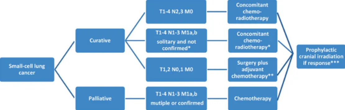
Approximately 5% of patients with SCLC present as T1, 2 N0, 1 M0 tumours (Figure1). These patients have more favourable outcomes with 5-year survival rates in the order of 50% [11,12]. Most series report on patients having been treated with surgery for a coin lesion without pathological diagnosis. A surgical approach in this group of patients is justified after ruling out mediastinal lymph node involvement (i.e. negative lymph nodes on CT scan, PET-CT scan or EBUS and/or mediastinoscopy if enlarged) [V, C]. Postoperatively, four cycles of adjuvant chemotherapy should be administered [III, C]. In the case of unforeseen N2 or N1 or in patients who have not undergone systematic nodal dissection, postoperative radiotherapy should be considered [V, C]. There is no role for surgery after induction chemotherapy in N2 disease [II, B]. In the absence of
randomised trials, due to frequent early dissemination and because total gross tumour volume has shown to be an
Table 2. Tumour stage grouping.
Occult carcinoma TX N0 M0 Stage 0 Tis N0 M0 Stage IA T1a,b N0 M0 Stage IB T2a N0 M0 Stage IIA T2b N0 M0 T1a,b N1 M0 T2a N1 M0 Stage IIB T2b N1 M0 T3 N0 M0
Stage IIIA T1a,b, T2a,b N2 M0
T3 N1, N2 M0
T4 N0, N1 M0
Stage IIIB T4 N2 M0
Any T N3 M0
Stage IV Any T Any N M1
Lababede O, Meziane M, Rice T. Seventh Edition of the Cancer Staging Manual and Stage Grouping of Lung Cancer. Chest 2011; 139: 183–189. Reproduced with permission from the American College of Chest Physicians.
Table 1. Tumour node metastasis classification.
TX Positive cytology only
T1 ≤3 cm
T1a ≤2 cm
T1b >2 to 3 cm
T2 Main bronchus≥2 cm from carina invades visceral pleura, partial
atelectasis
T2a >3–5 cm
T2b >5–7 cm
T3 >7 cm; chest wall, diaphragm, pericardium, mediastinal pleura,
main bronchus <2 cm from carina, total atelectasis, separate nodule(s) in the same lobe
T4 Mediastinum, heart, great vessels, carina, trachea, esophagus,
vertebra; separate tumour nodule(s) in a different ipsilateral lobe
N1 Ipsilateral peribronchial, ipsilateral hilar
N2 Subcarinal, ipsilateral mediastinal
N3 Contralateral mediastinal or hilar, scalene or supraclavicular
M1 Distant metastasis
M1a Separate tumour nodule(s) in a contralateral lobe; pleural nodules
or malignant pleural, or pericardial effusion
M1b Distant metastasis
Lababede O, Meziane M, Rice T. Seventh Edition of the Cancer Staging Manual and Stage Grouping of Lung Cancer. Chest 2011; 139: 183–189. Reproduced with permission from the American College of Chest Physicians.
independent prognostic factor leading to improved outcomes irrespective of the local treatment modality, patients with T1, 2 N0, 1 M0 may alternatively be treated with combined
concurrent chemoradiotherapy [III, C] [13]. This treatment is recommended as thefirst option in patients who are at increased risk for perioperative complications (e.g. significant concomitant medical illnesses) [II, C]. All patients with T1, 2 N0, 1 M0 should be considered for prophylactic cranial irradiation (PCI) if they have responded to initial treatment using the same dose and fractionation as for patients with stage III SCLC.
All other patients with T1-4, N0-3 M0 tumours who are in a good performance status (PS) should be treated with concurrent chemotherapy and thoracic radiotherapy [I, A]. Several
radiotherapy schedules have been studied. One phase III trial of 471 patients reported a superior 5-year overall survival (OS) with twice-daily radiotherapy (1.5 Gy twice-daily, 30 fractions) compared with once-daily (1.8 Gy, 25 fractions) of 26% versus 16% (P = 0.04) [10]. The inconvenience of the twice-daily administration and the significantly increased rate of transient grade 3 oesophagitis were, however, the main reasons why this regimen was not widely adopted. This current accelerated standard schedule is being compared with 70 Gy in daily fractions as an experimental arm in ongoing North American and European phase III trials in patients in which the lung dose can be kept within safe limits. Outside of a clinical trial, a twice-daily 1.5 Gy in 30-fraction regimen should be considered infit patients who are willing to accept temporarily increased toxicity [I, B]. The chemotherapy schedule consists of four cycles of cisplatin–etoposide or 4–6 cycles if a once-daily radiotherapy schedule is used [I, B].
The optimal timing of the concurrent radiotherapy has been studied extensively. Seven older trials assessing the timing of thoracic radiotherapy were analysed in two meta-analyses, with the conclusion that thoracic radiotherapy should be initiated as early as possible beginning with thefirst or second cycle when cisplatin-based chemotherapy was used [14,15]. In addition, an analysis of four of these studies which reported 5-year survival rates and used two concurrent arms with cisplatin–etoposide treatment found improved 5-year survival rates if the time between thefirst day of chemotherapy and the last day of
radiotherapy was <30 days [hazard ratio (HR): 0.62, 95% confidence interval (CI) 0.49–0.80, P = 0.0003] [16]. An update of an American trial reached the same conclusion [17]. On the other hand, a recent randomised trial did not show any survival difference when radiotherapy was administered with the third as opposed to thefirst cycle with less toxicity in the late arm in an Asian population [18]. Starting chest radiotherapy within 30 days after the beginning of chemotherapy is preferred [II, B]. When the general condition of the patient does not allow for the immediate administration of concurrent treatment or lung constraints preclude the target radiotherapy dose, chest irradiation may be postponed until the start of the third cycle of chemotherapy [II, B].
The optimal target volume remains to be defined. Omission of elective node irradiation based on CT scans should be used with caution as this strategy may result in nodal failures [III, C]. Whether selective node irradiation based on pre-treatment PET-CT scans can replace elective node irradiation has been addressed in two small studies [19,20]. Both studies, one prospective and the other one retrospective, have shown promisingly low nodal recurrence rates. This strategy, however, needs further prospective evaluation although it has been adopted already in some national guidelines [III, D]. Elective nodal volumes are not well-defined but may include the involved lymph node regions and one adjacent region and supraclavicular regions depending on the location of the primary tumour and the N2 or N3 nodes.
RECIST criteria are not well-suited to determine tumour response after radiotherapy. Patients in a reasonably good PS without progression should be offered PCI. The recommended dose is 25 Gy in 10 daily fractions [I, A]. Although PCI increases long-term survival, patients >65 years and/or with important vascular disease have a slightly elevated risk (HR 1.04) of developing neurocognitive side-effects [21,22].
management of metastatic disease
first-line treatment
Treatment of stage IV SCLC is palliative, and combination chemotherapy has been the main treatment option for more
Figure 1. Small-cell lung cancer (SCLC) treatment algorithm.
than three decades. Despite response rates (RRs) close to 70%, outcomes remain poor with a median progression-free survival (PFS) of only 5.5 months and a median OS of <10 months [22,23].
A meta-analysis of 19 randomised trials with a total of 4054 patients demonstrated prolonged OS of patients receiving a cisplatin-containing regimen compared with older
chemotherapy combinations [25]. Another meta-analysis of 36 trials reported an OS benefit in favour of etoposide alone or in combination with cisplatin compared with regimens that did not contain one of the two drugs [26]. These results led to the adoption of etoposide–cisplatin as a standard treatment regimen. A recent individual patient data meta-analysis including four randomised clinical trials comparing cisplatin versus carboplatin-based combination chemotherapy
demonstrated no difference in efficacy outcomes including RR, PFS and OS [24]. In the carboplatin group, increased
haematological toxicity rates were observed, whereas higher renal and neurotoxicity was seen with cisplatin. According to these results, cisplatin can be substituted by carboplatin in patients with metastatic SCLC [I, B]. Due to the limited number of only 663 patients included in this analysis, there was limited statistical power to draw conclusions in important subgroups such as patients with localised disease and young patients. In these subgroups, etoposide–cisplatin is recommended [II, B].
Studies with 3-drug regimens and the administration of increased dose intensity regimens, using increased dose or non-cross-resistant regimens, have not consistently reported improvement in OS. In addition, they have frequently been associated with significant toxicity in this usually co-morbid patient population [27]. Such regimens are not recommended as first-line treatment [II, C].
A recent literature-based meta-analysis of seven randomised studies showed an improved OS, but not PFS with irinotecan– platinum compared with etoposide–platinum. Irinotecan led to more gastrointestinal toxic effects, while more haematological toxic effects were observed with etoposide [28]. The results, however, were primarily driven by Asian studies, and pharmacogenomic differences between Asian and Western populations possibly contributing to these differential outcomes have previously been described [29]. No chemotherapy doublet has yet been shown to be superior to i.v. etoposide–platinum in a Western population. Randomised phase III trials which compared irinotecan–cisplatin, gemcitabine–carboplatin (in poor prognostic patients only) or i.v. or oral topotecan–cisplatin to etoposide–platinum have demonstrated non-inferiority for survival [30–33]. These regimens are recommended as alternative treatment options in the case of contraindications to etoposide [II, C].
Continuation of chemotherapy beyond 4–6 cycles has been assessed in at least 14 randomised, controlled trials. Although a significant OS benefit was reported in a literature-based review including 11 trials (HR 0.89, 95% CI: 0.81–0.92; P = 0.02), the benefit was small and high heterogeneity among the included trials was observed [34]. Similarly, a previous meta-analysis found a small OS benefit of 4% at 2 years with maintenance therapy [35]. However, the majority of the randomised, controlled trials did not show any significant OS benefit, and a properly designed large clinical trial to address this question is
lacking. In addition, there is a considerable risk of increased toxicity with prolonged platinum-based chemotherapy. Continuing chemotherapy beyond 4–6 cycles of first-line treatment is not recommended [II, B].
PCI significantly decreases the risk of symptomatic brain metastases (from 40.4% to 14.6% at 1 year) and increases OS (HR 0.68; 95% CI, 0.52–0.88) [36]. Of note, in this trial initial pre-treatment brain imaging was not required. PCI is associated with adverse effects such as fatigue and hair loss, and health-related quality of life may be negatively affected as well [37]. Patients with any response tofirst-line treatment and who have a reasonably good PS should be evaluated for PCI [II, B]. The PCI dose may be 25 Gy in 10 daily fractions or 20 Gy in 5 fractions.
Due to the often centrally located primary tumours, symptoms such as dyspnoea, infections due to atelectasis, chest pain or superior vena cava syndrome are frequent and make the incorporation of thoracic radiotherapy into the initial treatment algorithm an appealing concept. A four-arm randomised phase III trial has demonstrated a survival benefit of concurrent thoracic radiotherapy in patients whose primary tumours have responded after three cycles of cisplatin–etoposide and whose metastatic sites were in complete remission (OS: 17 versus 11 months,P = 0.041) [38]. This single centre trial was however small (54 patients per arm), and the concurrent
chemoradiotherapy treatment used does not correspond to the current standard approach. The routine use of thoracic irradiation in patients with metastatic SCLC is not recommended and the results of the Dutch phase III trial (CREST study) testing this concept should be awaited [II, C].
second-line treatment
RRs to second-line treatment depend on the treatment-free interval and are usually in the order of 10% in resistant disease (i.e. progression-free interval <3 months) and 20% in sensitive disease (i.e. interval >3 months). In refractory patients (i.e. patients not responding or progressing during chemotherapy) and resistant patients with early relapse (<6 weeks), outcomes are poor and the clinical benefit of further systemic therapy is uncertain. For these patients, participation in a clinical trial or best supportive care is recommended [II, C]. Oral topotecan led to better symptom control including slower time to quality of life deterioration and improved survival compared with best supportive care in a study in which half of the patients had resistant disease [39]. Prior to topotecan development, anthracycline-based regimes have been commonly used, including cyclophosphamide, doxorubicin and vincristine (CAV). In 1999, a trial of i.v. topotecan and CAV demonstrated equal efficacy, with similar RRs, time-to-progression, and OS, and better tolerance when compared with CAV [40]. Oral and i.v. topotecan have shown to be equally effective [41], but with differing toxicity profiles. Either oral or i.v. topotecan are recommended for patients having resistant or sensitive relapse with CAV being an alternative option [II, B]. Only patients with sensitive disease derive benefit from rechallenge with first-line therapy (usually platinum–etoposide) [V, C].
A recent randomised, phase III trial failed to show a survival benefit of amrubicin versus topotecan, despite a higher RR and
Table 3. Summary of recommendations.
Diagnosis • Pathological diagnosis should be made according to the World Health Organisation (WHO) classification
• Biopsies are best obtained by bronchoscopy. A biopsy from a metastatic lesion is preferred if the location of the metastasis can be easily and safely accessed to biopsy (e.g. liver, skin)
• No predictive molecular marker for treatment selection is currently available
Staging and risk assessment • Initial assessment should include smoking history, physical examination, complete blood count, liver enzymes, sodium,
potassium, calcium, glucose, lactate dehydrogenase levels and lung (if localised disease) and renal function tests • A computed tomography (CT) scan with contrast of the chest and abdomen is recommended
• In localised disease or if symptoms or clinical findings suggest involvement, additional bone scintigraphy and CT or MRI of the brain are recommended
• 2-fluor-2-desoxy-D-glucose positron-emission-tomography (FDG-PET CT) scan is optional in localised disease. PET
findings, which modify treatment decisions, should be pathologically confirmed [III, C]
• A bone marrow aspiration and biopsy should be carried out in the case of abnormal blood counts suggesting involvement, particularly in localised disease [V, C]
• Version 7 of the TNM staging system according to the Union for International Cancer Control (UICC) should be used (Tables 1and2) [I, A]
Treatment strategy • Figure1summarises the treatment algorithm of patients with SCLC
• In localised disease, a bimodality treatment approach is curative and chemotherapy plus radiotherapy result in 5-year survival rates of 20%–25%
• Treatment of stage IV SCLC is palliative and various combination chemotherapy regimens demonstrate similarly high response rates (RRs) of 60%–70%. Due to frequent rapid relapse and limited activity of second-line treatment, overall survival (OS) remains poor (<10 months)
• All SCLC patients responding to first-line treatment should be evaluated for prophylactic cranial irradiation (PCI) Treatment of localised
disease
• A small subset of patients who present with T1, 2 N0, 1 M0 tumours have a more favourable outcome and 5-year survival rates of 50% have been reported with surgery. These patients should receive four cycles of adjuvant chemotherapy [III, C] and postoperative thoracic radiotherapy if staged pN1 or pN2 [V, C]
• All other patients with T1-4, N0-3 M0 tumours who are in a good performance status (PS) should be treated with concurrent chemotherapy and thoracic radiotherapy [I, A]
• The best OS rates in fit patients were demonstrated with twice-daily 1.5 Gy in 30 fractions given concurrently with four cycles of cisplatin and etoposide [I, B]
• Patients who are not fit enough for twice-daily radiotherapy or are unwilling to accept increased toxic effects may be treated with a once-daily radiotherapy schedule with 4–6 cycles of concurrent etoposide–cisplatin [I, B]
• In good PS patients, thoracic radiotherapy should be initiated with the first or second cycle (i.e. within 30 days) of chemotherapy [II, B]
• All patients with T1-4, N0-3 M0 disease without disease progression after treatment and a reasonably good PS should be offered PCI [I, A]
First-line treatment of metastatic disease
• 4–6 cycles of etoposide plus cisplatin or carboplatin are recommended [I, B]
• In young patients and patients with localised disease, etoposide–cisplatin is recommended [II, B]
• Irinotecan–cisplatin, gemcitabine–carboplatin (in poor prognostic patients only) and i.v. or oral topotecan–cisplatin are alternative options if etoposide is contraindicated [II, C]
• Patients in a reasonably good PS with any response to first-line treatment should be evaluated for PCI [II, B] • The routine use of thoracic irradiation in patients with metastatic SCLC is not recommended [II, C] Second-line treatment of
metastatic disease
• For refractory patients and resistant patients with early relapse (<6 weeks), participation in a clinical trial or best supportive care is recommended [II, C]
• Oral or i.v. topotecan are recommended for patients having resistant or sensitive relapse with CAV being an alternative option [II, B]
• Patients with sensitive relapse may derive benefit from reintroduction of the first-line regimen (usually platinum– etoposide) [V, C]
Follow-up and long-term implications
• The occurrence of second malignancies, particularly if smoking is continued, is of concern in survivours and smoking cessation counselling is essential
• Two to three-monthly CT scans are recommended in patients with metastatic disease potentially qualifying for further treatments [V, C]
• Six-monthly CT scans for 2 years with lengthening of intervals thereafter are recommended for patients with non-metastatic disease who have received potentially curative treatment [V, C]
improved quality of life with amrubicin [42]. The subgroup of refractory patients derived a small survival benefit from amrubicin. Amrubicin is currently not available in Western countries.
personalised medicine
In this disease setting, more research is needed to identify molecular markers which could lead to advances in personalised medicine.
follow-up and long-term implications
All patients with metastatic SCLC and approximately three-quarters of patients with localised disease will progress. In survivors, the occurrence of second malignancies, particularly if smoking is continued, is of concern and smoking cessation counselling is essential. The main goal of regular follow-up is to detect recurrence early, while the patient is still in a good PS [43]. The frequency of follow-up visits depends on the availability of treatment options. Although there is no clinical trial evaluating the benefit of regular follow up, 2–3-monthly CT scans are recommended in patients with metastatic disease potentially qualifying for further treatments. Patients with localised disease who have received potentially curative treatment should undergo 3–6-monthly CT scans for two years with lengthening of intervals thereafter. Due to the high risk of secondary primary lung cancer, annual low-dose CT scans after 5 years might be considered [V, C]. Summary of
recommendations is provided in Table3.
note
Levels of evidence and grades of recommendation have been applied using the system shown in Table4. Statements without grading were considered justified standard clinical practice by the experts and the ESMO faculty.
con
flict of interest
Dr Peters has reported consultancy/honoraria from Roche, Eli Lilly, AstraZeneca, Pfizer, Boehringer-Ingelheim, Bristol-Myers Squibb, Daiichi-Sankyo, and Tesaro. Dr Felip has reported consultancy/honoraria from Lilly, GlaxoSmithKline, Pfizer, Roche, Boehringer Ingelheim. The other authors have declared no potential conflicts of interest.
references
1. Jemal A, Bray F, Center MM et al. Global cancer statistics. CA Cancer J Clin 2011;
61: 69–90.
2. Sant M, Allemani C, Santiaquilani M et al. EUROCARE-4. Survival of cancer patients diagnosed in 1995-1999. Results and commentary. Eur J Cancer 2009;
45: 931–991.
3. Govindan R, Page N, Morgensztern D et al. Changing epidemiology of small-cell lung cancer in the United States over the last 30 years: analysis of the surveillance, epidemiologic, and end results database. J Clin Oncol 2006; 24:
4539–4544.
4. Parsons A, Daley A, Begh R et al. Influence of smoking cessation after diagnosis of
early stage lung cancer on prognosis: systematic review of observational studies with meta-analysis. BMJ 2010; 340: b5569.
5. Cuffe S, Moua T, Summerfield R et al. Characteristics and outcomes of small
cell lung cancer patients diagnosed during two lung cancer computed tomographic screening programs in heavy smokers. J Thorac Oncol 2011; 6:
818–822.
6. Lababede O, Meziane M, Rice T. Seventh edition of cancer staging manual and stage grouping of lung cancer: quick reference chart and diagrams. Chest 2011;
139: 183–189.
7. Shepherd FA, Crowley J, Van Houtte P et al. The International Association for the study of lung cancer lung cancer staging project: proposals regarding the clinical staging of small cell lung cancer in the forthcoming (seventh) edition of the tumor,
node, metastasis classification for lung cancer. J Thorac Oncol 2007; 2:
1067–1077.
8. Thomson D, Hulse P, Lorigan P et al. The role of positron emission tomography
in management of small cell lung cancer. Lung Cancer 2011; 73: 121–126.
9. van Meerbeeck JP, Fennell DA, De Ruysscher DK. Small-cell lung cancer. Lancet
2011; 378: 1741–1755.
10. Turrisi AT, 3rd, Kim K, Blum R et al. Twice-daily compared with once-daily thoracic radiotherapy in limited small-cell lung cancer treated concurrently with cisplatin
and etoposide. N Engl J Med 1999; 340: 265–271.
Table 4. LOE and GOR adapted from the Infectious Diseases Society of America-United States Public Health Service Grading System† Levels of evidence
I Evidence from at least one large randomised control trial of good methodological quality (low potential for bias) or meta-analyses of well-conducted
randomised trials without heterogeneity
II Small randomised trials or large randomised trials with a suspicion of bias (lower methodological quality) or meta-analyses of such trials or of trials with
demonstrated heterogeneity
III Prospective cohort studies
IV Retrospective cohort studies or case-control studies
V Studies without control group, case reports, experts opinions
Summary of recommendations
A Strong evidence for efficacy with a substantial clinical benefit, strongly recommended
B Strong or moderate evidence for efficacy but with a limited clinical benefit, generally recommended
C Insufficient evidence for efficacy or benefit does not outweigh the risk or the disadvantages (adverse events, costs,.), optional
D Moderate evidence against efficacy or for adverse outcome, generally not recommended
E Strong evidence against efficacy or for adverse outcome, never recommended
†Dykewicz CA. Summary of the guidelines for preventing opportunistic infections among hematopoietic stem cell transplant recipients. Clin Infect Dis 2001; 33: 139-144. By permission of the Infectious Diseases Society of America.
11. Yu JB, Decker RH, Detterbeck FC et al. Surveillance epidemiology and end results evaluation of the role of surgery for stage I small cell lung cancer. J Thorac Oncol
2010; 5: 215–219.
12. Schreiber D, Rineer J, Weedon J et al. Survival outcomes with the use of surgery in limited-stage small cell lung cancer: should its role be re-evaluated? Cancer
2010; 116: 1350–1357.
13. Reymen B, Van Loon J, van Baardwijk A et al. Total gross tumor volume is an independent prognostic factor in patients treated with selective nodal irradiation for stage I to III small cell lung cancer. Int J Radiat Oncol Biol Phys 2013; 85:
1319–1324.
14. Fried DB, Morris DE, Poole C et al. Systematic review evaluating the timing of thoracic radiation therapy in combined modality therapy for limited-stage small-cell
lung cancer. J Clin Oncol 2004; 22: 4837–4845.
15. Pijls-Johannesma M, De Ruysscher D, Vansteenkiste J et al. Timing of chest radiotherapy in patients with limited stage small cell lung cancer: a systematic review and meta-analysis of randomised controlled trials. Cancer Treat Rev 2007;
33: 461–473.
16. De Ruysscher D, Pijls-Johannesma M, Bentzen SM et al. Time between thefirst
day of chemotherapy and the last day of chest radiation is the most important predictor of survival in limited-disease small-cell lung cancer. J Clin Oncol 2006;
24: 1057–1063.
17. Blackstock AW, Bogart JA, Matthews C et al. Split-course versus continuous
thoracic radiation therapy for limited-stage small-cell lung cancer:final report of a
randomized phase III trial. Clin Lung Cancer 2005; 6: 287–292.
18. Sun JM, Ahn YC, Choi EK et al. Phase III trial of concurrent thoracic radiotherapy
with eitherfirst- or third-cycle chemotherapy for limited-disease small-cell lung
cancer. Ann Oncol 2013; 24: 2088–2092.
19. van Loon J, De Ruysscher D, Wanders R et al. Selective nodal irradiation on basis of (18)FDG-PET scans in limited-disease small-cell lung cancer: a prospective study. Int J Radiat Oncol Biol Phys 2010; 77:
329–336.
20. Shirvani SM, Komaki R, Heymach JV et al. Positron emission tomography/ computed tomography-guided intensity-modulated radiotherapy for limited-stage small-cell lung cancer. Int J Radiat Oncol Biol Phys 2012; 82:
e91–e97.
21. Le Péchoux C, Laplanche A, Faivre-Finn C et al. Clinical neurological outcome and quality of life among patients with limited small-cell cancer treated with two different doses of prophylactic cranial irradiation in the intergroup phase III trial (PCI99-01, EORTC 22003-08004, RTOG 0212 and IFCT 99-01). Ann Oncol
2011; 22: 1154–1163.
22. Wolfson AH, Bae K, Komak R et al. Primary analysis of a phase II randomized trial Radiation Therapy Oncology Group (RTOG) 0212: impact of different total doses and schedules of prophylactic cranial irradiation on chronic neurotoxicity and quality of life for patients with limited-disease small-cell lung cancer. Int J Radiat
Oncol Biol Phys 2011; 81: 77–84.
23. Foster NR, Qi Y, Shi Q et al. Tumor response and progression-free survival as potential surrogate endpoints for overall survival in extensive stage small-cell lung
cancer:findings on the basis of North Central Cancer Treatment Group trials.
Cancer 2011; 117: 1262–1271.
24. Rossi A, Di Maio M, Chiodini P et al. Carboplatin- or cisplatin-based chemotherapy
infirst-line treatment of small-cell lung cancer: the COCIS meta-analysis of
individual patient data. J Clin Oncol 2012; 30: 1692–1698.
25. Pujol JL, Carestia L, Daurès JP. Is there a case for cisplatin in the treatment of small-cell lung cancer? A meta-analysis of randomized trials of a cisplatin-containing regimen versus a regimen without this alkylating agent. Br J Cancer
2000; 83: 8–15.
26. Mascaux C, Paesmans M, Berghmans T et al. A systematic review of the role of etoposide and cisplatin in the chemotherapy of small cell lung cancer
with methodology assessment and meta-analysis. Lung Cancer 2000; 30:
23–26.
27. Popat S, O’Brien M. Chemotherapy strategies in the treatment of small cell lung
cancer. Anticancer Drugs 2005; 16: 361–372.
28. Shao N, Jin S, Zhu W. An updated meta-analysis of randomized controlled trials comparing irinotecan/platinum with etoposide/platinum in patients with previously
untreated extensive-stage small cell lung cancer. J Thorac Oncol 2012; 7: 470–472.
29. Lara PN, Jr, Natale R, Crowley J et al. Phase III trial of irinotecan/cisplatin compared with etoposide/cisplatin in extensive-stage small-cell lung cancer: clinical and pharmacogenomic results from SWOG S0124. J Clin Oncol 2009; 27:
2530–2535.
30. Zatloukal P, Cardenal F, Szczesna A et al. A multicenter international randomized phase III study comparing cisplatin in combination with irinotecan or etoposide in previously untreated small-cell lung cancer patients with extensive disease. Ann
Oncol 2010; 21: 1810–1816.
31. Fink TH, Huber RM, Heigener DF et al. Topotecan/cisplatin compared with
cisplatin/etoposide asfirst-line treatment for patients with extensive disease
small-cell lung cancer:final results of a randomized phase III trial. J Thorac Oncol 2012;
7: 1432–1439.
32. Eckardt JR, von Pawel J, Papai Z et al. Open-label, multicenter, randomized, phase III study comparing oral topotecan/cisplatin versus etoposide/cisplatin as treatment for chemotherapy-naive patients with extensive-disease small-cell lung
cancer. J Clin Oncol 2006; 24: 2044–2051.
33. Lee SM, James LE, Qian W et al. Comparison of gemcitabine and carboplatin versus cisplatin and etoposide for patients with poor-prognosis small cell lung
cancer. Thorax 2009; 64: 75–80.
34. Rossi A, Garassino MC, Cinquini M et al. Maintenance or consolidation therapy in small-cell lung cancer: a systematic review and meta-analysis. Lung Cancer 2010;
70: 119–128.
35. Bozcuk H, Artac M, Ozdogan M et al. Does maintenance/consolidation chemotherapy have a role in the management of small cell lung cancer (SCLC)? A meta-analysis of the published controlled trials. Cancer 2005; 104:
2650–2657.
36. Slotman BJ, Faivre-Finn C, Kramer GW et al. Prophylactic cranial irradiation in
small-cell lung cancer. N Engl J Med 2007; 357: 664–672.
37. Slotman BJ, Mauer ME, Bottomley A et al. Prophylactic cranial irradiation in extensive disease small-cell lung cancer: short-term health-related quality of life and patient reported symptoms: results of an international phase III randomized controlled trial by the EORTC Radiation Oncology and Lung Cancer Groups. J Clin
Oncol 2009; 27: 78–84.
38. Jeremic B, Shibamoto Y, Nikolic N et al. Role of radiation therapy in the combined-modality treatment of patients with extensive disease small-cell lung cancer: a
randomized study. J Clin Oncol 1999; 17: 2092–2099.
39. O’Brien ME, Ciuleanu TE, Tsekov H et al. Phase III trial comparing supportive care
alone with supportive care with oral topotecan in patients with relapsed small-cell
lung cancer. J Clin Oncol 2006; 24: 5441–5447.
40. von Pawel J, Schiller JH, Shepherd FA et al. Topotecan versus cyclophosphamide, doxorubicin, and vincristine for the treatment of recurrent small-cell lung cancer. J
Clin Oncol 1999; 1 and 427: 658–667.
41. Eckardt JR, von Pawel J, Pujol JL et al. Phase III study of oral compared with intravenous topotecan as second-line therapy in small-cell lung cancer. Clin Oncol
2007; 25: 2086–2092.
42. Jotte R, Von Pawel J, Spigel D et al. Randomized phase III trial of amrubicin versus topotecan (Topo) as second-line treatment for small cell lung cancer (SCLC). J Clin Oncol 2012; 29 (Suppl.): abstr 7000.
43. Sugiyama T, Hirose T, Hosaka T et al. Effectiveness of intensive follow-up after response in patients with small cell lung cancer. Lung Cancer 2008; 59:
255–261.