RESEARCH OUTPUTS / RÉSULTATS DE RECHERCHE
Author(s) - Auteur(s) :
Publication date - Date de publication :
Permanent link - Permalien :
Rights / License - Licence de droit d’auteur :
researchportal.unamur.be
University of Namur
High TMEM45A expression is correlated to epidermal keratinization
Hayez, Aurélie; Malaisse, Jérémy; Roegiers, Edith; Reynier, Marie; Renard, Chantal; Haftek,
Marek; Geenen, Vincent; Serre, Guy; Simon, Michel; Lambert de Rouvroit, Catherine;
Michiels, Carine; Poumay, Yves
Published in: Experimental dermatology DOI: 10.1111/exd.12403 Publication date: 2014 Document Version
Early version, also known as pre-print
Link to publication
Citation for pulished version (HARVARD):
Hayez, A, Malaisse, J, Roegiers, E, Reynier, M, Renard, C, Haftek, M, Geenen, V, Serre, G, Simon, M, Lambert de Rouvroit, C, Michiels, C & Poumay, Y 2014, 'High TMEM45A expression is correlated to epidermal
keratinization', Experimental dermatology, vol. 23, no. 5, pp. 339-344. https://doi.org/10.1111/exd.12403
General rights
Copyright and moral rights for the publications made accessible in the public portal are retained by the authors and/or other copyright owners and it is a condition of accessing publications that users recognise and abide by the legal requirements associated with these rights. • Users may download and print one copy of any publication from the public portal for the purpose of private study or research. • You may not further distribute the material or use it for any profit-making activity or commercial gain
• You may freely distribute the URL identifying the publication in the public portal ?
Take down policy
If you believe that this document breaches copyright please contact us providing details, and we will remove access to the work immediately and investigate your claim.
For Review Only
High TMEM45A expression is correlated to epidermal keratinization
Journal: Experimental Dermatology Manuscript ID: Draft
Manuscript Type: Regular Article Date Submitted by the Author: n/a
Complete List of Authors: Hayez, Aurélie; University of Namur, Cell and Tissue Laboratory-URPHYM-Narilis
Malaisse, Jérémy; University of Namur, Cell and Tissue Laboratory-URPHYM-Narilis
Roegiers, Edith; University of Namur, Research Unit for Cell biology (URBC), Narilis
Reynier, Marie; CNRS-INSERM-University of Toulouse, Epidermal differentiation and rheumatoid autoimmunity, UMR5165/U1056 Renard, Chantal; University of Liège, Center of Immunoendocrinology, GIGA-I3
Haftek, Marek; University of Lyon, Laboratoire de Recherche Dermatologique
Geenen, Vincent; University of Liège, Center of Immunoendocrinology, GIGA-I3
SERRE, Guy; UMR5165 CNRS-Toulouse III University, Epidermis Differentiation and Rheumatoid Autoimmunity
Simon, Michel; CNRS-INSERM-University of Toulouse, Epidermal differentiation and rheumatoid autoimmunity, UMR5165/U1056 Lambert de Rouvroit, Catherine; University of Namur, Cell and Tissue Laboratory-URPHYM-Narilis
Michiels, Carine; University of Namur, Research Unit for Cell biology (URBC), Narilis
Poumay, Yves; University of Namur, Cell and Tissue Laboratory-URPHYM-Narilis
Keywords: epidermis, keratinization, keratinocyte, differentiation, Golgi apparatus
For Review Only
Word count (not including abstract, tables, figures & references) : 3,025 words Number of display items : 4Supporting Information :
- Supplementary figures : 5
- Supplementary material
- Supplementary reference
Title
High TMEM45A expression is correlated to epidermal keratinization
Authors
Aurélie Hayez1, Jérémy Malaisse1, Edith Roegiers2, Marie Reynier3, Chantal Renard4, Marek Haftek5, Vincent Geenen4, Guy Serre3, Michel Simon3, Catherine Lambert de Rouvroit1, Carine Michiels2 and Yves Poumay1
1
Cell and Tissue Laboratory-URPhyM-NARILIS, University of Namur, Namur, Belgium; 2 Research Unit for Cell biology-NARILIS, University of Namur, Namur, Belgium; 3 UMR5165/U1056 CNRS-INSERM-University of Toulouse, UDEAR, Toulouse, France; 4 Center of Immunoendocrinology, GIGA-I3, University of Liège, Liège, Belgium; 5 Laboratoire de Recherche Dermatologique, University of Lyon, Lyon, France
Correspondence
Yves Poumay, Cell and Tissue Laboratory-URPhyM-NARILIS, University of Namur, Rue de Bruxelles, 61, B-5000 Namur, Belgium. E-mail : [email protected]
3 4 5 6 7 8 9 10 11 12 13 14 15 16 17 18 19 20 21 22 23 24 25 26 27 28 29 30 31 32 33 34 35 36 37 38 39 40 41 42 43 44 45 46 47 48 49 50 51 52 53 54 55 56 57 58 59
For Review Only
Abstract
TMEM45A (DERP7, DNAPTP4 or FLJ10134) gene, belonging to the TMEM family encoding predicted transmembrane proteins, is highly expressed in epidermal keratinocytes. To investigate the potential involvement of TMEM45A during the differentiation and keratinization processes, its expression has been characterized in normal human keratinocytes and the protein subcellular localization has been studied in this cell type, both in vitro and in vivo. TMEM45A expression is upregulated with differentiation, either induced by cultured keratinocyte confluence or enhanced Ca2+ concentration in medium. In vivo, TMEM45A mRNA and protein are mostly found in the granular layer of the epidermis. TMEM45A expression is linked to keratinization, since accumulation of the protein is detected in native and reconstructed epidermis as well as in thymic Hassal bodies, but not in non-keratinized stratified epithelia. At the subcellular level, co-detection with ER and Golgi markers reveals that TM protein 45A is associated with the Golgi apparatus, and more specifically with the trans-Golgi/trans-Golgi network in vitro and in granular layer in vivo. The protein is neither related to lysosomes nor transported within corneodesmosin-containing lamellar bodies. As the expression of TMEM45A is strongly correlated to keratinization, these data indicate an important role played by TM protein 45A in this process.
Abbreviations
TM protein 45A, transmembrane protein 45A; IVL, involucrin; KRT, keratin; FLG, filaggrin; KLK7, kallikrein 7; CDSN, corneodesmosin; CALN, calnexin; ER, endoplasmic reticulum.
Key words
Epidermis – keratinization – keratinocyte – differentiation - Golgi apparatus
3 4 5 6 7 8 9 10 11 12 13 14 15 16 17 18 19 20 21 22 23 24 25 26 27 28 29 30 31 32 33 34 35 36 37 38 39 40 41 42 43 44 45 46 47 48 49 50 51 52 53 54 55 56 57 58
For Review Only
Introduction
The epidermis is a squamous stratified epithelium where keratinocytes follow a unique program of terminal differentiation and cell death that leads to the formation of the cornified layer, the outermost skin barrier. During this so-called keratinization process, the cell content including all organelles is replaced by compacted keratin filaments. At the cytoplasmic side of plasma membrane, various proteins are enzymatically cross-linked to form a cornified envelope. Finally, modifications of lipids secreted in the intercellular spaces occur, leading to the formation of a hydrophobic matrix [1]. Currently, our knowledge about gene expression during keratinization remains incomplete [2]. However, recent transcriptomic studies allowed identification of yet unknown although potentially major actors of this process. Indeed, a transcriptomic study based on reconstructed human epidermis classified the still poorly characterized TMEM45A gene in a cluster particularly highly expressed at the late differentiation stage [3]. This cluster also contains numerous genes of the epidermal differentiation complex and functionally gathers genes implicated in ectoderm and epidermis development, morphogenesis and keratinization. Moreover, according to BioGPS [4], skin is the tissue expressing TMEM45A mRNA at the highest level.
TMEM45A gene (also called DERP7, DNAPTP4 or FLJ10134) belongs to the large family of genes encoding uncharacterized predicted transmembrane (TMEM) proteins. To date, almost all available information about TMEM45A is coming from transcriptomic studies, databases or bioinformatics predictive tools. Seven transcripts have been reported, but only three isoforms of transmembrane protein 45A (TM protein 45A) have been detected : one contains 191 amino acids with 21.9 kDa MW, a second contains 275 amino acids with 31.7 kDa MW and a third isoform contains 291 amino acids with 33.5 kDa MW, according to Ensemble [5]. The isoforms contain three, five or seven predicted transmembrane domains, according to PredictProtein [6] and SOSUI [7], thus they are suspected to be integral proteins according to Uniprot [8]. Subcellular localization and functions remain unknown. Recently, we briefly reported selective induction of TMEM45A expression in differentating keratinocytes, as well as elevated expression in normal human epidermis [9], data later confirmed by others [10]. Finally, in an approach using keratinocytes fractions purified from normal epidermis, TMEM45A had been reported as a differentiation-associated gene, because of its significantly higher mRNA level in granular and cornified keratinocytes-enriched fraction compared to its level in basal keratinocytes-enriched fraction [11].
3 4 5 6 7 8 9 10 11 12 13 14 15 16 17 18 19 20 21 22 23 24 25 26 27 28 29 30 31 32 33 34 35 36 37 38 39 40 41 42 43 44 45 46 47 48 49 50 51 52 53 54 55 56 57 58 59
For Review Only
In addition, our current knowledge about TMEM45A is also incomplete in pathological conditions. Regarding skin pathology, no significant variation in TMEM45A mRNA was observed between healthy skin biopsies and atopic dermatitis, basal cell carcinoma, squamous cell carcinoma, or melanoma biopsies, whereas a significant upregulation was observed in psoriasis and actinic keratosis [10, 12]. In cancer cells, TMEM45A expression was reported to be implicated in hypoxia-induced resistance against drug-hypoxia-induced apoptosis by a still unknown mechanism [9]. Contrarily, the gene was shown suppressing progression of ductal carcinoma into invasive breast cancer, again by an unknown mechanism [13].
Here, we further characterized the expression of the new potential epidermal actor TMEM45A in
vitro and in vivo in order to assess its particular involvement in the keratinization process. Next, we studied subcellular localization of the TM protein 45A in keratinocytes in order to get more insight into its potential functions.
Materials & Methods
Antibodies, immunogenic peptide and chemicals
The list is available in the supplementary Material.
Isolation of normal human keratinocytes and induction of differentiation in autocrine cultures
Primary keratinocytes were isolated from normal human adult abdominal skin collected at plastic surgery after written informed consent of patients (Dr. B. Bienfait, Clinique St Luc, Namur, Belgium). Cells were cultured in autocrine conditions as described [14]. All experiments were carried out according to the Declaration of Helsinki Principles and were approved by the Medical Ethical Committee of Clinique St Luc.
Calcium-induced differentiation of keratinocytes
Keratinocytes monolayers were seeded at very low cell density (5,000 cells/cm2) and then incubated with the usual medium containing 0.06 mM Ca2+ (control) or with a medium supplemented with Ca2+ (1.5 mM) for 48 hours before extraction of total RNA.
Brefeldin A incubation
Confluent monolayers of keratinocytes were incubated with 10 µg/ml brefeldin A for 30 minutes.
3 4 5 6 7 8 9 10 11 12 13 14 15 16 17 18 19 20 21 22 23 24 25 26 27 28 29 30 31 32 33 34 35 36 37 38 39 40 41 42 43 44 45 46 47 48 49 50 51 52 53 54 55 56 57 58
For Review Only
Reconstruction of human epidermis and processing for histology
Reconstructed human epidermis were produced and analyzed as described [15], except fixation in 70% ethanol (100%), 10% formalin and 5% glacial acetic acid.
Human normal mouth epithelium, abdominal skin and thymus sections
A lip fragment was obtained thanks to Pr. Pierre Garin and Alain Koninckx (Laboratoire d’anatomie, University of Namur, Namur, Belgium) from two individuals at autopsy. Abdominal skins were collected from plastic surgery (Dr. B. Bienfait, Clinique St. Luc, Namur, Belgium). A normal thymus fragment was obtained from a young child undergoing corrective cardiovascular surgery for congenital cardiomyopathy. Tissues were embedded in OCT compound and serial cryosections of 6-8 µm thick were obtained.
Immunofluorescence and immunoperoxidase labelling on tissues and cells
Protocols are available in the supplementary Material.
Real time RT-PCR for keratinocyte monolayers and reconstructed human epidermis
Total RNA of monolayers were isolated using High Pure RNA isolation kit (Roche, Basel, Switzerland), according to manufacturer’s instructions. Total mRNA of Reconstructed Human Epidermis was isolated using RNeasy mini kit (Qiagen, Hilden, Germany), using spin technology for animal cells according to manufacturer’s instructions. 1 µg of total RNA was reverse transcribed using SuperScript II kit (Life technologies, Carlsbad, California, US), according to the manufacturer’s instructions. Amplification reaction assays contained FastStart Universal SYBR Green Master (Rox) (Roche, Basel, Switzerland) and 300nM of primers for real-time PCR. Primer sequences are available in the supplementary Material. The geometric mean of RPLP0 and TBP house-keeping genes values was used for normalization [16]. mRNA expression level was quantified using the threshold cycle method on a 7300 real-time PCR machine (Life technologies, Carlsbad, California, US).
Epidermal dissociation
Four fractions (T1, T2, T3 and T4) were obtained from normal human abdominal epidermis after successive dissociations of keratinocytes by serial incubations in trypsin-EDTA, as described in [2]. Total RNA was extracted from fractions using the RNeasy extraction kit (Qiagen, Hilden, Germany). Three hundred ng of total RNA of each fraction was reverse transcribed using a mixture
3 4 5 6 7 8 9 10 11 12 13 14 15 16 17 18 19 20 21 22 23 24 25 26 27 28 29 30 31 32 33 34 35 36 37 38 39 40 41 42 43 44 45 46 47 48 49 50 51 52 53 54 55 56 57 58 59
For Review Only
of dNTPs and random hexamer primers. Amplification assays were performed with the ABI prism 7000 Sequence Detection System and analyzed with the corresponding software (Life technologies, Carlsbad, California, US) using the Maxima Probe/ROX qPCR master mix (Thermo Fisher Scientific, Waltham, MA, US). Fluorescence was quantified as threshold cycle values. Primer sequences are available in the supplementary Material. mRNA levels were normalized to HPRT1 house-keeping gene expression levels. This work was performed on skin from two different donors.
Statistical analysis
Independent experiments were performed at least in triplicates except for mRNA analysis of epidermal dissociation. Statistical significance was evaluated by one-way Repeated Measures analysis of variance with a Holm-Sidak correction, except for analysis of calcium-induced differentiation for which we used the paired t-test. Data values were expressed as relative quantification level with error bars representing 95% confidence intervals.
Results
TMEM45A is upregulated during keratinocyte differentiation in vitro
In autocrine cultures of human epidermal keratinocytes, confluent cells lose their clonogenicity [17] and initiate a limited differentiation program, as shown by the expression of early and late markers [14]. Relative levels of TMEM45A mRNA were quantified at different time points of culture, before and after confluence (Figure 1a). A very highly significant increase in TMEM45A expression was observed after confluence with an expression profile similar to the differentiation markers keratin-10 (KRT10) and involucrin (IVL). The TM protein 45A expression was then analyzed with an affinity-purified peptide antibody as already described [9]. The specificity of this antibody was first further validated using competition with the immunogenic peptide (Figure S1). Then, the transmembrane protein 45A was labelled in autocrine cultures of keratinocytes at sub-confluence, confluence and post-confluence (Figure 1b). Our data illustrate an increase in immunoreactivity at culture confluence and later. Concomitant detections of the TM protein 45A and both of KRT10 and IVL in differentiating keratinocytes were also established using immunofluorescence co-labelling after culture confluence (Figure 1c). In order to analyze whether TMEM45A gene expression is related to differentiation rather than to the high cell density obtained at confluence, its mRNA levels were measured after induction of differentiation by an elevated Ca2+ concentration in culture medium. Keratinocytes at low cell density were cultured with a high (1.5
3 4 5 6 7 8 9 10 11 12 13 14 15 16 17 18 19 20 21 22 23 24 25 26 27 28 29 30 31 32 33 34 35 36 37 38 39 40 41 42 43 44 45 46 47 48 49 50 51 52 53 54 55 56 57 58
For Review Only
mM) or low (0.06 mM) calcium concentration for 48 hours. A very highly significant increase in KRT10 and IVL expression confirmed the commitment towards differentiation induced by high calcium concentration in the medium (Figure S2). Simultaneously, TMEM45A expression was also significantly increased, further demonstrating its link with epidermal differentiation.
TMEM45A is predominantly expressed by the most differentiated living keratinocytes of the epidermis and is related to keratinization
TM protein 45A immunoreactivity was found in all epidermal layers and in dermal cells in normal human abdominal skin; however the strongest signal in epidermis was observed in the granular layer (Figure 1d-e). To overcome the limited resolution of chromogenic immunohistochemistry, observations using confocal microscopy were performed after immunofluorescence labelling (Figure 1f-g). In granular layer, a punctuated cytoplasmic signal was observed, whereas a rather lamellar signal was localized in the lower part of the cornified layer. Furthermore, TMEM45A relative mRNA levels were quantified in the four fractions of enriched keratinocytes (T1, T2, T3 and T4) obtained after serial incubations of epidermal fragments with trypsin solution [2]. T1 fraction was enriched in undifferentiated keratinocytes and T4 fraction in keratinocytes from granular and cornified layers; T2 and T3 contained keratinocytes with an intermediate phenotype. This experiment was performed twice with epidermis from different donors (one representative experiment is shown in Figure 1h). KRT14 as a marker of basal keratinocytes together with kallikrein 7 (KLK7) and corneodesmosin (CDSN) as markers of granular cells were used as internal controls in order to assess successful enrichment. The highest relative TMEM45A mRNA level was observed in T4, confirming that TMEM45A expression is predominant in differentiating keratinocytes.
A model of epidermal reconstruction in vitro was then used in order to follow TMEM45A expression during morphogenesis and keratinization in culture [15]. No statistically significant variation in TMEM45A relative mRNA level was observed during morphogenesis (Figure S3a), indicating that TMEM45A transcript abundance increases concomitantly with the amount of transcripts encoded by house-keeping genes during tissue reconstruction. The immunoreactivity for transmembrane protein 45A (Figure S3b) was detected in suprabasal (spinous and granular) layers, from three days of reconstruction on, when corneocytes appear at top of the tissue, at the air-liquid interface. 3 4 5 6 7 8 9 10 11 12 13 14 15 16 17 18 19 20 21 22 23 24 25 26 27 28 29 30 31 32 33 34 35 36 37 38 39 40 41 42 43 44 45 46 47 48 49 50 51 52 53 54 55 56 57 58 59
For Review Only
To determine whether TM protein 45A accumulation in stratified epithelia relates to keratinization, immunohistochemical detection of the protein was performed on samples of human normal mouth epithelia from two different donors. In parallel to the absence of keratinization in this epithelium, no TM protein 45A immunoreactivity was found in suprabasal layers, conversely to the epidermis (Figure 2a-d). These data indicate that, in non-keratinized keratinocytes, TMEM45A expression is not enhanced like in the epidermis. Interestingly, TMEM45A expression was analyzed in one sample of normal human thymus, where epithelial cells undergo keratinization as in epidermal keratinocytes when they form Hassal bodies. Accordingly, TM protein 45A immunoreactivity was indeed restricted to Hassal bodies (Figure 2e). Co-detections of TM protein 45A with KRT14, KRT10 or FLG in the thymus revealed that TM protein 45A signal was detected in association with KRT10 and FLG in Hassal bodies only, whereas it was surrounded by KRT14 localized outside keratinized Hassal bodies as described [18] (Figure 2e).
TM protein 45A partially accumulates in the trans-Golgi/trans-Golgi network of keratinocytes and does not reach the lysosomal compartment
The subcellular localization of TM protein 45A was analyzed using confocal microscopy. In keratinocytes grown as monolayers, TM protein 45A immunoreactivity was located in the cytoplasm, with a mostly perinuclear distribution (Figure 3a-b). Because TM protein 45A is a putative transmembrane protein, its location was compared to specific markers of the endomembrane system. CALN, GM130 and golgin-97 were investigated respectively as a molecular chaperone of the endoplasmic reticulum [19], as a cis-Golgi/cis-Golgi network matrix protein and as a trans-Golgi/trans-Golgi network protein [20] in cultured keratinocytes (Figure 3a). No co-localization of TM protein 45A with CALN was observed, whereas its frequent but partial co-localization with golgin-97 was found, indicating the likely localization of TM protein 45A in trans-Golgi/trans Golgi network. Interestingly and in perfect accordance with a localization of TM protein 45A in trans-Golgi compartment, the TM protein45A signal was often interlocked with GM130 immunoreactivity, suggesting that TM protein 45A is localized in an organelle intimately associated with the cis-Golgi/cis-Golgi network compartment. The same results were obtained at all cell densities (data not shown). To confirm a relationship between TM protein 45A and the Golgi apparatus, brefeldin A was used to interfere with the structural organization of this organelle (Figure 3b). As expected, CALN localization was unaffected by incubation with brefeldin A, whereas the localization of both GM130 and golgin-97 was disrupted. In accordance with
3 4 5 6 7 8 9 10 11 12 13 14 15 16 17 18 19 20 21 22 23 24 25 26 27 28 29 30 31 32 33 34 35 36 37 38 39 40 41 42 43 44 45 46 47 48 49 50 51 52 53 54 55 56 57 58
For Review Only
association of TM protein 45A with the Golgi apparatus, the localization of TM protein 45A was disrupted by brefeldin A.
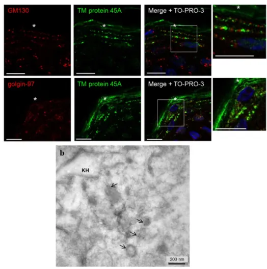
To confirm this result in vivo, co-detections of TM protein 45A with GM130 and golgin-97 were also performed on histological sections of normal human skin (Figure 4a). TM protein 45A signal was mostly located in granular layer of the epidermis, often side by side with GM130 in the granular layer and frequently organized like «Rosary Beads». However, TM protein 45A was also observed not associated with GM130 in more superficial locations. This seems to correspond to its presence in cornified keratinocytes. Similarly, TM protein 45A immunoreactivity partially co-localized with golgin-97 in locations corresponding to granular keratinocytes.
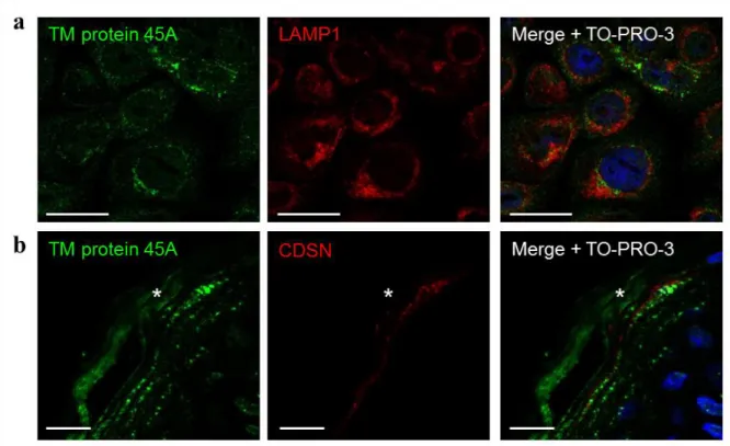
Thus, association of TM protein 45A with the trans-Golgi/trans-Golgi network was demonstrated but since this was not its only location, a potential association with the lysosomal compartment was also investigated through co-detection with LAMP1 in cultured keratinocytes (Figure S5a). Using a validated antibody against LAMP1 [21], no co-localization was observed, indicating that TM protein 45A does not reach the lysosomal compartment.
Ultrastructural analysis of epidermal TM protein 45A
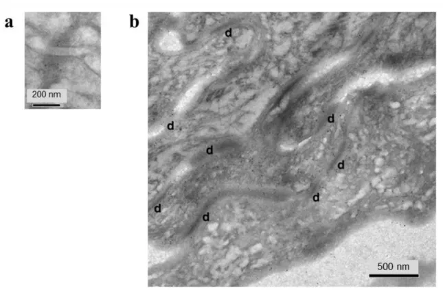
Immunogold-labelling of TM protein 45A was used to further determine its location in normal epidermis by electron microscopy. In granular keratinocytes, gold particles were often detected in small intracellular membrane vesicles (Figure 4b). Some labelling was also observed associated with intermediate filaments in the vicinity of keratohyalin granules (Figure 4b), as well as close to desmosomes (Figure S4a). Inside corneocytes, gold-labelling was dispersed, sometimes associated with anchorage sites of intermediate filaments to corneodesmosomes (Figure S4b). Those localizations concur with the immunofluorescence labelling shown on Figure 1d-g.
TM protein 45A is not transported with corneodesmosin in lamellar bodies
Several observations indicate that TM protein 45A could be related to lamellar bodies with namely a punctuated distribution in granular layer, a predicted transmembrane localization of the protein, an possible involvement during keratinization, its detection in trans-Golgi/trans-Golgi network and its ultrastructural detection in particular vesicles. To test this hypothesis, co-detection of TM protein 45A was performed in normal epidermis with CDSN, one of cargoes of lamellar bodies, using a validated antibody [22]. No or very few co-localization could be observed in the granular layer
3 4 5 6 7 8 9 10 11 12 13 14 15 16 17 18 19 20 21 22 23 24 25 26 27 28 29 30 31 32 33 34 35 36 37 38 39 40 41 42 43 44 45 46 47 48 49 50 51 52 53 54 55 56 57 58 59
For Review Only
(Figure S5b), indicating that TM protein 45A is likely not transported with CDSN in lamellar bodies. However, because cargoes are known to be transported as distinct aggregates [23], this result does not exclude potential relationship of TM protein 45A with lamellar bodies.
Discussion
This study confirms that TMEM45A expression in human keratinocytes is upregulated during epidermal differentiation, either induced by culture confluence or enhancement of Ca2+ concentration in medium [9, 10]. It also demonstrates strong connection between TMEM45A expression and keratinization, both in epidermis in vivo or during reconstruction in culture, but also in Hassal bodies inside the thymus. TMEM45A gene expression is increased in parallel with differentiation in cultured keratinocytes and its protein product accumulates during epidermal morphogenesis in vitro. Finally, TM protein 45A accumulation culminates in the granular layer in
vivo. Altogether, these data establish TMEM45A expression as a late marker of epidermal differentiation and suggest that it could be an actor of keratinization. These results are consistent with published literature identifying TMEM45A gene as a novel human keratinization-associated gene [11], strongly expressed in late stage of keratinization [3] and as a novel gene selectively and highly expressed in normal human skin [10]. This study allows to compile dispersed but important findings about the link between TMEM45A expression and keratinization that still requests further investigation.
Other members of the heterogeneous TMEM family are still poorly characterized, but new findings show potential importance in epidermis functionality or in chemoresistance. Tmem79 (Matt) gene is linked to Golgi apparatus and to lamellar body secretion; altered cornified barrier formation and dermatitis are observed in Tmem79ma/ma mutant mice [24]. Transmembrane protein 205 is associated with drug-resistance of squamous carcinoma, probably by altered membrane trafficking and secretion [25].
For the first time, TM protein 45A precise subcellular localization is reported here in keratinocytes. It partially accumulates in trans-Golgi/trans-Golgi network, both in vitro and in the granular layer in
vivo, in perfect accordance with its localization close to cis-Golgi/cis-Golgi network. Moreover, its localization is disrupted by brefeldin A, supporting strong relationship with Golgi apparatus. However, other localizations may be suspected, whereas our data exclude lysosomes as well as CDSN-containing lamellar bodies. Nevertheless, transport with other cargoes cannot be excluded.
3 4 5 6 7 8 9 10 11 12 13 14 15 16 17 18 19 20 21 22 23 24 25 26 27 28 29 30 31 32 33 34 35 36 37 38 39 40 41 42 43 44 45 46 47 48 49 50 51 52 53 54 55 56 57 58
For Review Only
Using electron microscopy, TM protein 45A immunoreactivity was associated to membrane vesicles in granular keratinocytes. The signal was also found at the level of insertion sites of intermediate filaments in the vicinity of keratohyalin granules and desmosomes in the granular layer, as well as in anchorage sites of intermediate filaments to corneodesmosomes in the cornified layer. This result suggests a possible association with cytoskeleton.
In conclusion, the strong relationship between TMEM45A expression and keratinization suggests that the TM protein 45A plays an important role in this process while being linked to Golgi apparatus.
Acknowledgements
AH is a research fellow funded by FRIA. ER is financed by Televie grant (FNRS). The study received also financial support from FNRS and FRFC (contract grants 1.5.033.06F and 2.4.522.10F) to YP. The authors thank Dr. Bernadette Bienfait, Pr. Pierre Garin, Alain Koninckx, Noëlle Ninane, Catherine Demazy, Marc Dieu, Lionel Flamant, Dominique Desnoeck, Benoît Balau, Daniel Van Vlaender, Frédéric Minner and Françoise Herphelin for their help in this study.
Conflict of interest
The authors state no conflict of interest.
References
1. Eckhart, L., et al., Cell death by cornification. Biochim Biophys Acta, 2013. 1833(12): p. 3471-80.
2. Toulza, E., et al., Large-scale identification of human genes implicated in epidermal barrier
function. Genome Biol, 2007. 8(6): p. R107.
3. Taylor, J.M., et al., Dynamic and physical clustering of gene expression during epidermal
barrier formation in differentiating keratinocytes. PLoS One, 2009. 4(10): p. e7651. 4. BioGPS, accessed 27 November 2013, available from
http://biogps.org/#goto=genereport&id=55076 5. Ensembl, accessed 27 November 2013, available from
http://www.ensembl.org/Homo_sapiens/Gene/Summary?g=ENSG00000181458;r=3:100211 463-100296288.
6. PredictProtein, accessed 30 October 2013, available from https://www.predictprotein.org/get_results?req_id=452078
7. SOSUI, accessed 27 November 2013, available from http://bp.nuap.nagoya-u.ac.jp/sosui/. 8. Uniprot, accessed 27 November 2013, available from
http://www.uniprot.org/uniprot/Q9NWC5. 3 4 5 6 7 8 9 10 11 12 13 14 15 16 17 18 19 20 21 22 23 24 25 26 27 28 29 30 31 32 33 34 35 36 37 38 39 40 41 42 43 44 45 46 47 48 49 50 51 52 53 54 55 56 57 58 59
For Review Only
9. Flamant, L., et al., TMEM45A is essential for hypoxia-induced chemoresistance in breast
and liver cancer cells. BMC Cancer, 2012. 12: p. 391.
10. Gerber, P.A., et al., Systematic identification and characterization of novel human
skin-associated genes encoding membrane and secreted proteins. PLoS One, 2013. 8(6): p. e63949.
11. Mattiuzzo, N.R., et al., A large-scale multi-technique approach identifies forty-nine new
players of keratinocyte terminal differentiation in human epidermis. Exp Dermatol, 2011.
20(2): p. 113-8.
12. Hudson, L.G., et al., Microarray analysis of cutaneous squamous cell carcinomas reveals
enhanced expression of epidermal differentiation complex genes. Mol Carcinog, 2010.
49(7): p. 619-29.
13. Lee, S., et al., Differentially expressed genes regulating the progression of ductal carcinoma
in situ to invasive breast cancer. Cancer Res, 2012. 72(17): p. 4574-86.
14. Minner, F., F. Herphelin, and Y. Poumay, Study of epidermal differentiation in human
keratinocytes cultured in autocrine conditions. Methods Mol Biol, 2010. 585: p. 71-82. 15. Frankart, A., et al., Epidermal morphogenesis during progressive in vitro 3D reconstruction
at the air-liquid interface. Exp Dermatol, 2012. 21(11): p. 871-5.
16. Minner, F. and Y. Poumay, Candidate housekeeping genes require evaluation before their
selection for studies of human epidermal keratinocytes. J Invest Dermatol, 2009. 129(3): p. 770-3.
17. Poumay, Y. and M.R. Pittelkow, Cell density and culture factors regulate keratinocyte
commitment to differentiation and expression of suprabasal K1/K10 keratins. J Invest Dermatol, 1995. 104(2): p. 271-6.
18. Hale, L.P. and M.L. Markert, Corticosteroids regulate epithelial cell differentiation and
Hassall body formation in the human thymus. J Immunol, 2004. 172(1): p. 617-24.
19. Ireland, B.S., M. Niggemann, and D.B. Williams, In vitro assays of the functions of calnexin
and calreticulin, lectin chaperones of the endoplasmic reticulum. Methods Mol Biol, 2006.
347: p. 331-42.
20. Goud, B. and P.A. Gleeson, TGN golgins, Rabs and cytoskeleton: regulating the Golgi
trafficking highways. Trends Cell Biol, 2010. 20(6): p. 329-36.
21. Sarafian, V., R. Jans, and Y. Poumay, Expression of lysosome-associated membrane protein
1 (Lamp-1) and galectins in human keratinocytes is regulated by differentiation. Arch Dermatol Res, 2006. 298(2): p. 73-81.
22. Caubet, C., et al., Degradation of corneodesmosome proteins by two serine proteases of the
kallikrein family, SCTE/KLK5/hK5 and SCCE/KLK7/hK7. J Invest Dermatol, 2004. 122(5): p. 1235-44.
23. Ishida-Yamamoto, A., et al., Epidermal lamellar granules transport different cargoes as
distinct aggregates. J Invest Dermatol, 2004. 122(5): p. 1137-44.
24. Sasaki, T., et al., A homozygous nonsense mutation in the gene for Tmem79, a component
for the lamellar granule secretory system, produces spontaneous eczema in an experimental model of atopic dermatitis. J Allergy Clin Immunol, 2013. 132(5): p. 1111-1120 e4.
25. Shen, D.W. and M.M. Gottesman, RAB8 enhances TMEM205-mediated cisplatin resistance. Pharm Res, 2012. 29(3): p. 643-50. 3 4 5 6 7 8 9 10 11 12 13 14 15 16 17 18 19 20 21 22 23 24 25 26 27 28 29 30 31 32 33 34 35 36 37 38 39 40 41 42 43 44 45 46 47 48 49 50 51 52 53 54 55 56 57 58
For Review Only
Figure 1. Upregulation of TMEM45A during differentiation of keratinocyte autocrine monolayers and predominance of TMEM45A mRNA/protein accumulation in the granular layer of human epidermis. (a) Relative quantification of TMEM45A mRNA 2 days before confluence (C-2d), at confluence (C), 2 days (C+2d) and 4 days after confluence (C+4d). Results were normalized to geometric mean values of RPLP0 and TBP house-keeping genes. The “C-2d” condition level was arbitrarily fixed at 1. Error bars represent 95% confidence intervals (n=3, one-way Repeated Measures ANOVA, *P<0.05, **P<0.01, ***P<0.001).
(b) Immunoperoxidase labelling of transmembrane protein 45A 3 days before confluence (C-3d), at confluence (C) and 2 days after confluence (C+2d). Hemalun staining of nuclei (scale bars: 100 µm).
(c) Co-immunofluorescence labelling of transmembrane protein 45A with KRT10 or IVL 4 days after confluence. Nuclei were stained with Hoechst (scale bars: 50 µm).
(d-e) Immunoperoxidase labelling of TM protein 45A in skin. Hemalun staining of nuclei (scale bars: 50 µm). Epidermis and dermal cells show immunoreactivity (arrows). Asterisks indicate cornified layer. Negative
control was incubated without primary antibody (e).
3 4 5 6 7 8 9 10 11 12 13 14 15 16 17 18 19 20 21 22 23 24 25 26 27 28 29 30 31 32 33 34 35 36 37 38 39 40 41 42 43 44 45 46 47 48 49 50 51 52 53 54 55 56 57 58 59
For Review Only
(f-g) Immunofluorescence labelling of TM protein 45A in skin. TO-PRO-3 staining of nuclei. Pictures show the same field in different Z-planes (scale bars: 50 µm).
(h) Relative mRNA levels of KRT14, KLK7, CDSN and TMEM45A genes in T1, T2, T3 and T4 fractions obtained by trypsin dissociation of epidermal keratinocytes. mRNA levels were normalized to HPRT1
house-keeping gene. The level in T1 was fixed at 1. Results from one representative experiment (n=2). 398x530mm (72 x 72 DPI) 3 4 5 6 7 8 9 10 11 12 13 14 15 16 17 18 19 20 21 22 23 24 25 26 27 28 29 30 31 32 33 34 35 36 37 38 39 40 41 42 43 44 45 46 47 48 49 50 51 52 53 54 55 56 57 58
For Review Only
Figure 2. TM protein 45A accumulates in thymic Hassal bodies but is not detected in non-keratinized stratified epithelium.
Immunofluorescence labelling of TM protein 45A in normal human non-keratinized mouth epithelium (a-b) and in normal human skin (c-d). Negative controls were produced by incubation without primary antibody (b
and d). Nuclei were stained with Hoechst 33258 (scale bars: 100 µm). Epidermis and dermal cells show immunoreactivity (arrows).
(e) Immunofluorescence labelling of TM protein 45A, KRT14, KRT10 and FLG in thymus. Nuclei were stained with Hoechst 33258 (scale bars: 50 µm).
391x434mm (72 x 72 DPI) 3 4 5 6 7 8 9 10 11 12 13 14 15 16 17 18 19 20 21 22 23 24 25 26 27 28 29 30 31 32 33 34 35 36 37 38 39 40 41 42 43 44 45 46 47 48 49 50 51 52 53 54 55 56 57 58 59
For Review Only
Figure 3. Association of TM protein 45A with the Golgi apparatus in normal human epidermal keratinocytes. (a) Immunolabelling of TM protein 45A, CALN, GM130 and golgin-97 (scale bars: 10 µm).
(b) Immunolabelling of CALN, GM130, golgin-97 and TM protein 45A of confluent cultures incubated without (control) or with brefeldin A 10 µg/ml for 30 minutes. Nuclei were stained with TO-PRO-3 (scale bars: 25
µm). 387x466mm (72 x 72 DPI) 3 4 5 6 7 8 9 10 11 12 13 14 15 16 17 18 19 20 21 22 23 24 25 26 27 28 29 30 31 32 33 34 35 36 37 38 39 40 41 42 43 44 45 46 47 48 49 50 51 52 53 54 55 56 57 58
For Review Only
Figure 4. Transmembrane protein 45A localization in epidermis.
(a) Immunohistochemical staining of GM130, golgin-97 and TM protein 45A in human epidermis. Nuclei were stained with TO-PRO-3. The asterisks indicate the cornified layer. The right panel is a magnification of the
merge (scale bars: 10 µm).
(b) Post-embedding immuno-electron microscopy of normal human epidermis using Lowicryl K4M resin and 5 nm immunogold-labeled secondary antibody. The picture illustrates a granular keratinocyte with
keratohyalin granule (KH). Arrows indicate labelled membrane vesicles. 391x376mm (72 x 72 DPI) 3 4 5 6 7 8 9 10 11 12 13 14 15 16 17 18 19 20 21 22 23 24 25 26 27 28 29 30 31 32 33 34 35 36 37 38 39 40 41 42 43 44 45 46 47 48 49 50 51 52 53 54 55 56 57 58 59
For Review Only
1
Supplementary figures
Figure S1. Validation of anti-transmembrane protein 45A antibody by
immunofluorescence labelling in human skin. Nuclei were stained with Hoechst 33258
(scale bars: 50 µm).
(a) Incubation with primary antibody anti-TM protein 45A was followed by incubation with
secondary antibody.
(b) Primary antibody anti-TM protein 45A was incubated with 30 fold excess immunogenic
peptide before incubation with tissue. This was followed by incubation with secondary
antibody.
(c) Tissue was incubated with secondary antibody only.
Figure S2. Upregulation of TMEM45A expression during Ca
2+-induced differentiation
in cultures of human keratinocytes cultured as monolayers.
Subconfluent cultures at low cell density were incubated with high calcium concentration (1.5
mM Ca
2+) during 48 hours. Cultures incubated with the usual calcium concentration (0.06
mM Ca
2+) were used as reference. mRNA levels were normalized to geometric mean values
of two house-keeping genes (RPLP0 and TBP). The mRNA level in the 0.06 mM Ca
2+condition was used as reference and arbitrarily fixed at 1. Error bars represent 95%
confidence intervals (n=3, paired t-test, ***P<0.001).
3 4 5 6 7 8 9 10 11 12 13 14 15 16 17 18 19 20 21 22 23 24 25 26 27 28 29 30 31 32 33 34 35 36 37 38 39 40 41 42 43 44 45 46 47 48 49 50 51 52 53 54 55 56 57 58 59 60
For Review Only
2
Figure S3. TMEM45A expression during epidermal morphogenesis at air-liquid
interface on polycarbonate filter. The reconstructed human epidermis was analyzed one day
after seeding the culture (1d), and then every second day until the eleventh day of
reconstruction (11d).
(a) mRNA levels were normalized to geometric mean values of TBP and RPLP0
house-keeping genes. Level after one day of reconstruction (1d) was arbitrarily fixed at 1. Error bars
represent 95% confidence intervals (n=4, one-way Repeated Measures ANOVA). No
statistically significant variation was observed.
(b) Immunofluorescent labelling of TM protein 45A. Reconstructed human epidermis stained
with Hemalun Erythrosin (HE) or labelled for TM protein 45A detection, followed by
DNA-staining using Hoechst 33258 (scale bars: 50 µm).
3 4 5 6 7 8 9 10 11 12 13 14 15 16 17 18 19 20 21 22 23 24 25 26 27 28 29 30 31 32 33 34 35 36 37 38 39 40 41 42 43 44 45 46 47 48 49 50 51 52 53 54 55 56 57 58 59 60
For Review Only
3
Figure S4. Transmembrane protein 45A localization in epidermis. Post-embedding
immuno-electron microscopy of normal human epidermis using Lowicryl K4M resin and 5
nm immunogold-labeled secondary antibody. (a) Desmosome in the granular layer. (b)
Cornified layer with corneodesmosomes (d).
3 4 5 6 7 8 9 10 11 12 13 14 15 16 17 18 19 20 21 22 23 24 25 26 27 28 29 30 31 32 33 34 35 36 37 38 39 40 41 42 43 44 45 46 47 48 49 50 51 52 53 54 55 56 57 58 59 60
For Review Only
4
Figure S5. Co-detections of TM protein 45A with LAMP1 in vitro and with CDSN in the
granular layer of epidermis.
(a) Immunolabelling of TM protein 45A and LAMP1 in normal human epidermal
keratinocytes cultured as autocrine monolayers. Nuclei were stained with TO-PRO-3 (scale
bars: 25 µm).
(b) Immunolabelling of TM protein 45A and CDSN in human normal epidermis. Nuclei were
stained with TO-PRO-3. The asterisks indicate the cornified layer (scale bars: 10 µm).
3 4 5 6 7 8 9 10 11 12 13 14 15 16 17 18 19 20 21 22 23 24 25 26 27 28 29 30 31 32 33 34 35 36 37 38 39 40 41 42 43 44 45 46 47 48 49 50 51 52 53 54 55 56 57 58 59 60
For Review Only
5
Supplementary material
Antibodies, immunogenic peptide and chemicals
Rabbit TM protein 45A antibody n° HPA024082 (dilution 1:50 or 1:100), mouse
anti-IVL antibody n° I9018 (dilution 1:200) and brefeldin A n° B7651 were purchased from
Sigma-Aldrich (Saint-Louis, MO, US). Mouse anti-GM130 antibody n° 610822 (dilution
1:100 for cells and 1:300 for tissues) was obtained from BD (Franklin Lakes, NJ, US). Mouse
anti-CALN antibody n° MA3-027 (dilution 1:500) came from Thermo Fisher Scientific
(Waltham, MA, USA). Mouse anti-golgin-97 antibody n° A-21270 (dilution 1:100), Hoechst
33258, TO-PRO-3, Alexa fluor 488-conjugated goat anti-rabbit antibody (dilution 1:200) and
Alexa fluor 546-conjugated goat anti-mouse antibody (dilution 1:200 or 1:500) were
purchased from Life technologies (Carlsbad, CA, US). Mouse anti-KRT10 antibody n°
M7002 (dilution 1:100) and DAB system n° K3468 were obtained from Dako (Glostrup,
Denmark). Mouse anti-LAMP1 antibody (H4A3) (dilution 1:50) was from DSHB (Iowa City,
Iowa, US). Mouse anti-FLG antibody n° MS-449-P1 (dilution 1:75) was obtained from
Neomarkers (Fremont, CA, US). Mouse anti-KRT14 antibody n° LL002 (dilution 1:50) was
purchased from Santa Cruz (Dallas, Texas, USA). Vectastain ABC Kit peroxidase was
purchased from Vector laboratories (Burlingame, CA, US). TMEM45A immunogenic peptide
was given by ATLAS ANTIBODIES (Stockholm, Sweden). Mouse anti-CDSN G36-19
antibody (dilution 1:3,000) was given by Guy Serre (UMR5165/U1056
CNRS-INSERM-University of Toulouse, UDEAR, Toulouse, France).Primers for real-time PCR (keratinocyte monolayers and reconstructed human
epidermis)
Gene
Forward primer (5’-3’)
Reverse primer (3’-5’)
IVL
TGAAACAGCCAACTCCAC TTCCTCTTGCTTTGATGGGKRT10
AATCA GATTCTCAACCTAACAAC CTCATCCAGCA CCCTACGKRT14
CGATGGCAAGGTGGTGTC GGGTGAAGCAGGGTCCAGRPLP0
ATCAACGGGTACAAACGAGTC CAGATGGATCAGCCAAGAAGGTPB
TCAAACCCAGAATTGTTCTCCTTAT CCTGAATCCCTTTAGAATAGGGT AGATMEM45A
TTATGCAGTAACCATTGTCATCGTT TGATTCTTGTTCTCGTTCAGCATTPrimers for real-time PCR (epidermal dissociation)
Gene
Forward primer (5’-3’)
Reverse primer (3’-5’)
KRT14
GAGGAGACCAAAGGTCGCTA CTGGATGACTGCGATCCAGAKLK7
CTCATGCTCGTGAAGCTCAA GGTCAGAGGGAAAGGTCACACDSN
CGTATCACCTCCCCTAACGA AGGAGTAGCTGACCTGGGAAHPRT1
ACCCCACGAAGTGTTGGATA AAGCAGATGGCCACAGAACTTMEM45A
TTATGCAGTAACCATTGTCATCGTT TGATTCTTGTTCTCGTTCAGCATT3 4 5 6 7 8 9 10 11 12 13 14 15 16 17 18 19 20 21 22 23 24 25 26 27 28 29 30 31 32 33 34 35 36 37 38 39 40 41 42 43 44 45 46 47 48 49 50 51 52 53 54 55 56 57 58 59 60
For Review Only
6
Immunofluorescence labelling on tissues and cells
Paraffin-embedded sections were deparaffinized and rehydrated. For TM protein 45A
detection, heat-induced antigen retrieval was performed by incubating sections in 10 mM
citrate buffer at pH6 and 95°C for 20 minutes. Cryosections and coverslips with seeded cells
were fixed for 15 min in PBS- PFA 4% buffered at pH 7.2. After rinsing in PBS-CaCl
2(0.01%), cryosections, paraffin sections and coverslips were incubated in 0.1 M glycine.
Paraffin sections were blocked for 30 minutes in PBS-BSA (0.2%)-CaCl
2(0.01%).
Cryosections and coverslips were blocked and permeabilized in PBS-BSA (0.2%)-Triton
X-100 (0.02%)-CaCl
2(0.01%) for 30 minutes. Incubation (from 45 minutes to 2 hours or
overnight) with primary antibody diluted in PBS-BSA-Triton X-100-CaCl
2(cryosections and
coverslips) or PBS-BSA-CaCl
2(paraffin sections) was performed. Negative controls were
obtained by incubating sections and coverslips in buffer without primary antibody. Sections
and coverslips were incubated for one hour with secondary Alexa fluor. Nuclei were stained
with Hoechst 33258 or TO-PRO-3. Labelled tissues and cells were analyzed under Olympus
AX70 microscope (Tokyo, Japan) or Leica TCS SP5II confocal microscope (Solms,
Germany).
Peroxidase labelling on tissues and cells
Cryosections and coverslips with seeded cells were fixed for 15 minutes in PBS- PFA 4%
buffered at pH 7.2. After rinsing in PBS-CaCl
2(0.01%), they were incubated in 0.1 M
glycine. Cryosections and coverslips were incubated in 3% H
2O
2for 10 minutes. They were
blocked and permeabilized in PBS-BSA (0.2%)-Triton X-100 (0.02%)-CaCl
2(0.01%) for 30
minutes. Cryosections and coverslips were incubated for 1-2 hours or overnight with primary
antibody diluted in PBS-BSA-Triton X-100-CaCl
2. Negative controls were obtained by
incubating sections and coverslips in PBS-BSA-Triton X-100-CaCl
2without primary
antibody. Cryosections and coverslips were incubated for 45 min with biotinylated secondary
antibody diluted 1:100 in PBS-BSA-Triton X-100. After washes, sections and coverslips were
incubated for 30 minutes with streptavidin-peroxidase solution. Detection of HRP was
performed using the high-sensitivity DAB system. Reaction was stopped in distilled water.
Counterstaining with hemalun was perfomed. For observation, an Olympus AX70 microscope
(Tokyo, Japan) was used.
Immunoelectron microscopy
Normal human skin fixed in 3% paraformaldehyde/PBS and low-temperature embedded in
Lowicryl K4M (Leica, Germany) was used for on-section immunogold labeling [1]. Ultrafine
sections, harvested on nickel grids, were first pretreated for 10 min. at 60°C with 10% sodium
metaperiodate for surface etching and optimum antigen retrieval. After washes in water and
incubation with a blocking buffer containing 5% normal goat serum, the sections were
exposed to the primary antibody diluted 1:50, in PBS supplemented with 0.1% goat serum
and 0.01% gelatin, at 4°C overnight. After washes, the primary antibody binding sites were
revealed using goat anti-rabbit 5 nm immunogold-labeled secondary antibody (diluted 1:10,
1h at r.t.; British Biocell International, Cardiff, UK). After final washes, the sections were
counterstained with uranyl acetate for observation in TEM.
3 4 5 6 7 8 9 10 11 12 13 14 15 16 17 18 19 20 21 22 23 24 25 26 27 28 29 30 31 32 33 34 35 36 37 38 39 40 41 42 43 44 45 46 47 48 49 50 51 52 53 54 55 56 57 58 59 60
For Review Only
7
Supplementary reference
1.
Haftek, M., et al., Expression of corneodesmosin in the granular layer and stratum
corneum of normal and diseased epidermis. Br J Dermatol, 1997. 137(6): p. 864-73.
3 4 5 6 7 8 9 10 11 12 13 14 15 16 17 18 19 20 21 22 23 24 25 26 27 28 29 30 31 32 33 34 35 36 37 38 39 40 41 42 43 44 45 46 47 48 49 50 51 52 53 54 55 56 57 58 59 60