HAL Id: tel-02889524
https://tel.archives-ouvertes.fr/tel-02889524v2
Submitted on 15 Jul 2020HAL is a multi-disciplinary open access archive for the deposit and dissemination of sci-entific research documents, whether they are pub-lished or not. The documents may come from teaching and research institutions in France or abroad, or from public or private research centers.
L’archive ouverte pluridisciplinaire HAL, est destinée au dépôt et à la diffusion de documents scientifiques de niveau recherche, publiés ou non, émanant des établissements d’enseignement et de recherche français ou étrangers, des laboratoires publics ou privés.
Dental development and replacement in Lagomorpha
Ludivine Bertonnier-Brouty
To cite this version:
Ludivine Bertonnier-Brouty. Dental development and replacement in Lagomorpha. Embryology and Organogenesis. Université de Lyon, 2019. English. �NNT : 2019LYSEN018�. �tel-02889524v2�
Numéro National de Thèse : 2019LYSEN018
THESE de DOCTORAT DE L’UNIVERSITE DE LYON
opérée parl’Ecole Normale Supérieure de Lyon
Ecole DoctoraleN°340
Biologie Moléculaire, Intégrative et Cellulaire Discipline : Biologie
Soutenue publiquement le 03/07/2019, par :
Ludivine BERTONNIER-BROUTY
Etude du développement et du
remplacement dentaire chez les
lagomorphes
Devant le jury composé de :
Pr. Buchtova, Marcela - Masaryk University Rapporteure Pr. Jernvall, Jukka - University of Helsinki Rapporteur Pr. Bleicher, Françoise - Université Claude Bernard Examinatrice Dr. Michon, Frédéric - Université de Montpellier Examinateur Pr. Tucker, Abigail - King’s College London Examinatrice Dr. Charles, Cyril – ENS de Lyon Directeur de thèse Dr. Joly, Thierry – ISARA Lyon Invité
3
A
CKNOWLEDGMENTS
As the tradition, let’s start this thesis manuscript with acknowledgments. I want to thank all the people that helped and supported me during this PhD. I would like to start by thanking my PhD supervisor, Cyril. You offered me the opportunity to do an internship at the end of my bachelor degree, and here I am 6 years later, finishing my PhD under your supervision. Thank you for having trusted me and having made me discover a research topic that I did not leave since. Thank you Laurent, even without being officially my supervisor during all these years, you always kept an eye on my research and gave relevant advice and valuable comments.
I want to thank my PhD committee, Pr. Françoise Bleicher, Dr. Vincent Lazzari and Dr. Thierry Joly who helped me to clarify my project over the years and gave me useful guidance and suggestions and who agreed to examine this thesis manuscript. I also thank the member of my jury, Pr. Marcela Buchtova, Pr. Jukka Jernvall, Dr. Frédéric Michon and Pr. Abigail Tucker who agreed to evaluate this thesis manuscript.
I also want to thank all the people that help me to obtain all the specimens used during my PhD. Thank you Thierry for providing us with the rabbit embryos. Thanks to Aline for providing us with irradiated samples. Thanks to all the museums and the people who gave me access to the collections: Eileen Westwig (AMNH), Cécile Callou (MNHN), François Vigouroux (Musée des Confluences) and Emmanuel Robert (Geological Collections, University Lyon 1).
I have been really happy to prepare my PhD at the IGFL. I have met and worked with great people these last years. Thanks to the administration team and more specifically Martine for its effectiveness in managing all administrative matters, always with the smile. Thank to Christian Lamy, responsible for the glassware, always benevolent. Thank you Pierre for the help in histology and microscopy. Thanks to all the other PhD of the institute for scientific
4 exchanges as well as the evening out of the lab. Finally, thanks to all the people from the institute for every interaction we had during these last years.
I want to warmly thank all the past and present members of the “Evolution of vertebrate dentition” team: Thank you Laurent and Beatrice for your infectious laughter that were as important as your scientific help during this thesis. Jérémie, we started at the same time and am very happy to be able to taunt you by finishing (a bit) before you. Thank you for making us laugh (but also exasperated) regularly. Thomas thank you for having experimented with me the joys and disappointments of molecular biology but also for your melodious whistles as background music. Thank you Pauline M. for teaching me so much during my first internship by always being in a good mood and caring. Finally, thank you Mathilde for your help with the lab work and the microtomography but also for all our lunch breaks together.
During my PhD, I moved in the “Biomodeling” team. Thank you Nicolas for welcoming us to your team. Thank you Louise, official roommate at each congress, for always having been there to discuss (or drink beers). Thank you Fidji to defend with me the real teeth with the help of Roland against the conodontologists surrounding us. Thanks to the conodontologists, Pauline G., Sam and Nick for promoting a good atmosphere in the office. Also, thank you Pauline J. for your help in statistics but especially for sing all your favorite Disney songs all the day and your great cakes.
I want to thank my parents and family. Thank you for having believed in me and having supported me, for doing everything to understand the strange world of research and to encourage me at every step. Thanks to my little brother to ask me at each family meeting "so, what's new about lagomorphs?”. Antonin, thank you for being here every day to support me and take the time to listen me speaking (or complain) too much about my thesisand my crazy holiday dreams. Finally, thanks to all my friends, and especially Cynthia and Elise for all the shared moments and the laughs. Thanks a lot!
5
TABLE OF CONTENTS
LIST OF THE TABLES AND FIGURES ... 7
ABBREVIATIONS ... 11
ABSTRACT ... 13
RÉSUMÉ SUBSTANTIEL EN FRANÇAIS ... 17
GENERAL INTRODUCTION &LITERATURE REVIEW ... 23
Tooth development in Amniota ... 25
Tooth replacement in Amniota ... 28
State of the art regarding dental replacement in polyphyodont reptiles ... 28
State of the art regarding dental replacement in diphyodont mammals ... 30
State of the art regarding dental replacement in monophyodont mammals ... 37
Rabbit as a model for tooth Evo-Devo studies ... 42
Rabbit tooth development ... 43
Characteristics of extant Lagomorpha ... 43
Evolutionary history of Lagomorpha ... 45
PART 1 ... 46
TOOTH DEVELOPMENT AND REPLACEMENT IN ORYCTOLAGUS CUNICULUS ... 46
Chapter 1.1 – Morphological characteristics of tooth development and replacement in Oryctolagus cuniculus ... 50
Article 1: Morphological features of tooth development and replacement in the rabbit Oryctolagus cuniculus ... 52
Chapter 1.2 – Spatio-temporal regulation of tooth replacement in Oryctolagus cuniculus ... 84
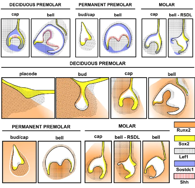
Article 2: Expression patterns of Runx2, Sox2, Lef1, Shh and Sostdc1 in Oryctolagus cuniculus tooth development and replacement ... 86
6
PART 2 ... 110
TOOTH SHAPE MODIFICATIONS DURING LAGOMORPHA EVOLUTION ... 110
Chapter 2.1 – Oryctolagus cuniculus cuspidogenesis ... 114
Article 3: Million years of mammalian tooth evolution revisited in 4 days of development ... 116
Chapter 2.2 – Lagomorpha crown shape variations during development and evolution ... 130
Article 4: Ontogenetic modifications of tooth crown shape unravel mechanisms of evolution of the dentition in Lagomorpha (Glires, Mammalia) ... 132
GENERAL CONCLUSION &PERSPECTIVES ... 172
Morphological features of tooth development in Oryctolagus cuniculus ... 174
Dentin holes in the upper ever-growing incisor ... 175
Genetic basis involved in rabbit tooth replacement ... 175
Rudimentary successional dental lamina in the molars... 176
Transcriptomic study of the tooth replacement ... 176
The rabbit as model for human pathologies linked to radiation therapies ... 177
Developmental heterochronies in rabbit ondontogenesis ... 180
Conclusion ... 181
REFERENCES ... 182
ANNEXES ... 198
Annex 1: Plasticity within the niche ensures the maintenance of a Sox2+ stem cell population in the mouse incisor ... 200
Annex 2: A giant step backward, molar replacement in mutant mice ... 226
Annex 3: Collection numbers of the lagomorph specimens coming from natural history museums ... 244
Annex 4: Denture of Oryctolagus cuniculus ... 256
Annex 5: 3D reconstructions of dental epithelium during Oryctolagus cuniculus embryonic development. ... 262
7
L
IST OF THE TABLES AND FIGURES
GENERAL INTRODUCTION &LITERATURE REVIEW……….…...23
Figure I. 1 - Histo-morphological stages and genetic control of tooth development in the mouse.………....26 Figure I. 2 - Tooth renewal capacity varies among species………27 Figure I. 3 - Tooth development and continuous dental replacement in Squamates…………..29 Figure I. 4 - Initiation of tooth replacement in the ferret, the fruit bat and the minipig………...31 Figure I. 5 - 3D-reconstructions of the dental replacement in the fruit bat……….33 Figure I. 6 - Tooth number abnormalities in humans: hyperdontia and agenesis………. 36 Figure I. 7 - Rudimentary successional dental lamina in the developing mouse molar………..38 Figure I. 8 - Signaling pathways involved in tooth number regulation in the mouse………...40 Figure I. 9 - Schematic representation of the rabbit skull and its dental formula………42 Figure I. 10 - Molecular phylogeny of the extant Lagomorpha……….…….44
ARTICLE 1: Morphological features of tooth development and replacement in the rabbit
Oryctolagus cuniculus……….52
Figure 1. 1 - 3D-reconstructions of the epithelial part of the upper incisors during development………...58 Figure 1. 2 - 3D-reconstructions of the mineralized part of the rabbit teeth……….59 Figure 1. 3 - Hole repartition in the upper incisor………...62 Figure 1. 4 - 3D-reconstructions of the epithelial tissues and histology in rabbit upper cheek teeth from 14 dpf to 28 dpf………..…64 Figure 1. 5 - 3D-reconstructions of the epithelial tissues and histology in rabbit lower cheek teeth from 14 dpf to 28 dpf………..66 Figure 1. 6 - Summary of the dental replacement progression in rabbit……….…………70 Figure 1. 7 - Chronology of dental development and replacement for each rabbit tooth from 12 dpf to 4 dpn……….73
8 Supplementary 1. 1 - Histological sections of the rabbit upper incisors and lower cheek teeth. ………..………..………81 Supplementary 1. 2 Views of the 3D reconstructions of the upper incisors mineralized tissues and virtual sections of the incisors………..………..…..82
ARTICLE 2: Expression patterns of Runx2, Sox2, Lef1, Shh and Sostdc1 in Oryctolagus
cuniculus tooth development and replacement……….…….86
Figure 2. 1 - Morphological features of tooth development and replacement in the rabbit…...90 Figure 2. 2 - Runx2 localization during tooth development and replacement………..93 Figure 2. 3 – Sox2 localization during tooth development and replacement………95 Figure 2. 4 - Lef1 localization during tooth development and replacement……….….97 Figure 2. 5 - Shh and Sostdc1 expression during tooth development and replacement………100 Figure 2. 6 - Synthesis of expression patterns and hypothetical regulatory events……...…..102
ARTICLE 3: Million years of mammalian tooth evolution revisited in 4 days of development………..….…….116
Table 3. 1-Cusp presence in the M1 during odontogenesis………...120
Figure 3. 1 - Cusp pattern and area of the crown of the M1 during odontogenesis………...….121
Figure 3. 2 - Molar cusp pattern variations during mammalian evolution and rabbit development………..………..…….124
ARTICLE 4: Ontogenetic modifications of tooth crown shape unravel mechanisms of evolution
of the dentition in Lagomorpha (Glires, Mammalia)………..………..…...132
Figure 4. 1 - Terminology of the M1 occlusal surface………..…139
Figure 4. 2 - Variation of M1 occlusal tooth pattern in function of the wear stage in Ochotona
princeps and Palaeolagus haydeni………...141
9 Figure 4. 4 - Variation of crenulation pattern in Oryctolagus cuniculus and frequency of the crenulation wavelength in micrometers………145 Figure 4. 5 - Variation of the number of crenulations between the occlusal and growth zone in controls and irradiated rabbits………..148 Figure 4. 6 - Simplified phylogeny of extant genera and crenulation pattern of the occlusal surface in adult……….153 Table 4. 1 - Variation of the number of crenulations in function of the species among Lagomorpha……….150 Figure 4. 7 - Chronology of morphological variations of the M1 during lagomorph evolution
and Oryctolagus cuniculus development………..156 Supplementary figure 4. 1 - Variation of the M1 from the occlusal to the growth part in
Ochotona pallasi, Palaeolagus haydeni, Oryctolagus cuniculus and Lepus capensis………169
Supplementary figure 4. 2 -Variations of the crenulation number in function of the skull size in Oryctolagus cuniculus and Lepus capensis………...………...170 Supplementary table 4. 1-List of the species studied……….171
GENERAL CONCLUSION &PERSPECTIVES……….………...……….172
11
A
BBREVIATIONS
MUSEUMS
AMNH: American Museum of Natural History, New York City Confluences: Musée des Confluences, Lyon
MNHN: Muséum national d'Histoire naturelle, Paris
UCBL-FSL: Geological Collections, University Lyon 1, Lyon
TOOTH NOMENCLATURE I: Incisor P: Premolar M: Molar d: deciduous Xx: Upper tooth Xx: Lower tooth STAGES dpf: Days post-fertilization dpn: Days post-natal
DENTAL MORPHOLOGY DESCRIPTION
CL: cervical loops
HERS: Hertwig epithelial root sheath IEE: Inner enamel epithelium
OEE: Outer enamel epithelium
RSDL: Rudimentary successional dental lamina
TECHNIQUES
CRISPR: Clustered Regularly Interspaced Short Palindromic Repeats LIPUS: Low intensity pulsed ultrasound
13
A
BSTRACT
Français
Le développement dentaire est essentiellement étudié chez la souris, modèle mammifère le plus commun en biologie. Cependant, contrairement à la majorité des mammifères, les souris ne remplacent pas leurs dents. Ainsi, les mécanismes impliqués dans le remplacement dentaire mammalien sont encore inconnus. Au cours de cette thèse, nous nous sommes intéressés au développement et remplacement dentaire mammalien en utilisant le lapin Oryctolagus
cuniculus comme modèle d’étude. Le lapin étant déjà séquencé, utilisé en recherche
biomédicale, avec une période de gestation courte et remplaçant ses dents, il semblait être un modèle pertinent en odontologie. Le lapin était un modèle méconnu du point de vue du développement dentaire, j’ai donc d’abord réalisé une étude histo-morphologique afin de caractériser la mise en place des dents déciduales et permanentes. Des reconstructions 3D des tissus mous ont été réalisés à différents stades embryonnaires afin d’obtenir une chronologie du développement et remplacement dentaire. Cette chronologie commence aux premières observations morphologiques de l’initiation du développement des premières dents jusqu’à la minéralisation des dernières dents à se développer. Puis, suite à l’identification dans la bibliographie de gènes candidats potentiellement impliqués dans le remplacement dentaire, j’ai étudié les profils d’expressions de ces gènes afin de mieux comprendre la régulation spatio-temporelle du remplacement dentaire chez le lapin. Nous avons ensuite replacé nos résultats sur le développement chez le lapin dans un contexte évolutif. Ainsi, j’ai réalisé une étude d’anatomie comparée chez les lagomorphes actuels et quelques fossiles afin d’identifier des variations morphologiques dentaires au cours de leur histoire évolutive. Nous nous sommes particulièrement intéressé à la mise en place des cuspides au cours de l’odontogénèse ainsi
14 qu’aux variations de la surface occlusale des dents supérieure tout au long de la vie des lapins et autres lagomorphes. En comparant les variations au cours de l’évolution avec celles observées lors de l’ontogénie dentaire chez le lapin nous avons identifié des processus d’hétérochronies du développement. Nous avons montré que la molaire actuelle du lapin suit un processus de péramorphose, donc de surdéveloppement, en comparaison aux lagomorphes fossiles. Le lapin est ainsi un modèle animal prometteur en biologie du développement et en évolution afin de mieux comprendre la mise en place du remplacement dentaire mammalien ainsi que les variations de forme dentaire au cours de l’évolution des mammifères.
English
Tooth development is essentially studied in mice, the favorite mammalian model in biology. However, mice do not replace their teeth on contrary to numerous mammals. So, the mechanisms involved in mammalian tooth replacement are still unknown. During this thesis, we focused on mammalian dental development and replacement using the European rabbit
Oryctolagus cuniculus as animal model. The European rabbit is already sequenced, used in
biomedical field, has a short gestation time and replaced its teeth, so rabbit seemed to be a relevant model in dental research. Rabbit dental development was not defined, so I first performed a histo-morphological study to characterize the development of deciduous and permanent teeth. 3D soft tissue reconstructions were performed at different embryonic stages to obtain a chronology of tooth development and replacement. This chronology begins with the first morphological observations of the initiation of the development of the first tooth until the mineralization of the last tooth to develop. Then, we identified in the bibliography candidate genes potentially involved in dental replacement. I studied the expression profiles of these genes in order to better understand the spatio-temporal regulation of tooth replacement in rabbits.We then returned our results to rabbit development in an evolutionary context. Thus, I performed a
15 comparative anatomy study in the current lagomorphs and some fossils in order to identify dental morphological variations during their evolutionary history. We were particularly interested in the setting of cusps during odontogenesis as well as in the variations of the occlusal surface of the upper cheek teeth throughout the life of rabbits and other lagomorphs. Comparing changes during evolution with those observed during dental ontogeny in rabbits allow us to identify heterochronous processes of development. We have shown that the current molar rabbit follows a process of peramorphosis, so an overdevelopment compared to fossil lagomorphs. The rabbit is thus a promising animal model in developmental and evolutionary biology to better understand the implementation of mammalian tooth replacement and tooth shape variations during mammalian evolution.
Key words
Tooth development, mammalian dental replacement, rabbit teeth, peramorphosis, heterochrony, odontongenesis, cusp
17
R
ESUME SUBSTANTIEL EN FRANÇAIS
Les dents, une innovation chez les vertébrés, se sont diversifiés en forme, en nombre et en capacité de remplacement. En paléontologie, les dents sont très étudiées car elles sont les tissus minéralisés les plus solides et donc les mieux conservés durant la fossilisation. L’étude d’anatomie comparée des dents fossiles donne de nombreuses informations en évolution et taxonomie. De plus, les dents sont sujettes à une forte pression de sélection puisque la forme des dents corrèle avec le régime alimentaire. Ainsi, les variations morphologiques dentaires entre les espèces sont étudiées pour comprendre les conséquences des modifications de formes en évolution et écologie (i.e Gómez Cano et al., 2013; Renvoisé et al., 2012). Les dents font aussi partie des organes ectodermaux les mieux conservés chez les vertébrés. La dentition est ainsi un modèle pertinent en biologie évolutive du développement chez les vertébrés (Evo-Devo). L’étude des dents dans un contexte d’Evo-Devo nécessite d’identifier les mécanismes moléculaires impliqués dans le développement et la morphogenèse dentaire afin de comprendre les variations morphologiques entre les espèces, éteintes ou actuelles. De même, l’étude des fossiles et les acquisitions morphologiques au cours de l’évolution aident à comprendre la morphogénèse dentaire. Le développement dentaire est étudié chez de nombreuses espèces comme les poissons cartilagineux (i.e. Rasch et al., 2016), les reptiles (i.e. Richman and Handrigan, 2011) et les mammifères (i.e. Renvoisé and Michon, 2014). Même si ces espèces sont phylogénétiquement séparés depuis approximativement 450 million d’années, les voies de signalisations impliqués dans le développement dentaire sont très conservés entre les vertébrés (Rasch et al., 2016). Les vertébrés ancestraux avaient la capacité de remplacer leurs dents de manière continue tout au long de leur vie, ils étaient polyphyodontes. La majorité des vertébrés actuels sont polyphyodontes, mais les mammifères ont perdu cette capacité de remplacement et la majorité d’entre eux possèdent deux générations dentaires au cours de leur vie
18 (diphyodontie), corrélé avec une augmentation de complexité de la morphologie dentaire. Chez les mammifères, la majorité des études d’odontogénèse utilisent la souris comme modèle. Cependant, la souris ne remplace pas ses dents donc les mécanismes du remplacement dentaire chez les mammifères sont mal connus. Nous avons donc décidé pour cette thèse d’utiliser le lapin (Oryctolagus cuniculus) comme modèle animal pour étudier (1) les mécanismes impliqués dans le développement et le remplacement dentaire chez les mammifères et (2) l’évolution des dents chez les lagomorphes.
Un bon animal modèle doit pouvoir être élevé en laboratoire, donner de nombreux embryons, avoir une période de gestation assez courte et être séquencé. Dans le cadre de notre thématique de recherche, le remplacement dentaire est aussi un critère essentiel. Le lapin respecte tous ces critères, de plus il est déjà utilisé comme modèle animal en recherche biomédicale et orthodontie (Abtahi et al., 2018). La littérature disponible sur le développement dentaire chez le lapin est assez ancienne et incomplète, obtenues grâce à des techniques désormais obsolètes et avec un manque de connaissances sur certaines dents et stades de développement (Horowitz et al., 1973; Navarro et al., 1976; Ooë, 1980). Nous avons donc décidé de mettre à jour les connaissances sur la morphogénèse dentaire du remplacement dentaire chez le lapin en utilisant les techniques actuelles. Nous avons étudié chaque dent du lapin depuis l’initiation de la dent déciduale à la minéralisation de la dent de remplacement, soit entre 12 jours post fertilisation et 4 jours post natal, représentant 23 jours de développement. Nous avons réalisé des reconstructions 3D des tissus dentaires épithéliaux pour chaque dent à chaque stage en utilisant la microtomographie à rayons X. En complément, une étude histologique a été réalisée afin d’obtenir une description complète de la chronologie de l’odontogénèse. Nous avons ainsi obtenu une chronologie complète de la morphogénèse dentaire chez le lapin Oryctolagus cuniculus afin de pouvoir cibler les stades de développement importants pour l’étude moléculaire du remplacement dentaire mammifère. Au cours de cette
19 étude, nous avons aussi identifié une caractéristique des incisives de lapin non décrite jusque-là. Ainsi, à la naissance, les incisives supérieures de lapin présentent des trous dans la dentine qui ouvrent la cavité pulpaire. Ces trous sont rapidement réparés par les odontoblastes préexistants qui continuent de secréter de la dentine malgré la perturbation.
Les voies de signalisations supposément impliqués dans le remplacement dentaire sont majoritairement étudiés chez les souris transgéniques (Ahn et al., 2010; Popa et al., 2019) ou chez les espèces polyphyodontes (Tucker and Fraser, 2014; Whitlock and Richman, 2013). Seulement quelques données ont été collectés chez des espèces mammifères diphyondontes comme le furet ou le cochon nain (Jussila et al., 2014; Wang et al., 2019). Mais pour le moment, aucune étude ne suit le pattern d’expression de gènes candidats à chaque étape du remplacement dentaire. Nous avons donc utilisé le lapin comme animal modèle pour étudier la régulation spatio-temporelle du développement et remplacement dentaire chez les mammifères. Nous avons identifié dans la littérature cinq gènes candidats qui pourraient être impliqués dans le remplacement dentaire : Shh, Sostdc1, Runx2, Lef1 et Sox2. Nous avons suivi leur profil d’expression par hybridation in situ et la localisation des protéines par immunohistochimie à chaque étape clé du remplacement dentaire. La localisation des proteines Runx2 et Sostdc1 correlent avec un mechanisme d’inhibition du remplacement. De plus, comme chez la souris, Shh et Sostdc1 sont localisés dans l’epithelium adamantin interne et pourraient réguler le pattern des cuspides. Le lapin semble ainsi être un modèle pertinent pour étudier le développement et le remplacement dentaire.
En Evo-Devo, l’étude du développement peut permettre de mieux comprendre les processus évolutifs. Par exemple, les changements dans la vitesse ou la durée des évènements développementaux induisent des changements de taille ou de forme des organes au cours de l’évolution, ce sont les hétérochronies du développement. Ces hétérochronies peuvent être identifiées en étudiant les dents fossiles ainsi que l’odontogénèse des espèces actuelles. Les
20 différentes espèces fossiles de lagomorphes sont principalement définies par leurs dents. Ainsi, il y a eu des variations nettes de morphologie dentaire au cours de l’évolution des lagomorphes. Les dents des lagomorphes au Paléocène étaient modérément hyposondontes avec des racines alors que les lagomorphes actuels ont des dents hyselondontes à croissance continue (Kraatz et al., 2010). Les relations phylogéniques entre les lagomorphes sont souvent basées sur la morphologie de la troisième prémolaire inférieure (Čermák et al., 2015a; Hibbard, 1963) mais les modifications de forme des autres dents sont peu étudiées. Habituellement, la première molaire supérieure est la dent utilisée en anatomie comparée chez les mammifères. Nous avons donc décidé de suivre les variations de forme de la première molaire supérieure au cours de l’évolution des lagomorphes et de l’ontogénie du lapin Oryctolagus cuniculus. Chez les lapins adultes, les faces occlusales des dents jugales sont complètement plates. Cependant nous avons montré que les dents de lapins possèdent transitoirement des cuspides sur leur face occlusale. Les cuspides sont un caractère essentiel pour étudier l’histoire évolutive à travers l’étude de l’homologie des cuspides chez les mammifères. Les terminologies utilisées pour définir les cuspides des lagomorphes dans la littérature sont nombreuses et il est difficile de trouver un consensus (Kraatz et al., 2010). Afin de mieux définir les cuspides chez les lagomorphes nous avons étudié leur mise en place au cours de l’odontogénèse chez le lapin. Nous avons suivi l’ordre d’apparition des cuspides et nous les avons nommées d’après la nomenclature définie par Butler (1956). Nous avons observé au cours de la cuspidogénèse chez le lapin des millions d’années d’évolution dentaire par homologie de forme. Ainsi, au cours du développement de la molaire supérieure du lapin nous avons clairement identifié des homologies de forme avec le pattern tribosphénique commun chez les mammifères ancestraux.
Les changements de formes dentaires continuent au cours de l’ontogénie chez les lapins. Nous avons donc étudié les changements de formes de la première molaire supérieure au cours de l’ontogénie du lapin et de l’évolution des lagomorphes. Afin de caractériser les changements
21 de la morphologie dentaire en relation avec l’âge nous avons étudiés la variabilité morphologique des molaires supérieures chez différentes espèces de la naissance à l’âge adulte. Puisque les dents de lagomorphes actuels sont à croissance continue, nous avons pu extrapoler la surface occlusale à différents stades d’usures en réalisant des sections virtuelles de reconstructions 3D de dents obtenues par microtomographie 3D. Nous avons observé que les changements de forme dentaire pendant l’évolution des lagomorphes et l’odontogénèse chez le lapin semblent corréler, indiquant possiblement un mécanisme d’hétérochronie du développement. Ainsi, il semblerait que les molaires supérieures de lapin ont évolué par péramorphose, avec une rétention des caractères ancestraux au début de l’odontogénèse ainsi que par l’apparition de nouveaux caractères, les crénulations. La mise en place des crénulations semble liée à la prolifération cellulaire à la base de la dent dans la zone de croissance. Ainsi, en perturbant la prolifération cellulaire par des irradiations, le pattern de crénulations est affecté. Toutefois, nous n’avons pas identifié la fonction de ces crénulations dans les dents de lapins.
Pour conclure, le lapin semble être un modèle pertinent en recherche dentaire. Nous avons montré dans cette thèse que les lapins pouvaient être un modèle utile pour étudier la morphogénèse dentaire. L’étude du développement et du remplacement dentaire chez les lapins pourraient permettre de comprendre les mécanismes de remplacement dentaires chez les mammifères. Nous avons montré que les protocoles de biologie moléculaires utilisés chez la souris pouvaient être facilement transposés aux tissus de lapin. De plus, le développement de la dentition du lapin semble suivre l’évolution des dents des mammifères, les dents de lapins pourraient donc être un modèle pertinent dans les thématiques d’Evo-Devo dentaires.
23
G
ENERAL
I
NTRODUCTION
&
24
T
rue teeth are an innovation in vertebrates and they then diversified in shape, number and replacement abilities among extant and extinct vertebrates. Teeth are highly studied in paleontology, first because teeth are the hardest mineralized tissues of the body and so are well preserved during fossilization. Using comparative anatomy, tooth fossils give information to study evolution and taxonomy. Then, teeth are subject to strong selective constraints due to the correlation between tooth shape and diet. So morphological variations of teeth between species are studied for understanding consequences of shape modifications in evolution and ecology (Gómez Cano et al., 2013; Renvoisé et al., 2012). Moreover, teeth are among the most conserved ectodermal organs throughout vertebrates. So, teeth are useful models to study vertebrate Evo-Devo. In tooth Evolutionary Developmental Biology, we need to identify the molecular mechanisms involved in tooth development and dental morphogenesis in order to understand the morphological variations of the teeth between extant and extinct vertebrates. Similarly, the study of fossils and morphological acquisitions during evolution helps to understand the dental morphogenesis. Tooth development is studied in numerous extant species as cartilaginous fishes (i.e. Rasch et al., 2016), reptiles (i.e. Richman and Handrigan, 2011) and mammals (i.e. Renvoisé and Michon, 2014). Even if these species are separated since approximately 450 million years, gene pathways involved in tooth development are highly conserved among vertebrates (Rasch et al., 2016). Basal vertebrates had the ability to replace their teeth during all their life span. Majority of the extant vertebrates still have a continuous tooth replacement system but mammals lost this ability and most of them possess two dental generations over their life, correlated with an increase in the dental shape complexity. In mammals, most of odontogenesis studies use the mouse as a model species. However, the mouse has no dental replacement. So, for now, the mechanisms of tooth replacement in mammals are poorly known. We thus decided in the present thesis to use the European rabbit25 (Oryctolagus cuniculus) as a new model to study (1) mechanism underlying mammal tooth development and replacement and (2) Lagomorph tooth evolution. I here review in the scientific literature the actual knowledge about signaling events involved in tooth development and replacement in Amniota and summarize the knowledge available before this thesis about the rabbit tooth development and the evolutionary history of lagomorphs.
Tooth development in Amniota
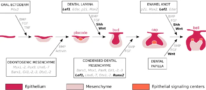
Teeth are ectodermal organs: they are originating from two distinct layers of tissues, the epithelium and the mesenchyme. Teeth develop from the interaction of mesenchymal cells derived from the neural crest and oral epithelium cells (Chai et al., 2000). The signaling events involved in initiation of the tooth development are common with the other ectodermal organs as the hair or the salivary glands despite the diversity in form and function (Pispa and Thesleff, 2003). Tooth development is commonly divided into the following stages: the thickening, the bud, the cap, the bell, and finally the maturation. Numerous signaling pathways between the epithelial and the mesenchymal cells have been identified in mice to explain all the stage transitions (Figure I. 1, Balic and Thesleff, 2015; Sharir and Klein, 2016).
The first observation of tooth development initiation is a thickening of the oral epithelium, giving rise to the dental lamina. The surrounding mesenchyme condensates around the dental lamina, which forms a dental placode. During all the tooth morphogenesis, epithelial-mesenchymal interactions occur to regulate the development. In the placode, an early epithelial signaling center grouping five signaling families has been identified to give at the epithelium an odontogenic potential: the Wnt, Shh, BMP, FGF, and Edar signaling pathways (Balic & Thesleff, 2015). All these signals regulate the proliferation and the budding of the placode; the
26 tooth begins its morphogenesis. The epithelial bud is more invaginated in the mesenchyme that continues to condensate around it. The bud also possesses a signaling center at the tip that is not proliferating: the primary enamel knot (Vaahtokari et al., 1996). Signals coming from the enamel knot induce the formation by the dental epithelium of the cervical loops; it defines the bud to cap stages transition. Cervical loops contain epithelial dental stem cells. At the cap stage, the primary enamel knot matures and is morphologically identifiable. Cells begin to histo-differentiate; we can identify the different cell types that constitute the tooth germ. Then, at the bell stage, cells finish to differentiate, giving layers of functional ameloblasts and odontoblasts. For multicuspid teeth, secondary enamel knots appear and regulate the patterning of the tooth cusps. When cells begin to secrete mineralized tooth tissues, tooth finishes its morphogenesis and the maturation stage begins. The ameloblasts secrete enamel and the odontoblasts secrete dentin, the mineralized tissues of the tooth crown. Then, the cervical loops of the tooth close and differentiate in the Hertwig epithelial root sheath (HERS) cells to form roots (Zeichner-David et al., 2003). Roots are then covered with dentin followed by an apposition of cementum.
Figure I. 1. Histo-morphological stages and genetic control of tooth development in the mouse. In
color, the mouse tooth development stages. In the boxes and arrows, some of the epithelium-mesenchyme interactions during mouse tooth development from initiation to crown morphogenesis. The genes in black are studied in this thesis (from Thesleff, 2004, colors modified). The color code is the same for all the figures.
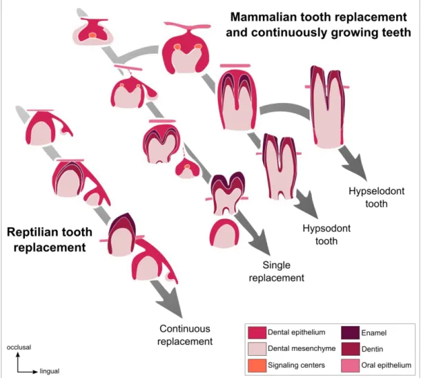
27 In mammals, by comparing the height of the crown, rooted teeth are separated in two categories: Brachyodont teeth have a short crown whereas hypsodont teeth have a high crown. However, not all the teeth develop roots. Hypselodont teeth are defined by their ability to grow during all the life of the animal (Figure I. 2). In hypselodonts, the cervical loops at the basis of the tooth never close, inducing a dental stem cell maintenance and so a continuous renewal of the dental tissues (Renvoisé and Michon, 2014). Some epithelial stem cells markers have been identified in the cervical loops of the ever-growing teeth such as Sox2 (Juuri et al., 2012), Bmi1 (Biehs et al., 2013) or Lgr5 (Sanz-Navarro et al., 2018, see Annex 1, p187). Tooth ever-growing ability in some mammals could compensates for the loss of the capacity to continuously replacing their teeth (Jernvall and Thesleff, 2012).
Figure I. 2. Tooth renewal capacity varies among species. Reptiles have a continuous tooth
replacement whereas majority of the mammals have a single tooth replacement and only some of the mammals have continuously growing teeth. (From Jernvall and Thesleff, 2012, colors modified).
28
Tooth replacement in Amniota
Tooth replacement in Amniota is divided in categories in function of the number of tooth generations: the polyphyodonts continuously replace their teeth, the diphyodonts replace their teeth only once, and the monophyodonts do not replace their teeth (Bertin et al., 2018). The polyphyodonty is the ancestral condition of all vertebrate dentitions (Maisey et al., 2014; Rucklin et al., 2012).
State of the art regarding dental replacement in polyphyodont reptiles
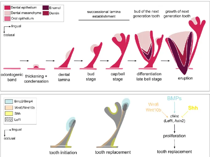
Tooth replacement is mainly studied in polyphyodont species, which have the ability to replace their teeth during all their life span. Reptiles are polyphyodonts and some species can be raised in animal facilities. The ball python (Python regius) has a continuous tooth replacement throughout its life. The tooth replacement in the ball python begins by the formation of the replacement dental lamina (also named successional lamina) at the lingual extremity of the dental lamina (Figure I. 3). The replacement teeth bud from the vestibular side of this replacement dental lamina. The preservation of the replacement dental lamina is essential to maintain the continuous tooth replacement.
Handrigan and Richman (2010b) showed that Shh is necessary for tooth initiation and morphogenesis as described in mouse (Hardcastle et al., 1998) but is not involved in the successional dental lamina formation. These authors also studied the Wnt signaling activity and the BMP pathway in the dental tissues of the ball python in order to identify the signaling pathways involved in tooth replacement (Handrigan and Richman, 2010a). Using LEF1 staining, they showed that the canonical Wnt pathway is persistently active at the growing tip of the dental lamina and replacement dental lamina (Figure I. 3). They also showed that the Shh
29 expression domain and the canonical Wnt domain are complementary in the tooth tissues and that SHH may restricts Wnt activity. They suggest that canonical Wnt signaling could order tooth replacement by promoting proliferation of the replacement dental lamina tip in snakes.
Dental stem cells have been identified in the lingual side of the dental lamina of the leopard gecko (Eublepharis macularius) (Handrigan et al., 2010). These stem cells are Lgr5+,
Dkk3+ and IGfbp5+, genes already known as hair bulge stem cells (Jaks et al., 2008; Tumbar et al., 2004). These cells have low proliferation rates and are not directly involved in tooth morphogenesis. However, they may play a role in the maintenance of the replacement dental lamina.
Figure I. 3. Tooth development and continuous dental replacement in Squamates. At the top, the tooth
development stages in Squamates (adapted from Richman and Handrigan, 2011). At the bottom, the signaling pathways involved in tooth initiation and continuous tooth replacement (adapted from Handrigan and Richman, 2010a). In bold, the genes that are studied in this thesis.
30 Juuri et al. (2013) identified another dental stem cell population in reptile teeth, the
Sox2+ cells. They localized Sox2+ cells in the replacement dental lamina of five reptile species: the American alligator (Alligator mississippiensis), the green iguana (Iguana iguana), the leopard gecko (Eublepharis macularius), the ball python (Python regius) and the corn snake (Elaphe guttata). They showed that Sox2 is expressed in the replacement dental lamina excepted in the tip. So, dental stem cells involved in maintenance of ever-growing tooth ability are also present in the replacement dental lamina allowing continuous tooth replacement.
To sum up, in reptiles, the active canonical Wnt signaling in the replacement dental lamina and the presence of dental stem cells are involved in the maintenance of the dental renewal capacity.
State of the art regarding dental replacement in diphyodont mammals
Majority of the mammals are diphyodonts, their dentitions are limited to two dental generations. Diphyodonty is a character derived from the polyphyodonty, mammals shifted from numerous teeth with simple shape to more complex teeth with a limited replacement capacity (Luo et al., 2008). Studies done in tooth replacement in diphyodont mammals are reviewed below.
Dental replacement in the ferret
Ferrets (Mustela putorius furo) are diphyodonts, their genome is sequenced (MusPutFur1.0) and they are already used as model in biomedical field (Ball, 2006; Pillet et al., 2009). Due to these characteristics, the ferret can be a good model for tooth replacement studies. Järvinen et al. (2009) described the tooth replacement of the canine and the premolars from 34
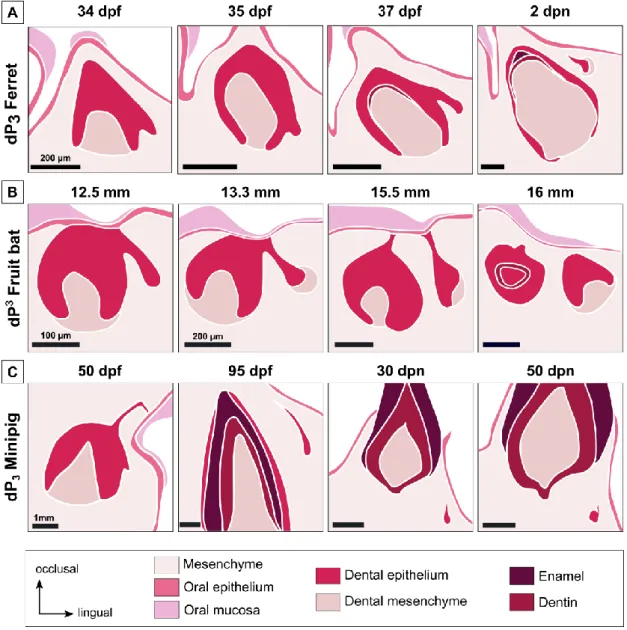
31 days post fertilization (dpf) to 2 days post-natal (dpn). They showed that the tooth replacement begins at the lingual part of the deciduous tooth by a budding of the dental lamina, giving the replacement dental lamina (Figure I. 4A). Then this replacement dental lamina elongates. In the canine, the permanent tooth develops from the replacement dental lamina when it is still connected to the deciduous tooth. In premolars, the dental lamina first detaches from the deciduous tooth and then gives rise in the permanent premolars.
Figure I. 4. Initiation of tooth replacement in the ferret, the fruit bat and the minipig. Initiation of the
tooth replacement of the lower third premolar for the ferret (A) and the minipig (C) and the upper third premolar for the fruit bat (B). Timing of development is indicated in days post fertilization or post-natal for the ferret and the minipig and in embryo size for the fruit bat. Summary of the dental replacement
32 has been obtained using the histological sections of ferret (Järvinen et al., 2009), fruit bat (Popa et al., 2016) and minipig (Wang et al., 2014a).
At the molecular level, Järvinen et al. (2009) showed that Sostdc1, a Wnt/BMP inhibitor, is expressed in the dental epithelium at the intersection between the deciduous tooth and the replacement dental lamina. They also showed that Shh has a similar expression pattern than in the mouse with an expression in the enamel knot and then in the inner enamel epithelium but no expression in the replacement region. Sox2+ stem cells have been identified at the lingual part of the replacement dental lamina with a free area of Sox2 in the tip of the replacement dental lamina (Juuri et al., 2013). Jussila et al. (2014) completed the study of tooth replacement in ferret by studying more developmental stages. By following Edar expression, they suggested that this signaling pathway is not involved in tooth replacement.
These results provided the first molecular observations of the mechanisms of tooth replacement in mammals. They identified the same signaling events than in the reptiles with a regulation of the Wnt pathway by SOSTDC1 and the presence of Sox2+ cells, indicating the presence of epithelial dental stem cells. However, few developmental stages were studied in ferrets due to the difficulty to obtain embryos at the correct stage and the presence of seasonal estrus.
Dental replacement in the fruit bat
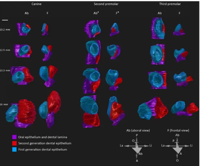
3D reconstructions and histological sections has been published to illustrate tooth replacement from the budding of the replacement dental lamina to the morphogenesis of the permanent tooth in the fruit bat embryos, Eidolon helvum (Popa et al., 2016). The authors showed that tooth replacement in the fruit bat always begins with a budding of the replacement dental lamina from the dental lamina at the lingual side of the deciduous tooth, as in the ferret
33 (Figure I. 4B). Then the separation of the replacement dental lamina from the deciduous tooth and the spatial arrangement between the deciduous and the permanent teeth can vary throughout the jaw (Figure I. 5). So, even if the tooth replacement always begins similarly, the spatial arrangement is variable and specific for each tooth. 3D reconstructions allow visualizing the spatial configuration of teeth during their development. This configuration is probably determined by specific signals according to the teeth. However, authors indicated that this species is no longer available for experimental purposes and that specimens studied were captured in 1972-73, so no molecular analyses can be realized.
Figure I. 5. 3D-reconstructions of the dental replacement in the fruit bat. 3D-reconstructions of the
canine, the second and the third premolar tooth replacement. In the fruit bat, variations of the spatial configuration of teeth during their development and replacement are observed. In function of the tooth, the replacement tooth develops in various position compared to their deciduous teeth: directly on the
34 lingual side for the canine, directly posterior for the second premolar and completely separated from the third premolar before starting its morphogenesis (figure from Popa et al., 2016).
Dental replacement in the minipig
Wang et al. (2014a) described tooth development in the minipig Sus scrofa as a model for diphyodonty. By their size and morphology, the minipig deciduous teeth are pretty similar to humans. The authors studied the development from 40 dpf to 90 dpn, so for approximately 164 days (Wang et al., 2014a). The tooth replacement in the minipig begins as in the ferret by a budding of the dental lamina at the lingual part of the deciduous tooth. In the minipig, the replacement dental lamina is detected when the deciduous tooth is already at a late bell stage, later than for the ferret and the fruit bat. So, the timing of tooth replacement initiation varies from a species to another. In the minipig, the replacement lamina elongates, detaches from the deciduous tooth, and then starts its tooth morphogenesis (Figure I. 4C). At least 100 days are necessary from the appearance of the replacement dental lamina to the mineralization of the replacement tooth.
Some molecular studies have been done in the minipig. Ki67 staining showed that more cells are proliferating in the lingual part of the replacement dental lamina (Wang et al., 2014a). The authors correlate this asymmetry of proliferation with the inclined growth of the replacement dental lamina in the minipig. Global transcriptome has been obtained for various stages during early morphogenesis of teeth; this transcriptome could be very useful to identify signaling events correlated with tooth replacement (Wang et al., 2014b). The authors indicated in the supplementary files that Wnt10B expression is up regulated between 40 and 50 dpf, stage were the replacement dental lamina begins its development in the minipig, indicating a role of the Wnt signaling in tooth replacement. It has also been shown that in vitro studies of tooth
35 development are possible in the minipig by transplanting the tooth germ in the mouse subrenal capsule (Wang et al., 2019). However, the slow development, the size of the animal and ethical concerns are significant obstacles to study tooth development in the minipig.
Dental replacement in humans
Humans are diphyodonts: incisors, canines and premolars are replaced once. Tooth development and replacement timelines are well known in humans. Histological sections that illustrate tooth replacement in human show that the permanent tooth is developing at the lingual position of the deciduous tooth (Kumar, 2014). However, very few studies give information about molecular mechanisms involved in human tooth development.
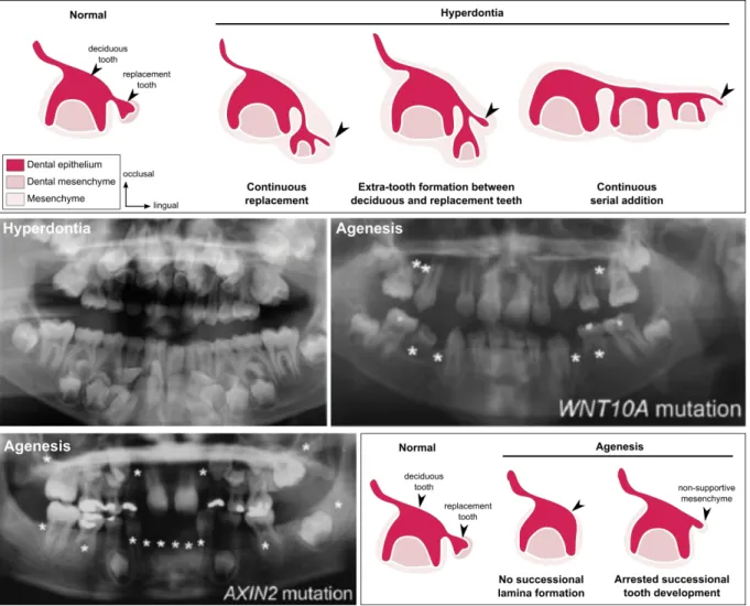
Juuri & Balic (2017) reviewed the molecular processes involved in tooth number abnormalities in humans. Tooth agenesis, the absence of at least one tooth, can give information about tooth replacement when it affect only permanent teeth. Juuri & Balic summarize that 50% of the tooth agenesis are attributed to WNT10A mutations, and that mutations of the Wnt inhibitor AXIN2 affect the permanent tooth formation (Figure I. 6). Other genes have been identified in agenesis syndrome, as Pitx2, Msx1 or the EDA signaling pathway (Juuri & Balic, 2017). Humans can also have supernumerary tooth formation, named hyperdontia. Developmental causes of supernumerary tooth formation are still unclear (Figure I. 6). Juuri & Balic listed mutated genes associated with supernumerary teeth. They indicate that Sox2, Runx2, IL11RA and Wnt pathway mutations are correlated with supernumerary teeth (Juuri & Balic, 2017).
So, in humans numerous genes have been identified to be associated with tooth developmental failure. As in reptiles and ferret, the Wnt pathway seems to play an essential role
36 in the tooth replacement. However, molecular and cellular studies are not possible in humans. We need a model to clearly identify the role of these genes and how their mutations disrupt tooth development and replacement.
Figure I. 6. Tooth number abnormalities in humans: hyperdontia and agenesis. At the top, schematics
of possible mechanisms leading to tooth hyperdontia: continuous replacement, extra-tooth formation or continuous serial addition. Panoramic radiograph showing hyperdontia of a child (9 years old) with cleidocranial dysplasia. Panoramic radiographs of children (10 and 8 years old) showing agenesis with homozygous WNT10A mutation and AXIN2 mutation. At the bottom, schematics of possible mechanisms leading to tooth agenesis: no successional lamina formation or arrested successional tooth development. Missing teeth marked by asterisks (adapted from Juuri & Balic, 2017).
From studies of various diphyodont species, we possess histological description of the tooth replacement and some candidate genes. However, studies are always limited by the constraints of the animal models. For now, no ideal diphyodont animal has been found to study the molecular basis of tooth development and replacement in mammals.
37 State of the art regarding dental replacement in monophyodont mammals
To better understand dental developmental processes, majority of the genes involved in human tooth disorders have been studied in transgenic mice. Mice are monophyodonts (only one set of teeth), but some transgenic mice have been used to study tooth replacement.
The mouse possesses one incisor and three molars per quadrant. The incisors have the ability to grow during the entire animal’s life due to stem cells maintenance in the incisor growing area. The molars develop from a continuous dental lamina, have roots and are never replaced. However, during molar development in the mouse we can observe a rudimentary successional dental lamina (RSDL) in the lingual part of the tooth germ (Dosedělová et al., 2015). This RSDL structure is visible in the mouse molars from 16 dpf to 10 dpn and some
Sox2+ cells can be identify at the lingual part of the RSDL (Figure I. 7). The RSDL will then regress, correlated with a loss of Sox2 signal and a decrease of proliferation in the RSDL (Dosedělová et al., 2015).
38
Figure I. 7. Rudimentary successional dental lamina in the developing mouse molar.
Hematoxylin-eosin staining and 3D-reconstructions of the dental lamina in the first lower molar. Arrow, rudimentary successional dental lamina; oe, oral epithelium; E, embryonic day; P, post-natal. Scale bars: 100 µm. (From Dosedělová et al., 2015).
The RSDL in wild type mouse does not have odontogenic potential (Popa et al., 2019). However, some transgenic mice are able to develop a new tooth from the RSDL. Popa et al. (2019) showed that by stabilizing the Wnt/β-catenin signaling in the Sox2+ cells during embryo
development, so in the lingual RSDL, they were able to maintain the proliferation of the RSDL. By isolating the RSDL in culture, they were even able to obtain a mineralized tooth. They showed that the RSDL could continue its development in transgenic mice thanks to the presence of Wnt signaling at the tip of the lamina when the RSDL is isolated from the main tooth. So, the deciduous molar seems to have the capacity to inhibit the development of the RSDL. By stabilizing Wnt/ β-catenin signaling, they observed expression of odontogenic markers as Shh,
Fgf4, Fgf3, Bmp4 and Sostdc1 in the RSDL that are not expressed in controls. So, activation of
the Wnt/ β-catenin signaling induces the odontogenic potential of the RSDL.
Transgenic mice can also have variation in their tooth number without affecting directly the RSDL; some mutations induce the development of rudimentary teeth. Ahn et al. (2010) showed that inactivation of Sostdc1 in mouse induce elevated Wnt and Spry2/4 signaling and
39 the formation of supernumerary teeth. Sprouty mutants also present supernumerary teeth (Klein et al., 2006, see Annex 2, p213). On the contrary, the overexpression of Sostdc1 induces a
diminution of the tooth number. Munne et al. (2009) described the Sostdc1 expression pattern during normal tooth development and indicated an intensive expression in the lingual side of the incisor, where a new incisor is developing in Sostdc1 deficient mutants. They hypothesize that theses extra-incisors are replacement teeth. So, SOSTDC1 seems to inhibit the tooth development and replacement in wild type mice (Figure I. 8A). This gene is described as an inhibitor of the Wnt signaling (Itasaki et al., 2003). Inhibiting Sostdc1 induces tooth formation, indicating that Wnt signaling plays a role as activator of tooth development.
40
Figure I. 8. Signaling pathways involved in tooth number regulation in the mouse. (A) Sostdc1
signaling identified in mouse (from Ahn et al., 2010). (B) Signaling pathways involved in the development of a lingual bud in the mouse (from Togo et al., 2016, colors modified). (C) Role of the Wnt signaling in tooth induction and initiation (from Järvinen et al., 2018).
On the contrary, some mutants are unable to develop teeth, as Runx2 null mice (Togo et al., 2016). At birth, the mutants do not have mineralized molars (they stopped their development during embryogenesis). However, during odontogenesis the Runx2 null mice can present a lingual bud with Sox2+ cells (Figure I. 8B). Interestingly, by inactivating both Runx2 and
Sostdc1, the dental phenotype is rescuing in 25% of the mice at birth and this double mutant
present less supernumerary teeth than Sostdc1 mutants. Togo et al. (2016) suggested that
41 suggested that inhibition of Runx2 could arrest primary tooth development but stimulates the formation of a secondary tooth (Wang et al., 2005).
Moreover, by expressing β-catenin in oral and dental epithelium, the tooth morphogenesis is disturbed. At the beginning of tooth development in mouse mutants, Järvinen et al. (2006) observed numerous small buds of teeth that possess each an enamel knot instead of a normal tooth bud with a unique enamel knot. In this mutant, the expression of Sostdc1 is not modified between mutants and wild type mice. By isolating the numerous buds in the mutant, they showed that all these buds had the capacity to give a mineralized tooth. These results suggest a direct role of the epithelial Wnt signaling in the formation of supernumerary teeth (Figure I. 8C). On the contrary, activation of Wnt signaling in the mesenchyme inhibits the sequential formation of the molars, indicating an opposite effect of increased epithelial and mesenchymal Wnt/ β-catenin signaling (Järvinen et al., 2018).
To conclude, these various mutants indicate that the Wnt signaling seems to be a key regulator of the tooth number in mouse as previously shown in other species. SOSTDC1 is identified as an inhibitor of the Wnt signaling (Figure I. 8A), Runx2 plays a dual role in the tooth development and the presence of Sox2+ cells seems necessary to induce a tooth replacement (Figure I. 8B). The suggested signaling pathways that could regulate the tooth numbers are summarized in Figure I. 8C. These signaling pathways include numerous candidate genes identified in ferret but also those related to human pathologies.
However, it is necessary to keep in mind that these results come from mutants with abnormal development of extra-teeth. This is why it is necessary to follow these candidate genes during the tooth development and replacement in a wild-type diphyodont mammal.
42
Rabbit as a model for tooth Evo-Devo studies
We thus decided to use the rabbit (Oryctolagus cuniculus) as a new model to study tooth development and replacement. The rabbit is already an important model for biomedical research (Bosze and Houdebine 2006) and the whole genome has been sequenced (OryCun2.0). On contrary to the ferret, the rabbit does not have seasonal estrus, gives numerous embryos per litter, and when the environmental conditions are stabilized (temperature, food) the gestation time does not vary (31 days of gestation only). Rabbit teeth are sufficiently small to perform all the conventional molecular studies primarily designed for mice. Moreover, more complex gene-targeting technology have been successfully applied in rabbit, as CRISPR/Cas9 system (Yan et al., 2014).
The rabbit dentition is diphyodont with a replacement of one incisor and all premolars (Figure I. 9, Horowitz et al., 1973, see annex 4 p256). Rabbits are heterodont: they possess on each side two uppers and one lower incisors, three upper and two lower premolars and three upper and lower molars.
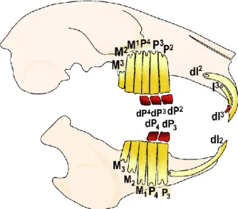
Figure I. 9. Schematic representation of the rabbit skull and its dental formula. In red, the teeth that
are replaced, in yellow the permanent teeth (replacement teeth and deciduous teeth not replaced). I, incisor; P, premolar; M, molar.
43 In rabbit, the deciduous premolars have a limited growth with root formation whereas permanent teeth are continuously growing (Sych & Reade, 1987). Concerning the incisors, the dI2 and the permanent I3 are ever-growing but the dI3 has roots.
Rabbit tooth development
Rabbit tooth development has been studied during the 70-80’s. Horowitz and collaborators did a complete chronology of deciduous tooth eruption using radiography, giving information on the mineralized part of the teeth (Horowitz et al., 1973). Navarro et al. did histological studies of the postnatal development in maxillary (1975) and mandibular cheek teeth (1976). However at birth the deciduous incisors and premolars are fully developed. In embryos, Ooë (1980) and Hirschfeld (1973) have studied the incisor development, but nothing has been reported for the cheek teeth development. All these studies give a composite chronology of the tooth development in rabbit and no molecular data has been acquired. The aim of this thesis is to characterize the precise chronology of the tooth morphogenesis in the rabbit and to follow the spatio-temporal regulation of the tooth replacement. By studying the tooth morphogenesis during ontogeny, we also point out possible links between rabbit tooth development and the evolution of the Lagomorpha cheek teeth.
Characteristics of extant Lagomorpha
The lagomorph order contains extant rabbits, hares and pikas. Lagomorpha is the sister group of Rodentia, named together the Glires. Lagomorphs are defined by their dentition: they possess two upper incisors one behind the other (Rose, 2006). In extant species, all the permanent teeth are hypselodont and adapted for feeding on grass and other vegetation. Lagomorphs are divided in two families, the Leporidae (rabbits and hares) and the Ochotonidae
44 (pikas). The Ochotonidae family contains only one living genus, Ochotona, which includes 30 species living in North America and Central Asia (Hoffmann and Smith 2005). Ochotonidae have only two upper molars instead of Leporidae. Leporidae includes 11 living genera and 61 species that have colonized Africa, Eurasia and North and South America. The European rabbit,
Oryctolagus cuniculus, is the only extant species of the Oryctolagus genus. The common names
hare and rabbit are used to designate different leporid genera (Figure I. 10).
Figure I. 10. Molecular phylogeny of the extant Leporidae. Phylogeny obtained using seven gene
fragments. Values above and below nodes represent the nodal support (MP=maximum parsimony bootstrap; ML=maximum likelihood bootstrap; and BI=bayesian inference posterior proba-bility) for associations among the 25 ingroup taxa. An asterisk indicates that the node was not supported by>50% bootstrap support. ( Simplified from Matthee et al., 2004)
45 Evolutionary history of Lagomorpha
Duplicidentata mirorder contains the lagomorphs and their sister group, the mimotonids. Lagomorpha is a monophyletic group whereas recent analyses show that mimotonids are a paraphyletic group that contains the stem lagomrpha ancestor. Mimotonids, known from the late Paleocene to the middle Eocene, have two incisors in both upper and lower jaws and their cheek teeth are unilaterally hypsodont and rooted (Rose, 2006). As mimotonids, the stem lagomorphs group was originated from Asia, and they diversified then in different continents. Stem Lagomorpha, as Dawsonolagus from late early Eocene, had hypsodont rooted cheek teeth and are described as morphologically transitional between mimotonids and lagomorphs (Li et al. 2007). The Oligocene marks the separation of the Lagomorpha in two families, the Ochotonidae and the Leporidae. At the end of the Miocene, the Leporidae are represented by the genera Alilepus, and especially Alilepus hibbardi, a suggested ancestor of the extant genera of Leporids (Jin et al. 2010).
Lagomorpha teeth evolved from moderate hypsodont rooted cheek teeth in late Paleocene to hypselodont teeth. The shape variations of the P3 is highly studied because this
tooth bears informative characters about phylogenetic relationships among Lagomorpha (Hibbard 1963; Cermák 2015). We decided in the present thesis to study the variation of the M1 morphology during European rabbit ontogeny and lagomorph evolution notably because this tooth shows the typical bilophodont pattern of the jugal dentition of modern lagomorph and because it is the mostly used tooth for comparative anatomy purposes in mammals (Hershkovitz 1971).
46
P
ART
1
TOOTH DEVELOPMENT AND REPLACEMENT IN
48
S
ince decades, tooth development mechanisms in mammals are highly studied using mouse as model (i.e. Balic and Thesleff, 2015; Chavez et al., 2012; Lan et al., 2014). However, the mouse does not replace its teeth. As a consequence, mammal tooth replacement is poorly known. As previously presented in introduction, various species have been studied in order to identify a good animal model for dental replacement studies (Järvinen et al., 2009; Popa et al., 2016; Wang et al., 2014a). However, for now, any model answer both morphological and molecular questions about mammal tooth replacement. A good mammal animal model has to replace its teeth, be easy to maintain, breed in large numbers in a laboratory facility, have the shorter generation time possible and be sequenced. If possible, it is even more interesting to have a model animal that has already been used for genetic manipulation techniques or in vitro studies.In this thesis, we decided to study tooth development and replacement using the European rabbit, Oryctolagus cuniculus as model. The European rabbit is already used as model in biomedical research, is sequenced, has already be used for CRISPR-CAS9 genetic manipulation and in vitro organ culture (Bosze and Houdebine, 2006; Glasstone, 1938; Yan et al., 2014). The European rabbit is currently used in dentistry research to study orthodontic process (Abtahi et al., 2018). Studying rabbit dental development with modern techniques is necessary, the available studies in literature being rather old and incomplete (Horowitz et al., 1973; Navarro et al., 1976; Ooë, 1980). We show in this first part that the European rabbit is an efficient model to study both morphological and molecular mechanisms involved in tooth development and replacement.
50
Chapter 1.1 – Morphological characteristics of tooth development and
replacement in Oryctolagus cuniculus
The review of publications about rabbit tooth development and replacement leads to a composite developmental chronology, with a lack of knowledge about some tooth types and developmental stages. Moreover, the studies already carried out are about 50 years old, some techniques used in these papers are now obsolete (Glasstone, 1938; Horowitz et al., 1973; Yardin, 1968). Most of the current studies on rabbit dentition are about orthodontic or veterinary cares, with few studies on rabbit embryos (Abtahi et al., 2018; Meredith, 2007).
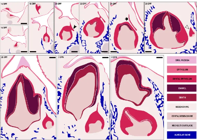
In the following paper, we decided to update the knowledge on the dental morphogenesis in the European rabbit using modern techniques. We studied each rabbit tooth from the initiation of the development to the mineralization of the replacement teeth, so from 12 days post-fertilization to 4 days post-natal, representing 23 days of development. We performed 3D reconstructions of dental epithelial tissues for each tooth using X-ray microtomography associated with a histological study to follow the rabbit dental development. We obtained the complete description of the histo-morphological chronology of the tooth development and replacement in rabbit. During this study, we also observed an undescribed rabbit incisor characteristic: at birth, the growing upper incisors of the rabbit present holes in the dentin, opening the pulp cavity. These holes are quickly repaired with a new apposition of dentin from the pre-existing odontoblasts that continue to secrete despite the disruption.
The new dental morphogenesis chronology of the rabbit Oryctolagus cuniculus presented in the following paper will allow further molecular studies to better understand mammal tooth replacement. Moreover, the 3D epithelium surface reconstructions are available online in Morphomuseum for who is interested to re-use these surfaces for other scientific works.