HAL Id: hal-02114312
https://hal.archives-ouvertes.fr/hal-02114312
Submitted on 25 May 2020
HAL is a multi-disciplinary open access
archive for the deposit and dissemination of
sci-entific research documents, whether they are
pub-lished or not. The documents may come from
teaching and research institutions in France or
abroad, or from public or private research centers.
L’archive ouverte pluridisciplinaire HAL, est
destinée au dépôt et à la diffusion de documents
scientifiques de niveau recherche, publiés ou non,
émanant des établissements d’enseignement et de
recherche français ou étrangers, des laboratoires
publics ou privés.
Distributed under a Creative Commons Attribution| 4.0 International License
putative plasmid
Julie Reveillaud, Sarah Bordenstein, Corinne Cruaud, Alon Shaiber, Mylène
Weill, Patrick Makoundou, Karen Lolans, Andrea Watson, Ignace
Rakotoarivony, Seth Bordenstein, et al.
To cite this version:
Julie Reveillaud, Sarah Bordenstein, Corinne Cruaud, Alon Shaiber, Mylène Weill, et al.. The
Wol-bachia mobilome in Culex pipiens includes a putative plasmid. Nature Communications, Nature
Publishing Group, 2019, 10 (1), pp.1051. �10.1038/s41467-019-08973-w�. �hal-02114312�
ARTICLE
The
Wolbachia mobilome in Culex pipiens includes
a putative plasmid
Julie Reveillaud
1
, Sarah R. Bordenstein
2
, Corinne Cruaud
3
, Alon Shaiber
4,5
, Özcan C. Esen
5
, Mylène Weill
6
,
Patrick Makoundou
6
, Karen Lolans
5
, Andrea R. Watson
5
, Ignace Rakotoarivony
1
, Seth R. Bordenstein
2,7,8
&
A. Murat Eren
4,5,9
Wolbachia is a genus of obligate intracellular bacteria found in nematodes and arthropods
worldwide, including insect vectors that transmit dengue, West Nile, and Zika viruses.
Wolbachia’s unique ability to alter host reproductive behavior through its temperate
bacteriophage WO has enabled the development of new vector control strategies. However,
our understanding of
Wolbachia’s mobilome beyond its bacteriophages is incomplete. Here,
we reconstruct near-complete
Wolbachia genomes from individual ovary metagenomes of
four wild
Culex pipiens mosquitoes captured in France. In addition to viral genes missing
from the
Wolbachia reference genome, we identify a putative plasmid (pWCP), consisting
of a 9.23-kbp circular element with 14 genes. We validate its presence in additional
Culex
pipiens mosquitoes using PCR, long-read sequencing, and screening of existing metagenomes.
The discovery of this previously unrecognized extrachromosomal element opens additional
possibilities for genetic manipulation of
Wolbachia.
https://doi.org/10.1038/s41467-019-08973-w
OPEN
1ASTRE, INRA, CIRAD, University of Montpellier, Montpellier 34398, France.2Department of Biological Sciences, Vanderbilt University, Nashville 37235 TN,
USA.3Commissariat à l’Energie Atomique et aux Energies Alternatives (CEA), Institut de Biologie François Jacob, Genoscope, Evry 91057, France.
4Graduate Program in the Biophysical Sciences, University of Chicago, Chicago, IL 60637, USA.5Department of Medicine, University of Chicago, Chicago
60637 IL, USA.6Institut des Sciences de l’Evolution de Montpellier (ISEM), UMR CNRS-IRD-EPHE-Université de Montpellier, Montpellier 34095, France.
7Department of Pathology, Microbiology, and Immunology, Vanderbilt University, Nashville 37235 TN, USA.8Vanderbilt Institute for Infection, Immunology,
and Inflammation, Vanderbilt University, Nashville 37235 TN, USA.9Josephine Bay Paul Center for Comparative Molecular Biology and Evolution, Marine
Biological Laboratory, Woods Hole 02543 MA, USA. These authors contributed equally: Julie Reveillaud, Sarah R. Bordenstein. Correspondence and requests for materials should be addressed to J.R. (email:[email protected]) or to A.M.E. (email:[email protected])
123456789
M
osquitoes are major vectors of disease-causing
patho-gens worldwide including viruses such as dengue, West
Nile, Chikungunya, Zika, and yellow fever
1–3. In the
absence of effective vaccines and the off-target effects of
insecti-cides used to control mosquitoes, novel vector biocontrol efforts
are the focus of intense study
4. Over the past decade, the widely
distributed endosymbiotic alphaproteobacteria Wolbachia has
gained attention as a promising mosquito-control strategy. The
basic reasons for its heightened attention is that Wolbachia
can cause reproductive parasitism whereby the intracellular
bacteria in the reproductive tissues can alter sexual reproduction
to enhance its maternal spread through host populations at the
expense of host
fitness
5–7. Second and notably, native Wolbachia
reduce RNA virus replication in the fruit
fly Drosophila
8–10, and
Wolbachia transinfections into mosquito species (i.e., Aedes
aegypti, Ae. albopictus, Ae. polynesienses, and Anopheles stephensi)
similarly result in mosquito lines refractory to various types
of pathogenic RNA viruses
11–14. The combination of a selfish
drive system and pathogen blocking by Wolbachia has led to
successful pilot trials for suppression of mosquito population
size and replacement of infected mosquito populations so they
can no longer transmit pathogens
15–17.
Wolbachia are transovarially transmitted from the mother to
offspring
6,18–20. In some arthropods, including the naturally
infected vector species complex Culex pipiens, Wolbachia
‘modify’
sperm in testes, leading to embryonic lethality if the infected male
mates with either an uninfected female or a female harbouring an
incompatible Wolbachia strain. When both male and female are
infected with the same Wolbachia, the modification is ‘rescued’,
and compatibility is restored
21. This reproductive alteration,
termed
‘cytoplasmic incompatibility’ (CI), comprises the most
common form of Wolbachia-induced parasitism and, as studied
in C. pipiens, can lead to highly diverse unidirectional and
bidirectional incompatibility phenotypes
22. CI is currently used in
vector control studies for population replacement by
Wolbachia-infected strains that block arbovirus transmission
17or population
suppression that reduces the number of mosquito vectors
23.
Notably, the genes responsible for CI
24–26occur in the eukaryotic
association module of Wolbachia’s temperate phage WO
25–27,
which laterally transfers between Wolbachia coinfections and
evolves rapidly
28–32. Overall, these
findings emphasize the
importance of further investigating mobile genetic elements
in Wolbachia.
While recent studies shed light on the role of phage WO in
Wolbachia genome evolution and CI, other extrachromosomal
elements, such as plasmids, have not been detected in the
sym-biont. Notably, over half of the species in the closely related
Rickettsia genus have plasmids
33that play roles in DNA
repli-cation, partitioning, mobilization, and conjugation
34,35and offer
a potential tool for genetic manipulation of diverse members of
Rickettsia
36,37. Similar genetic manipulation strategies for
Wol-bachia are conceivable
27; however, previous efforts to search for
such extrachromosomal mobile genetic elements have not been
successful
38,39. The lack of isolates limit direct insights into
Wolbachia genomics, and most metagenomic approaches thus far
rely on pooled individuals grown in the laboratory environment
due to low infection densities
40,41. Since these limitations can
conceal naturally occurring genomic diversity among Wolbachia
populations, highly resolved analyses of individual mosquitoes
may reveal additional insights into the Wolbachia
‘mobilome’, the
pool of all mobile genetic elements associated with Wolbachia
populations.
Here we sequence ovary samples from four wild-caught C.
pipiens individuals captured in Southern France from a single
trap. Using genome-resolved metagenomic and pangenomic
analysis strategies, we were able to reconstruct and compare
near-complete Wolbachia genomes from each individual. Besides a
diverse set of virus-associated genes that were missing or absent
in the reference Wolbachia genome wPip Pel, our data reveal the
first lines of evidence for an extrachromosomal circular element
with genetic and functional hallmarks of a plasmid that we
ten-tatively name pWCP.
Results
A
Wolbachia metagenome-assembled genome (MAG) is
recovered in each sample. Shotgun sequencing of DNA
recov-ered from ovary samples of four C. pipiens individuals (O03, O07,
O11, O12) resulted in 65–78 million paired-end sequences after
quality
filtering. Metagenomic assembly of each sample
indivi-dually yielded 147K–183K contiguous DNA segments (contigs)
>1 kbp, which recruited 48.1–72.9% of the raw sequencing reads.
The relatively high fraction of unmapped reads were likely due to
challenges associated with the assembly of environmental
metagenomes
42,43, especially in the presence of eukaryotic host
genomic DNA. Supplementary Table 1 reports statistics for the
raw number of reads and assembly results for each sample. We
employed a metagenomic binning strategy that uses sequence
composition signatures and differential coverage statistics of
contigs across samples. For each ovary sample, we were able to
reconstruct a highly complete single bacterial MAG that resolved
to Wolbachia (Table
1
).
Wolbachia MAGs include highly covered non-phage contigs.
The relatively low number of single-nucleotide variants
(0.01–0.05%, Supplementary Table 2) suggested that each
Wol-bachia MAG represented a nearly monoclonal population of
bacterial cells in the ovary metagenomes. In addition, the high
average nucleotide identity across our MAGs and the 1482 kbp
reference genome for Wolbachia, wPip Pel
40(99.1–99.98%,
Supplementary Table 3) suggested a high degree of conservation
between the endosymbionts of different individuals. Our
meta-genomic read recruitment analyses using Wolbachia MAGs
revealed consistent coverage statistics averaging 168×–491×
for contigs that were enriched with bacterial genes. However,
a subset of contigs in each individual displayed approximately a
five-fold increase in coverage (Supplementary Table 4). Because
Wolbachia harbour prophage WO that can enter the lytic cycle
Table 1
Wolbachia MAG estimates
MAG Percent completion (PC) Percent redundancy (PR) Number of contigs (N) Number of genes (n)
Length (total number of nucleotides) GC content (%) O03 94.24 0.72 93 1091 1,213,072 33.83 O07 94.24 0.72 127 1181 1,317,313 33.78 O11 94.24 0 99 1085 1,208,099 33.84 O12 94.24 0 99 1113 1,237,800 33.95
Estimates include completion and redundancy estimates, number of contigs (N), number of genes (n), total number of nucleotides and percentage of GC. More than 90% completion and <10% redundancy based on the single occurrence of 139 single-copy genes (SCG) identified from the collection by authors in ref.77suggest high completion of the bins
and form phage particles
27,44,45, we postulated that some of the
contigs could be phage associated, which would explain coverage
inconsistencies. We used the
five prophage regions identified in
the wPip Pel reference genome
27,40to identify contigs enriched
with genes of phage origin. Contigs classified and validated
through homology searches against phage WO matched to
contigs that were highly covered in our MAGs, confirming that
most shifts in coverage could be attributed to phages of
Wolbachia. However, surprisingly,
five contigs in our MAGs
(Contig O12_A, Contig O11_A, Contig O07_A, Contig O07_A’,
and Contig O03_A, Supplementary Data 1) showed no
homol-ogy to prophage WO despite their remarkable coverage that
ranged between 720× and 2176×. Interestingly, the 5’ and 3’ ends
of these contigs showed homology to the non-coding
flanking
regions of wPip’s ISWpi12 transposable element (TE; WP0440,
WP1209, and WP1347) of the IS110-family
46. Given their
(1) high coverage in metagenomes, (2) lack of homology to
prophage WO, and (3) putative association with IS110 TE, we
hypothesized
that
these
contigs
could
represent
extra-chromosomal elements.
pWCP: a
Wolbachia-associated putative plasmid. Based on
homology between the ends of these
five contigs and the flanking
regions of IS110 TE, we predicted that the missing region was
the latter element. We artificially circularized Contig O11_A
(8037 bp) and inserted an IS110 TE (1386 bp) based on the
overlapping 5’ and 3’ ends (Fig.
1
). Metagenomic read
recruit-ment onto this artificially circularized contig, which we tentatively
name
‘pWCP’ (for plasmid of Wolbachia endosymbiont in
C. pipiens), showed consistent coverage over its entire length
except a clear two-fold coverage increase in a region that matched
to the IS110 TE in all four C. pipiens individuals in our study
(Supplementary Figure 1). These data suggested the IS110 TE
are located in the extrachromosomal pWCP while some others
could be integrated in the bacterial chromosome. The read
recruitment from three C. pipiens egg metagenomes generated
in a previous study
32confirmed the near identical presence of
pWCP in all three, including the increase in coverage matching
to the IS110 TE (Supplementary Figure 1).
To validate the circular and extrachromosomal nature of
pWCP independently of short-read recruitment- and
assembly-based strategies, we generated long reads from additional
C. pipiens complex samples using a MinION sequencer. Since
MinION sequencing occurs with no polymerase chain reaction
(PCR) or downstream assembly steps, we hypothesized that long
reads that match to pWCP should never be (1)
flanked by
genomic regions matching to the Wolbachia chromosome and
(2) longer than the pWCP itself. Our MinION sequencing
analysis resulted in 14,808 high-quality sequences that were
>5000 nucleotides. While a significant fraction of these reads
were eukaryotic contamination and the lambda phage DNA that
we used to pad our low-biomass samples (see Methods), a local
BLAST search of artificially circularized pWCP sequence against
long reads revealed 19 that aligned to pWCP with an e value of
<1e−20 (Supplementary Data 2). Thirteen of these 19 reads
covered >50% of the length of pWCP and contained no other
genomic region. In other words, each of the long reads were equal
to or shorter than the length of pWCP, as expected for an
extrachromosomal element (Fig.
2
a). Moreover, 6 of these 13
reads were exactly equal to the length of pWCP, covering its
entirety with non-identical start positions, confirming its
circularity (Fig.
2
a). The
final 6 long reads that covered <50%
of the length of pWCP had specific matches to the IS110 TE
(Fig.
2
b); this result is consistent with the multiple occurrences
of the IS110 TE in the Wolbachia genome and explains the
increase in coverage in metagenomic read recruitment results
(Supplementary Figure 1).
To further validate the extrachromosomal nature of pWCP, we
PCR screened genomic DNA from the wild caught individuals as
well as from tetracycline (TC)-treated laboratory lines (TC1–TC3;
negative controls). TC treatment eliminates Wolbachia from its
hosts and is commonly used to generate C. pipiens uninfected
laboratory lines
47. First, using LCO1490 and HCO2198
mito-chondrial cytochrome c oxidase subunit I (COI) invertebrate
primers
48, we detected the presence of a ~708-bp band in the four
C. pipiens individuals and the three Wolbachia-free Culex
quinquefasciatus controls treated with TC (TC1–TC3),
confirm-ing the presence of arthropod DNA in all samples (Fig.
1
b). Next,
we verified the presence of Wolbachia DNA in the first four
samples by PCR amplifying a 438-bp fragment of the Wolbachia
16S rRNA gene using Wspec-F and Wspec-R primers
49. No band
was observed in the TC samples, confirming the absence of
Wolbachia (Fig.
1
c). Critically, a ~1800-bp fragment amplified
with primers 263F and 2127R (Supplementary Table 5) designed
from the ends of the artificially circularized contig within DnaB
gene (and uniquely matching those sites), confirmed the circular
nature of the pWCP and its presence only in Wolbachia-infected
C. pipiens samples (Fig.
1
d). We also designed primers to Sanger
sequence across the circular gap (see Supplementary Table 5 for
primer sets EC_1–EC_7), and results confirmed the pWCP
sequence obtained with both Illumina and MinION sequencing.
Finally, we amplified IS110 TE with primers EC_4F and EC_4R
(Supplementary Table 5) in the four C. pipiens samples studied
herein while observing no band in the Wolbachia-free samples
(Fig.
1
e) in order to verify the strict association of IS110 TE with
Wolbachia (Fig.
1
f). These results were further confirmed using
additional rifampicin- and oxytetracycline-treated C. pipiens
samples (Supplementary Figure 2). Of note, we verified the
presence of pWCP in the originating TC1–TC3 and C. pipiens
laboratory stocks prior to antibiotic treatment.
The average nucleotide identity of the four independently
assembled pWCP sequences from individual mosquitoes was
99.65–100% (Supplementary Table 6). In addition, we identified a
8315-bp contig in the wPip JHB assembly (ABZA01000008.1
50),
which also was >99.53% identical to each of the four pWCP
sequences (Supplementary Table 6). The IS110 TE was 100%
identical across samples and confirmed as clonal. Our additional
read recruitment analyses from publicly available metagenomes
(SRR5810516, SRR5810517, SRR5810518)
32also revealed the
widespread occurrence of pWCP in C. pipiens individuals from
Turkey, Algeria, and Tunisia (Supplementary Note 1,
Supple-mentary Figure 3) at a similar 4.2–7.3-fold higher copy number
relative to the Wolbachia genome. We also confirmed that pWCP
and the Wolbachia genome display a similar tetranucleotide
composition (Supplementary Note 2, Supplementary Figure 4).
Overall, these
findings suggest that pWCP is maintained over the
long term with Wolbachia from these strains.
pWCP encodes an IS110 TE, 14 genes, and an intergenic repeat
region. The IS element is homologous to ISWpi12 of the IS110
family
46based on IS
finder platform search. Annotation of genes
in pWCP also revealed the presence of a disrupted DnaB-like
helicase, two RelBE loci, a ParA-like gene (each with identical AA
sequences across samples, Supplementary Note 3), and multiple
genes encoding hypothetical proteins (Fig.
1
f, Supplementary
Data 3). The ParA-like partitioning sequence showed amino acid
homology to the chromosome partitioning of plasmid protein
(ParA) identified in Ca. Caedibacter acanthamoebae (an
endo-symbiont of acanthamoebae; e value: 2 × 10
–46), the bacterium
Odyssella thessalonicensis (e value: 9 × 10
–45), and Rickettsia
raoultii (e value: 7 × 10
–41). The RelBE toxin–antitoxin (TA) locus
has been identified in multiple Wolbachia genomes
51and is often
associated with prophage WO regions (e.g., of wVitA, wHa,
wMel, wAu, wRi, wSuzi, wFol, wInc), specifically within the tail
and/or capsid modules. In other bacteria, this TA system can
promote the stability of its encoding mobile element, including
plasmids or pathogenicity islands, through post-segregational
killing of cells that have lost the antitoxin component of the TA
operon
52,53. Most remaining pWCP genes were hypothetical and
unique to either wPip and/or the B-Wolbachia phyletic
super-group
54(Supplementary Data 3), including GP-09 and GP-11
which showed a very weak homology to a Transcription Factor
and a Terminase, respectively (e value > 0 in SCOPe, Pfam, and
Clusters of Orthologous Group (COG), highlighting the need
for further functional characterization of this newly discovered
mobile genetic element. In particular, terminase proteins bind
and package DNA into the capsids of phage particles
55. These
data indicate that the extrachromosomal DNA could potentially
fall into three categories: a simple plasmid, a mini-chromosome
of Wolbachia, or a plasmid-like replicon that hijacks the capsids
of phage WO.
Beyond the putative coding regions, alignment of all contigs
revealed an intergenic variable number tandem repeat (VNTR)
region (Fig.
3
a, b, Supplementary Figure 5), characterized as 16-nt
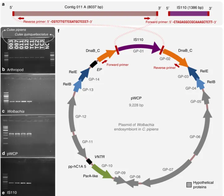
5′ 3′ 5′ IS110 (1386 bp) 3′ Reverse primer: 5′ Reverse primer Culex pipiens Culex quinquefasciatus Forward primer: 5′ Forward primer 3′ 3′ Contig 011 A (8037 bp) DnaB_C IS110 pWCP 9,228 bp Plasmid of Wolbachia endosymbiont in C. pipiens DnaB_C ReIE ReIB Hypothetical proteins ReIE Arthropod Wolbachia pWCP IS110 ReIB pp-hC1A 5 ParA-like GP-13 GP-12 GP-11 GP-10 GP-09 GP-08 GP-07 GP-06 GP-05 GP-04 GP-03 GP-02 GP-01 GP-14 EP VNTR
a
b
c
d
e
f
Fig. 1 The artificially circularized genome of putative plasmid pWCP. a illustrates Contig O11_A and IS110 transposable element (TE) identified in our assembly. The red rectangles are regions of 100% nucleotide identity between the two contigs. Outward PCR primers were designed to amplify and confirm circularity of the sequence. b–e Gels for PCR tests to confirm a Wolbachia-associated circular genome. To verify the presence of arthropod DNA in our fourCulex pipiens samples and the tetracycline-treated (TC) Culex quinquefasciatus samples, we PCR amplified a 708-bp sequence using LCO1490 and HCO2198 primers (b). A 438-bp fragment of theWolbachia 16S rRNA gene (c), an approximately 1800 bp sequence amplified with the outward primers designed to support circularity of the genome, as illustrated in top panel (d) and a 431-bp of IS110 TE (e) were obtained in wildC. pipiens samples O03-O07-O11-O12 while no amplification was observed in Wolbachia-free samples. NC corresponds to negative control. f illustrates the complete genome. Each arrow represents an open reading frame (ORF). ORFs with no homology to a known function are shown in grey.ParA-like (green), RelBEtoxin–antitoxin operon (blue), andDnaB_C replicative DNA helicase (orange) that is disrupted by the ISWpi12 TE of the IS110 family (purple) are represented by arrows (with ane value < e−12from an NCBI Conserved Domain or Pfam Search). Black squares represent the location of (1) the variable number tandem repeat (VNTR) and (2) the extragenic palindrome (EP) region
repeats adjacent to parA that vary in number among individual
pWCP sequences. Recent studies observed the same repeat
region, identified as pp-hC1A_5, and used it to genotype different
strains of Wolbachia
56,57, yet these have not been studied at an
individual level. The authors suggested that a deletion in the
intergenic polymorphic region could serve as a recombination or
horizontal gene transfer site
56. Alternatively, we hypothesize that
the direct repeats, as present in iteron plasmids, could indicate a
potential origin of replication and play a role in copy number
control
58. Our analysis of the pWCP sequence also revealed a
pWCP reference sequence
minION long read 299f27bf-04d3-46e2-8a09-9c06fa77a5fb 9.0 kb 9.0 kb 8.0 kb 8.0 kb 7.0 kb 7.0 kb 6.0 kb 6.0 kb 5.0 kb 5.0 kb 4.0 kb 4.0 kb 3.0 kb 3.0 kb 2.0 kb 2.0 kb 1.0 kb 1.0 kb 0.0b 0.0b pWCP reference sequence
minION long read fedbb183-ae1b-472a-a002-57f9809bf4ac 9.0 kb 8.0 kb 7.0 kb 6.0 kb 5.0 kb 4.0 kb 3.0 kb 2.0 kb 1.0 kb 8.0 kb 7.0 kb 6.0 kb 5.0 kb 4.0 kb 3.0 kb 2.0 kb 1.0 kb 0.0b pWCP reference sequence 9.0 kb 8.0 kb 7.0 kb 6.0 kb 5.0 kb 4.0 kb 3.0 kb 2.0 kb 1.0 kb 0.0b pWCP reference sequence 9.0 kb 8.0 kb 7.0 kb 6.0 kb 5.0 kb 4.0 kb 3.0 kb 2.0 kb 1.0 kb 0.0b 0.0b pWCP reference sequence
minION long read
9.0 kb 8.0 kb 7.0 kb 6.0 kb 5.0 kb 4.0 kb 3.0 kb 2.0 kb 1.0 kb 0.0b pWCP reference sequence
minION long read ea7fda64-6f55-4ea5-a7bf-427565890eb1 6.0 kb 5.5 kb 5.0 kb 4.5 kb 4.0 kb 3.5 kb 3.0 kb 2.5 kb 2.0 kb 1.5 kb 1.0 kb 500b 9.0 kb 8.0 kb 7.0 kb 6.0 kb 5.0 kb 4.0 kb 3.0 kb 2.0 kb 1.0 kb 0.0b 0.0b 7.0 kb 6.0 kb 5.0 kb 4.0 kb 3.0 kb 2.0 kb 1.0 kb 0.0b 7ff837c8-632f-4475-8d1b-b059ccbfbafe pWCP reference sequence
minION long read The start position of the
artificially circularized sequence of pWCP
Red triangles mark the start position, and the direction of a given long read sequence
9.0 kb 8.0 kb 7.0 kb 6.0 kb 5.0 kb 4.0 kb 3.0 kb 2.0 kb 1.0 kb 0.0b 7.0 kb 6.0 kb 5.0 kb 4.0 kb 3.0 kb 2.0 kb 1.0 kb 0.0b 0b33a74b-357d-4f63-b3b2-ae33c394101a pWCP reference sequence 9.0 kb 8.0 kb 7.0 kb 6.0 kb 5.0 kb 4.0 kb 3.0 kb 2.0 kb 1.0 kb 0.0b pWCP reference sequence 9.0 kb 8.0 kb 7.0 kb 6.0 kb 5.0 kb 4.0 kb 3.0 kb 2.0 kb 1.0 kb 0.0b pWCP reference sequence 9.0 kb 8.0 kb 7.0 kb 6.0 kb 5.0 kb 4.0 kb 3.0 kb 2.0 kb 1.0 kb 0.0b pWCP reference sequence 9.0 kb 8.0 kb 7.0 kb 6.0 kb 5.0 kb 4.0 kb 3.0 kb 2.0 kb 1.0 kb 0.0b pWCP reference sequence 9.0 kb 8.0 kb 7.0 kb 6.0 kb 5.0 kb 4.0 kb 3.0 kb 2.0 kb 1.0 kb 0.0b
minION long read ad56146c-2f09-4385-bdca-69acca953820 9.0 kb 8.0 kb 7.0 kb 6.0 kb 5.0 kb 4.0 kb 3.0 kb 2.0 kb 1.0 kb 0.0b
minION long read
7.0 kb 6.0 kb 5.0 kb 4.0 kb 3.0 kb 2.0 kb 1.0 kb 0.0b 9ee4afe1-4891-49bd-b5ee-57a43cc121dct
minION long read 7.0 kb 8.0 kb 9.0 kb 6.0 kb 5.0 kb 4.0 kb 3.0 kb 2.0 kb 1.0 kb 0.0b a475551c-dca9-4791-8a4b-43b14fe5fc64t minION long read
0.0b 500b 1.0 kb 1.5 kb 2.0 kb 2.5 kb 3.0 kb 3.5 kb 4.0 kb 4.5 kb 5.0 kb
46fb29a9-47c6-4504-b739-91aafccb1b0ft minION long read
292bbdac-b01d-43d7-a16d-d1c4429fa3e5 7.0 kb 8.0 kb 9.0 kb 6.0 kb 5.0 kb 4.0 kb 3.0 kb 2.0 kb 1.0 kb 0.0b
minION long read ba0cb8e0-8002-476b-b9fd-91f1c7447035 7.0 kb 8.0 kb 9.0 kb 6.0 kb 5.0 kb 4.0 kb 3.0 kb 2.0 kb 1.0 kb 0.0b 8.0 kb 7.0 kb 6.0 kb 5.0 kb 4.0 kb 3.0 kb 2.0 kb 1.0 kb 0.0b minION long read 2d9e2bec-45ef-442f-86c2-394a17893a5f
pWCP
IS110
EP
VNTR
Long-reads that cover > 50% of pWCP
Long-reads that cover < 50% of pWCP Purple arrows mark the location of the IS110 element in pWCP pWCP reference sequence 9.0 kb 8.0 kb 7.0 kb 6.0 kb 5.0 kb 4.0 kb 3.0 kb 2.0 kb
minION long read 6a511b03-f20c-4777-9579-44516a5b6342 7.0 kb 8.0 kb 6.0 kb 5.0 kb 4.0 kb 3.0 kb 2.0 kb 1.0 kb 0.0b pWCP reference sequence 9.0 kb 8.0 kb 7.0 kb 6.0 kb 5.0 kb 4.0 kb 3.0 kb 2.0 kb
minION long read 12ef229a-23d0-4738-91a1-c1a0335a13a9t 16 kb 14 kb 12 kb 10 kb 8.0 kb 6.0 kb 4.0 kb 2.0 kb 0.0b pWCP reference sequence 9.0 kb 8.0 kb 7.0 kb 6.0 kb 5.0 kb 4.0 kb 3.0 kb 2.0 kb
minION long read 804e3920-9157-442e-ae02-9c609841cfb0t
8.0 kb 7.0 kb 6.0 kb 5.0 kb 4.0 kb 3.0 kb 2.0 kb 1.0 kb 0.0b
pWCP reference sequence pWCP reference sequence
9.0 kb 8.0 kb 7.0 kb 6.0 kb 5.0 kb 4.0 kb 3.0 kb 2.0 kb 4.0 kb 5.0 kb 6.0 kb 7.0 kb 8.0 kb 9.0 kb 0.0b 1.0 kb 2.0 kb 3.0 kb 4.0 kb 5.0 kb 6.0 kb 7.0 kb 3.0 kb 2.0 kb
minION long read b5256c98-91b6-47e9-958c-a95c19159872
minION long read b2d5d65f-25f7-4a19-b3cb-f5c0ba1ac225 0.0b 500b 1.0 kb 2.0 kb 1.5 kb 2.5 kb 3.0 kb 3.5 kb 4.0 kb 4.5 kb 5.0 kb
a
b
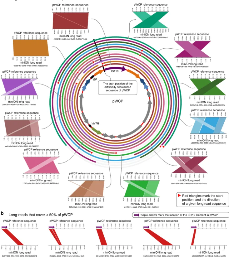
Fig. 2 Alignment of MinION long reads to pWCP. a The alignment of long reads that cover >50% of the pWCP genome (only 12 of 13 total long reads are shown; we omitted 1 from this display solely due to space considerations). Each rectangularfigure shows high scoring pairs (HSPs) and their alignments between pWCP and long reads. The broken HSPs that are parallel to each other are due to low-quality regions in long read sequences, and they are shown in different shades. Concentric circles around pWCP demonstrate the alignment of each long read and their start and stop positions.b The alignment of long reads that cover <50% of the pWCP genome (only 5 of 6 are shown). Every long read shown in thefigure has a hit to the IS110 TE
209-bp extragenic palindrome (EP) region with two palindromes
(Fig.
3
c). Although the role of these sequences is not clear, the
closely related plasmid of Rickettsia monacensis (pRM) harbours
four perfect and four imperfect palindromes
34.
The
Wolbachia metapangenome reveals novel viral genes. The
assembly and binning of individual mosquitoes from the wild also
enabled comparison of the diversity and the gene content of
prophage WO regions in our Wolbachia genomes vs. the wPip Pel
reference genome. We performed a metapangenomic analysis
of the four Wolbachia MAGs and wPip Pel in conjunction with
the four metagenomes from individual mosquitoes. By linking
genes to their abundances in C. pipiens metagenomes, we aimed
at tying genomic and environmental data. To determine gene
coverages, we used the Wolbachia MAG O07 as reference for read
recruitment since (1) it was the largest MAG in size with most
number of genes (Table
1
) and (2) all MAGs were over 99.8%
identitical (Supplementary Table 3).
The Wolbachia pangenome contained 1166 gene clusters (that
is, groups of homologous predicted open reading frames (ORFs)
based on amino acid sequence identity), the majority of which
were conserved across all
five genomes (Fig.
4
, Supplementary
Figure 6). wPip Pel and Wolbachia MAG O07 carried the largest
number of unique gene clusters (Supplementary Data 4). Genes
that were unique to wPip Pel (n
= 41) encoded functions
including several transposases, bacteriophage capsid protein
coding genes, and other phage-related sequences, most of which
were associated with known Wolbachia prophages.
Gene clusters unique to MAG O07 (n
= 56) included a gene
coding for an ankyrin and tetratricopeptide repeat protein
previously identified in phage WO from Nasonia vitripennis
wasps
27. Ankyrin and tetratricopeptide repeats mediate a broad
range of protein–protein interactions (apoptosis, cell signaling,
inflammatory response, etc.) within eukaryotic cells and are
commonly associated with effector proteins of certain
intracel-lular pathogens
59,60. There was also a Retron-type reverse
transcriptase and genes coding for Transposases (COG3293 and
a Transposase InsO and inactivated derivatives gene). Although
most remaining unique O07 gene clusters had no functional
annotation, about a third matched to eukaryotic viral genes based
on homology searches in the NCBI’s non-redundant protein
sequence database (Supplementary Data 5).
These data add to previous studies showing that regions of
genomic diversity between closely related Wolbachia genomes
are often virus associated
50,61–63. Note that most gene clusters
with genes that were unique to MAG O07 did recruit reads from
the three other metagenomes (Fig.
4
, Supplementary Figure 6),
suggesting that, even though they were not characterized in our
other MAGs, they did occur in C. pipiens metagenomes. Absence
of these genes from our MAGs are most likely due to (1) assembly
artifacts that result in fragmented contigs that are too short to be
considered for binning or (2) mutations in the gene context that
affect the gene caller to identify them properly. Gene clusters that
matched to pWCP also occurred only in our Wolbachia MAGs and
were missing in wPip Pel (Supplementary Figure 6), which is
expected since the wPip Pel reference genome is solely composed
of the Wolbachia bacterial chromosome
40. Overall, the
metapan-genome sheds light on a substantial amount of viral genetic
diversity, revealing almost as many virus-associated genes as the
ones that were previously recognized in the reference genome wPip.
Unlike our Wolbachia MAGs, wPip Pel is a high-quality
genome assembled into a single scaffold. Even though wPip Pel is
not completely closed
40, it offers a more complete representation
of the synteny of genes in comparison to our MAGs. Hence, in
addition to ordering gene clusters based on their distribution
patterns across all genomes (Supplementary Figure 6), we took
advantage of the wPip Pel genome to determine the order of gene
clusters in the metapangenome based on the order of wPip genes
in the wPip Pel genome (Fig.
4
). This
‘forced synteny’ allowed us
to investigate the diversity and abundance of phage genes in the
context of the
five previously identified prophage WO regions in
wPip Pel (Fig.
4
). Some gene clusters within prophage regions
appeared to be unique to wPip Pel and were not detected in our
metagenomes. It is possible that these genes were not recovered
from our MAGs due to small contig size (that is, contigs were too
short to be considered for binning). However, our MAGs often
carried upstream and downstream phage genes in these regions,
suggesting that while some phage genes were conserved across
all genomes, others differed significantly from their wPip Pel
counterparts (Fig.
4
, Supplementary Data 4). It is possible that a
set of new phage-associated genes only found in MAGs (Fig.
4
,
Supplementary Data 5) have functional homologues in wPip Pel.
Previous studies indeed show WO genes that have distinct
nucleotide sequences yet similar predicted functions
30. However,
it is also possible that some genes detected only in our our MAGs
at the sequence level may also be encoding unique functions
compared to known phage genes; for instance, eukaryotic-like
homologues were recently shown as constituents of phage WO
27.
ParA-like Culex 003 Culex 007 Culex 011 Culex 012 36 bp spacer Palindrome 1 Palindrome 1 Palindrome 1 Palindrome 2 Palindrome 2 Palindrome 2 Probability High Medium Low 33 bpspacer
pp-hC1A 5 Hypothetical protein GP-11
GP-10 VNTR
a
b
c
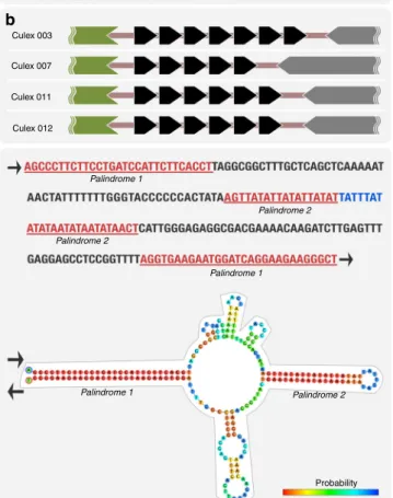
Fig. 3 pWCP contains a variable number tandem repeat (VNTR) region and extragenic palindrome (EP) sequence.a A VNTR region is located between parA and uncharacterized gene in the circular genome. While the number of repeats varies across individuals (b), the 36- and 33-bp spacers are conserved. Each black arrow represents a 16-nt repeat. The predicted DNA structure of the EP sequence is illustrated inc where color indicates probability of each base pairing. Red represents the strongest probability, whereas blue is the lowest
Metagenomic read recruitment revealed between a 1. and
5-fold increase in coverage between pWCP and some structural
phage genes (e.g., WP0415 and WP0446 Tail genes) in our
metagenomes compared to the coverage of the bacterial
chromosome in all four C. pipiens individuals (Supplementary
Data 6). The forced synteny organization of gene clusters also
revealed that a single prophage WO region could include both
high- and low-coverage phage genes (Fig.
4
). The multi-copy
occurrence of pWCP could explain its increased coverage
(Supplementary Figure 1), and differential coverage regimes for
genes within a single prophage region could be explained by at
least two different models. First, increased coverage of some
prophage genes could be attributed to lytic activity: the prophage
genes displaying lower bacterial-like coverage are not part of the
virion, while those with higher coverage correspond to phage
genes that are replicated and packaged into phage capsids. This
lytic model is consistent with the observation of phage particles in
C. pipiens mosquitoes
44,45, observed lytic activity of Wolbachia
phages in Nasonia testes, and the sequencing of WO genomes
from purified phage particles
27. The partial replication and
packaging of prophage WO genes could result from either a
‘less
than headful’ mechanism of packaging, as described in model
Phage WO assignment
O07
NCBI COGs Known (858) Unknown (312)
Wolbachia MAG O07 has one or more genes matching to this gene cluster Wolbachia MAG O07 do not show any gene matching to this wPip gene within this gene cluster.
The wPip gene in this gene cluster matches to prophage WO Pip 5 The coverage of the wolbachia MAG O07 gene in this gene cluster in the culex pipiens 007 metagenome
None of the genes in this gene cluster matches to any known NCBI COGs WO Pip 5 (17) WO Pip 1 (43) WO Pip 2 (11) WO Pip 3 (8) WO Pip 4 (28) O03 O12 O11 84 gene clusters found only in MAGs
1082 gene clusters ordered by their synteny in the wPip reference genome
wPip
Fig. 4Wolbachia metapangenome in the context of wPip Pel genome synteny. The figure shows the presence–absence of 1166 gene clusters in the pangenome of fourWolbachia metagenome-assembled genomes (MAGs) and the reference genome wPip Pel. The gene clusters (i.e., groups of homologous genes based on amino acid sequence identity) are organized based onwPip Pel synteny. Each MAG is represented by two layers, where the first layer indicates the presence or absence of a gene cluster in a given MAG, and the other shows the average coverage of each Wolbachia MAG O07 gene cluster in the correspondingC. pipiens metagenome. The second to last layer shows whether genes in a given gene cluster have a match in NCBI’s COGs. The outermost layer associates gene clusters with previously identified prophage regions in the wPip Pel genome. The number of gene clusters assigned to WOPip prophage regions are indicated in parenthesis
phages P1 and T4
64,65, or it could represent active vs. degenerate
prophage variants in the wPip Pel chromosome. Second, some
genes in prophage WO genomes could be copied throughout
the Wolbachia chromosome explaining the increase in coverage
due to their multi-copy occurrence. This model is supported
by the prophage duplication events observed in the wRi and wSuz
genomes
61,66as well as the presence of TEs within and/or
flanking prophage variants
27,40that could enable genomic
rearrangement and duplication. These models may not be
mutually exclusive, and the system could involve both duplication
of prophage genomes and the induction of phage particles.
Discussion
Shotgun metagenomes from individual C. pipiens ovary samples
allowed us to de novo reconstruct Wolbachia genomes from
single mosquitoes and compare these MAGs to each other as well
as to the reference wPip Pel genome through pangenomic
stra-tegies. Our data reveal an extensive diversity of previously
undetected Wolbachia phage WO and other viral genes and
notably the
first indication that Wolbachia harbours a candidate
plasmid, shedding new light on the richness of the Wolbachia
mobilome. The definition of a plasmid varies; here we adhere to
the typical characterization of a plasmid as a small hereditary,
extrachromosomal, circular element.
Even though a Wolbachia plasmid has not been reported
before, we did
find evidence of its occurrence in C. pipiens
metagenomes in previous studies. It is likely that the previous
efforts overlooked this element due to computational challenges
associated with the assembly of metagenomic short reads. Beyond
the IS110 TE that occurred both in the plasmid and Wolbachia
chromosome, pWCP contained a region of intergenic VNTR that
differed across individuals in our study. This suggests that the
co-assembly of pooled individuals may yield fragmented contigs.
The same VNTR sequences were found in C. pipiens from
Australia, Argentina, USA, Italy, Japan, Israel, and Greece
56, and
we confirmed the presence of pWCP in C. pipiens samples from
countries of the Mediterranean basin
32. The high similarity
of pWCP sequences across C. pipiens metagenomes in addition
to its global prevalence suggest evolutionary constraints and a
possible functional role in Wolbachia symbiosis. However, we
did not detect the plasmid in other Wolbachia strains through
screening available metagenomes from the
fly Drosophila
mela-nogaster, the planthopper Laodelphax striatella, and Anopheles
gambiae mosquito.
Our work demonstrates that the combination of
genome-resolved metagenomics, long-read sequencing, and pangenomic
strategies provide an effective computational framework to
investigate the diversity and distribution of mobile genetic
elements in endosymbionts that are challenging to cultivate.
Furthermore, it shows the importance of studying distinct
indi-viduals from wild mosquito populations, in parallel with
con-trolled experiments in laboratory settings, to improve our
understanding of the Wolbachia mobilome. The fragmented
nature of Wolbachia MAGs in our study emphasizes the critical
need for harnessing the power of emerging long-read sequencing
technologies to characterize complex genomic variations of
Wolbachia and its mobilome at
finer scales. Wolbachia has been
so far recalcitrant to`genetic modification, but the discovery of
pWCP and phage WO may create new avenues for effective
genome-editing strategies.
Methods
Sample collection. We collected mosquito specimens using a carbon dioxide mosquito trap located in Languedoc, Herault, France in May 2017 (Camping l’Europe de Vic La Gardiole, EID Méditerranée). Specimens were transported alive to the laboratory immediately upon recovery. We anesthetized adult females for
4 min at−20 °C and proceeded to species-level identification. To remove potential contaminants from the insect surface, we gently vortexed specimens for 1 min in 1 ml cold (4 °C) 96% ethanol. We then transferred them to a new 1.5 ml tube with 1 ml sterile cold (4 °C) phosphate-buffered saline (PBS) 1× solution and gently vortexed them again for 10 s to avoid DNA precipitation with ethanol. Finally, we transferred specimens onto a sterile microscope slide with sterile PBS 1× on top of a cold plate and dissected two ovaries from four specimens using sterilized tweezers. We preserved ovary samples at−80 °C until further processing. C. pipiens complex controls. We obtained Wolbachia uninfected mosquitoes by treating the host with antibiotics. For antibiotics treatment we either used TC, which inhibits protein synthesis, or rifampicin, which interferes with nucleic acid synthesis67. The TC-treated lines47shown in Fig.1(C. quinquefasciatus SLAB-TC
lines, ISEM, France) were raised at least 1 year without TC in standard laboratory conditions before beginning experiments. The rifampicin and oxytetracycline-treated C. pipiens lines were kindly provided by Maria del Mar Fernandez de Marco (Animals Plant Health Agency, UK).
Metagenomic library preparation and sequencing. We extracted total genomic DNA from each ovary sample, hereafter referred to as O03, O07, O11, and O12, using the MoBio PowerFecal DNA Isolation Kit (QIAGEN Inc., Germantown, MD, USA). We used an E220 Covaris instrument (Covaris, Woburn, MA, USA) to sonicate 3.8–5.7 ng of genomic DNA. We end-repaired and 3’-adenylated resulting fragments and used the NEBNext Ultra II DNA Library Prep Kit for Illumina (New England Biolabs, Ipswich, MA, USA) to add NEXTflex PCR-free barcode adapters (Bioo Scientific, Austin, TX, USA). We purified ligation products by Ampure XP (Beckman Coulter, Brea, CA, USA) and PCR-amplified DNA frag-ments (>200 bp; 2 PCR reactions, 14 cycles) using Illumina adapter-specific pri-mers and NEBNext Ultra II Q5 Master Mix (NEB). After library profile analysis using an Agilent 2100 Bioanalyzer (Agilent Technologies, Santa Clara, CA, USA) and quantitative PCR quantification using the KAPA Library Quantification Kit for Illumina Libraries (KapaBiosystems, Wilmington, MA, USA), we sequenced the library using a HiSeq4000 Illumina sequencer (Illumina, San Diego, CA, USA) at the Genoscope in Evry, France, generating 151 bp paired-end reads. To remove the least reliable data, wefiltered the raw sequencing results using cluster intensity and chastityfilter as described in ref.68.
Metagenomic assembly and binning. We used illumina-utils v1.4.469to
quality-filter raw paired-end reads with the program ‘iu-quality-filter-quality-minoche’ with default parameters, IDBA_UD v1.1.270to assemble paired-end reads into contigs,
and Bowtie2 v2.2.971for all read recruitment analyses. We processed our contigs
that are >1000 nts and read recruitment results with anvi’o v572to recover MAGs
from C. pipiens metagenomes. Briefly, we used the program ‘anvi-gen-contigs-database’ to generate anvi’o contigs databases for each four individual assemblies, during which anvi’o calculates and stores tetranucleotide frequency values and Prodigal v2.6.373identifies ORFs in each contig. We ran default anvi’o HMM
profiles on resulting contigs databases using the program ‘anvi-run-hmms’ and assigned functions to genes using the program‘anvi-run-ncbi-cogs’, which searches gene amino acid sequences against the December 2014 release of the COG data-base74using blastp v2.3.0+75. We profiled the short reads of our contigs recruited
from each four individual metagenomes onto the four assemblies using the pro-gram‘anvi-profile’, which generates anvi’o profile databases that store the coverage and detection statistics of each contig within each sample independently. We then merged resulting anvi’o profile databases for each sample using the program ‘anvi-merge’. For an initial coarse binning, we used the CONCOCT76algorithm
through the program‘anvi-cluster-with-concoct’ and confined the number of clusters tofive using the parameter ‘–num-clusters-requested 5’. We then used the program‘anvi-script-get-collection-info’ to identify bins with a bacterial popula-tion genome, manually refined them to identify bacterial genomes using the program‘anvi-refine’, and assigned taxonomy to resulting MAGs using NCBI’s non-redundant protein sequence database with amino acid sequences of core genes. The program‘anvi-refine’ allows the identification and refinement of population genome bins through an interactive interface by offering a number of tools including (1) guiding hierarchical clustering dendrograms of contigs (including one based on tetranucleotide frequencies and differential coverage statistics across samples), (2) real-time completion and redundancy estimates of binned contigs based on bacterial single-copy core genes77, (3) interfaces that
display nucleotide-level coverage statistics per gene and contig, (4) functional annotations and synteny of genes, and (5) online sequence homology search options for gene and contig sequences. These tools collectively help minimize the inclusion of non-target contigs (such as eukaryotic contamination) infinal bins, especially in complex metagenomes78. Except for the manual refinement step, we
automated the processing of our raw sequencing data with a snakemake79
work-flow anvi’o v5 implements ( http://merenlab.org/2018/07/09/anvio-snakemake-workflows/), and the Code availablility section reports necessary configuration files to fully reproduce this analysis.
Sample preparation and long-read sequencing. To investigate the circularity of pWCP, we prepared for a long-read sequencing strategy. We extracted and pooled
DNA from the ovaries of 7 C. pipiens and 15 C. quinquefasciatus mosquitoes. As the total DNA quantity from this step was only 5.83 ng, we also extracted DNA from the abdomens of 15 C. quinquefasciatus that left us with a total of 76.63 ng. To acquire high-molecular weight DNA from these low-biomass samples, we used an in-house Phenol Chloroform protocol for total genomic DNA extraction. Briefly, we used a chloridric acid-washed pestle adapted to the size of a micro-centrifuge tube to gently grind tissue in cetyltrimethylammonium bromide buffer (10 ml) mixed with betamercaptoethanol (20 µL) that we incubated for 15 min at 60 °C. We then added 200 µL of phenol:chloroform:isoamyl alcohol and incubated each microcentrifuge tube at room temperature while rotation shaking for 5 min. To minimize inclusion of proteins, we centrifuged the tubes for 10 min at 8000 rpm (5939 × g), removed the aqueous phase (the top layer), and placed it into fresh tubes. We repeated those steps with chloroform:isoamyl alcohol and lastly with isopropanol. We incubated microcentrifuge tubes for 10 min at room temperature and mixed by inversion every 3 min. We then centrifuged those tubes for 10 min at 10,000 rpm (9279 × g), removed the top layer, and kept the sole DNA at the bottom of the microcentrifuge tube. We next washed DNA with 400 µL of freshly prepared 70% ethanol and centrifuged tubes for 10 min at 10,000 rpm (9279 × g). Finally, we kept microcentrifuge tubes in a speed vacuum for 10 min and eluted DNA with 20 uL of molecular grade water. During all laboratory steps, we performed smooth and slow pipetting with cut pipette tips to avoid further shearing the DNA molecules. To prepare mosquitoes' total DNA for sequencing, we used the Rapid Barcoding Kit (SQK-RBK004) by Oxford Nanopore Technologies. We followed the manufacturer protocols, except for one additional step, in which we supplemented samples that did not meet the manufacturer DNA input mass recommendations (400 ng) with linear double-stranded lambda DNA (New England Biolabs) for ‘padding’. We then used 1× Agencourt AMPure XP beads (A63882, Beckman Coulter) for sample clean-up and concentration of pooled barcoded samples. A MinION with a pristine R9.4/FLO-MIN106flow cell (Oxford Nanopore Tech-nologies) sequenced thefinal prepared library with a starting voltage of −180 mV and a run time of approximately 20 h. While theflow cell had 1293 active pores prior to the run, only 200 were active at the beginning of the run, dropping down to 2 active pores by 20 h. We were not able to determine whether the low number of initial pores and their rapid decline was due to remaining chemical residues from our in-house Phenol Chloroform extraction protocol or a defectiveflow cell. We used MinKNOW (v1.15.4) for live base-calling and Albacore (v2.1.7) to de-multiplex our samples and convert raw FAST5files to FASTQ files. For downstream analyses, we only used reads with a minimum quality score of 7. Pangenomic analysis and the metapangenome. Our analysis of the Wolbachia pangenome followed the workflow outlined in ref.80and athttp://merenlab.org/ 2016/11/08/pangenomics-v2/. Briefly, we first generated an anvi’o genomes storage database using the program‘anvi-gen-genomes-storage’ for our four Wolbachia MAGs and the wPip reference genome wPip Pel40(NCBI Accession ID
NC_010981.1). We then used the program‘anvi-pan-genome’ using the flag ‘–use-ncbi-blast’, and parameters ‘–minbit 0.5’, and ‘–mcl-inflation 5’. This program (1) uses the blastp program all vs. all to create a graph of similarities between pairs of amino acid sequences, (2) removes bad hits using the‘minbit heuristic’, (3) uses the Markov Cluster algorithm to determine gene clusters, (4) calculates the occurrence of gene clusters across genomes and the total number of genes they contain, (5) realizes a hierarchical clustering analysis for gene clusters (based on their distribution across genomes) and for genomes (based on shared gene clus-ters), andfinally (6) generates an anvi’o pangenomic database that stores all results for downstream analyses. We used the program‘anvi-display-pan’ to visualize gene clusters and their distribution across thefive genomes in anvi’o interactive interface and summarized our manual selection of gene clusters using the program ‘anvi-summarize’. To identify ‘phage-like’ gene clusters, we used blastn to search for genes homologous to thefive phage regions27in the wPip Pel genome (hereafter
referred to as WOPip1–5). We finally used a custom R script (see the Code availability section) to recover coverage values of each gene cluster using our four metagenomes. We visualized the metapangenome using the program ‘anvi-display-pan’.
Computing the density of single-nucleotide variants per genome and meta-genome. To infer the extent of heterogeneity in metagenomes for each Wolbachia MAG, we used the anvi’o program ‘anvi-gen-variability-profile’ to recover single-nucleotide variants from each merged profile without any filters (following the tutorial athttp://merenlab.org/2015/07/20/analyzing-variability/). We then kept only single-nucleotide variants that fall into the context of complete gene calls to minimize erroneous variants that often emerge from mappig artefacts around beginnings and ends of contigs and calculated the percentage of nucleotide posi-tions in each genome that was not clonal in the metagenome.
Computing the average nucleotide identity between genomes. To compute the level of similarity between the four Wolbachia MAGs and the wPip Pel reference genome, we used the program‘anvi-compute-ani’, which called PyANI v0.2.781in
‘ANIb’ mode to align 1 kbp long fragments of the input genome sequences with the NCBI’s blastn program to summarize the average nucleotide identity and align-ment coverage scores.
PCR amplification of DNA. We used a set of specific primers on the four C. pipiens individuals and three Wolbachia-free C. quinquefasciatus controls gen-erated by TC treatment using standard techniques. The presence of arthropod DNA was verified by amplifying a ca. 708-bp fragment of the Cytochrome Oxidase I gene from arthropod mitochondria using LCO1490: (5’- GGT CAA CAA ATC ATA AAG ATA TTG G -3’) and HCO2198: (5’- TAA ACT TCA GGG TGA CCA AAA AAT CA -3’) primers48. We checked for the presence of
Wolbachia DNA by PCR amplifying a ca. 438-bp fragment of the Wolbachia 16S rDNA gene using Wspec-F: CAT ACC TAT TCG AAG GGA TAG and Wspec-R: AGC TTC GAG TGA AAC CAA TTC primers49. The PCR conditions for both
sets of primers included a temperature regime of 2 min at 94 °C, followed by 29 cycles of 30 s at 94 °C, 45 s at 49 °C, 1 min at 72 °C, and afinal elongation of 10 min at 72 °C. To detect the circularity of the plasmid-like genome, we designed new primers (listed in Results) and used the following cycling para-meters: 2 min at 95 °C, followed by 34 cycles of (94 °C for 30 s, 58 °C for 45 s, and 72 °C for 2 min) with afinal elongation of 5 min at 72 °C. Finally, to confirm the presence of the IS110 TE, we used primers described in the Results section under the following PCR conditions: 2 min at 95 °C, followed by 34 cycles of (94 °C for 30 s, 55 °C for 45 s, and 72 °C for 30 s) and afinal elongation of 5 min at 72 °C. Amplifications by PCR were performed using Platinium Taq DNA Polymerase High Fidelity (5 U/μL, Invitrogen).
Plasmid characterization. We used the Glimmer software within Geneious v8.1.9 (Biomatters, Ltd) to identify ORFs and used the NCBI databases for non-redundant protein sequences and conserved domains82, SMART83, and HHpred84
(including SCOPe, Pfam, and COG) to manually annotate putative functions based on amino acid sequence homology searches. We used the ISfinder platform (https://isfinder.biotoul.fr/) for IS homology search. The nucleotide analysis plugin in Geneious for EMBOSS identified and illustrated the EP in the plasmid. Figures. We visualized BLAST hits between pWCP and long read sequences using Kablammo85(http://kablammo.wasmuthlab.org/) andfinalized all figures using
the open-source vector graphics editor Inkscape (https://inkscape.org/). Reporting summary. Further information on experimental design is available in the Nature Research Reporting Summary linked to this article.
Code availability. The URLhttp://merenlab.org/data/2019_Reveillaud_and_ Bordenstein_et_al_Wolbachiagives access to a reproducible bioinformatics workflow document and ad hoc scripts used for all computational analyses.
Data availability
The raw sequencing data for shotgun metagenomes are available in the European Nucleotide Archive via accession codePRJEB26028. Sequencing data for the MinION run is available athttps://doi.org/10.6084/m9.figshare.7306784. We also made available FASTAfiles for individual metagenomic assemblies (https://doi.org/10.6084/m9. figshare.6263867), the four metagenome-assembled Wolbachia genomes (https://doi.org/ 10.6084/m9.figshare.6292040), artificially circularized individual plasmid sequences (https://doi.org/10.6084/m9.figshare.6380015), as well as anvi’o merged profile databases (https://doi.org/10.6084/m9.figshare.6263876) and anvi’o files for the Wolbachia pangenome (https://doi.org/10.6084/m9.figshare.6291650).
Received: 10 August 2018 Accepted: 6 February 2019
Published online: 05 March 2019
References
1. Gould, E., Pettersson, J., Higgs, S., Charrel, R. & de Lamballerie, X. Emerging arboviruses: Why Today? One Health 4, 1–13 (2017).
2. Shragai, T., Tesla, B., Murdock, C. & Harrington, L. C. Zika and chikungunya: mosquito-borne viruses in a changing world. Ann. NY Acad. Sci. 1399, 61–77 (2017).
3. Boyer, S., Calvez, E., Chouin-Carneiro, T., Diallo, D. & Failloux, A. B. An overview of mosquito vectors of Zika virus. Microbes Infect. 20, 646–660 (2018).
4. Flores, H. A. & O’Neill, S. L. Controlling vector-borne diseases by releasing modified mosquitoes. Nat. Rev. Microbiol. 16, 508–518 (2018).
5. Taylor, M. J., Bordenstein, S. R. & Slatko, B. Microbe profile: Wolbachia: a sex selector, a viral protector and a target to treatfilarial nematodes. Microbiology 164, 1345–1347 (2018).
6. LePage, D. & Bordenstein, S. R. Wolbachia: can we save lives with a great pandemic? Trends Parasitol. 29, 385–393 (2013).
7. Stouthamer, R., Breeuwer, J. A. J. & Hurst, G. D. D. Wolbachia pipientis: microbial manipulator of arthropod reproduction. Annu. Rev. Microbiol. 53, 71–102 (1999).
8. Hedges, L. M., Brownlie, J. C., O’Neill, S. L. & Johnson, K. N. Wolbachia and virus protection in insects. Science 322, 702 (2008).
9. Teixeira, L., Ferreira, Á. & Ashburner, M. The bacterial symbiont Wolbachia induces resistance to RNA viral infections in Drosophila melanogaster. PLoS Biol. 6, 2753–2763 (2008).
10. Jeffries, C. L. & Walker, T. Wolbachia biocontrol strategies for arboviral diseases and the potential influence of resident Wolbachia strains in mosquitoes. Curr. Trop. Med. Rep. 3, 20–25 (2016).
11. Joubert, D. A. & O’Neill, S. L. Comparison of stable and transient Wolbachia infection models in Aedes aegypti to block Dengue and West Nile viruses. PLoS Negl. Trop. Dis. 11, 1–14 (2017).
12. McMeniman, C. J. et al. Stable introduction of a life-shortening Wolbachia infection into the mosquito Aedes aegypti. Sci. 323, 141–144 (2009). 13. Blagrove, M. S. C., Arias-Goeta, C., Failloux, A. B. & Sinkins, S. P. Wolbachia
strain wMel induces cytoplasmic incompatibility and blocks dengue transmission in Aedes albopictus. Proc. Natl Acad. Sci. 109, 255–260 (2012).
14. Andrews, E. S., Crain, P. R., Fu, Y., Howe, D. K. & Dobson, S. L. Reactive oxygen species production and Brugia pahangi survivorship in Aedes polynesiensis with artificial Wolbachia infection types. PLoS Pathog. 8, e1003075 (2012).
15. Bourtzis, K. et al. Harnessing mosquito-Wolbachia symbiosis for vector and disease control. Acta Trop. 132, S150–S163 (2014).
16. Mains, J. W., Brelsfoard, C. L., Rose, R. I. & Dobson, S. L. Female adult Aedes albopictus suppression by Wolbachia-infected male mosquitoes. Sci. Rep. 6, 1–7 (2016).
17. O’Neill, S. L. et al. Scaled deployment of Wolbachia to protect the community from Aedes transmitted arboviruses. Gates Open Res. 2, 36 (2018). 18. Ferree, P. M. et al. Wolbachia utilizes host microtubules and dynein for
anterior localization in the Drosophila oocyte. PLoS Pathog. 1, 0111–0124 (2005).
19. Serbus, L. R. & Sullivan, W. A cellular basis for Wolbachia recruitment to the host germline. PLoS Pathog. 3, 1930–1937 (2007).
20. Funkhouser-Jones, L. J., van Opstal, E. J., Sharma, A. & Bordenstein, S. R. The maternal effect gene Wds controls Wolbachia titer in Nasonia. Curr. Biol. 28, 1692–1702.e6 (2018).
21. Werren, J. H. Biology of Wolbachia. Annu. Rev. Entomol. 124, 587–609 (1997).
22. Atyame, C. M. et al. Wolbachia divergence and the evolution of cytoplasmic incompatibility in Culex pipiens. PLoS. One. 9, 21–26 (2014).
23. Atyame, C. M. et al. Cytoplasmic incompatibility as a means of controlling Culex pipiens quinquefasciatus mosquito in the islands of the South-Western Indian Ocean. PLoS Negl. Trop. Dis. 5, 20–22 (2011).
24. Beckmann, J. F., Ronau, J. A. & Hochstrasser, M. A. Wolbachia
deubiquitylating enzyme induces cytoplasmic incompatibility. Nat. Microbiol. 2, 17007 (2017).
25. LePage, D. P. et al. Prophage WO genes recapitulate and enhance Wolbachia-induced cytoplasmic incompatibility. Nature 543, 243 (2017).
26. Shropshire, J. D., On, J., Layton, E. M., Zhou, H. & Bordenstein, S. R. A single prophage WO gene rescues cytoplasmic incompatibility in Drosophila melanogaster. Proc. Natl. Acad. Sci. 115, 4987–4991 (2018).
27. Bordenstein, S. R. & Bordenstein, S. R. Eukaryotic association module in phage WO genomes from Wolbachia. Nat. Commun. 7, 1–10 (2016). 28. Chafee, M. E., Funk, D. J., Harrison, R. G. & Bordenstein, S. R. Lateral phage
transfer in obligate intracellular bacteria (Wolbachia): verification from natural populations. Mol. Biol. Evol. 27, 501–505 (2010).
29. Bordenstein, S. R. & Wernegreen, J. J. Bacteriophageflux in endosymbionts (Wolbachia): infection frequency, lateral transfer, and recombination rates. Mol. Biol. Evol. 21, 1981–1991 (2004).
30. Kent, B. N., Funkhouser, L. J., Setia, S. & Bordenstein, S. R. Evolutionary genomics of a temperate bacteriophage in an obligate intracellular bacteria (Wolbachia). PLoS ONE 6, e24984 (2011).
31. Kent, B. N. et al. Complete bacteriophage transfer in a bacterial endosymbiont (Wolbachia) determined by targeted genome capture. Genome Biol. Evol. 3, 209–218 (2011).
32. Bonneau, M. et al. Culex pipiens crossing type diversity is governed by an amplified and polymorphic operon of Wolbachia. Nat. Commun. 9, 319 (2018).
33. El Karkouri, K., Pontarotti, P., Raoult, D. & Fournier, P. E. Origin and evolution of rickettsial plasmids. PLoS ONE 11, 1–17 (2016).
34. Baldridge, G. D., Burkhardt, N. Y., Felsheim, R. F., Kurtti, T. J. & Munderloh, U. G. Transposon insertion reveals pRM, a plasmid of Rickettsia monacensis. Appl. Environ. Microbiol. 73, 4984–4995 (2007).
35. Ogata, H. et al. The genome sequence of Rickettsia felis identifies the first putative conjugative plasmid in an obligate intracellular parasite. PLoS Biol. 3, e248 (2005).
36. Burkhardt, N. Y. et al. Development of shuttle vectors for transformation of diverse Rickettsia species. PLoS ONE 6, e29511 (2011).
37. Wood, D. O. et al. Establishment of a replicating plasmid in Rickettsia prowazekii. PLoS ONE 7, 1–6 (2012).
38. Sun, L. V. et al. Determination of Wolbachia genome size by pulsed-field gel electrophoresis. J. Bacteriol. 183, 2219–2225 (2001).
39. Woolfit, M. et al. Genomic evolution of the pathogenic Wolbachia strain, wMelPop. Genome Biol. Evol. 5, 2189–2204 (2013).
40. Klasson, L. et al. Genome evolution of Wolbachia strain wPip from the Culex pipiens group. Mol. Biol. Evol. 25, 1877–1887 (2008).
41. Iturbe-Ormaetxe, I., Woolfit, M., Rancès, E., Duplouy, A. & O’Neill, S. L. A simple protocol to obtain highly pure Wolbachia endosymbiont DNA for genome sequencing. J. Microbiol. Methods 84, 134–136 (2011). 42. Sczyrba, A. et al. Critical Assessment of Metagenome Interpretation - a
benchmark of metagenomics software. Nat. Methods 14, 1063–1071 (2017). 43. Delmont, T. O. et al. Nitrogen-fixing populations of Planctomycetes and
Proteobacteria are abundant in surface ocean metagenomes. Nat. Microbiol. 3, 804–813 (2018).
44. Sanogo, Y. O. & Dobson, S. L. WO bacteriophage transcription in Wolbachia-infected Culex pipiens. Insect Biochem. Mol. Biol. 36, 80–85 (2006). 45. Wright, J. D., Sjostrand, F. S., Portaro, J. K. & Barr, A. R. The ultrastructure
of the rickettsia-like microorganism Wolbachia pipientis and associated virus-like bodies in the mosquito Culex pipiens. J. Ultra. Res. 63, 79–85 (1978). 46. Cerveau, N., Leclercq, S., Leroy, E., Bouchon, D. & Cordaux, R. Short- and
long-term evolutionary dynamics of bacterial insertion sequences: Insights from Wolbachia endosymbionts. Genome Biol. Evol. 3, 1175–1186 (2011). 47. Duron, O. et al. Tracking factors modulating cytoplasmic incompatibilities in
the mosquito Culex pipiens. Mol. Ecol. 15, 3061–3071 (2006).
48. Folmer, O., Black, M., Wr, H., Lutz, R. & Vrijenhoek, R. DNA primers for amplification of mitochondrial Cytochrome C oxidase subunit I from diverse metazoan invertebrates. Mol. Mar. Biol. Biotechnol. 3, 294–299 (1994). 49. Werren, J. H. & Windsor, D. M. Wolbachia infection frequencies in insects:
evidence of a global equilibrium? Proc. R. Soc. B Biol. Sci. 267, 1277–1285 (2000).
50. Salzberg, S. L., Puiu, D., Sommer, D. D., Nene, V. & Lee, N. H. Genome sequence of the Wolbachia endosymbiont of Culex quinquefasciatus JHB. J. Bacteriol. 191, 1725 (2009).
51. Singhal, K. & Mohanty, S. Comparative genomics reveals the presence of putative toxin–antitoxin system in Wolbachia genomes. Mol. Genet. Genomics 293, 525–540 (2018).
52. Shao, Y. et al. TADB: A web-based resource for Type 2 toxin-antitoxin loci in bacteria and archaea. Nucleic Acids Res. 39, 606–611 (2011).
53. Harms, A., Brodersen, D. E., Mitarai, N. & Gerdes, K. Toxins, targets, and triggers: an overview of toxin-antitoxin biology. Mol. Cell 70, 768–784 (2018). 54. Lo, N., Casiraghi, M., Salati, E., Bazzocchi, C. & Bandi, C. How many
Wolbachia supergroups exist? Mol. Biol. Evol. 19, 341–346 (2002). 55. Shen, X. et al. Functional identification of the DNA packaging terminase from
Pseudomonas aeruginosa phage PaP3. Arch. Virol. 157, 2133–2141 (2012). 56. Petridis, M. & Chatzidimitriou, D. Characterization of an intergenic
polymorphic site (pp-hC1A_5) in Wolbachia pipientis (wPip). Mol. Ecol. Resour. 11, 753–756 (2011).
57. Riegler, M., Iturbe-Ormaetxe, I., Woolfit, M., Miller, W. J. & O’Neill, S. L. Tandem repeat markers as novel diagnostic tools for high resolution fingerprinting of Wolbachia. BMC Microbiol. 12, S12 (2012). 58. Konieczny, I., Bury, K., Wawrzycka, A. & Wegrzyn, K. Iteron plasmids.
Microbiol. Spectr. 2, 1–16 (2014).
59. Pan, X., Lührmann, A., Satoh, A., Laskowski-Arce, M. A. & Roy, C. R. Ankyrin repeat proteins comprise a diverse family of bacterial type IV effectors. Science 320, 1651–1654 (2008).
60. Cerveny, L. et al. Tetratricopeptide repeat motifs in the world of bacterial pathogens: role in virulence mechanisms. Infect. Immun. 81, 629–635 (2013). 61. Klasson, L. et al. The mosaic genome structure of the Wolbachia wRi strain
infecting Drosophila simulans. Proc. Natl. Acad. Sci. 106, 5725–5730 (2009). 62. Siozios, S. et al. Draft genome sequence of the Wolbachia endosymbiont of
Drosophila suzukii. Genome Announc. 1, e00032–13 (2013).
63. Ishmael, N. et al. Extensive genomic diversity of closely related Wolbachia strains. Microbiology 155, 2211–2222 (2009).
64. Coren, J. S., Pierce, J. C. & Sternberg, N. Headful packaging revisited: the packaging of more than one DNA molecule into a bacteriophage P1 head. J. Mol. Biol. 249, 176–184 (1995).
65. Leffers, G. & Basaveswara Rao, V. A discontinuous headful packaging model for packaging less than headful length DNA molecules by bacteriophage T4. J. Mol. Biol. 258, 839–850 (1996).
66. Conner, W. R. et al. Genome comparisons indicate recent transfer of wRi-like Wolbachia between sister species Drosophila suzukii and D. subpulchrella. Ecol. Evol. 7, 9391–9404 (2017).
67. Kohanski, M. A., Dwyer, D. J. & Collins, J. J. How antibiotics kill bacteria: from targets to networks. Nat. Rev. Microbiol. 8, 423–435 (2010). 68. Alberti, A. et al. Viral to metazoan marine plankton nucleotide sequences