Robert Goetti Gudrun Feuchtner Paul Stolzmann Lotus Desbiolles
Michael Alexander Fischer Christoph Karlo Stephan Baumueller Hans Scheffel Hatem Alkadhi Sebastian Leschka Received: 9 February 2010 Revised: 3 May 2010 Accepted: 7 May 2010 Published online: 29 June 2010 # European Society of Radiology 2010
High-pitch dual-source CT coronary
angiography: systolic data acquisition
at high heart rates
Abstract Objective To assess the
effect of systolic data acquisition for electrocardiography (ECG)-triggered high-pitch computed tomography (CT) on motion artefacts of coronary arteries in patients with high heart
rates (HRs). MethodsEighty
consec-utive patients (15 women, age 67±
14years) with HR≥70bpm underwent
CT angiography of the thoracic aorta (CTA) on 128-slice dual-source CT in ECG-triggered high-pitch acquisition mode (pitch=3.2) set at 60% (group A, n=40) or 30% (group B, n=40) of the RR interval. Two blinded readers graded coronary artery image quality on a three-point scale. Radiation
doses were calculated.Results
Inter-observer agreement in grading image quality of the 1,154 coronary
seg-ments was good (κ=0.62). HRs
were similar in groups A and B (85±
13bpm vs 85±14bpm, p not signi
fi-cant). Significantly fewer coronary
segments with non-diagnostic image quality occurred (i.e. score 3) in group B than in group A [2.8% (16/ 579) vs 8.3% (48/575), p<0.001]. Seventeen patients (42.5%) of group A and 12 patients (30.0%) of group B had at least one non-diagnostic seg-ment. Effective radiation doses were
2.3±0.3mSv for chest CTA.
Conclu-sionA systolic acquisition window
for high-pitch dual-source CTA in
patients with high HRs (≥70bpm)
significantly improves
coronary artery image quality at a low radiation dose.
Keywords High-pitch . Computed tomography . Systolic
reconstruction . Coronary arteries . Heart rate . Radiation dose
Introduction
Electrocardiography (ECG)-synchronised CT coronary angiography (CTCA) is a non-invasive tool for with a high diagnostic accuracy for the detection of coronary
arterial stenoses [1–6]. However, at high heart rates (HRs),
motion artefacts are still a limiting factor for the conclusive assessment of the coronary arteries at CTCA
[5, 7, 8]. In order to minimise motion artefacts, it is
essential to time CTCA data acquisition to take place in a phase of the cardiac cycle with relatively slow coronary artery motion. Periods of lowest relative coronary artery motion were shown to be at systolic and mid- to
end-diastolic phases at 45–50% and at 80–85% of the RR
interval of the cardiac cycle, respectively [9]. The optimal
timing of image acquisition to minimise coronary arterial
motion artefacts—especially of the mid to distal right
coronary artery (RCA)—may shift to systolic intervals in
patients with high HRs as diastolic diastasis shortens and
eventually disappears with increasing HRs [9, 10]. It has
been demonstrated with 16- and 64-detector single-source as well as dual-source CT that the best results regarding coronary artery image quality are achieved at mid-diastolic intervals in patients with low HRs and at systolic
intervals in patients with high HRs [11–18].
The recently introduced second generation of dual-source CT (DSCT) systems combines two X-ray tubes and corresponding detector systems with 64 detector rows, R. Goetti
:
G. Feuchtner:
P. Stolzmann:
L. Desbiolles
:
M. A. Fischer:
C. Karlo:
S. Baumueller:
H. Scheffel:
H. Alkadhi:
S. LeschkaInstitute of Diagnostic Radiology, University Hospital Zurich, Raemistrasse 100, 8091 Zurich, Switzerland
P. Stolzmann
:
H. Scheffel:
H. Alkadhi Cardiac MR PET CT Program, Massachusetts General Hospital and Harvard Medical School, 100 Charles River Plaza, Boston, MA 02114, USA S. Leschka ()) Institute of Radiology, Kantonsspital St. Gallen, Rorschacherstrasse 95, 9007 St. Gallen, Switzerland e-mail: sebastian.leschka@kssg.ch Tel.: +41-71-4941111 Fax: +41-71-4946479each simultaneously acquiring 128 slices by means of a
z-flying focal spot [19]. With this system, data acquisition
for gapless image reconstruction at pitch values of up to
3.2 has become possible by filling the gaps of the first
detector with the data acquired by the second detector
[19]. A gantry rotation time of 0.28 s enables a temporal
resolution of 75 ms. Owing to the increased detector width in second-generation dual-source CT systems, high-pitch imaging allows the acquisition of all axial planes that comprise the heart (12 cm) in approximately 280 ms, hence capturing the heart during a fraction of a single cardiac cycle. High-pitch CTCA with the cranial-most slice acquired at 60% of the RR interval enables coronary artery assessment with high diagnostic accuracy in patients with
HRs equal to or below 60 bpm at doses <1 mSv [6].
To our knowledge, it has not yet been investigated whether a systolic acquisition window in high-pitch dual-source CTA yields improved coronary image quality at higher HRs. The purpose of our study, therefore, was to
assess the influence of diastolic and systolic acquisition
windows on motion artefacts of the coronary arteries in prospectively ECG-triggered high-pitch CTA of the chest
in patients with HRs of ≥70 bpm.
Materials and methods
Patient population
Eighty consecutive patients [65 men, 15 women, age 67± 14 years (mean ± standard deviation), BMI 24.7±2.9 kg/
m2(range, 16.4–29.9 kg/m2)] with mean HR≥70 beats per
minute (bpm) who were referred to our institution for clinically indicated chest CTA were prospectively included in this study. Patients with previous coronary artery interventions, i.e. stenting and/or coronary artery bypass grafts, as well as patients with HRs <70 bpm or a body
mass index (BMI)≥30 kg/m2 were not included. General
exclusion criteria for contrast-enhanced CT were
nephr-opathy with a serum creatinine level above 150 μmol/l,
known hypersensitivity to iodine-containing contrast media, untreated hyperthyroidism and pregnancy. Institu-tional review board (IRB) approval was obtained. The written informed consent requirement was waived by the IRB because all CT studies were clinically indicated and
the patients had the direct benefit of the low radiation
exposure associated with the high-pitch protocol.
The CTA of the chest was performed for postoperative follow-up after vascular surgery (n=52), preoperative evaluation before vascular or aortic valve surgery (n= 16), follow-up evaluation of conservatively treated ascending aorta aneurysm (n=5) and suspected aortic dissection (n=7).
CT data acquisition
All examinations were performed with a
second-gener-ation dual-source CT system (Somatom Definition Flash,
Siemens Healthcare, Forchheim, Germany). As intra-venous contrast material, 100 ml iopromide (Ultravist 300, 300 mg/ml; Bayer Schering Pharma, Berlin,
Ger-many) was injected at a flow rate of 5 ml/s followed by
60 ml saline solution. Contrast material application was controlled by bolus tracking in the ascending aorta with a signal attenuation threshold set at 100 HU with the formal CT data acquisition beginning with a delay of 9 s after this threshold was reached. CT parameters were: detector collimation 2×64×0.6 mm with a resulting slice
collima-tion of 2×128×0.6 mm by means of a z-flying focal spot
[19]; gantry rotation time 280 ms; pitch 3.2; reference tube
current-time product set at 320 mAs per rotation with attenuation-based tube current modulation; and a tube potential of 100 kV, because all patients had a BMI
<30 kg/m2. CT data acquisition was cranio-caudal. The
level 2 cm below the tracheal carina was manually set as the target for image acquisition at 60% of the RR interval
for thefirst 40 patients (group A) and at 30% of the RR
interval for the following 40 patients (group B). The start time of CT acquisition at the most cranial position of the chest was then automatically calculated by the CT software in accordance with the electrocardiogram (ECG), in order to time the data acquisition to arrive at the indicated level (i.e. 2 cm below the carina) at 60% or 30% of the RR interval.
Images were reconstructed with a slice thickness of 0.75 mm and an increment of 0.5 mm, using a medium smooth tissue convolution kernel (B26f) and a sharp tissue convolution kernel (B46f). One radiologist not involved in the subsequent analyses noted the average, minimum and maximum HR during ten heartbeats before the start of CTA data acquisition. All images were anonymised and transferred to an external workstation (Multi-Modality Workplace, Siemens Healthcare, Forchheim, Germany).
Assessment of coronary artery motion artefacts
For the assessment of coronary artery motion artefacts,
two experienced readers (with 2 and 5 years’ experience
in cardiovascular imaging), blinded to each other as well as to the HR and acquisition window, evaluated all coronary artery segments on cross-sectional source images and multiplanar reformations applying a semi-quantitative three-point scale: score 1 (excellent image quality, no motion artefacts), score 2 (moderate, however diagnostic image quality with minor blurring of the vessel wall) and score 3 (non-diagnostic image quality due to severe blurring or doubling of the vessel wall). For the assess-ment of the individual segassess-ments, the coronary tree was
subdivided according to the modified 16-segment model
proposed by the American Heart Association (AHA) [20].
Segments missing because of normal anatomical variants as well as segments with a diameter of less than 1 mm at their origin, as measured with an electronic calliper tool, were excluded from analysis. For any disagreement on image quality scores 1 and 2, a consensus reading was performed by the two radiologists after 1 week. Segments
non-diagnostic for further analysis, independent of the rating of the other reader.
Measurement of image noise and contrast-to-noise ratio (CNR)
As an objective parameter of image quality, image noise was measured and CNR was calculated by one independ-ent radiologist, who was blinded to the results of the qualitative image analysis. Vessel contrast of the proximal right coronary artery (RCA) and the left main artery (LM)
was defined as the difference in mean attenuation (in HU)
between the contrast-enhanced lumen of the vessel and the perivascular tissue. Attenuations were measured by man-ually placing a region of interest (ROI) in the proximal
segment of the RCA and in the LM avoiding calci
fica-tions, plaques and stenoses. Image noise was defined as
the standard deviation of the attenuation value in a ROI placed in the ascending aorta. The contrast-to-noise ratio
(CNR) was defined as the ratio of vessel contrast and
image noise.
Estimation of radiation dose
The effective radiation dose delivered at chest CT was calculated by applying a method proposed by the Euro-pean Working Group for Guidelines on Quality Criteria
for CT [21] using the dose-length product (DLP) and a
conversion coefficient of 0.017 mSv/(mGy × cm) [22].
The DLP was obtained from the patient protocol, which summarised the individual radiation exposure parameters of each CT.
Statistical analyses
Continuous variables were expressed as means ± standard deviations and categorical variables were expressed as frequencies or percentages. A p value below 0.05 was
considered statistically significant. All statistical analyses
were performed with commercially available software (SPSS, release 17; SPSS, Chicago, Ill., USA).
The sample size of the two groups of 40 patients with 600 coronary artery segments was determined by power analysis. It was calculated for a power level of greater than 80% for detecting a difference of 5% using the pessimistic assumption of 15% non-evaluable segments and an alpha error level of 0.05.
HR variability (HRV) was defined, as previously
described [23], as the difference between minimum and
maximum HR in ten heartbeats before image acquisition divided by ten. Differences in the average HR and HRV were compared between the two protocol groups with the unpaired t-test.
Inter-observer agreement regarding the presence and severity of motion artefacts in coronary artery segments
was evaluated using Cohen’s kappa statistics (κ>0.81:
excellent agreement; κ=0.61–0.80: good agreement; κ=
0.41–0.60: moderate agreement; κ=0.21–0.40: fair
agree-ment; κ<0.20: poor agreement).
Differences in image quality regarding motion artefacts between the two protocol groups were assessed with the Mann-Whitney U-test. Differences in the proportions of non-diagnostic segments between the two protocol groups
were evaluated with the χ2 test. Image noise and CNR
were compared between the two protocol groups with the unpaired t-test. Differences of image quality between
patients with HR ranges of 70–89 bpm and ≥90 bpm in
each group were assessed with the Mann-Whitney U-test.
Results
The average HR was 85±14 bpm (range, 70–140 bpm)
and the average HRV before the scan was 3.1±3.7 bpm
(range, 0.1–14.5 bpm). There were no significant
differ-ences in HR and HRV between the two groups (p not
significant). Table 1 provides an overview of the results
for the two patient groups.
Motion artefacts
Inter-observer agreement for image quality grading of the total of 1,154 coronary artery segments (group A: 575
segments, group B: 579 segments) was good (group A:κ=
0.62, group B:κ=0.67).
Image quality was rated as being excellent (score 1) in 62.1% (357/575) in group A and 73.7% (427/579) in group B, moderate in 29.6% (170/575) in group A and 23.5% (136/579) in group B, and non-diagnostic in 8.3% (48/575) in group A and 2.8% (16/579) in group B. Non-diagnostic image quality occurred in 17 patients (42.5%) of group A and in 12 patients (30.0%) of group B. Image
examples of the different scores are displayed in Fig. 1.
Non-diagnostic image quality was most often found in the
RCA in both groups (Fig. 2). Tables 2and 3 summarise
the image quality scoring results in both groups.
Image quality regarding motion artefacts was significantly
lower in group A than in group B (1.46±0.65 vs 1.29±0.52, p<0.001), and the proportion of non-diagnostic segments
was significantly higher in group A than in group B (48/575
vs 16/579, p<0.001). Image quality was slightly lower in
patients with HR≥90 bpm than in those with HR 70–89 in
both groups; however, this was not significant [group A:
1.60±0.48 (n=10) vs 1.43±0.41 (n=40), p=0.36; group B: 1.32±0.26 (n=11) vs 1.28±0.24 (n=29), p=0.68].
Image noise and CNR
The average image noise and CNR were 32.0±7.4 HU
(range 23–60 HU) and 13.7±3.7 (range 7.1–23.2) in group
A and 31.7±8.1 HU (range 21–55 HU) and 15.0±5.2 (range
6.5–29.4) in group B. There were no significant differences
in image noise and CNR between the two groups (Table1).
Radiation dose estimates
The effective radiation dose of CT angiography of the
Discussion
Our results demonstrate that motion artefacts of the coronary arteries at high-pitch CTA of the chest are
significantly lower in patients with high HRs (≥70 bpm)
when cardiac image acquisition begins at 30% of the RR interval rather than at 60%.
Influence of the HR on coronary artery image quality
Leschka et al. [6] recently found 99% evaluable coronary
segments in CTCA performed in the dual-source
high-pitch acquisition mode in patients with HR≤60 bpm when
triggering the initiation of the imaging at 60% of the RR
interval and a sensitivity of 94% and a specificity of 96%
for the detection of significant coronary artery stenoses.
Similarly, Achenbach et al. [24] reported only 0.5%
non-evaluable coronary artery segments in patients with HR≤
60 bpm. In patients undergoing dual-source high-pitch CTA for the evaluation of atypical chest pain, with
acquisition of the heart beginning at 50–60% of the RR
interval, Lell et al. [25] found diagnostic image quality of
all coronary artery segments in patients with HR≤70 bpm.
At higher HRs, however, motion artefacts leading to non-diagnostic image quality occurred, predominantly affect-ing the mid RCA. The optimal timaffect-ing of image acquisition
to minimise coronary arterial motion artefacts—especially
of the mid to distal RCA—may shift to systolic intervals
in patients with high HRs as diastolic diastasis shortens
and eventually disappears with increasing HRs [9,10]. In
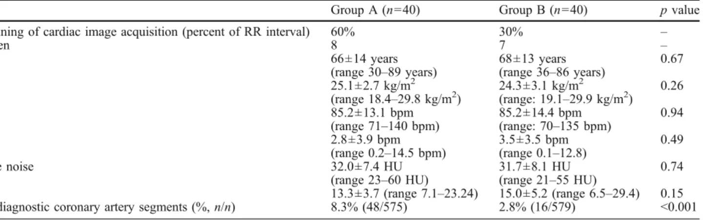
Table 1 Characteristics of patients in groups A and B (BMI body mass index, HR heart rate, HRV heart rate variability, defined as the difference between minimum and maximum HR in ten heartbeats
before image acquisition divided by ten, Image noise standard deviation of attenuation measured in the ascending aorta, CNR contrast-to-noise ratio)
Group A (n=40) Group B (n=40) pvalue Beginning of cardiac image acquisition (percent of RR interval) 60% 30% –
Women 8 7 –
Age 66±14 years 68±13 years 0.67
(range 30–89 years) (range 36–86 years)
BMI 25.1±2.7 kg/m2 24.3±3.1 kg/m2 0.26 (range 18.4–29.8 kg/m2) (range: 19.1–29.9 kg/m2) HR 85.2±13.1 bpm 85.2±14.4 bpm 0.94 (range 71–140 bpm) (range: 70–135 bpm) HRV 2.8±3.9 bpm 3.5±3.5 bpm 0.49 (range 0.2–14.5 bpm) (range 0.1–12.8) Image noise 32.0±7.4 HU 31.7±8.1 HU 0.74
(range 23–60 HU) (range 21–55 HU)
CNR 13.3±3.7 (range 7.1–23.24) 15.0±5.2 (range 6.5–29.4) 0.15
Non-diagnostic coronary artery segments (%, n/n) 8.3% (48/575) 2.8% (16/579) <0.001
Fig. 1 Imaging examples of the different image quality scores. Curved multiplanar reformations of the right coronary arteries of (a) a 63-year-old man from group B (HR 74 bpm) showing no motion artefacts (score 1), (b) an 85-year-old man from group A (HR 76 bpm)
with slight motion artefacts of the distal segment of the RCA (score 2, white arrowhead) and (c) a 66-year-old woman from group A (HR 75 bpm) with non-diagnostic image quality of all segments due to severe motion artefacts (score 3, white arrows)
retrospectively ECG-gated 16-detector CTCA, Herzog et
al. [11] found the optimal reconstruction window for the
mid RCA to be at 59.4% of the RR interval at HR< 67 bpm and at 28.3% of the RR interval at HR>67 bpm. Similarly, a shift of optimal image quality from diastolic to systolic reconstruction intervals in patients with high HRs is reported for 64-detector single-source as well as 64-detector dual-source retrospectively ECG-gated CTCA
[12–18]. Using retrospectively ECG-gated DSCT, Adler et
al. [17] found the optimal systolic phase of image
reconstruction to be between 35% and 50% of the RR
interval in patients with HR>65 bpm and Araoz et al. [16]
reported that optimal image sharpness of the coronary
arteries is achieved at 65–70% of the RR interval in
patients with HR≤70 bpm and at 35–40% of the RR
interval in patients with HR>70 bpm.
For systolic image acquisition, we chose the 30% interval for initiation of imaging of the cranial-most slices of the heart, in order to achieve data acquisition of the mid
to distal RCA—the segments most affected by motion
artefacts [13]—within a range of 35–50% of the RR
interval. Similarly, for diastolic image acquisition, initia-tion of CT data acquisiinitia-tion of the heart was chosen to begin at 60% of the RR interval.
Despite the fact that diagnostic image quality can be achieved in 97.2% of coronary segments by determining the beginning of cardiac data acquisition at 30% of the RR interval, a note of caution must be made. We found at least one non-diagnostic coronary segment due to motion artefacts in a relatively high percentage (30%) of patients. Thus, we recommend the following strategies in patients with higher HRs. When the primary indication of the CT study is the evaluation of coronary arteries, beta-blockers should be given to lower the HR, and the start of data acquisition should be set at 60% of the RR interval, as
previously shown [6,26]. When the coronary arteries are
not the primary purpose of the study in patients with high HRs, or when the administration of beta blockers is contraindicated or if the patient does not respond to beta blockers, then a start of data acquisition in systole at 30% of the RR interval would be a valuable option.
Radiation dose estimates
The minimised overlapping data acquisition at a high pitch value of 3.2 leads to a low radiation dose exposure of 2.3 mSv for a complete chest CTA. This radiation exposure is considerably lower than that obtained in
routine non-ECG-gated chest CT of about 5–7 mSv [27,
28]. Furthermore, radiation exposure with high-pitch
ECG-triggered CT is substantially lower than that with conventional 64-slice ECG-gated CT of the entire chest
with reported values up to 17.8 mSv [29,30].
Limitations
First, we applied a protocol with a tube voltage of 100 kV
for all patients, and patients with a BMI of >30 kg/m2
were excluded from the study. Obese patients may require adjustments in tube voltage or current in order to reduce image noise and obtain diagnostic image quality. Second, the semi-quantitative image quality scoring system may be
influenced by subjectivity bias. However, image quality
was evaluated by two independent readers, segments were
defined as non-diagnostic regardless of opposing readings
and the kappa values of 0.62 and 0.67 for groups A and B correspond to a good inter-observer agreement. Third, only motion artefacts as qualitative factors of impaired image quality were assessed. Blooming artefacts of
calcifications may also lead to non-diagnostic image
quality; however, the presence of calcifications is not
attributable to the acquisition window, and this study was Fig. 2 Imaging example of a 42-year-old man (HR 73 bpm) referred
for suspected aortic dissection. Cardiac image acquisition was triggered at 30% of the RR interval (group B). The left anterior descending (a) and circumflex (b) arteries show excellent image
quality. However the right coronary artery (c) shows slight blurring in the mid segment (score 2, white arrowhead) and non-diagnostic image quality of the distal segment (score 3, white arrow)
T able 3 Coronary artery image quality of high-pitch CT A in group B (i.e. start of image acquisition at 30% of the RR interval) RCA LM LAD CX IA Segment 1234 5 6789 1 0 1 1 1 2 1 3 1 4 1 5 1 6 Evaluated segments 40 39 38 38 40 40 40 40 40 39 40 37 39 38 26 5 Score 1 77.5% 51.3% 57.9% 78.9% 100% 90% 60% 70% 67.5% 82.1% 62.5% 75.7% 76.9% 84.2% 73.1% 60% Score 2 20% 33.3% 26.3% 21.1% -10% 40% 30% 32.5% 17.9% 32.5% 24.3% 20.5% 15.8% 26.9% 40% Score 3 2.5% 15.4% 15.8% -5 % -2.6% -T able 2 Coronary artery image quality of high-pitch CT A in group A (i.e. start of image acquisition at 60% of the RR interval). RCA right coronary artery , LM left main artery , LAD left anterior descending artery , CX circum fl ex artery ,IA intermediate artery RCA LM LAD CX IA Segment 1 2 3 4 5 6 7 8 9 10 1 1 12 13 14 15 16 Evaluated segments 40 40 40 39 40 40 40 40 40 40 39 37 38 34 23 5 Score 1 55% 30% 42.5% 69.3% 87.5% 75% 60% 62.5% 67.5% 60% 59% 51.4% 55.3% 79.4% 87% 80% Score 2 25% 30% 27.5% 17.9% 10% 25% 40% 37.5% 32.5% 40% 35.9% 43.2% 39.4% 20.6% 13% 20% Score 3 20% 40% 30% 12.8% 2.5% -5.1% 5.4% 5.3%
-aimed at the assessment of differences between two data acquisition windows. Finally, we did not assess diagnostic performance for the detection of coronary artery stenoses, as no cardiac catheterisation studies were available for comparison. Therefore, only differences in the presence and severity of motion artefacts between the two protocols could be evaluated and we cannot prove the accuracy of our scoring threshold of diagnostic image quality.
Conclusion
Our results suggest that a systolic window for data acquisition for high-pitch dual-source CTA in patients
with high HRs (≥70 bpm) significantly improves the
quality of coronary artery imaging. The radiation dose using the high-pitch acquisition mode is low at 2.3 mSv for CTA of the entire chest.
References
1. Raff GL, Gallagher MJ, O’Neill WW, Goldstein JA (2005) Diagnostic accuracy of noninvasive coronary angiography using 64-slice spiral computed tomography. J Am Coll Cardiol 46:552–557
2. Scheffel H, Alkadhi H, Plass A et al (2006) Accuracy of dual-source CT coronary angiography:first experience in a high pre-test probability population without heart rate control. Eur Radiol 16:2739–2747
3. Weustink AC, Meijboom WB, Mollet NR et al (2007) Reliable high-speed coronary computed tomography in symptomatic patients. J Am Coll Cardiol 50:786–794
4. Johnson TR, Nikolaou K, Busch S et al (2007) Diagnostic accuracy of dual-source computed tomography in the diagnosis of coronary artery disease. Invest radiol 42:684–691
5. Ropers U, Ropers D, Pflederer T et al (2007) Influence of heart rate on the diagnostic accuracy of dual-source computed tomography coronary angiography. J Am Coll Cardiol 50:2393–2398
6. Leschka S, Stolzmann P, Desbiolles L et al (2009) Diagnostic accuracy of high-pitch dual-source CT for the assessment of coronary stenoses:first experience. Eur Radiol 19:2896–2903
7. Nieman K, Rensing BJ, van Geuns RJ et al (2002) Non-invasive coronary angiography with multislice spiral computed tomography: impact of heart rate. Heart 88:470–474
8. Hoffmann MH, Shi H, Manzke R et al (2005) Noninvasive coronary
angiography with 16-detector row CT: effect of heart rate. Radiology 234:86– 97
9. Achenbach S, Ropers D, Holle J, Muschiol G, Daniel WG, Moshage W (2000) In-plane coronary arterial motion velocity: measurement with electron-beam CT. Radiology 216:457–463 10. Chung CS, Karamanoglu M, Kovacs SJ
(2004) Duration of diastole and its phases as a function of heart rate during supine bicycle exercise. Am J Physiol Heart Circ Physiol 287:H2003–H2008
11. Herzog C, Arning-Erb M, Zangos S et al (2006) Multi-detector row CT coronary angiography: influence of reconstruction technique and heart rate on image quality. Radiology 238:75–86 12. Wintersperger BJ, Nikolaou K, von
Ziegler F et al (2006) Image quality, motion artifacts, and reconstruction timing of 64-slice coronary computed tomography angiography with 0.33-second rotation speed. Invest radiol 41:436–442
13. Leschka S, Wildermuth S, Boehm T et al (2006) Noninvasive coronary angiography with 64-section CT: effect of average heart rate and heart rate variability on image quality. Radiology 241:378–385
14. Weustink AC, Mollet NR, Pugliese F et al (2008) Optimal electrocardiographic pulsing windows and heart rate: effect on image quality and radiation exposure at dual-source coronary CT
angiography. Radiology 248:792–798 15. Seifarth H, Wienbeck S, Pusken M et al
(2007) Optimal systolic and diastolic reconstruction windows for coronary CT angiography using dual-source CT. AJR Am J Roentgenol 189:1317–1323 16. Araoz PA, Kirsch J, Primak AN et al
(2009) Optimal image reconstruction phase at low and high heart rates in dual-source CT coronary angiography. Int J Cardiovasc Imaging 25:837–845 17. Adler G, Meille L, Rohnean A, Sigal-Cinqualbre A, Capderou A, Paul JF (2009) Robustness of end-systolic reconstructions in coronary dual-source CT angiography for high heart rate patients. Eur Radiol. doi:10.1007/ s00330-009-1642-9
18. Bamberg F, Sommer WH, Schenzle JC et al (2009) Systolic acquisition of coronary dual-source computed tomography angiography: feasibility in an unselected patient population. Eur Radiol. doi:10.1007/s00330-009-1680-3
19. Petersilka M, Bruder H, Krauss B, Stierstorfer K, Flohr TG (2008) Technical principles of dual source CT. Eur J Radiol 68:362–368
20. Austen WG, Edwards JE, Frye RL et al (1975) A reporting system on patients evaluated for coronary artery disease. Report of the Ad Hoc Committee for Grading of Coronary Artery Disease, Council on Cardiovascular Surgery, American Heart Association. Circ 51(4 Suppl):5–40
21. Menzel H, Schibilla H, Teunen D (2000) European guidelines on quality criteria for computed tomography, Publication no. EUR 16262 EN. European Commission, Luxembourg 22. Morin RL (1988) Monte Carlo
simulation in the radiological sciences. CRC Press, Boca Raton
23. Halliburton SS, Sola S, Kuzmiak SA et al (2008) Effect of dual-source cardiac computed tomography on patient radiation dose in a clinical setting: comparison to single-source imaging. J Cardiovasc Comput Tomogr 2:392–400 24. Achenbach S, Marwan M, Ropers D et
al (2009) Coronary computed tomography angiography with a consistent dose below 1mSv using prospectively electrocardiogram-triggered high-pitch spiral acquisition. Eur Heart J 31(3):340–346
25. Lell M, Hinkmann F, Anders K et al (2009) High-pitch electrocardiogram-triggered computed tomography of the chest: initial results. Invest radiol 44:728–733
26. Goetti R, Baumuller S, Feuchtner G et al (2010) High-pitch dual-source CT angiography of the thoracic and abdominal aorta: is simultaneous coronary artery assessment possible? AJR Am J Roentgenol 194:938–944 27. Gerber TC, Kuzo RS, Morin RL (2005)
Techniques and parameters for estimating radiation exposure and dose in cardiac computed tomography. Int J Cardiovasc Imaging 21:165–176 28. McCollough CH, Primak AN, Saba O et
al (2007) Dose performance of a 64-channel dual-source CT scanner. Radiology 243:775–784
29. Shapiro MD, Dodd JD, Kalva S et al (2009) A comprehensive
electrocardiogram-gated 64-slice multidetector computed tomography imaging protocol to visualize the coronary arteries, thoracic aorta, and pulmonary vasculature in a single breath hold. J Comput Assist Tomogr 33:225–232
30. Johnson TR, Nikolaou K, Becker A et al (2008) Dual-source CT for chest pain assessment. Eur Radiol 18:773–780