HAL Id: hal-03211118
https://hal.archives-ouvertes.fr/hal-03211118
Submitted on 28 Apr 2021
HAL is a multi-disciplinary open access
archive for the deposit and dissemination of sci-entific research documents, whether they are pub-lished or not. The documents may come from teaching and research institutions in France or abroad, or from public or private research centers.
L’archive ouverte pluridisciplinaire HAL, est destinée au dépôt et à la diffusion de documents scientifiques de niveau recherche, publiés ou non, émanant des établissements d’enseignement et de recherche français ou étrangers, des laboratoires publics ou privés.
Experimental models for the development of new
medical treatments in prostate cancer
Anne Chauchereau
To cite this version:
Anne Chauchereau. Experimental models for the development of new medical treatments in prostate cancer. European Journal of Cancer, Elsevier, 2011, 47 (Suppl 3), pp.S200-S214. �10.1016/S0959-8049(11)70166-6�. �hal-03211118�
EXPERIMENTAL MODELS FOR THE DEVELOPMENT OF NEW MEDICAL TREATMENTS IN PROSTATE CANCER
Anne Chauchereau,
Prostate Cancer Group, INSERM U981, Institut Gustave Roussy, Villejuif, F-94805, France
Anne CHAUCHEREAU, Prostate Cancer Group, INSERM Unit 981,
12th floor, room 507, Institut Gustave Roussy,
114 rue Edouard Vaillant, 94805 Villejuif, France Tel : 33-142116607
Fax : 33-142116094
GENERAL OUTLINES
Although prostate cancer is a major cause of death and morbidity in western countries, one major obstacle in the progress of prostate cancer studies has been the limited number of relevant preclinical models. Even so, these models constitute the major preclinical tool required for the further development of new therapeutic strategies. Nevertheless, current limitations have hampered the transition of scientific findings from these models to human clinical trials. Each xenograft and genetically engineered mouse model possess inherent advantages and limitations regarding the mimicking of the natural history of the prostate cancer disease. Xenografts are the most commonly used models in drug development and are able to maintain several biological properties of the human tumours they are derived from, particularly when orthotopically transplanted. Osteoblastic bone metastases can also be modelled. Non-invasive methods of imaging have recently been developed and have improved the follow-up of the tumour growth and response to therapy in these models. In contrast, genetic models are very useful for target validation, but are less suitable for drug evaluation. Finally, some models are more relevant for the study of specific pathways as androgen receptor signalling, or bone metastases development, and others are useful in developing strategies targeting prostate cancer stem cells or mechanisms of resistance to chemotherapy or radiotherapy. As the perfect preclinical model does never exist, efforts are needed to better define questions and endpoints before choosing one particular animal preclinical model.
1. INTRODUCTION TO PRECLINICAL MODELS IN ONCOLOGY
a. Models in animals
The modern drug development technologies increase the number of candidate therapeutics, and subsequently increase the costs for preclinical and clinical testing. Thus the improvement of the ability of preclinical models to predict clinical efficacy can have a high impact by lowering the cost of drug development and by helping to prioritize compounds for clinical investigation. While the vast contribution of mouse models to advancing our understanding of cancer biology is indisputable, there have been ongoing concerns about the value of mouse models for predicting drug efficacy and usefulness in humans, and the predictive value of tumour xenograft models in cancer research is still a matter of debate.
Preclinical cancer models remain a highly valuable tool in the drug discovery process. As we continue to research and develop molecularly targeted therapies, xenografts are ideal systems for modelling biomarker responses in target tissue and toxicity in the host organism. Despite the debate around these models, there is an obvious need to develop better tumour models, and importantly, there is agreement that there is also a need to better define questions and endpoints for which animal models are used. Thus, selecting the right model is of crucial importance during the in vivo characterization of novel therapeutics.
The ideal prostate cancer model is one that reproduces the natural history of the disease, namely prostate tumourigenesis able to generate long-term spontaneous metastases in lymph node, lung and bone. This ideal model does not exist. It is therefore necessary to choose the most appropriate model for the signalling pathway targeted by the new drug to be tested.
b. Available preclinical models
1. Xenograft models
To pharmacologically evaluate molecularly targeted therapies, human cancer cell lines that exhibit a wide spectrum of prostate cancer characteristics can be implanted subcutaneously in immunodeficient mice. Such models offer good throughput by using cell lines that are readily replenished in tissue culture and by allowing assessment of tumour burden by simple calliper
measurements of the superficial tumours. Xenograft models offer a unique opportunity to evaluate the impact of drug candidates on tumour growth, pharmacodynamic target modulation and downstream signalling. In the case of pathway dependence, these models can be powerful tools to predict drug exposures required to completely inhibit pathway signalling. Importantly, combinations of specific targeted agents can also be used in human cancer cell line xenografts to inhibit multiple pathways that may be aberrantly regulated in the same tumour. However, these traditional subcutaneous xenograft models have many limitations which have led to the development of new models that are clinically more relevant. The major drawback of the subcutaneous xenograft models is the absence of an intact host immune system during cancer progression. Additionally, even if they may reproduce certain aspects of the tumour–host microenvironment, xenografts also lack several key features of the tumour environment and the absence of spontaneous metastases to clinically relevant sites. To address these concerns, many groups have developed orthotopic models implanting tumour cell lines or tumour fragments directly into the anatomical location of origin.
The orthotopic model is achieved by implanting tumour cells in mouse prostate glands, and although it’s more difficult to do compared to subcutaneous xenografts, it has the advantage of frequently developing metastatic disease, thus mimicking disease observed in patients more closely. In many cases, orthotopic implantation of tumour cells has been found to greatly enhance metastasis compared to the same cells implanted subcutaneously. Nevertheless, the primary tumour often reaches maximum limits before metastases occur. Experimental modelling of metastasis can also be accomplished by directly inoculating tumour cells intravenously or intracardiactically. However, these approaches bypass the initial steps of metastasis, including local-regional invasion. Together, these results suggest that orthotopic transplant models may be better than subcutaneous transplant models, at least in reproducing certain aspects of the tumour– host interactions. Studying metastatic disease is possible in orthotopic xenografts, but it remains uncertain whether these models are more suitable than subcutaneous xenografts in predicting therapeutic benefit.
Non-invasive methods, including small animal imaging, have been developed recently for quantifying tumour burden for most disease sites. One important imaging modality, bioluminescence imaging (BLI), relies on the detection of light produced from cells tagged with luciferase and has improved the relevance of preclinical cancer models. The preclinical animal models can now be evaluated with a combination of imaging approaches that allow a rapid evaluation of tumour location and overall tumour burden, including metastases. This provides a number of benefits in the context of experimental therapeutic evaluation. As orthotopic xenograft models have been shown to provide a more faithful reproduction of human disease biology, the bioluminescent imaging constitutes an essential tool to non-invasively monitor tumour burden and response to therapy. Finally, since they are of human origin, orthotopic xenografts may be more likely to recapitulate molecular events involved in human prostate tumourigenesis than other experimental models.
• Direct implantation of tumour material into mice
Prostate cancer xenografts can be established in immune deficient mice by the subcutaneous implantation of fresh human prostate cancer explants surgically removed from patients with locally advanced or metastatic prostate cancer. The tissue from primary prostate tumours as well as from metastatic sites (lymph node, lung, bone and other organ metastases) is implanted into any subcutaneous site. Once established, the xenograft tumours grow and can be serially transplanted into other animals. Two important characteristics are required to use a prostate cancer heterotransplant as an experimental model. First, the resemblance between the serially heterotransplanted tumour in the host mouse and the initial patient specimen must be determined. Second, the fidelity with which the heterotransplant model reproduces the clinical outcome observed in patients must be characterized. This defines the predictive value of the
heterotransplant model which ultimately has to predict how human beings will respond to newly developed therapies (1).
The established heterotransplanted prostate tumours retained the biological properties of the original tumour, such as morphology, the degree of differentiation, pathology, secretory activity, and the expression of tumour markers. Human prostate tumour heterotransplants have considerable experimental advantages over cell culture following xenotransplantation because the human tumour architecture is preserved, allowing the stromal-epithelial cell crosstalk. On the other hand, the human stroma and vasculature are substituted as serial passaging progresses. The drawback of these models is the need for continued transplantation in animals, these models having been shown to be difficult to grow in in vitro culture. Additionally, because of the high transplantation failure rate, experiments involving serial passages in vivo require a large number of animals, which is expensive and laborious. Serial heterotransplantation results in androgen-dependent and androgen-inandrogen-dependent heterotransplants, and accurately reproduces the clinical transition from androgen-dependent to androgen-independent disease. In this context, the heterotransplants have been used to evaluate new androgen deprivation agents (2-3).
• Tissue reconstitution models
In order to avoid some of the drawbacks of the models described above, an original in vivo model has been developed by several groups, consisting in the tissue slice graft of solid cancer, termed “tumourgrafts”, implanted under the renal capsule of immunocompromised mice (4-6). Recently, Zhao et al (7) have improved the method using precision-cut tissue slices to establish the prostate cancer models in one month that were maintained up to three months with a high engraftment rate. All the stages of the disease from premalignant lesions to well-differentiated cancer can be modelled, and prostate tissues responded to androgen ablation. It has been used to investigate the responses of prostate tissue to androgen manipulations. Thus it could be used to assess the antitumour activity of new drugs.
The tissue slice grafts implanted under the renal capsule retain the major histopathological and immunohistochemical characteristics of the original tissues and preserve an intact tissue microenvironment, thus providing the opportunity to address questions such as the role of androgen signalling in primary prostate cancers in a realistic in vivo setting. Potential applications of the tumourgrafts include preclinical testing of patients’ tumour responses to various chemotherapeutic regimens, evaluation of novel therapeutic agents and diagnostic methods, analysis of tumour progression at cellular and molecular levels, and the identification of new therapeutic targets. As for heterotransplanted prostate cancer models, a limitation is the tedious work of creating these explant models; human tumours must be freshly implanted. Furthermore, it appears imperative to utilize tumour specimens that do not just contain tumour cells to preserve the tissue environment influence of tumour growth. Importantly, these models are restricted to the study of localized prostate cancer and are not suitable for studying bone metastatic prostate cancer.
• Genetically engineered models
In cancer xenograft models, the study of human material grafted into the mouse has certain limitations, including a compromised immune system, differences in species specificity of proteins and an inability to capture early events in tumourigenesis. To overcome these limitations, the use of genetically modified animals provides another possibility for modelling prostate cancer and many genetically engineered models (GEM) in which tumour formation is driven by clinically relevant oncogenes, or loss of tumour suppressors, has been developed in the recent years. The ability to reproduce human disease in a mouse model has also contributed to discovering and assessing possible therapies. Although genetic differences between mice and humans exist, many molecular pathways involved in human tumourigenesis are conserved in mice and may serve as a proof of principle for the role of a given gene or pathway in oncogenesis. Such models have contributed
significantly to our understanding of the molecular and pathological aspects of prostate cancer initiation and progression. The initial and best characterized transgenic mouse models, TRAMP and LADY, generated over 10 years ago, were developed by inserting the SV40 viral genes upon the control of the prostate-specific rat probasin promoter. As the relevance of viral proteins in the development and progression of human prostate cancer is still unclear, newly generated transgenic models have been produced, involving overexpression or knockdown of gene generally suspected of being involved in oncogenesis. Because several recent reviews report a detailed description of available transgenic mouse models in prostate cancer, they will be not presented here (8-10). Some of them that mimic the physiopathological and molecular features of human malignancy exist now. For example, the prostate-specific deletion of the murine PTEN gene leads to the development of PIN within six weeks and invasive androgen resistant adenocarcinoma within nine weeks (11). Another transgenic model has been generated in which the dual inactivation of the tumour suppressor gene Nkx3.1 and PTEN results in an invasive carcinoma with androgen independence and lymph node metastasis (12-13). However this model is only obtained within a year. Invasive carcinomas were also obtained by the synergistic inactivation of p53 and Rb in mice presenting highly metastatic tumours that are resistant to androgen ablation and share several molecular features seen in advanced stage human prostate cancer (14).
The greatest advantage in studying tumourigenesis in the immunocompetent host is the ability to evaluate the role of the tumour microenvironment on the initiation, maintenance, and progression of the disease. Together with freshly transplanted human material, allografts of murine tumours from GEMs have resulted in the greater heterogeneity of tumour models that more closely mimic tumour-stromal architecture observed in the clinic. Depending on the homology between mouse and human of the pathway being studied, questioning tumour–stromal interactions in xenografts has been possible. Furthermore, these models enable the detailed study of the early stages of prostate cancer but are less accurate for studying the late stages of the metastatic disease. In addition, the major limitations of these models are the long latency of tumour progression and the limited metastasis in the single-gene mutation models. These models are also generally very expensive, and despite their unlimited potential, none truly encompasses the whole spectrum of prostate cancer.
2. EXPERIMENTAL MODELS TO TARGET ANDROGEN RECEPTOR AND AR PATHWAYS
The androgen receptor (AR) is the key molecule in both normal prostate development and prostate cancer. AR is a transcription factor belonging to the nuclear receptor family that activates the transcription of androgen-regulated genes such as the PSA gene. Upon binding of androgens, such as 5α-dihydrotestosterone (DHT), AR dimerises, then is translocated into the cell's nucleus, and modulates the transcription of the target-gene after binding on the androgen-regulated element located in the promoter region of these genes.
Since the first observation by Huggins et al. in 1941 (15), androgen deprivation therapy (ADT) has become the major therapeutic option for advanced prostate cancer. Although initially effective in blocking tumour growth, ADT eventually fails, leading to a state of castration-resistant prostate cancer (CRPC). The AR paradox is that androgen independent tumour growth occurs even though the AR continues to be expressed in most cells of the CRPC. A recent study demonstrating that increased expression of the AR is the most common event associated with castration-resistant growth confirms the first observations (16). The detailed mechanisms involved in the conversion from androgen dependence (AD) to androgen independence in prostate cancer remain unclear. Subsequent castration-resistant growth of prostate cancer has been attributed to a variety of mechanisms that include AR mutation, with AR acquiring promiscuous binding to other steroids; imbalance of AR co-regulators, causing abnormal alteration of AR transactivation; alteration of selective androgen/AR signal transduction pathways through non-androgen induction; activation by receptor tyrosine kinases from growth factors, loss of cell cycle regulators or changes in the surrounding microenvironment and stromal cells (17). Moreover, recent studies of cell lines and prostate cancers have identified several alternative splice forms of the AR which arise after
castration and play a role in the progression of prostate cancer (18) (Table I). These AR variants have somewhat different structures, although each variant lacks portions of the ligand-binding domain (LBD), a feature predicted to produce a constitutively active receptor or dominant negative receptor. All the biologically active AR variants require full-length AR to activate endogenous
androgen target genes and confer castration-resistant growth (39). A recently discovered ARV567ES
variant is constitutively active and able to increase expression of full-length AR in the absence of ligand (40). These findings suggest that AR variants could emerge as a primary cause of resistance to castration even though how they are generated is not yet known. The fact that the full-length AR is required for the AR variant function validates continuing efforts to develop even better anti-androgens targeting the AR ligand-binding domain.
• Models to target AR signalling
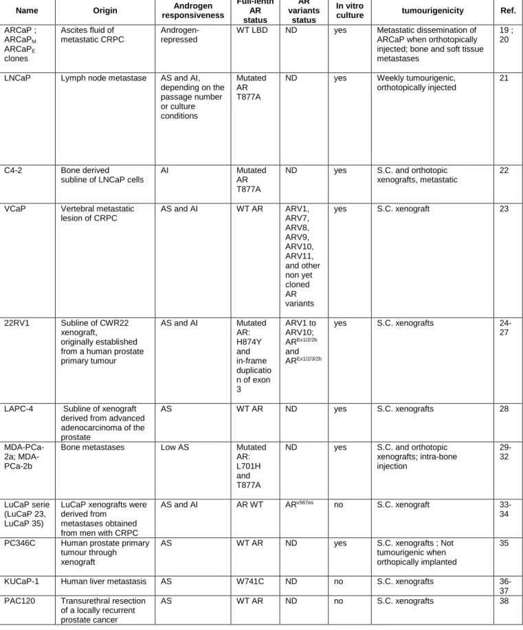
Taking into account the physiopathological functions of AR in prostate cancer, it seems crucial to choose the best appropriate model to develop new AR-targeting drugs. Data obtained solely from in vitro studies of human prostate cancer cell lines might not reliably predict the in vivo AR functions in prostate cancer progression and metastasis, in particular because several current available human prostate cancer cell lines were generated from long-term culture in the absence of androgen. That points up the importance of conducting in vivo animal studies of prostate tumours to delineate the pathologic functions of AR in prostate cancer progression and metastasis. However, the availability of a model that can be used both in vitro and in animals was clearly an advantage for studying AR-targeting drugs. It should be noted that some preclinical models derived from an original cell line could have been extensively manipulated, thus potentially introducing inaccurate genetic modifications. The most widely used human prostate cancer models that enable in vivo preclinical studies as xenografts to target AR signalling are shown in Table I. Note that many of them were propagated into xenografts and were not possible in vitro. As AR and AR variants are of central importance in the castration resistance progression, the appropriate models are still quite limited and the AR variant expression have not yet been identified in all of these models.
By analyzing a range of prostate models, the Charles Sawyers group has identified several structurally different AR variants with divergent biologic activity. All of those variants required full-length AR through dimerisation to activate endogenous AR target genes and confer castration-resistant growth (39). The new antiandrogen MDV3100 acting on the AR ligand-binding domain was shown to be active on CRPC patients in clinic (41-42). This compound abolishes the growth of LNCaP tumours expressing the AR-V7 variant, suggesting that MDV3100 could reverse the growth advantage of AR variants even if they were devoid in the ligand-binding domain and that AR variants require wild-type AR to confer gain of function through heterodimer formation (39). These results thus validate efforts to develop even better antiandrogens targeting the AR ligand-binding domain.
3. EXPERIMENTAL MODELS TO TARGET BONE METASTASES
As CRPC progresses, approximately 90% of the patients will develop bone metastases, and 20% will develop soft tissue metastases, most commonly in the lung, liver or lymph nodes. Currently, prostate cancer with bone metastases is not considered curable. The median survival of patients with bone metastases is approximately 20 months and is increasing with chemotherapy and other novel agents (43). Management of the quality of life in this patient group is therefore especially important and is likely to improve with the emergence of new bone-specific therapies. In patients with CRPC, bone metastases cause skeletal-related events that are responsible for substantial morbidity, and assessing bone health is an important aspect of clinical monitoring. PSA is the most widely used and best-characterized biomarker in CRPC; however, it does not provide accurate information regarding the extent of bone metastasis. In prostate cancer, bone metastases are predominantly osteoblastic; however, high bone turnover and consequent excess bone resorption are also characteristic features.
In normal bone, the remodelling process is balanced; however, the presence of tumour cells disrupts the process, causing bone lesions that are detectable by radiographic imaging or radionuclide bone scanning. Lesions result from an imbalance between osteoclast-mediated bone resorption and osteoblast-mediated bone formation. Osteoclasts adhere to the bone surface and resorb the bone matrix by secreting proteases and acid, and in turn, osteoblasts secrete collagen fibrils that become mineralised, eventually forming bone. However, cross talk between tumour cells and the bone microenvironment causes imbalance in the remodelling process, leading to a pathological “vicious cycle.” Specifically, tumour cells secrete factors that stimulate osteoclast-mediated bone destruction, and factors that were immobilized within the bone matrix are released, stimulating the growth of cancer cells and promoting further bone destruction. Once established in bone, the tumour cells interact with the bone microenvironment in a reciprocal fashion via cytokine mediators to form osteoblastic, osteolytic or mixed lytic/blastic lesions (44-45).
• Prostate cancer models to target bone metastases
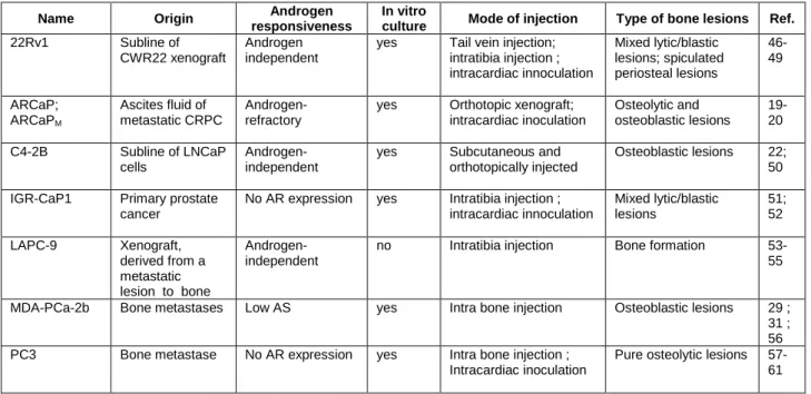
At present, the molecular mechanism underlying the bone tropism of prostate cancer metastasis is largely unknown. This gap in knowledge is due in part to difficulties in obtaining metastatic tissue from patients, as well as in generating mouse models that display bone metastasis. Experimental models for investigating bone metastases are limited to a small range of xenograft models that typically rely on the intracardiac injection of tumour cells. Prostate cancer cells can also be introduced into the mouse bone marrow through direct inoculation. While these models of human prostate cancer to mouse bone fail to reproduce the early steps of the invasion-metastatic cascade, they often can generate bone metastases at high frequency and have proven to be valuable for elucidating the later steps of this progression. Moreover, these tumours do closely resemble prostate tumours at the histopathologic level that are commonly encountered in the oncology clinic. A list of the models which can be used in preclinical studies targeting the bone is shown in Table II, with the nature of the bone lesions that are generated. In addition, the dynamic monitoring of the metastatic development with multimodality imaging technology such as bioluminescence, SPECT-CT and CT-scan provides a tremendous advantage for studying bone metastases in xenograft models. However, the availability of luciferase-expressing models to realise the follow up of bone metastasis development by BLI is still quite limited. To date there are no genetically modified mouse models that reliably generate bone metastases at an appreciable frequency.
For example, the significant decrease in tumour progression with focal adhesion kinase inhibitor PF562,271 was shown in both the PC3M-luc-C6 subcutaneous local implant and bone metastasis xenograft models, using bioluminescent imaging techniques (59). By using direct intra-tibia injection of PC3 cells, Yang et al (60) showed a reduction of tumour invasion in the bone matrix using the specific Src family kinase inhibitor saracatinib. The PC3-ML cell derivatives were injected directly into the blood system for studying the efficacy of the IMC-3G3 compound targeting PDGFRα on metastatic dissemination (61). With another preclinical model of intracardiac-injected 22Rv1 prostate cancer cells, the efficacy of Atrazentan, an endothelin receptor A antagonist, was evaluated in mixed osteoblastic/osteolytic bone lesions (49).
4. EXPERIMENTAL MODELS TO TARGET RESISTANCE TOWARDS MICROTUBULE-TARGETED CHEMOTHERAPY
• The use of microtubule-targeted chemotherapy in CRPC and resistance to
chemotherapy
Although only 5% of prostate cancers are diagnosed today with metastases, we know that more than half of high-risk localised prostate cancers will relapse with metastatic lesions. Since 2004, the standard of treatment is chemotherapy with Docetaxel when prostate cancer becomes
resistant to castration. Docetaxel has demonstrated its efficacy with a gain in survival demonstrated in two clinical trials, TAX 327 and SWOG 99-16 (62-63) and is now the treatment of choice in CRPC. Despite the survival benefit afforded by Docetaxel, almost half of patients treated with this agent develop resistance to chemotherapy either immediately or subsequently. Intrinsic and acquired resistances to anticancer agents are the first obstacles to effective cancer therapies. Patients who are refractory to treatment often exhibit multiple resistances to various anticancer agents that may have different structures and different functions. This clinical resistance, comparable to the experimental phenomenon called multidrug resistance (MDR), is probably multifactorial and heterogeneous, involving multiple molecular mechanisms. The mechanisms of this intrinsic or acquired resistance of tumour cells to drugs remain poorly understood and controversial. Efforts should therefore be made to better understand and overcome this serious problem.
• Cellular mechanism of resistance toward microtubule-targeted drugs
Microtubules have a diverse set of roles in cellular physiology, including providing structural integrity as part of the cytoskeleton, and a transport system for proteins, vesicles and organelles. They are critically important in the formation of the mitotic spindle, which dictates the proper segregation of chromosomes during mitosis, making them one of the best known targets in cancer therapy. Microtubules are formed by the polymerisation of monomers into linear structures. The combined assembly and disassembly processes yield a state of dynamic instability that allows the
growing or shortening of individual microtubules. Vinca-alkaloids bind free β-tubulin monomers,
preventing their incorporation into growing microtubules. In contrast, Taxanes and Epothilones bind to β-tubulin already incorporated into microtubules, preventing disassembly, stabilising the mitotic spindle and triggering apoptotic pathways.
Drug resistance in metastatic CRPC is multifactorial and complex. It includes cellular mechanisms of resistance to Taxanes, mechanisms related to the interactions between cancer cells and the surrounding microenvironment and mechanisms related to impaired drug delivery to cancer cells (64). There is also evidence for subpopulations of cancer cells with different proliferative capacity in prostate cancer as cancer stem cells which are considered to be rare multidrug-resistant cells, which repopulate the tumour during and after treatment and may be responsible for treatment failure. At the cellular level, the mechanisms of resistance to Docetaxel and other tubulin-binding chemotherapeutic agents are multifactorial and have been studied in resistant-cell lines and in clinical samples. The mechanisms related to the tubulin and the microtubule systems that mediate resistance to anti-mitotics are complex but they are beginning to be unravelled (65). Overall, Taxanes can be exported outside the cell by the ABC transporters (including ABCB1/MDR1), thus minimising their intracellular concentration and their effect; the expression of tubulin isoforms and microtubule composition may promote the expression of
isoforms that are less sensitive to taxanes; mutations of β-tubulin isoforms may be acquired,
converting cells into taxane-resistant cells; alterations at mitotic cycle checkpoints may minimise the impact of taxanes on microtubule stabilisation by preventing mitotic catastrophe and apoptotic cell death (65). However, unlike data obtained in breast cancer, the general mechanisms of resistance to Taxanes have not convincingly been demonstrated in prostate cancer. Finally, despite published results on biomarker candidates obtained in small series of tumours, there is still no biomarker predictive of response to Docetaxel for clinical use in prostate cancer. In an effort to identify predictive biomarkers, a standard definition of taxane refractory disease is needed that should enable the possibility of correlating biomarker expression and clinical response to microtubule-targeted chemotherapies.
• Available models of resistance to microtubule-targeted chemotherapy
Despite the increasing prevalence of taxane-resistant prostate cancer, there are limited studies in this setting, and very few models of microtubule-targeted chemotherapy resistant prostate cancers are available. Drug-resistant cell lines were generated by exposing the parental
cell line to increasing drug concentrations for a long period. Acquired in vitro chemoresistance takes a relatively long time depending of the cell line and must be maintained during successive passages of the drug. Several laboratories have obtained Paclitaxel or Docetaxel-resistant PC3, DU145 and LNCaP derivatives (66-71). In some studies cross-resistance has been assessed (66). The molecular mechanism conferring resistance to docetaxel in vitro was shown to be cell-specific (71). Although several drug-resistant cell lines can be generated in vitro, the establishment of a drug-resistant model in animals seems very tricky. Docetaxel-resistant PC3 cells were grown in xenografts to study the targeting of clusterin in a docetaxel-refractory prostate cancer preclinical model (72). With another strategy, the efficacy of the combination of estramustine with docetaxel and the appearance of docetaxel resistance was evaluated in-vivo in the PAC120 androgen-dependent human xenograft model (73).
5. EXPERIMENTAL MODELS TO TARGET PROSTATE CANCER STEM CELLS
• The theory of cancer stem cells
According to the cancer stem cell hypothesis, tumour cells are heterogeneous, and only a subset of cells, known as cancer stem cells (CSCs) are responsible for both self-renewing, giving rise to differentiated cells and producing the remaining cellular heterogeneity represented in the tumour (74). The CSCs exhibit higher resistance to cytotoxic drugs and radiation compared to bulk tumour cells. The survival of few CSCs would result in subsequent tumour regrowth and disease relapse. The concept of cancer as a stem cell disease has the potential to change our view of its treatment. Most therapies are directed at the fast-growing tumour mass but not the slow-dividing cancer stem cells, which may thus explain the frequent ineffectiveness of these therapeutics and suggest that effective therapeutics should target rare CSCs that sustain tumour malignancy. Research is now focusing on understanding the growth of CSCs as well as identifying CSCs antigens in order to discover new targeted approaches. The cancer stem cell hypothesis does not contradict the established clonal evolution view of cancer, but instead suggests that clonal progression of cancer could operate through the stem cell compartment and highlights the importance of an aberrant differentiation programme in tumourigenesis.
• Application of the CSC hypothesis in prostate cancer
Evidence for the existence of a stem cell subpopulation in the prostate has been accumulating since the 1980s. There has also been an increasing interest in the role of CSCs in prostate cancer development and progression. However, the cellular origin of CSCs is still controversial. The prostate is comprised of several types of epithelial cells that may serve as targets for cancer initiation, including basal, luminal, and neuroendocrine cells. Normal human prostate stem cells are thought to reside in the basal cell layer of the ducts (75), and several studies have utilised flow cytometry-based approaches to isolate candidate prostate epithelial stem cells directly from primary human tissue and have shown that these stem cells are basal (76). However, a lineage marking methodology has recently been used to identify a rare luminal epithelial population with stem cell properties during prostate regeneration (77).
Prostate cancers have been assumed to originate from luminal cells as prostate cancer is characterised by an expansion of epithelial luminal cells and an absence of epithelial basal cells. The available data on the identity and characterisation of CSCs in localized and advanced prostate cancer were limited. The putative stem cell marker CD133 was shown to isolated prostate cancer cells with stem-cell-like properties in vitro (78). Although contested, some studies suggest that normal and prostate cancer stem cells may not express the androgen receptor, implying that prostate cancers may become castration resistant through the survival and expansion of cancer-initiating cells that lack the functional androgen receptor. This theory was confirmed very recently by work performed in a novel human prostate cancer model, showing that only AR-negative basal cells can propagate a tumour which reproduces the histological and molecular features of prostate adenocarcinomas (79).
Future studies are necessary to determine if CSCs are responsible for tumourigenic activity and can be specifically targeted. The goal of existing therapies, such as androgen ablation, has been to eradicate the bulk of cells within a tumour. However by targeting the AR-positive population, resistance occurs in most patients. Mechanisms such as AR amplification do occur, but the resultant tumour may well arise from a more primitive AR-negative clone. We should therefore aim to develop therapeutics that can selectively target CSCs, rather than more differentiated cancer cells. Studying stem cells and their properties will hopefully lead both to a better understanding of cell survival and proliferation in androgen-depleted conditions and to the development of new targets for advanced disease. It is expected that CSCs will be a main target for cancer therapy in the near future.
• Available PCSC models
The gold standard assay to demonstrate this concept involves xenotransplantation of flow-sorted populations of primary cancer cells into immunodeficient mice. In this assay, CSCs are defined as a subpopulation of cells within a primary tumour that can initiate tumour formation in mice following transplantation, unlike the remaining tumour cells. Whereas patient samples are the gold standard, they are difficult to obtain regularly. Flow-sorted cells obtained from stable cancer cell lines constitute an alternative and are simple to use but have been selected in a long-term culture. Ectopic xenografts are however limited in analysing CSCs from solid tumours because of the loss of the human stromal microenvironment, which might provide a niche for normal stem cells or for CSCs. Orthotopic models might more faithfully maintain the tumour–host interaction, giving CSCs a greater chance to interact with and obtain stimulation from a mouse environment. The use of more highly immunodeficient mice, orthotopic xenotransplantation and tissue reconstitution with prostate stromal cells may all improve the efficiency and specificity of tumour-initiation assays. The study of CSCs may best be evaluated in xenografts of primary patient isolates that maintain the heterogeneity and architecture of the original patient tumour, but no prostate CSCs have been identified until now using this assay. Avoiding the transplant altogether by generating GEM models of tumours may better maintain the tumour–host and CSC–niche interactions. However, due to the practical difficulty of modelling the multitude of genetic changes in a tumour, mouse prostate cancer models have not been extensively evaluated to date (80).
Cancer stem cell populations are commonly defined by the presence of various combinations of cell-surface proteins, named CSC-markers, and are easily identified and isolated by flow cytometry and/or fluorescence-activated cell sorting (FACS). Isolated CSCs have shown their ability to form spheres in soft agar culture conditions and are more resistant in vivo and in vitro to common chemotherapeutic drugs due to high expression of drug efflux transporters. Another feature of CSCs is the activation of gene and pathways that are commonly involved in stem cell properties such as the Wnt, Hedgehog and Notch pathways. Thus, prostate cancer cell lines exhibiting these stem-like properties provide cellular models to study the CSC pathways and can be used in xenograft transplantations (Table III). However, it should be kept in mind that, for some of these markers, their specificity for identifying CSC populations has been questioned in some reports, and their biological function is still unknown (80;84).
In this field, PC3 and DU145 xenografts were used to assess the activity of a combination therapy targeting both CSCs and differentiated cell propulations in prostate carcinoma (82). It was shown that treatment of prostate tumour xenografts with a combination of the standard chemotherapy (docetaxel) with the NVP-BEZ235 compound targeting CD133+/CD44+ progenitors leads to near complete tumour regression compared to single agent treatments. Thus this study suggests that combination strategies based on bulk tumour reduction and CSC-specific pathway inhibition offer a promising treatment modality. Interestingly, acquired mutations in PTEN appeared selectively in the CD133+/CD44+ cell subset of tumour relapses suggesting that acquired PTEN mutations can serve as a mechanism of drug resistance in CSC (82).
6. EXPERIMENTAL MODELS TO INVESTIGATE THE EFFECT OF RADIOTHERAPY
The aim of curative radiotherapy is to sterilise all clonogenic tumour cells which represent a small subpopulation of all malignant cells in a tumour and may lead to recurrence if they survive following therapy. Whether a tumour can be cured by ionising radiation depends to a large extent on the number and radiation sensitivity of clonogenic tumour cells. The most important treatment parameters that affect local control are the total radiation dose and the toxicity of surrounding normal tissue. However, other factors contribute to the radiotherapy response: cells with the capacity to expand and to form a recurrence, the cancer stem cells, have to be killed. It thus seems important to understand the underlying molecular mechanisms of radiation resistance that give the ability to repopulate to the clonogenic tumour cells, to reoxygenate and to recover from sublethal damage during fractionated irradiation. This should lead to the identification of molecules driving these processes that can constitute new therapeutic targets. It has recently been suggested that CSCs might be more radioresistant than non-tumourigenic cells (93). This issue is of major importance because biomarkers that are under development for predicting the outcome of radiotherapy, including bio-imaging assays, measure the bulk of non-stem cells and not the subpopulation of cancer stem cells.
• Combining molecular-targeted agents with irradiation in prostate cancer preclinical
models
Combining molecular-targeted agents with irradiation is a promising strategy for patient care. The combinations can be easily studied in preclinical prostate cancer models. In fact, these molecular targets are often differentially expressed in tumours and normal tissue, offering the option of therapeutic gain and they are expressed in only a few tumour stem cells. Endpoints which reflect the killing of CSCs for experiments combining radiotherapy with molecular-targeted drugs using in vivo tumour models have been reviewed (94).
The evaluation of irradiation was often done on ectopic xenografts. The combination of radiation with the receptor tyrosine kinase inhibitor AG-013736 was assessed in DU145 prostate cancer cells implanted in the left hind leg of nude mice (95). Multimodality imaging was used in
subcutaneous xenografts from luciferase-expressing PC3 cells for the evaluation of 177Lu-AMBA
radionuclide-based therapy (96). As the use of an orthotopic model is essential in prostate cancer because of tumour–stromal interactions, which are particularly important to consider when treatments in combination with androgen deprivation, some studies have evaluated the combination of androgen deprivation and pelvic radiation on intraprostatic xenograft models. The combination of high doses of bicalutamide and radiation was evaluated in orthotopic xenografts from MDA-PCa-2b cells (32), and the effects of antisense-MDM2 on androgen deprivation in combination with radiation was investigated in orthotopic xenografts LNCaP cells (97). In such models tumour response was assessed by magnetic resonance imaging-based tumour volume or human PSA measurements. In order to address the interaction of anti-tumour immune therapy and radiotherapy, the autochthonous murine TRAMP model of prostate cancer has been used to test whether radiotherapy could modify the CD4 T-cell response to a tumour vaccine (98).
7. GENERAL CONCLUSION
The development of better cancer therapeutics continues, and it has become increasingly evident that selection of the appropriate mouse cancer model is essential during the drug discovery process when interrogating novel biological processes in tumourigenesis. Facing continued criticism regarding the predictive value of mouse models, the in vivo cancer pharmacologist is challenged to develop models that best enable the study of a specific scientific question. A growing number of research programs are now yielding compounds with demonstrated anti-tumour activity in vitro and possible efficacy as in vivo agents. The confirmation of therapeutic efficacy therefore requires trials in living, tumour-bearing animals to demonstrate selective anti-tumour activity while remaining safe to non-tumour tissues. Animal models are essential in developing early in vivo
biomarkers predictive of therapeutic response of pathway-specific drugs, a field that will see rapid expansion as the costs of undertaking full scale clinical trials escalate. There is a need for improved animal models of human cancer. As our understanding of human disease becomes more comprehensive, the need for more accurate and sophisticated models increases. Experimental therapeutics that target a particular signalling pathway or tumour-specific molecules must be validated in well characterised and suited models that enable the testing of the experimental hypothesis. Sophisticated tumour imaging methodologies are now available to facilitate analysis of anti-tumour efficacy and drug development as bioimaging, use of fluorescent-specific probes, CT- scan as well as technologies such as F-18-labeled compounds followed by PET imaging to selectively label and detect tumour cells and drug biodistribution.
By establishing models that more closely reproduce prostate tumours, and by asking more specific questions with such models, animal models will continue to play an important role in validating cancer targets and evaluating putative cancer therapies. In addition to measuring anti-tumour efficacy, in vivo models provide a valuable opportunity to assess pharmacokinetics, pharmacodynamics, drug-drug interactions and preclinical toxicity, as well as initial development of in vivo biomarkers as predictors for anti-tumour efficacy. The most important thing is to use the right models to ask the right questions about the right therapies.
CONFLICT OF INTEREST STATEMENT
The new IGR-CaP1 cell line was the subject of an International patent pending entitled “Prostate
cancer cell lines and their use in screening method” deposed on the 14th April 2009. Biological
material has been deposited at the Pasteur Institut (Paris) (CNCM I-4126). The patented material will be available under a Material Transfer Agreement for research use.
REFERENCES
1. Lopez-Barcons LA. Serially heterotransplanted human prostate tumours as an experimental model. J Cell Mol Med 2010, 14, 1385-1395.
2. Schmidt LJ, Regan KM, Anderson SK, Sun Z, Ballman KV, Tindall DJ. Effects of the 5 alpha-reductase inhibitor dutasteride on gene expression in prostate cancer xenografts. Prostate 2009, 69, 1730-1743.
3. Khan N, Asim M, Afaq F, Abu Zaid M, Mukhtar H. A novel dietary flavonoid fisetin inhibits androgen receptor signaling and tumor growth in athymic nude mice. Cancer Res 2008, 68, 8555-8563.
4. Staack A, Kassis AP, Olshen A, et al. Quantitation of apoptotic activity following castration in human prostatic tissue in vivo. Prostate 2003, 54, 212-219.
5. Lee CH, Xue H, Sutcliffe M, et al. Establishment of subrenal capsule xenografts of primary human ovarian tumors in SCID mice: potential models. Gynecol Oncol 2005, 96, 48-55. 6. Wang Y, Revelo MP, Sudilovsky D, et al. Development and characterization of efficient xenograft models for benign and malignant human prostate tissue. Prostate 2005, 64,149-159.
7. Zhao H, Nolley R, Chen Z, Peehl DM. Tissue slice grafts: an in vivo model of human prostate androgen signaling. Am J Pathol 2010, 177, 229-239. 8. Kasper S, Smith JA Jr. Genetically modified mice and their use in developing therapeutic strategies for prostate cancer. J Urol 2004, 172, 12-19. 9. Jeet V, Russell PJ, Khatri A. Modeling prostate cancer: a perspective on transgenic mouse models. Cancer Metastasis Rev 2010, 29, 123-142. 10. Shen MM, Abate-Shen C. Molecular genetics of prostate cancer: new prospects for old challenges. Genes Dev 2010, 24, 1967-2000.
11. Wang S, Gao J, Lei Q, et al. Prostate-specific deletion of the murine Pten tumor suppressor gene leads to metastatic prostate cancer. Cancer Cell 2003, 4, 209-221.
12. Kim MJ, Cardiff RD, Desai N, et al. Cooperativity of Nkx3.1 and Pten loss of function in a mouse model of prostate carcinogenesis. Proc Natl Acad Sci U S A 2002, 99, 2884-2889.
13. Abate-Shen C, Banach-Petrosky WA, Sun X, et al. Nkx3.1; Pten mutant mice develop invasive prostate adenocarcinoma and lymph node metastases. Cancer Res 2003, 63, 3886-3890.
14. Zhou Z, Flesken-Nikitin A, Corney DC, et al. Synergy of p53 and Rb deficiency in a conditional mouse model for metastatic prostate cancer. Cancer Res 2006, 66, 7889-7898. 15. Huggins C, Stevens RE Jr, Hodges CV. Studies on prostate cancer. II. The effect of castration on advanced carcinoma of the prostate gland. Arch Surg 1941, 43, 209-223. 16. Chen CD, Welsbie DS, Tran C, et al. Molecular determinants of resistance to antiandrogen therapy. Nat Med 2004, 10, 33-39.
17. Scher HI, Sawyers CL. Biology of progressive, castration-resistant prostate cancer: directed therapies targeting the androgen-receptor signaling axis. J Clin Oncol 2005, 23, 8253-8261.
18. Guo Z, Yang X, Sun F, et al. A novel androgen receptor splice variant is up-regulated during prostate cancer progression and promotes androgen depletion-resistant growth. Cancer Res 2009, 69, 2305-2313.
19. Zhau HY, Chang SM, Chen BQ, et al. Androgen-repressed phenotype in human prostate cancer. Proc Natl Acad Sci U S 1996, 936,15152-15157.
20. Xu J, Wang R, Xie ZH, et al. Prostate cancer metastasis: role of the host microenvironment in promoting epithelial to mesenchymal transition and increased bone and adrenal gland metastasis. Prostate 2006, 66, 1664-1673.
21. Horoszewicz JS, Leong SS, Kawinski E, et al. LNCaP model of human prostatic carcinoma. Cancer Res 1983, 43, 1809-1818.
22. Thalmann GN, Anezinis PE, Chang SM, et al. Androgen-independent cancer progression and bone metastasis in the LNCaP model of human prostate cancer. Cancer Res 1994, 54, 2577-2581.
23. Korenchuk S, Lehr JE, MClean L, et al. VCaP, a cell-based model system of human prostate cancer. In Vivo 2001, 15,163-168.
24. Chlenski A, Nakashiro K, Ketels KV, Korovaitseva GI, Oyasu R. Androgen receptor expression in androgen-independent prostate cancer cell lines. Prostate 2001, 47, 66-75. 25. Tepper CG, Boucher DL, Ryan PE, et al. Characterization of a novel androgen receptor mutation in a relapsed CWR22 prostate cancer xenograft and cell line. Cancer Res 2002, 62, 6606-6614.
26. Dehm SM, Schmidt LJ, Heemers HV, Vessella RL, Tindall DJ. Splicing of a novel androgen receptor exon generates a constitutively active androgen receptor that mediates prostate cancer therapy resistance. Cancer Res 2008, 68, 5469-5477.
27. Marcias G, Erdmann E, Lapouge G, et al. Identification of novel truncated androgen receptor (AR) mutants including unreported pre-mRNA splicing variants in the 22Rv1 hormone-refractory prostate cancer (PCa) cell line. Hum Mutat 2010, 31, 74-80.
28. Klein KA, Reiter RE, Redula J, et al. Progression of metastatic human prostate cancer to androgen independence in immunodeficient SCID mice. Nat Med 1997, 3, 402-408. 29. Navone NM, Olive M, Ozen M, et al. Establishment of two human prostate cancer cell lines derived from a single bone metastasis. Clin Cancer Res 1997, 3, 2493-2500. 30. Zhao XY, Peehl DM, Navone NM, Feldman D. 1alpha,25-dihydroxyvitamin D3 inhibits prostate cancer cell growth by androgen-dependent and androgen-independent mechanisms. Endocrinology 2000, 141, 2548-2556.
31. Fizazi K, Sikes CR, Kim J, et al. High efficacy of docetaxel with and without androgen deprivation and estramustine in preclinical models of advanced prostate cancer. Anticancer Res 2004, 24, 2897-2903.
32. Le Moulec S, de Crevoisier R, Huard J, et al. Concomitant administration of bicalutamide with radiotherapy leads to an important decrease of prostate tumor growth in mice orthotopic xenografts. ASCO Prostate Symposium, Orlando 2007, p78
33. Ellis WJ, Vessella RL, Buhler KR, et al. Characterization of a novel androgen-sensitive, prostate-specific antigen-producing prostatic carcinoma xenograft: LuCaP 23. Clin Cancer Res 1996, 2,1039-1048.
34. Corey E, Quinn JE, Buhler KR, Nelson PS, Macoska JA, True LD, Vessella RL. LuCaP 35: a new model of prostate cancer progression to androgen independence. Prostate 2003, 55, 239-246.
35. van Weerden W., Romijn JC. Use of nude mouse xenograft models in prostate cancer research. Prostate 2000, 43, 263-271.
36. Yoshida T, Kinoshita H, Segawa T, et al. Antiandrogen bicalutamide promotes tumor growth in a novel androgen-dependent prostate cancer xenograft model derived from a bicalutamide-treated patient. Cancer Res 2005, 65, 9611-9616.
37. Terada N, Shimizu Y, Yoshida T, et al. Antiandrogen withdrawal syndrome and alternative antiandrogen therapy associated with the W741C mutant androgen receptor in a novel prostate cancer xenograft. Prostate 2010, 70, 252-261.
38. de Pinieux G, Legrier ME, Poirson-Bichat F, et al. Clinical and experimental progression of a new model of human prostate cancer and therapeutic approach. Am J Pathol 2001, 159, 753-764.
39. Watson PA, Chen YF, Balbas MD, et al. Constitutively active androgen receptor splice variants expressed in castration-resistant prostate cancer require full-length androgen receptor. Proc Natl Acad Sci U S A 2010, 107, 16759-16765.
40. Sun S, Sprenger CC, Vessella RL, et al. Castration resistance in human prostate cancer is conferred by a frequently occurring androgen receptor splice variant. J Clin Invest 2010, 120, 2715-2730.
42. Scher HI, Beer TM, Higano CS, et al. Antitumour activity of MDV3100 in castration-resistant prostate cancer: a phase 1-2 study. Lancet 2010, 375, 1437-1446. 43. Moore CN, George DJ. Update in the management of patients with hormone-refractory prostate cancer. Curr Opin Urol 2005, 15,157-162.
44. Vessella RL, Corey E. Targeting factors involved in bone remodeling as treatment strategies in prostate cancer bone metastasis. Clin Cancer Res 2006, 12, 6285s-6290s. 45. Kingsley LA, Fournier PG, Chirgwin JM, Guise TA. Molecular biology of bone metastasis. Mol Cancer Ther 2007, 6, 2609-2617.
46. Holleran JL, Miller CJ, Edgehouse NL, Pretlow TP, Culp LA. Differential experimental micrometastasis to lung, liver, and bone with lacZ-tagged CWR22R prostate carcinoma cells. Clin Exp Metastasis 2002,19,17-24.
47. Drake JM, Gabriel CL, Henry MD. Assessing tumor growth and distribution in a model of prostate cancer metastasis using bioluminescence imaging. Clin Exp Metastasis 2005, 22, 674-684.
48. Henry MD, Silva MD, Wen S, et al. Spiculated periosteal response induced by intraosseous injection of 22Rv1 prostate cancer cells resembles subset of bone metastases in prostate cancer patients. Prostate 2005, 65, 347-354.
49. Drake JM, Danke JR, Henry MD. Bone-specific growth inhibition of prostate cancer metastasis by atrasentan. Cancer Biol Ther 2010, 9, 607-614.
50. Wu TT, Sikes RA, Cui Q, et al. Establishing human prostate cancer cell xenografts in bone: induction of osteoblastic reaction by prostate-specific antigen-producing tumors in athymic and SCID/bg mice using LNCaP and lineage-derived metastatic sublines. Int J Cancer 1998, 77, 887-894.
51. Chauchereau A, Al Nakouzi N, Gaudin C, et al. Stemness markers characterize IGR-CaP1, a new cell line derived from primary epithelial prostate cancer. Exp Cell Res 2011, 317, 262-275.
52. Al Nakouzi N, Bawa O, Opolon P, et al. A novel preclinical model of prostate cancer bone metastasis derived from a human primary tumor. AACR meeting, Orlando 2011, p1597. 53. Craft N, Chhor C, Tran C, et al. Evidence for clonal outgrowth of androgen-independent prostate cancer cells from androgen-dependent tumors through a two-step process. Cancer Res 1999, 59, 5030-5036.
54. Corey E, Quinn JE, Bladou F, et al. Establishment and characterization of osseous prostate cancer models: intra-tibial injection of human prostate cancer cells. Prostate 2002, 52, 20-33.
55. Lee Y, Schwarz E, Davies M, et al. Differences in the cytokine profiles associated with prostate cancer cell induced osteoblastic and osteolytic lesions in bone. J Orthop Res 2003, 21, 62-72.
56. Yang J, Fizazi K, Peleg S, et al. Prostate cancer cells induce osteoblast differentiation through a Cbfa1-dependent pathway. Cancer Res 2001, 61, 5652-5659.
57. Kaighn ME, Narayan KS, Ohnuki Y, Lechner JF, Jones LW. Establishment and characterization of a human prostatic carcinoma cell line (PC-3). Invest Urol 1979, 17,16-23. 58. Nemeth JA, Yousif R, Herzog M, et al. Matrix metalloproteinase activity, bone matrix turnover, and tumor cell proliferation in prostate cancer bone metastasis. J Natl Cancer Inst 2002, 94, 17-25.
59. Sun H, Pisle S, Gardner ER, Figg WD. Bioluminescent imaging study: FAK inhibitor, PF-562,271, preclinical study in PC3M-luc-C6 local implant and metastasis xenograft models. Cancer Biol Ther 2010, 10, 38-43
60. Yang JC, Bai L, Yap S, Gao AC, Kung HJ, Evans CP. Effect of the specific Src family kinase inhibitor saracatinib on osteolytic lesions using the PC-3 bone model. Mol Cancer Ther 2010, 9,1629-1637.
61. Russell MR, Liu Q, Fatatis A. Targeting the {alpha} receptor for platelet-derived growth factor as a primary or combination therapy in a preclinical model of prostate cancer skeletal metastasis. Clin Cancer Res 2010,16, 5002-5010.
62. Petrylak DP, Tangen CM, Hussain MH, et al. Docetaxel and estramustine compared with mitoxantrone and prednisone for advanced refractory prostate cancer. N Engl J Med. 2004, 351,1513-1520.
63. Tannock IF, de Wit R, Berry WR, et al. Docetaxel plus prednisone or mitoxantrone plus prednisone for advanced prostate cancer. N Engl J Med 2004, 351,1502-1512. 64. Seruga B, Ocana A, Tannock IF. Drug resistance in metastatic castration-resistant prostate cancer. Nat Rev Clin Oncol 2011, 8, 12-23.
65. Kavallaris M. Microtubules and resistance to tubulin-binding agents. Nat Rev Cancer 2010, 10,194-204.
66. Takeda M, Mizokami A, Mamiya K, et al. The establishment of two paclitaxel-resistant prostate cancer cell lines and the mechanisms of paclitaxel resistance with two cell lines. Prostate 2007, 67, 955-967.
67. Makarovskiy AN, Siryaporn E, Hixson DC, Akerley W. Survival of docetaxel-resistant prostate cancer cells in vitro depends on phenotype alterations and continuity of drug exposure. Cell Mol Life Sci 2002, 59,1198-1211.
68. Patterson SG, Wei S, Chen X, et al. Novel role of Stat1 in the development of docetaxel resistance in prostate tumor cells. Oncogene 2006, 25, 6113-6122. 69. Zhao L, Lee BY, Brown DA, et al. Identification of candidate biomarkers of therapeutic response to docetaxel by proteomic profiling. Cancer Res 2009, 69, 7696-7703. 70. Wiltshire C, Singh BL, Stockley J, et al. Docetaxel-resistant prostate cancer cells remain sensitive to S-trityl-L-cysteine-mediated Eg5 inhibition. Mol Cancer Ther 2010, 9, 1730-1739.
71. Desarnaud F, Geck P, Parkin C, Carpinito G, Makarovskiy AN. Gene expression profiling of the androgen independent prostate cancer cells demonstrates complex mechanisms mediating resistance to docetaxel. Cancer Biol Ther 2011, 11, 204-212.
72. Sowery RD, Hadaschik BA, So AI, et al. Clusterin knockdown using the antisense oligonucleotide OGX-011 re-sensitizes docetaxel-refractory prostate cancer PC-3 cells to chemotherapy. BJU Int 2008, 102, 389-397.
73. Dahmani A, de Plater L, Guyader C, et al. A preclinical therapeutic schedule optimizing docetaxel plus estramustine administration in prostate cancer. Anticancer Drugs 2010, 21, 927-931.
74. Reya T, Morrison SJ, Clarke MF, Weissman IL. Stem cells, cancer, and cancer stem cells. Nature 2001, 414, 105-111.
75. English HF, Santen RJ, Isaacs JT. Response of glandular versus basal rat ventral prostatic epithelial cells to androgen withdrawal and replacement. Prostate 1987, 11, 229–242. 76. Goldstein AS, Stoyanova T, Witte ON. Primitive origins of prostate cancer: in vivo evidence for prostate-regenerating cells and prostate cancer-initiatingcells. Mol Oncol 2010 , 4, 385-396.
77. Wang X, Kruithof-de Julio M, Economides KD, et al. A luminal epithelial stem cell that is a cell of origin for prostate cancer. Nature 2009, 461, 495-500. 78. Collins AT, Berry PA, Hyde C, Stower MJ, Maitland NJ. Prospective identification of tumorigenic prostate cancer stem cells. Cancer Res 2005, 65, 10946-10951. 79. Goldstein AS, Huang J, Guo C, Garraway IP, Witte ON. Identification of a cell of origin for human prostate cancer. Science 2010, 329, 568-571.
80. Wang ZA, Shen MM. Revisiting the concept of cancer stem cells in prostate cancer. Oncogene 2011, 30, 1261-1271.
81. Dubrovska A, Kim S, Salamone RJ, et al. The role of PTEN/Akt/PI3K signaling in the maintenance and viability of prostate cancer stem-like cell populations. Proc Natl Acad Sci USA 2009, 106, 268–273.
82. Dubrovska A, Elliott J, Salamone RJ, et al. Combination therapy targeting both tumor-initiating and differentiated cell populations in prostate carcinoma. Clin Cancer Res 2010, 16, 5692-5702.
83. Li H, Chen X, Calhoun-Davis T, Claypool K, Tang DG. PC3 human prostate carcinoma cell holoclones contain self-renewing tumor-initiating cells. Cancer Res 2008, 68, 1820-1825.
84. Pfeiffer MJ, Schalken JA. Stem cell characteristics in prostate cancer cell lines. Eur Urol 2010, 57, 246-254.
85. Stone KR, Mickey DD, Wunderli H, Mickey GH, Paulson DF. Isolation of a human prostate carcinoma cell line (DU 145). Int J Cancer 1978, 21, 274-281.
86. Wei C, Guomin W, Yujun L, Ruizhe Q. Cancer stem-like cells in human prostate carcinoma cells DU145, the seeds of the cell line? Cancer Biol Ther 2007, 6, 763-768. 87. Kallifatidis G, Labsch S, Rausch V, et al. Sulforaphane Increases Drug-mediated Cytotoxicity Toward Cancer Stem-like Cells of Pancreas and Prostate. Mol Ther 2011, 19, 188-195.
88. Rybak AP, He L, Kapoor A, Cutz JC, Tang D. Characterization of sphere-propagating cells with stem-like properties from DU145 prostate cancer cells. Biochim Biophys Acta 2011, 1813, 683-694.
89. Hurt EM, Kawasaki BT, Klarmann GJ, Thomas SB, Farrar WL. CD44+ CD24(-) prostate cells are early cancer progenitor/stem cells that provide a model for patients with poor prognosis. Br J Cancer 2008, 98, 756-765.
90. Liu T, Xu F, Du X, et al. Establishment and characterization of multi-drug resistant, prostate carcinoma-initiating stem-like cells from human prostate cancer cell lines 22RV1. Mol Cell Biochem 2010, 340, 265-273.
91. Lam JS, Yamashiro J, Shintaku IP, et al. Prostate stem cell antigen is overexpressed in prostate cancer metastases. Clin Cancer Res 2005, 11, 2591-2596.
92. Patrawala L, Calhoun-Davis T, Schneider-Broussard R, Tang DG. Hierarchical organization of prostate cancer cells in xenograft tumors: the CD44+alpha2beta1+ cell population is enriched in tumor-initiating cells. Cancer Res 2007, 67, 6796-6805.
93. Rich JN. Cancer stem cells in radiation resistance. Cancer Res 2007, 67, 8980-8984.
94. Krause M, Zips D, Thames HD, Kummermehr J, Baumann M. Preclinical evaluation of molecular-targeted anticancer agents for radiotherapy. Radiother Oncol 2006, 80,112-122. 95. Fenton BM, Paoni SF. The addition of AG-013736 to fractionated radiation improves tumor response without functionally normalizing the tumor vasculature. Cancer Res 2007, 67, 9921-9928.
96. Liu IH, Chang CH, Ho CL, et al. Multimodality imaging and preclinical evaluation of 177Lu-AMBA for human prostate tumours in a murine model. Anticancer Res 2010, 30, 4039-4048
97. Stoyanova R, Hachem P, Hensley H, et al. Antisense-MDM2 sensitizes LNCaP prostate cancer cells to androgen deprivation, radiation, and the combination in vivo. Int J Radiat Oncol Biol Phys 2007, 68, 1151-1160.
Name Origin Androgen responsiveness Full-lenth AR status AR variants status In vitro
culture tumourigenicity Ref.
ARCaP ; ARCaPM ARCaPE clones Ascites fluid of metastatic CRPC Androgen-repressed
WT LBD ND yes Metastatic dissemination of ARCaP when orthotopically injected; bone and soft tissue metastases
19 ; 20
LNCaP Lymph node metastase AS and AI, depending on the passage number or culture conditions Mutated AR T877A
ND yes Weekly tumourigenic, orthotopically injected
21
C4-2 Bone derived subline of LNCaP cells
AI Mutated AR T877A
ND yes S.C. and orthotopic xenografts, metastatic
22
VCaP Vertebral metastatic lesion of CRPC AS and AI WT AR ARV1, ARV7, ARV8, ARV9, ARV10, ARV11, and other non yet cloned AR variants yes S.C. xenograft 23 22RV1 Subline of CWR22 xenograft, originally established from a human prostate primary tumour AS and AI Mutated AR: H874Y and in-frame duplicatio n of exon 3 ARV1 to ARV10; AREx1/2/2b and AREx1/2/3/2b yes S.C. xenografts 24-27
LAPC-4 Subline of xenograft derived from advanced adenocarcinoma of the prostate AS WT AR ND yes S.C. xenografts 28 PCa-2a; MDA-PCa-2b
Bone metastases Low AS Mutated AR: L701H and T877A
ND yes S.C. and orthotopic xenografts; intra-bone injection 29-32 LuCaP serie (LuCaP 23, LuCaP 35)
LuCaP xenografts were derived from
metastases obtained from men with CRPC
AS and AI AR WT ARv567es no S.C. xenograft
33-34
PC346C Human prostate primary tumour through xenograft
AS WT AR ND yes S.C. xenografts ; Not tumourigenic when orthopically implanted
35
KUCaP-1 Human liver metastasis AS W741C ND no S.C. xenografts 36-37 PAC120 Transurethral resection
of a locally recurrent prostate cancer
AS WT AR ND no S.C. xenografts 38
Table I: Commonly used human AR expressing prostate cancer preclinical models
AR: androgen receptor expression; LBD: ligand-binding-domain; AS: androgen sensitive; AI: androgen independent; ND: not determined; S.C.: subcutaneous; WT: Wild-type.
Name Origin Androgen responsiveness
In vitro
culture Mode of injection Type of bone lesions Ref.
22Rv1 Subline of CWR22 xenograft
Androgen independent
yes Tail vein injection; intratibia injection ; intracardiac innoculation Mixed lytic/blastic lesions; spiculated periosteal lesions 46-49 ARCaP; ARCaPM Ascites fluid of metastatic CRPC Androgen-refractory
yes Orthotopic xenograft; intracardiac inoculation Osteolytic and osteoblastic lesions 19-20 C4-2B Subline of LNCaP cells Androgen-independent
yes Subcutaneous and orthotopically injected
Osteoblastic lesions 22; 50 IGR-CaP1 Primary prostate
cancer
No AR expression yes Intratibia injection ; intracardiac innoculation Mixed lytic/blastic lesions 51; 52 LAPC-9 Xenograft, derived from a metastatic lesion to bone Androgen-independent
no Intratibia injection Bone formation 53-55
MDA-PCa-2b Bone metastases Low AS yes Intra bone injection Osteoblastic lesions 29 ; 31 ; 56 PC3 Bone metastase No AR expression yes Intra bone injection ;
Intracardiac inoculation
Pure osteolytic lesions 57-61
Name Origin responsiveness Androgen CSC markers expression Graft sites expansion Ex vivo Ref. PC3 Bone metastase AR- PSCA; CD44+; α2β1-integrinhigh S.C. and prostate, direct intracardiac or intra-tibia-injections Spheroids; holoclone 81-84 DU145 Brain metastase AR- CD44+; CD133+ CXCR4+; α2β1integrinhigh;; BRCP; CD44+CD24+α2β1+
S.C.; bone injection Holoclone; spheroids 81; 84-88 IGR-CaP1 Primary prostate tumour AR- CD44+, CD133+, CXCR4+, α2β1integrinhigh S.C. and prostate, direct intracardiac or intra-tibia-injections Spheroids 50
LNCaP Lymph node metastase
AR+ CD44+/CD24- S.C. with matrigel Holoclone 84;
89 22RV-1 Subline of CWR22 xenograft AR+ CD117+/ABCG2+; CD133+ S.C.; direct intracardiac or intra-tibia-injections Holoclone 84; 90 LAPC-9 Xenograft AR+ PSCA;
CD44+/α2β1integrinhigh
S.C. Holoclone 91-92
Table III: Models of prostate cancer used to target prostate cancer stem cells.
AR: androgen receptor. Holoclones are round colonies with tightly packed cells that correspond to cells possessing self-renewal stem cell properties (84). Spheroids correspond in 3D structures obtained in anchorage-independent cell culture.