HAL Id: hal-02345754
https://hal.archives-ouvertes.fr/hal-02345754
Submitted on 1 Dec 2020
HAL is a multi-disciplinary open access
archive for the deposit and dissemination of
sci-entific research documents, whether they are
pub-lished or not. The documents may come from
teaching and research institutions in France or
abroad, or from public or private research centers.
L’archive ouverte pluridisciplinaire HAL, est
destinée au dépôt et à la diffusion de documents
scientifiques de niveau recherche, publiés ou non,
émanant des établissements d’enseignement et de
recherche français ou étrangers, des laboratoires
publics ou privés.
cerebellum and its motor- and affective-related behaviors
Rodrigo Muñoz-Castañeda, David Díaz, Leticia Peris, Annie Andrieux,
Christophe Bosc, José Muñoz-Castañeda, Carsten Janke, José Alonso,
Marie-Jo Moutin, Eduardo Weruaga
To cite this version:
Rodrigo Muñoz-Castañeda, David Díaz, Leticia Peris, Annie Andrieux, Christophe Bosc, et al..
Cy-toskeleton stability is essential for the integrity of the cerebellum and its motor- and affective-related
behaviors. Scientific Reports, Nature Publishing Group, 2018, 8 (1), �10.1038/s41598-018-21470-2�.
�hal-02345754�
Cytoskeleton stability is essential
for the integrity of the cerebellum
and its motor- and affective-related
behaviors
Rodrigo Muñoz-Castañeda
1,2, David Díaz
1,2, Leticia Peris
3,4, Annie Andrieux
3,4,5,
Christophe Bosc
3,4, José M. Muñoz-Castañeda
6, Carsten Janke
7,8,9, José R. Alonso
1,2,10,
Marie-Jo Moutin
3,4& Eduardo Weruaga
1,2The cerebellum plays a key role in motor tasks, but its involvement in cognition is still being considered. Although there is an association of different psychiatric and cognitive disorders with cerebellar
impairments, the lack of time-course studies has hindered the understanding of the involvement of cerebellum in cognitive and non-motor functions. Such association was here studied using the Purkinje Cell Degeneration mutant mouse, a model of selective and progressive cerebellar degeneration that lacks the cytosolic carboxypeptidase 1 (CCP1). The effects of the absence of this enzyme on the cerebellum of mutant mice were analyzed both in vitro and in vivo. These analyses were carried out longitudinally (throughout both the pre-neurodegenerative and neurodegenerative stages) and different motor and non-motor tests were performed. We demonstrate that the lack of CCP1 affects microtubule dynamics and flexibility, defects that contribute to the morphological alterations of the Purkinje cells (PCs), and to progressive cerebellar breakdown. Moreover, this degeneration led not only to motor defects but also to gradual cognitive impairments, directly related to the progression of cellular damage. Our findings confirm the cerebellar implication in non-motor tasks, where the formation of the healthy, typical PCs structure is necessary for normal cognitive and affective behavior. There is a consensus regarding the key role of the cerebellum in motor tasks, but its involvement in cognitive and affective functions is not well known1,2. Physiological and neuroimaging studies have shown cerebellar
activa-tion in non-motor tasks3–7, and also some cerebellar impairments have been related to different psychiatric and
neurodevelopmental disorders, such as schizophrenia, autism spectrum disorders or Niemann-Pick disease8–13.
The few animal studies so far performed indicate that the severity of the cerebellar abnormalities directly deter-mine the extent to which cognition is affected14,15. Thus, due to the lack of time-course studies it is not clear
how the nature of cerebellar defects influences the performance of non-motor behaviors16. Previous works have
demonstrated the importance of both migration and morphogenesis of Purkinje cells (PCs) for correct cerebellar function17,18, but the repercussion of progressive alterations of the cerebellar circuitry on the development of
non-motor behaviors is not well documented.
Microtubules are essential for normal neuronal development and function; they change their structure and dynamics during cellular development and fate. In the brain, this molecular plasticity is necessary for neuronal migration, but also enables neurons to modify their morphology during development and to maintain this mor-phology as mature neurons17–19. Microtubular alterations have been linked to different cognitive disorders, as
well as to cerebellar defects13,20–23. In this sense, microtubule dynamics has been proposed to be the basis of
1Laboratory of Neural Plasticity and Neurorepair. Institute for Neurosciences of Castile and Leon (INCyL), University
of Salamanca, E-37007, Salamanca, Spain. 2Institute for Biomedical Research of Salamanca (IBSAL), E-37007,
Salamanca, Spain. 3Inserm, U1216, F-38000, Grenoble, France. 4Université Grenoble Alpes, Grenoble Institut des
Neurosciences, GIN, F-38000, Grenoble, France. 5CEA, BIG-GPC, F-38000, Grenoble, France. 6Physics Department,
Aeronautics Engineering School, Polytechnic University of Madrid, E-28040, Madrid, Spain. 7Institut Curie,
F-91405, Orsay, France. 8Paris Sciences et Lettres Research University, F-75005, Paris, France. 9Centre National de la
Recherche Scientifique, UMR3348, F-91405, Orsay, France. 10Institute for Higher Research, University of Tarapaca,
Arica, Chile. Correspondence and requests for materials should be addressed to E.W. (email: ewp@usal.es) Received: 6 October 2017
Accepted: 29 January 2018 Published: xx xx xxxx
the cerebellar destruction in different animal models, such as the Purkinje Cell Degeneration (PCD) mutant mouse24,25. However, it is not known how microtubule dynamics shape cerebellar structure and function along
postnatal development.
In this context, the PCD mouse provides a good model to study the impact of cerebellar disorders on social and cognitive disorders. The PCD mouse suffers a mutation in the Nna1 gene, which encodes cytosolic car-boxypeptidase 1 (CCP1), an enzyme responsible for the de-polyglutamylation of microtubules24. This mutation
induces the degeneration of the Purkinje cells (PCs) of the cerebellum, but its effect on microtubule dynamics and structure remains to be determined. The progression of the death of PCs has been previously characterized and divided into two different stages: (1) an initial stage with cytoplasmic and nuclear alterations (from postnatal day 15, P15, to P18) and (2) a neurodegenerative process where all PCs die (P18–P45), leading to an aberrant cerebel-lum without the main projecting neurons26–28. Interestingly, the expression of Nna1 gene increases in the normal
cerebellum from P15 to P2529, just when Purkinje cells dendritic arbors are re-modeling30,31. Thus, CCP1 seems
to be closely related to Purkinje cells maturation. Furthermore, previous studies have demonstrated that no other neuronal populations are affected during cerebellar degeneration in the PCD mouse25,28,29, therefore making it a
good model for studying the progression of alterations in the cerebellar structure and its influence on motor, cog-nitive and social behaviors. Also, since neuronal degeneration of the PCD mouse is known to be associated with microtubule defects32, this model allows us to understand, first, the effect of polyglutamylation on microtubules
dynamics and structure and, second, how these alterations affect postnatal cognitive and social behaviors along the neurodegenerative process. Thus, in order to study the impact of the lack of the CCP1 enzyme on behavioral development and cerebellar histology we used this well-established model25,33,34. For the study of microtubules
dynamics and morphology we generated a CCP1 KO mouse line that mimics the pcd mutation32.
The main goal of this work is to understand the influence of microtubule dynamics and structure on cerebellar development and function, especially in Purkinje cells. To do this, we first studied the effects the lack of a func-tional CCP1 enzyme has on microtubule dynamics and structure in a generated CCP1 KO mouse line. We then analyzed the impact of microtubule alterations on PCs morphology and on general cerebellar structure in the original PCD mouse, which has a spontaneous deletion of the CCP1 enzyme. Finally, we analyzed in these PCD mice the effect of progressive cerebellum degeneration on motor, cognitive, and social processes by means of an animal model lacking the CCP1 enzyme.
Results
Effect of pcd mutation on microtubule dynamics and structure.
The lack of CCP1 induces anover-polyglutamylation of microtubules in the brain24, which may influence their interaction with different
pro-teins including severing and motor propro-teins35,36. However, the effect on microtubule dynamics is poorly
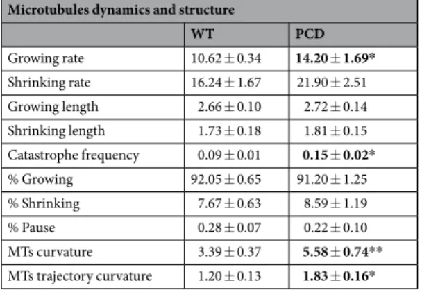
under-stood. To address this, we studied the microtubules of mouse embryonic fibroblasts (MEFs) lacking the CCP1 enzyme, which we named PCD microtubules (Fig. 1). Results showed (Table 1) that “growing rate” (p = 0.022) and “catastrophe frequency” (p = 0.012) were significantly increased in the PCD microtubules (Fig. 1A–C). Regarding microtubule curvature, PCD microtubules were more curved than the controls (p = 0.003; Fig. 1D–F). In parallel, the curvature of trajectories was also increased in PCD microtubules (p = 0.024; Fig. 1G–I). The lack of the CCP1 enzyme increased the instability of microtubules and influenced microtubule flexibility, thus affect-ing their trajectories.
Morphological changes in the main dendrite of PCs begin during the pre-neurodegeneration.
Microtubules are essential for maintaining neuronal shape13,37. Therefore, to understand the impact of these
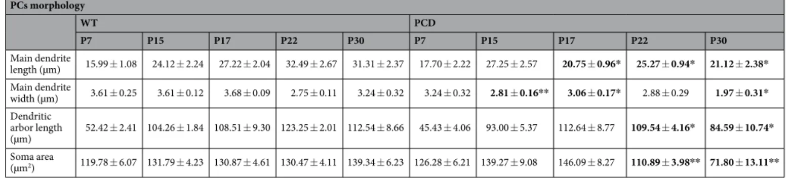
microtubular defects, we analyzed the morphology of PCs (Fig. 2A; Table 2) during different stages of PCD post-natal development: (1) P7, where no defects have been previously described, (2) P15 and P17, to characterize the pre-neurodegenerative stage and (3) P22 and P30, to characterize the neurodegenerative stage, when PCs disap-pear. Results showed that morphological changes of the main dendrite length (P7, p = 0.483; P15, p = 0.393; P17, p = 0.028; P22, p = 0.043; P30, p = 0.027) and width (P7, p = 0.379; P15, p = 0.007; P17, p = 0.017; P22, p = 0.695; P30, p = 0.020) began during pre-neurodegeneration (Fig. 2B,C), both parameters decreased in PCD mice. By contrast, a reduction of the dendritic arbor length - molecular layer thickness - (P7, p = 0.361; P15, p = 0.064; P17, p = 0.704; P22, p = 0.041; P30, p = 0.030) and of the soma size (P7, p = 0.484; P15, p = 0.466; P17, p = 0.085; P22, p = 0.008; P30, p = 0.001) was observed later, during the neurodegenerative stage (Fig. 2D,E). Hence, micro-tubule instability may induce morphological alterations of the PCs throughout late postnatal stages of cerebellar development, before the beginning of neurodegeneration.
TUNEL analysis revealed general neuronal death in the cerebellum of PCD mice.
The Nna1/Ccp1 gene mutation is known to affect all neuronal populations of the cerebellum24,25,27. However, apart from
PC loss, the progression of cell death of other cerebellar neuronal populations of the PCD mouse has not been previously characterized. Therefore, we monitored the death of the three main neuronal populations of the cerebellum: the molecular layer cells, the PCs and the granular layer cells (Fig. 3). Results confirmed that the PCs began to die in the PCD mouse from P22 and onwards, resulting in a process of cerebellar degeneration (P7, p = 0.276; P15, p = 0.073; P17, p = 0.444; P22, p = 0.004; P30, p = 0.012; Fig. 3D. We also found an increase in cellular death during the same period in both the molecular (P7, p = 0.168; P15, p = 0.411; P17, p = 0.302; P22, p < 0.001; P30, p = 0.010; Fig. 3C) and granular layers (P7, p = 0.581; P15, p = 0.960; P17, p = 0.346; P22, p = 0.029; P30, p < 0.001; Fig. 3E). The neuronal nature of apoptotic cells in the three cerebellar layers was con-firmed using TUNEL/NeuN and TUNEL/PV immunofluorescence colocalization (Fig. 3A,B). In summary, the initial symptoms of the breakdown of cerebellar structure are seen in the main dendrite of PCs. Following on, the
morphological defects extend to the whole neuron, altering both the general dendritic arbor and soma. At the same time, there is a progressive and general neuronal loss in the other two layers resulting in a general break-down of the cerebellum.
Figure 1. Effect of pcd mutation on microtubule dynamics and structure (mean ± SEM). (A and B) manual and automatic analyses of microtubules growth and shrinking rates; in both analysis PCD microtubules showed an increase in growing rate. (C) Catastrophe frequency analysis; an increase in the frequency of catastrophes in PCD microtubules is demonstrated. (D–F) Analysis of WT (D) and PCD (E) microtubules curvature and its graphical representation (F); an increase in the curvature of PCD microtubules can be observed. (G–I) Analysis of WT (G) and PCD (H) microtubule trajectory curvature and its graphical representation (I); an increase in the trajectory curvature of PCD microtubules can be observed. *p < 0.05.
Microtubules dynamics and structure
WT PCD Growing rate 10.62 ± 0.34 14.20 ± 1.69* Shrinking rate 16.24 ± 1.67 21.90 ± 2.51 Growing length 2.66 ± 0.10 2.72 ± 0.14 Shrinking length 1.73 ± 0.18 1.81 ± 0.15 Catastrophe frequency 0.09 ± 0.01 0.15 ± 0.02* % Growing 92.05 ± 0.65 91.20 ± 1.25 % Shrinking 7.67 ± 0.63 8.59 ± 1.19 % Pause 0.28 ± 0.07 0.22 ± 0.10 MTs curvature 3.39 ± 0.37 5.58 ± 0.74** MTs trajectory curvature 1.20 ± 0.13 1.83 ± 0.16*
Motor coordination is only affected during the neurodegeneration.
Data from the rota-rod test (Fig. 4A,B) showed that motor performance of the PCD mouse was not affected during pre-neurodegeneration (P15, p = 0.677 and P17, p = 0.398; Fig. 4B), but impaired during neurodegeneration; i.e. at P22 (p < 0.001) and P30 (p < 0.001; Fig. 4B). Thus, motor coordination is affected only during PC loss, but not prior to this.Home-cage behavior is altered in PCD mice before PC death.
Results of the home-cage behaviortest showed no differences between genotypes regarding the time spent displacing, during any of the develop-mental stages, which indicated that the pcd mutation did not affect general movement (Fig. 4D). Concerning grooming time, no differences were found during pre-neurodegeneration (Fig. 4E), but it did decrease during Figure 2. Morphological analysis of the PCs during postnatal development (mean ± SEM). (A) Micrographs of the PCs at different ages as seen with anti-calbindin immunofluorescence (Cb-28k; red). (B–E) Charts showing the quantification of the studied parameters; note that morphological alterations appear from P15 and onwards, with the soma area/size and the dendritic arbor length being the last to be significantly modified. n = 4 per experimental group. *p < 0.05; **p < 0.01.
neurodegeneration in the PCD mice (P15, p = 0.444; P17, p = 0.535; P22, p = 0.005; P30, p < 0.001). Interestingly, environmental exploratory behavior - rearings - was also reduced in PCD mice compared to the controls dur-ing both pre- (P15, p = 0.042; P17, p < 0.001) and neurodegeneration (P22, p = 0.985; P30, p = 0.005; Fig. 4F). Surprisingly, at P22 no changes were found in this exploratory behavior of PCD mice.
Long-term memory is only affected in late neurodegenerative stages.
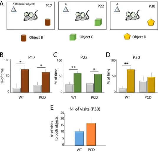
The results of the novelobject recognition test showed that wild-type (WT) mice at all ages spent a large percentage of time exploring new rather than familiar objects (Fig. 5; P17, p = 0.016; P22, p = 0.004; P30, p = 0.001), indicating a normal mem-ory recognition. Similarly, PCD mice also presented the same behavior at P17 (p = 0.016) and P22 (p = 0.010). However, at P30 no differences were detected in the PCD mice regarding the percentage of time spent exploring both types of objects (p = 0.497), suggesting a deficit in object memory recognition at this age.
The number of visits to both objects at P30 was further analyzed to avoid possible biases derived from motor alterations in PCD mice. Results did not show differences between the genotypes (Fig. 5E), thus discarding any effects of ataxia in PCD mice, with respect to moving within the plastic box and exploring the objects, and vali-dating the previous result.
Social preference is affected in PCD mice because of cerebellar pre-neurodegeneration.
Theresults of the social preference test revealed that WT mice at all ages spent a larger percentage of time explor-ing the chamber containexplor-ing other mouse rather than the chamber containexplor-ing an object (P15, p = 0.028; P17, p = 0.042; P22, p = 0.001; P30, p < 0.001; Fig. 6), indicating a preference for social contact. However, PCD mice spent the same percentage of time exploring both chambers at all ages (P15, p = 0.305; P17, p = 0.328; P22, p = 0.272; P30, p = 0.812; Fig. 6). Thus, the pre-neurodegenerative changes observed prior to the breakdown of cerebellar structure seem to be enough to induce social deficits.
In conclusion, the behavioral analysis of PCD mice strongly suggests a close relationship between cerebellar function and cognition. Moreover, the progression of the cerebellar alterations increasingly impaired the perfor-mance of cognitive and affective tasks.
Discussion
One of the main obstacles to establishing a relationship between the cerebellum and cognitive and social pro-cesses is the lack of studies analyzing the impact of progressive cerebellar defects on non-motor behavior. Also, this association has only been studied over short time frames. Here, to allow for a better understanding of this relationship, our results will be discussed chronologically based on their appearance throughout postnatal devel-opment in normal and PCD mice. The relevant order includes the first week of postnatal develdevel-opment (P7), then during the pre-neurodegenerative stage (P15–P17), and finally during the neurodegenerative stage (P22–P30). Thus, a temporal pattern is presented, characterizing the microtubule alterations observed in the PCD mouse, the progression of the cerebellar defects, and the influence of these alterations on motor and non-motor behaviors.
To understand the importance of the CCP1 enzyme in cerebellar development, the effect of the lack of this enzyme on microtubule dynamics and the structure of fibroblasts was initially studied, as the in vitro model available was able to provide insight into the structure and dynamics of this cellular component. While previous results have demonstrated that the lack of CCP1 enzyme increases microtubule polyglutamylation24, here it is
shown that the lack of this de-polyglutamylase affects microtubule stability and flexibility. Microtubules are more unstable at the growth cones, which consequently increases their catastrophe frequency, an essential process for normal neuronal development38,39. Additionally, certain factors related to neuronal migration and development
preferentially bind to curved microtubules40–43. Therefore, microtubule dynamics and curvature seem to have a
different pattern during neuronal development and maturation, being more dynamic and curved during develop-ment and stable and rigid during maturation. The microtubules of PCD mice are likely very similar to those found in developing neurons, in terms of dynamics and structure. In WT mice, CCP1 expression increases at P15 in the cerebellum29, that is, at the beginning of the pre-neurodegeneration stage of the PCD mouse26,29. Precisely, during
this short time frame, the growth of the dendritic arbor of PCs is completed and the process of maturation and remodeling takes place44,45. Thus, microtubule polyglutamylation seems to be essential during cerebellar
develop-ment, and a reduction of this post-translational modification seems to be necessary at the end of neuronal growth.
PCs morphology WT PCD P7 P15 P17 P22 P30 P7 P15 P17 P22 P30 Main dendrite length (µm) 15.99 ± 1.08 24.12 ± 2.24 27.22 ± 2.04 32.49 ± 2.67 31.31 ± 2.37 17.70 ± 2.22 27.25 ± 2.57 20.75 ± 0.96* 25.27 ± 0.94* 21.12 ± 2.38* Main dendrite width (µm) 3.61 ± 0.25 3.61 ± 0.12 3.68 ± 0.09 2.75 ± 0.11 3.24 ± 0.32 3.24 ± 0.32 2.81 ± 0.16** 3.06 ± 0.17* 2.88 ± 0.29 1.97 ± 0.31* Dendritic arbor length (µm) 52.42 ± 2.41 104.26 ± 1.84 108.51 ± 9.30 123.25 ± 2.01 112.54 ± 8.66 45.43 ± 4.06 93.00 ± 5.37 112.64 ± 8.77 109.54 ± 4.16* 84.59 ± 10.74* Soma area (µm2) 119.78 ± 6.07 131.79 ± 4.23 130.87 ± 4.61 130.47 ± 4.11 139.34 ± 6.23 126.28 ± 6.21 139.27 ± 9.08 146.09 ± 8.27 110.89 ± 3.98** 71.80 ± 13.11**
Then, the microtubule over-polyglutamylation occurring in the PCD mouse triggers the pre-neurodegeneration and the loss of the PCs from P18 and onwards25,26,29. Conversely, at P7 no differences were observed in the
mor-phology of the PCs between genotypes, the general structure of the cerebellum was apparently unaffected. Since neuronal migration and growth occur in the normal cerebellar cortex during this first postnatal week46, it appears
that the pcd mutation (i.e., microtubule over-polyglutamylation) does not affect the initial perinatal stages of cerebellar cortex formation. CCP1 activity is extremely low during the first two postnatal weeks (it significantly increases at P15)29 thus, our data support the idea that microtubule polyglutamylation is essential during early
cerebellar development, but deleterious for establishing a mature pattern. Therefore, the fine-tuning of microtubule polyglutamylation has a major role in cerebellar postnatal development.
In order to understand the influence of the cerebellum on cognitive and social behavior, different authors have analyzed the effects of different cerebellar alterations in animal models14,15. However, to our knowledge, there are
no studies that have attempted to analyze progressive cerebellar alterations throughout postnatal development to understand the normal cerebellar function.
Morphological defects in the PCs of different psychiatric and neurodevelopmental disorders have been recently described47–49, but the direct effect of alterations in the morphology of PCs on non-motor behavior is
not known. We demonstrate that prior to the breakdown of cerebellar structure (during pre-neurodegeneration, Figure 3. Analysis of apoptosis along cerebellar postnatal development. (A) Micrograph of a Purkinje cell labeled with parvalbumin (PV, red) and TUNEL (green) in a PCD mouse. (B) Micrograph of an interneuron of the molecular layer labeled with NeuN (red) and TUNEL (green) in a PCD mouse. (C–E) Charts showing the quantification of TUNEL-positive cells in the three cerebellar layers (mean ± SEM). An increase in the number of apoptotic cells can be observed from P22 and onwards in the three cerebellar layers of the PCD mice. n = 4 per experimental group. *p < 0.05; **p < 0.01.
when no motor defects were detected), morphological alterations in the main dendrite of PCs and social and environmental exploratory behavioral disabilities occur simultaneously (both at P15 and P17). In psychiatric and neurodevelopmental disorders, such as autism or schizophrenia that can often exhibit severe symptoms of social disability, have been related to cerebellar alterations8–12,15. Furthermore, PC loss is the most common
anatomo-pathological change found in autistic patients47, and even in those cases where no loss of PCs was observed,
mor-phological defects were still found48. Also, in accordance with our results, environmental exploratory defects have
been previously associated with cerebellar alterations50–52. A possible explanation for these impairments is that
alterations in dendritic morphology influence the electrophysiological activity of neurons, including the PCs53,54.
More precisely, defects in the PCs axial resistance, which directly depends on dendritic length and width, disrupt the synaptic integration in PCs53, as previously hypothesized55,56. Hence, it is plausible to relate the progressive
extinction of Purkinje cells in PCD mice to the progressive alteration of the behaviors analyzed (see below). During the cerebellar degeneration that occurs in PCD mice, neuronal death takes place throughout the three cerebellar layers. Previous studies assumed that in the PCD animal, the loss of neuronal populations of the gran-ular and molecgran-ular layers is residual and delayed in time, in comparison to the death of PCs25; however, the
Figure 4. Analysis of motor coordination and home-cage behavior along postnatal development (mean ± SEM). (A,B) Representation and quantification of the rota-rod test. (C–F) Representation and quantification of the parameters analyzed in the home-cage behavior test; i.e. time for grooming, time of environmental exploratory behavior and time displacing. Note the differences derived from the pcd mutation for all parameters, except for displacement. n = 8–9 per experimental group. *p < 0.05; **p < 0.01.
progression of their death has never been characterized. Our results demonstrate that this neuronal loss pro-gresses at the same time as the loss of PCs, indicating a simultaneous decline in all three layers in the cerebellum of the PCD mouse.
During neurodegeneration, the morphological alterations of the PCs worsen, with the most evident changes linked to the length and width of the main dendrite, the soma area and the dendritic arbor length. In parallel, the defects observed in social preference and environmental exploratory behavior also persist during this stage in the PCD mouse. Alterations in these behaviors have been described in both animal and human studies with cerebellar impairments8–10,57–59. It was observed that these alterations may be due both to neuronal loss and to PC
malfunction. Interestingly, we saw that ataxia did not bias the results of the cognitive/social tasks performed by the PCD mice, and during neurodegeneration grooming was found to be reduced. In contrast, this behavior has been previously shown to increase in animal models of specific PC loss15, being more so in models presenting a
general alteration of the cerebellum58. Accordingly, during grooming behavior there is an increase in the activity
of both granule cells60 and PCs59. Consequently, grooming may be affected differently depending on whether
the cerebellar alterations affect the PCs, granule cells or both. Thus, the severity of cerebellar defects helps to understand the relationship between the cerebellum and grooming. Although grooming is a very complex motor task, we cannot discard the possibility that the motor defects caused by cerebellar alterations have influenced the observed behavioral changes.
Interestingly, general changes in cerebellar structure in the PCD mouse, which disrupt recognition memory, only occurred at P30 when most of the PCs are lost. The cerebellum has been associated with procedural learn-ing and memory61,62, and current neuroimaging studies have also described activation of the cerebellum during
non-procedural memory tasks63–65. These works, however, did not study the impact of the progression of the
breakdown of cerebellar structure on recognition memory. According to our results, cerebellar alterations lead to the impairment of recognition memory in PCD mice, and during neurodegeneration both the cerebellar and behavioral defects become worse. Therefore, these results suggest a correlation between the progression of the breakdown of the cerebellar structure and the severity of the defects related to performing cognitive and affective tasks. Then, a healthy, typical cerebellar structure is necessary to execute specific cognitive and affective tasks. Figure 5. Analysis of the novel object recognition test along postnatal development. (A) Schematic
representation of the test and the objects employed. (B–D) Analysis of the percentage of time exploring familiar and novel objects at different ages for both WT and PCD mice (mean ± SEM); a lack of preference for the new object is seen in PCD mice at P30. (E) Quantification of the number of visits to both objects at P30; no differences were observed. n = 8–9 per experimental group. *p < 0.05; **p < 0.01.
In conclusion, polyglutamylation affects microtubules dynamics and structure, and its modulation is essential throughout normal cerebellar development. During the first two postnatal weeks, when the PCs are still migrat-ing and growmigrat-ing, microtubules instability is crucial. However, a pathologically extended instability of microtu-bules (triggered by the pcd mutation) affects the morphology and survival of PCs, which are vital for the correct functioning of the cerebellum. In this sense, cerebellum plays an important role in cognitive and affective func-tions, since the progressive breakdown of the cerebellar cortex in PCD mice is directly related to the increasing severity of cognitive and affective impairments.
Material and Methods
Animals.
Both WT and PCD mice from the C57/DBA strain were used and purchased from The Jackson Laboratory, Maine, USA. For in vitro studies, MEFs were prepared from both WT and CCP1-KO embryos (see below). The embryos were separated into groups depending on their genotype (WT or PCD; n = 3 embryos per group). For in vivo studies, animals were separated into groups depending on their genotype and age at the time of analysis: P7, P15, P17, P22 and P30 (n = 4 per age and genotype) for the immunohistochemical analyses; and P15, P17, P22 and P30 (n = 8–9 per genotype) for the behavioral analyses.Since PCD and CCP1-KO animals are not fertile, heterozygous mice were mated, and the embryos or new-born mice were genotyped (see below and28).
Animals were housed, handled and sacrificed per the guidelines established by European (2010/63/UE) and national legislations (Spanish RD53/2013 and Law 32/2007; French permit n°38 07 11). Every effort was made to Figure 6. Analysis of the social preference test during postnatal development. (A) Diagram of test performance. (B–E) Charts showing quantification of the percentage of time spent exploring the chambers containing either the intruder animal or an object for both WT and PCD mice at different ages (mean ± SEM). Note that PCD mice, contrary to the WT, spent the same amount of time exploring both chambers. n = 8–9 per experimental group. *p < 0.05; **p < 0.01.
minimize the number of animals used and their suffering. This study was approved by the Bioethics Committee of the University of Salamanca.
Generation of CCP1 KO mice.
The conditional mutant mouse line for CCP1 (on exons 20 and 21) was established and generated at the Mouse Clinical Institute (MCI, Illkirch, France) to study microtubules dynamics and structure. The targeting vector was constructed as follows. The 5′ (4.5 kb), 3′ (1.4 kb) and inter-loxP (1.9) fragments were polymerase chain reaction (PCR) amplified and sequentially subcloned into an MCI proprietary vector containing the LoxP sites and a Neo cassette flanked by Flippase Recognition Target (FRT) sites (Fig. 7). The linearized construct was electroporated in 129S2/SvPas mouse embryonic stem (ES) cells. After selection, tar-geted clones were identified by PCR using external primers and further confirmed by Southern blot with 5′ and 3′ external probes. Two positive ES clones were injected into blastocysts, and derived male chimaeras gave germline transmission. The excision of the neomycin-resistance cassette was performed in vivo by breeding the chimeras with a Flp deleter line (C57BL/6 N genetic background FLP under ACTB promoter). The Flp transgene was segre-gated by breeding the first germ line mice with a wild type C57BL/6 N animal. For generation ccp1 KO, ccp1 floxed mice were crossed with transgenic mice expressing Cre recombinase under the control of a CMV promoter.Genomic DNA isolated from mouse tail snip was analyzed by PCR. Mice were genotyped by PCR according to MCI protocols using GoTag polymerase (Promega) and 33 amplification cycles. The three primer pairs listed below were used to define the genotypes.
(1) Ef: GTACACAGACTTTTAGGCTAATCTC Er: ACTTCCACATCTGCACTGTGGACAG (2) Lf: CATAAGAACCAGGTTCATTCTGTCC Lr: TAGCATCATGTTAAAACTCCCTCCT (3) Lf: CATAAGAACCAGGTTCATTCTGTCC Er: ACTTCCACATCTGCACTGTGGACAG
Cell culture and transfection.
For in vitro analysis of the microtubules, MEFs from the CCP1 KO line were prepared as previously described66. To visualize the microtubules, MEFs were transfected using theNucleofectorTM Kits for MEFs (Amaxa Biosystems) with GFP-EB3 (to visualize microtubules (+)-ends; provided
by N. Galjart, Erasmus Medical Center, Rotterdam, The Netherlands) and m-cherry α-tubulin (to visualize the entire microtubules; provided by F. Saudou, Curie Institute, Paris, France) plasmids.
Video microscopy.
Video time-lapses of transfected MEFs were captured with an inverted microscope (Axiovert 200 M; Carl Zeiss, Oberkochen, Germany) and a 100× NA 1.3 Plan-Neofluar oil objective controlled by MetaMorph software (MDS Analytical Technologies, Sunnyvale, CA, USA). Images were taken every three seconds for 5 minutes. All images were captured with a charge-coupled device camera (CoolSNAP HQ; Roper Scientific, Sarasota, Florida, USA).Microtubule dynamics and structure analysis.
For the analysis of the microtubule dynamics the plusT-ipTracker software67 and the GFP-EB3 video time-lapse images were employed. We first adjusted the accuracy ofthe GFP-EB3 detection, and then analyzed the following parameters: growing and shrinking speed; growth and shrinking mean length; percentage of time in pause, growing and shrinking; distribution of microtubules based on their speed and length; catastrophe frequency and rescue frequency. These parameters were chosen based on Figure 7. Schematic representation in scale of the targeting vector used and all the possible alleles for CCP1 gene. Orange bar: genomic DNA. Black boxes: exons with their corresponding number. Green and purple arrowheads: LoxP and Flp sequences, respectively. White bar: neo cassette, with the neomycin resistance gene (white box). Blue lines: zone of sequence homology for homologous recombination, with the corresponding size in kbp. Black arrowheads: primers used for the PCR genotyping.
previous studies68,69. To ensure the accuracy of the analysis, we compared the growing speed data obtained both
automatically and manually.
The microtubule curvature analysis was performed using the first image of each video time-lapse with m-cherry α-tubulin. Additionally, with the GFP-EB3 labeling experiment we obtained the maximum projection of the entire video time-lapse, allowing us to reconstruct the trajectories of the microtubules, whose curvature was also analyzed. Thus, the curvature of microtubules was analyzed both in a static way and within their trajec-tory of movement.
Curvature (k) was determined using the Mathematica software (Wolfram Research Europe, UK). After obtain-ing the equation of each microtubule with a spline cubic interpolation, the general formula of curvature70 was
employed to analyze each microtubule (equation 1):
= ′ ″ − ′ ″ ′ + ′
k x y y x
x y
( 2 2 3/2) (1) where (equations 2, 3, 4 and 5)
′ = x dx dt, (2) ″ = x d x dt , (3) 2 2 ′ = y dy dt, (4) ″ = y d y dt , (5) 2 2
i.e., the first ( ′ ′x y, ) and second ( ′′ ′x y, ) derivatives at each point.
Tissue extraction and preparation for in vivo studies.
To analyze the effect of the pcd mutation on the PCs morphology and the general cerebellar structure in vivo, animals were anesthetized and intracardially perfused with Somogyi’s fixative without glutaraldehyde (5 ml/g body weight), as previously described 26Cerebellawere sectioned sagittally at 30 µm thick using a freezing microtome (Leica Jung SM 2000, Nussloch, Germany), as previously described71–73. The histological analyses were focused on the vermis using three sections per animal
(separated by 180 µm each), with a total of five animals per genotype and age. All values represent the mean count of the three sections analyzed.
Immunofluorescence.
Sections were incubated with the primary antisera at 4 °C during 72 h, as previously described74. The antibodies employed were mouse anti-calbindin (Cb-28k 1:1,000; Swant, Switzerland), mouseanti-parvalbumin (PV 1:1,000; Swant, Switzerland) and mouse anti-NeuN (1:8,000; Merck Millipore, Darmstadt, Germany). Appropriate secondary antibodies conjugated with Cy2 or Cy3 (1:500; Jackson Laboratories, West Grove, PA, USA) were used. Sections were counter-stained with DAPI (1:30,000; Sigma Aldrich) to identify the cell nuclei. Antibodies were selected based on their selectivity to identify different neuronal populations. Calbindin is a well-known calcium-binding protein that label the whole PCs46,75,76, allowing us to analyze the PCs
morphology. In addition, parvalbumin stains both PCs and interneurons in the molecular layer46. Finally, NeuN
was used to counterstain whole cerebellar nuclei during cell counting, ensuring no biases were made due to the lack of immunolabeling during degeneration.
Terminal Deoxynucleotidyl Transferase-Mediated Fluorescein dUTP Nick-End Labeling
(TUNEL) detection.
To study apoptosis throughout cerebellar postnatal development, the TUNEL tech-nique was used as previously described29,74. To determine the neuronal nature of apoptotic cells, the TUNEL wascombined with immunohistochemistry against NeuN or PV. Finally, both TUNEL and immunohistochemistry were revealed using Cy2-streptavidin and Cy3-secondary antibodies, respectively.
Morphological analysis of PCs.
Morphological analysis of the PCs was made using sections immunos-tained for Cb-28k from P7 to P30. To avoid possible biases, only those Purkinje cells containing the soma and a clear dendritic arbor were analyzed. Also, all Purkinje cells fitting this criterion were analyzed in three cerebellar sections per animal (separated by 180 µm each). The final value for each animal used to compare was the mean of all Purkinje cells analyzed in these three sections. The morphological parameters studied were selected in basis to its meaning for PCs function77,78: (1) the length and width of the main dendrite; (2) the length of thedendritic arbor of the PCs (measured indirectly through the molecular layer thickness); and (3) the soma size. Morphological analyses were carried out with the Neurolucida (MBF Bioscience, Williston, Vermont, USA) and ImageJ (NIH, USA) software, as previously described77.
Behavioral analyses.
We characterized the effects of progressive cerebellar defects on motor, cognitive, and social processes. Behavioral analyses were performed at P15, P17, P22 and P30, encompassing both the pre-neurodegenerative (P15–P18) and neurodegenerative (P18–P30) stages. All devices employed were cleaned with 96% (v/v) ethanol before and after each session.The rota-rod test was used to characterize motor coordination. It was performed as previously described79,
with an acceleration of 0.6 rpm/s, from 4 to 40 rpm, in 10 min.
The home-cage behavior analysis was used to characterize general behavior. Animals were placed each day in the home-cage for 10 min and allowed to freely explore. Afterwards, the animals’ behavior was observed for 10 min, during which time the following parameters were recorded manually: (1) grooming time (stereotyped behavior); (2) number of rearings (environmental exploratory behavior) and (3) time moving (general move-ment). Each animal was assigned to an individual home-cage to avoid the influence of odor from the other animals.
The novel object recognition test was used to analyze recognition memory. The test was performed in the same home-cage used described above to help the animals to become habituated. On the first day, at P15, animals were placed in the home-cage with two identical objects (A and A) for 10 min. Then, for each of the following sessions (at P17, P22 and P30) the animals were placed in the home-cage with: (1) the familiar object (A) and (2) a new object (B, C, and D, respectively for each of the different ages; see Fig. 5). The objects were placed in opposite corners of the home-cage, the time interacting with each object was measured and their ratio was calculated.
The social preference test was performed in a white Plexiglas
®
box (50 × 29 cm) divided into three chambers. The social preference test was performed as previously described80–82. On each test day, a mouse could explore thebox for 10 min. Then, the animal was removed from the box and a different mouse, of the same age and sex, was placed in one of the lateral chambers under a drilled pencil cup. On the other side of the box, an object was placed in the other lateral chamber, also under a drilled pencil cup. The original mouse was then returned to the box and the percentage of time spent exploring each lateral chamber and each pencil cup was analyzed.
Statistical analysis.
For data analysis, the Student’s t-Test was performed. For histological analysis, both genotypes were compared for each age. For behavioral analysis, the parameters analyzed and the different con-frontation variables are described in each behavioral procedure. All analyses were performed with the SPSS sta-tistical package software (IBM, NY, USA).References
1. Strick, P. L., Dum, R. P. & Fiez, J. A. Cerebellum and nonmotor function. Annu. Rev. Neurosci. 32, 413–34 (2009). 2. Ito, M. Control of mental activities by internal models in the cerebellum. Nat. Rev. Neurosci. 9, 304–313 (2008).
3. Allen, G. & Courchesne, E. Differential effects of developmental cerebellar abnormality on cognitive and motor functions in the cerebellum: An fMRI study of autism. Am. J. Psychiatry 160, 262–273 (2003).
4. Allen, G., Buxton, R. B., Wong, E. C. & Courchesne, E. Attentional activation of the cerebellum independent of motor involvement.
Science 275, 1940–1943 (1997).
5. Fatemi, S. H. et al. Consensus paper: Pathological role of the cerebellum in Autism. Cerebellum 11, 777–807 (2012).
6. Allen, G., Müller, R. A. & Courchesne, E. Cerebellar function in autism: Functional magnetic resonance image activation during a simple motor task. Biol. Psychiatry 56, 269–278 (2004).
7. Piven, J. et al. Magnetic resonance imaging in autism: measurement of the cerebellum, pons, and fourth ventricle. Biol. Psychiatry
31, 491–504 (1992).
8. Joseph, R. M., Tager-Flusberg, H. & Lord, C. Cognitive profiles and social-communicative functioning in children with autism spectrum disorder. J. Child Psychol. Psychiatry 43, 807–821 (2002).
9. Armstrong, D. D. Rett syndrome neuropathology review 2000. Brain Dev. 23, S72–S76 (2001).
10. Bauman, M. L. & Kemper, T. L. Neuroanatomic observations of the brain in autism: A review and future directions. Int. J. Dev.
Neurosci. 23, 183–187 (2005).
11. Walkley, S. U. & Suzuki, K. Consequences of NPC1 and NPC2 loss of function in mammalian neurons. Biochim. Biophys. Acta 1685, 48–62 (2004).
12. Andreasen, N. C. & Pierson, R. The Role of the Cerebellum in Schizophrenia. Biol. Psychiatry 64, 81–88 (2008).
13. Baas, P. W., Rao, A. N., Matamoros, A. J. & Leo, L. Stability properties of neuronal microtubules. Cytoskeleton 73, 442–460 (2016). 14. Galliano, E. et al. Synaptic transmission and plasticity at inputs to murine cerebellar Purkinje cells are largely dispensable for
standard nonmotor tasks. J. Neurosci. 33, 12599–618 (2013).
15. Tsai, P. T. et al. Autistic-like behaviour and cerebellar dysfunction in Purkinje cell Tsc1 mutant mice. Nature 488, 647–651 (2012). 16. Brossard-Racine, M., du Plessis, A. & Limperopoulos, C. Developmental Cerebellar Cognitive Affective Syndrome in Ex-preterm
Survivors Following Cerebellar Injury. Cerebellum 14, 151–164 (2015).
17. Faivre, C., Legrand, C. & Rabié, A. The microtubular apparatus of cerebellar purkinje cell dendrites during postnatal development of the rat: The density and cold-stability of microtubules increase with age and are sensitive to thyroid hormone deficiency. Int. J.
Dev. Neurosci. 3, 559–565 (1985).
18. Kim, J. et al. Dendritic planarity of Purkinje cells is independent of Reelin signaling. Brain Struct. Funct. 220, 2263–2273 (2014). 19. Liu, Y., Lee, J. W. & Ackerman, S. L. Mutations in the Microtubule-Associated Protein 1A (Map1a) Gene Cause Purkinje Cell
Degeneration. J. Neurosci. 35, 4587 LP–4598 (2015).
20. Andrieux, A. et al. The suppression of brain cold-stable microtubules in mice induces synaptic defects associated with neuroleptic-sensitive behavioral disorders. Genes Dev. 16, 2350–2364 (2002).
21. Maussion, G. et al. Convergent evidence identifying MAP/microtubule affinity-regulating kinase 1 (MARK1) as a susceptibility gene for autism. Hum. Mol. Genet. 17, 2541–2551 (2008).
22. Brown, A. S. et al. Increased stability of microtubules in cultured olfactory neuroepithelial cells from individuals with schizophrenia.
Prog. Neuro-Psychopharmacology Biol. Psychiatry 48, 252–258 (2014).
23. Cartelli, D. & Cappelletti, G. Microtubule Destabilization Paves the Way to Parkinson’s Disease. Mol. Neurobiol. 54, 6762–6774 (2016).
24. Rogowski, K. et al. A family of protein-deglutamylating enzymes associated with neurodegeneration. Cell 143, 564–578 (2010). 25. Wang, T. & Morgan, J. I. The Purkinje cell degeneration (pcd) mouse: An unexpected molecular link between neuronal degeneration
and regeneration. Brain Res. 1140, 26–40 (2007).
26. Baltanás, F. C. et al. Nucleolar disruption and cajal body disassembly are nuclear hallmarks of DNA damage-induced neurodegeneration in Purkinje cells. Brain Pathol. 21, 374–388 (2011).
27. Baltanás, F. C. et al. Purkinje Cell Degeneration in pcd Mice Reveals Large Scale Chromatin Reorganization and Gene Silencing Linked to Defective DNA Repair. J. Biol. Chem. 286, 28287–28302 (2011).
28. Valero, J., Berciano, M. T., Weruaga, E., Lafarga, M. & Alonso, J. R. Pre-neurodegeneration of mitral cells in the pcd mutant mouse is associated with DNA damage, transcriptional repression, and reorganization of nuclear speckles and Cajal bodies. Mol. Cell.
Neurosci. 33, 283–295 (2006).
29. Baltanás, F. C. et al. Differential glial activation during the degeneration of Purkinje cells and mitral cells in the PCD mutant mice.
Glia 61, 254–72 (2013).
30. Hashimoto, K. & Kano, M. Postnatal development and synapse elimination of climbing fiber to Purkinje cell projection in the cerebellum. Neurosci. Res. 53, 221–228 (2005).
31. Nedelescu, H. & Abdelhack, M. Comparative morphology of dendritic arbors in populations of purkinje cells in mouse sulcus and apex. Neural Plast. 2013, 948587 (2013).
32. Bosch Grau, M. et al. Alterations in the balance of tubulin glycylation and glutamylation in photoreceptors leads to retinal degeneration. J. Cell Sci. 130, 938–949 (2017).
33. Ghetti, B., Triarhou, L. C., Alyea, C. J., Dlouhy, S. R. & Karn, R. C. Unique cerebellar phenotype combining granule and Purkinje cell loss: morphological evidence for weaver* pcd double mutant mice. J. Neurocytol. 20, 27–38 (1991).
34. Mullen, R. J., Eicher, E. M. & Sidman, R. L. Purkinje cell degeneration, a new neurological mutation in the mouse. Proc. Natl. Acad.
Sci. USA 73, 208–212 (1976).
35. Lacroix, B. et al. Tubulin polyglutamylation stimulates spastin-mediated microtubule severing. J. Cell Biol. 189, 945–54 (2010). 36. Sirajuddin, M., Rice, L. M. & Vale, R. D. Regulation of microtubule motors by tubulin isotypes and post-translational modifications.
Nat. Cell Biol. 16, 335–44 (2014).
37. Sakakibara, A., Ando, R., Sapir, T. & Tanaka, T. Microtubule dynamics in neuronal morphogenesis. Open Biol. 3, 130061 (2013). 38. Dent, E. W., Callaway, J. L., Szebenyi, G., Baas, P. W. & Kalil, K. Reorganization and movement of microtubules in axonal growth
cones and developing interstitial branches. J. Neurosci. 19, 8894–8908 (1999).
39. Dubey, J., Ratnakaran, N. & Koushika, S. P. Neurodegeneration and microtubule dynamics: death by a thousand cuts. Front. Cell.
Neurosci. 9, 343 (2015).
40. Gleeson, J. G., Peter, T. L., Flanagan, L. A. & Walsh, C. A. Doublecortin is a microtubule-associated protein and is expressed widely by migrating neurons. Neuron 23, 257–271 (1999).
41. Bechstedt, S. & Brouhard, G. J. Doublecortin Recognizes the 13-Protofilament Microtubule Cooperatively and Tracks Microtubule Ends. Dev. Cell 23, 181–192 (2012).
42. Fu, X. et al. Doublecortin (Dcx) family proteins regulate filamentous actin structure in developing neurons. J. Neurosci. 33, 709–21 (2013).
43. Bechstedt, S., Lu, K. & Brouhard, G. J. Doublecortin Recognizes the Longitudinal Curvature of the Microtubule End and Lattice.
Curr. Biol. 24, 2366–2375 (2015).
44. Kaneko, M. et al. Remodeling of monoplanar Purkinje cell dendrites during cerebellar circuit formation. PLoS One 6, e20108 (2011). 45. Kawaguchi, K., Habara, T., Terashima, T. & Kikkawa, S. GABA modulates development of cerebellar Purkinje cell dendrites under
control of endocannabinoid signaling. J. Neurochem. 114, 627–638 (2010).
46. White, J. J. & Sillitoe, R. V. Development of the cerebellum: from gene expression patterns to circuit maps. Wiley Interdiscip. Rev. Dev.
Biol. 2, 149–64 (2013).
47. Sudarov, A. Defining the role of cerebellar purkinje cells in autism spectrum disorders. Cerebellum 12, 950–5 (2013). 48. Fatemi, S. H. et al. Purkinje cell size is reduced in cerebellum of patients with autism. Cell. Mol. Neurobiol. 22, 171–175 (2002). 49. Mavroudis, I. A. et al. Dendritic and spinal pathology of the purkinje cells from the human cerebellar vermis in alzheimer’s disease.
Psychiatr. Danub. 25, 221–226 (2013).
50. Dahhaoui, M., Caston, J., Lannou, J. & Avenel, S. Role of the cerebellum in habituation exploration behavior in the rat. Physiol.
Behav. 52, 339–344 (1992).
51. Pierce, K. & Courchesne, E. Evidence for a cerebellar role in reduced exploration and stereotyped behavior in autism. Biol. Psychiatry
49, 655–664 (2001).
52. Caston, J., Chianale, C., Delhaye-Bouchaud, N. & Mariani, J. Role of the cerebellum in exploration behavior. Brain Res. 808, 232–7 (1998).
53. Bekkers, J. M. Changes in dendritic axial resistance alter synaptic integration in cerebellar Purkinje cells. Biophys. J. 100, 1198–1206 (2011).
54. Krichmar, J. L., Nasuto, S. J., Scorcioni, R., Washington, S. D. & Ascoli, G. A. Effects of dendritic morphology on CA3 pyramidal cell electrophysiology: A simulation study. Brain Res. 941, 11–28 (2002).
55. Schmahmann, J. D. The role of the cerebellum in affect and psychosis. J. Neurolinguistics 13, 189–214 (2000).
56. Schmahmann, J. D. The cerebellar cognitive affective syndrome: clinical correlations of the dysmetria of thought hypothesis. Int. Rev.
Psychiatry 13, 313–322 (2001).
57. Ismail, M. M. T. et al. Studying Autism Spectrum Disorder with Structural and Diffusion Magnetic Resonance Imaging: A Survey.
Front. Hum. Neurosci. 10, 211 (2016).
58. Strazielle, C. & Lalonde, R. Grooming in Lurcher mutant mice. Physiol. Behav. 64, 57–61 (1998). 59. Ghosh, K. K. et al. Miniaturized integration of a fluorescence microscope. Nat. Methods 8, 871–878 (2011).
60. Hartmann, M. J. & Bower, J. M. Tactile responses in the granule cell layer of cerebellar folium crus IIa of freely behaving rats. J.
Neurosci. 21, 3549–3563 (2001).
61. Molinari, M. et al. Cerebellum and procedural learning: Evidence from focal cerebellar lesions. Brain 120, 1753–1762 (1997). 62. Torriero, S. et al. Changes in cerebello-motor connectivity during procedural learning by actual execution and observation. J. Cogn.
Neurosci. 23, 338–48 (2011).
63. Weis, S., Klaver, P., Reul, J., Elger, C. E. & Fernández, G. Temporal and Cerebellar Brain Regions that Support both Declarative Memory Formation and Retrieval. Cereb. Cortex 14, 256–267 (2004).
64. Cabeza, R., Kapur, S., Craik, F. I. M. & McIntosh, A. R. Functional neuroanatomy of recall and recognition: A PET study of episodic memory. J. Cogn. Neurosci. 9, 254–265 (1997).
65. Buckner, R. L. The cerebellum and cognitive function: 25 years of insight from anatomy and neuroimaging. Neuron 80, 807–815 (2013).
66. Erck, C. et al. A vital role of tubulin-tyrosine-ligase for neuronal organization. Proc. Natl. Acad. Sci. USA 102, 7853–7858 (2005). 67. Matov, A. et al. Analysis of microtubule dynamic instability using a plus-end growth marker. Nat. Methods 7, 761–768 (2010). 68. Peris, L. et al. Motor-dependent microtubule disassembly driven by tubulin tyrosination. J. Cell Biol. 185, 1159–1166 (2009). 69. Applegate, K. T. et al. PlusTipTracker: Quantitative image analysis software for the measurement of microtubule dynamics. J. Struct.
Biol. 176, 168–184 (2011).
70. Goetz, A. Introduction to differential geometry. Addison Wesley Pub. Co, New York, USA. ISBN 10: 0201024314 (1970).
71. Zhao, Z. et al. Genetic Ablation of PLA2G6 in Mice Leads to Cerebellar Atrophy Characterized by Purkinje Cell Loss and Glial Cell Activation. PLoS One 6, e26991 (2011).
72. Shimizu-Albergine, M. et al. Individual cerebellar Purkinje cells express different cGMP phosphodiesterases (PDEs): in vivo phosphorylation of cGMP-specific PDE (PDE5) as an indicator of cGMP-dependent protein kinase (PKG) activation. J. Neurosci.
73. Kaemmerer, W. F. et al. In vivo transduction of cerebellar Purkinje cells using adeno-associated virus vectors. Mol. Ther. 2, 446–57 (2000).
74. Díaz, D., Recio, J. S., Weruaga, E. & Alonso, J. R. Mild cerebellar neurodegeneration of aged heterozygous PCD mice increases cell fusion of Purkinje and bone marrow-derived cells. Cell Transplant. 21, 1595–1602 (2012).
75. Hoebeek, F. E., Khosrovani, S., Witter, L. & De Zeeuw, C. I. Purkinje cell input to cerebellar nuclei in tottering: ultrastructure and physiology. Cerebellum 7, 547–58 (2008).
76. Sillitoe, R. V. & Joyner, A. L. Morphology, molecular codes, and circuitry produce the three-dimensional complexity of the cerebellum. Annu. Rev. Cell Dev. Biol. 23, 549–77 (2007).
77. Donald, S. et al. P-Rex2 regulates Purkinje cell dendrite morphology and motor coordination. Proc. Natl. Acad. Sci. USA 105, 4483–4488 (2008).
78. Fujishima, K., Horie, R., Mochizuki, A. & Kengaku, M. Principles of branch dynamics governing shape characteristics of cerebellar Purkinje cell dendrites. Development 139, 3442–3455 (2012).
79. Muñoz-Castañeda, R., Díaz, D., Ávila-Zarza, C. A., Alonso, J. R. & Weruaga, E. Sex-influence of nicotine and nitric oxide on motor coordination and anxiety-related neurophysiological responses. Psychopharmacology (Berl). 231, 695–706 (2014).
80. Crawley, J. N. Designing mouse behavioral tasks relevant to autistic-like behaviors. Ment. Retard. Dev. Disabil. Res. Rev. 10, 248–258 (2004).
81. Panksepp, J. B. & Lahvis, G. P. Social reward among juvenile mice. Genes, Brain Behav. 6, 661–671 (2007).
82. Silverman, J. L., Yang, M., Lord, C. & Crawley, J. N. Behavioural phenotyping assays for mouse models of autism. Nat. Rev. Neurosci.
11, 490–502 (2010).
Acknowledgements
This work was supported by the MINECO (SAF2016-79668-R; to EW and MTM2014-57129-C2-1-P to JMMC), the Regional Government of Castile and Leon (SA178U13 and SA030P17 to EW and RMC), the Centre for Regenerative Medicine and Cell Therapy of Castile and Leon (EW), the Samuel Solórzano Barruso Memorial Foundation (FS/3 2014 to DD), the Foltra Foundation (DD), INSERM, ARC (Association pour la Recherche sur le Cancer grant n°SFI20111204053 to MJM), La Ligue contre le Cancer comités Savoie/Isère (MJM), ANR Blan07-2_187328 ‘TyrTIPs’ (AA) and INCA PLBIO10-030 ‘TetraTips’ (AA). The authors express their gratitude to E.J. Keck for the English language editing.
Author Contributions
R.M.-C., D.D., M.-J.M., A.A. and E.W. designed the experiments; C.J. designed the conditional mutant mouse line for CCP1; R.M.-C., M.-J.M., L.P. and C.B. did the in vitro experiments; R.M.-C. did in vivo experiments and behavioral tests; R.M.-C., D.D. and E.W. performed the histological analysis; R.M.-C. and J.M.M.-C. performed the mathematical analyses of microtubule curvature; R.M.-C. did statistical analyses; R.M.-C. and D.D. prepared the figures; R.M.-C., D.D., M.-J.M., A.A., J.R.A. and E.W. wrote the manuscript, and all authors revised the manuscript.
Additional Information
Supplementary information accompanies this paper at https://doi.org/10.1038/s41598-018-21470-2. Competing Interests: The authors declare no competing interests.
Publisher's note: Springer Nature remains neutral with regard to jurisdictional claims in published maps and institutional affiliations.
Open Access This article is licensed under a Creative Commons Attribution 4.0 International License, which permits use, sharing, adaptation, distribution and reproduction in any medium or format, as long as you give appropriate credit to the original author(s) and the source, provide a link to the Cre-ative Commons license, and indicate if changes were made. The images or other third party material in this article are included in the article’s Creative Commons license, unless indicated otherwise in a credit line to the material. If material is not included in the article’s Creative Commons license and your intended use is not per-mitted by statutory regulation or exceeds the perper-mitted use, you will need to obtain permission directly from the copyright holder. To view a copy of this license, visit http://creativecommons.org/licenses/by/4.0/.