HAL Id: hal-00527784
https://hal.archives-ouvertes.fr/hal-00527784
Submitted on 19 Sep 2018
HAL is a multi-disciplinary open access
archive for the deposit and dissemination of
sci-entific research documents, whether they are
pub-lished or not. The documents may come from
teaching and research institutions in France or
abroad, or from public or private research centers.
L’archive ouverte pluridisciplinaire HAL, est
destinée au dépôt et à la diffusion de documents
scientifiques de niveau recherche, publiés ou non,
émanant des établissements d’enseignement et de
recherche français ou étrangers, des laboratoires
publics ou privés.
Distributed under a Creative Commons Attribution| 4.0 International License
Prion replication in the hematopoietic compartment is
not required for neuroinvasion in scrapie mouse model
Corinne Loeuillet, Catherine Lemaire-Vieille, Philippe Naquet, Marie-France
Cesbron-Delauw, Jean Gagnon, Jean-Yves Cesbron
To cite this version:
Corinne Loeuillet, Catherine Lemaire-Vieille, Philippe Naquet, Marie-France Cesbron-Delauw, Jean
Gagnon, et al.. Prion replication in the hematopoietic compartment is not required for neuroinvasion
in scrapie mouse model. PLoS ONE, Public Library of Science, 2010, 5 (10), pp.e13166.
�10.1371/jour-nal.pone.0013166�. �hal-00527784�
Prion Replication in the Hematopoietic Compartment Is
Not Required for Neuroinvasion in Scrapie Mouse Model
Corinne Loeuillet1, Catherine Lemaire-Vieille1, Philippe Naquet2, Marie-France Cesbron-Delauw1, Jean Gagnon1, Jean-Yves Cesbron1*
1 Laboratoire Adaptation et Pathoge´nie des Micro-organismes, Centre National Recherche Scientifique UMR 5163, Universite´ Joseph Fourier, Grenoble, France, 2 Centre d’Immunologie de Marseille-Luminy, Institut National de la Sante´ et de la Recherche Me´dicale, Centre National Recherche Scientifique, Universite´ de La Me´diterrane´e, Marseille, France
Abstract
Fatal neurodegenerative prion diseases are caused by the transmissible PrPSc prion agent whose initial replication after peripheral inoculation takes place in follicular dendritic cells present in germinal centers of lymphoid organs. However, prion replication also occurs in lymphoid cells. To assess the role of the hematopoietic compartment in neuroinvasion and prion replication, we generated chimeric mice, on a uniform congenic C57/BL6J background, by bone marrow replacement with hematopoietic cells expressing different levels of PrP protein. Nine different types of chimeric mice were inoculated intraperitoneally either with the lymphotropic Rocky Mountain Laboratory (RML) strain or the non lymphotropic ME-7 scrapie strain, at different doses. Here, we clearly demonstrate that overexpression of PrP by the hematopoietic system, or the lack of PrP expression by the bone marrow derived cells, does not change the incubation time period of the disease, even when the mice are infected at limiting doses. We conclude that the hematopoietic compartment is more or less permissive to prion replication, both for RML and ME-7, but does not play a role in neuroinvasion.
Citation: Loeuillet C, Lemaire-Vieille C, Naquet P, Cesbron-Delauw M-F, Gagnon J, et al. (2010) Prion Replication in the Hematopoietic Compartment Is Not Required for Neuroinvasion in Scrapie Mouse Model. PLoS ONE 5(10): e13166. doi:10.1371/journal.pone.0013166
Editor: Jose Alejandro Chabalgoity, Universidad de la Republica, Uruguay
Received July 20, 2010; Accepted September 11, 2010; Published October 5, 2010
Copyright: ß 2010 Loeuillet et al. This is an open-access article distributed under the terms of the Creative Commons Attribution License, which permits unrestricted use, distribution, and reproduction in any medium, provided the original author and source are credited.
Funding: CL was supported by a non-permanent research position at the Centre National de la Recherche Scientifique. The funders had no role in study design, data collection and analysis, decision to publish, or preparation of the manuscript.
Competing Interests: The authors have declared that no competing interests exist. * E-mail: jean-yves.cesbron@ujf-grenoble.fr
Introduction
After oral exposure to prions, accumulation of infectivity is first detected in mucosal lymphoid organs. Neuroinvasion occurs later, and involves the translocation of PrPScvia peripheral nerves and its accumulation in the brain. PrP deficient mice are not susceptible to prion [1] and the expression levels of PrPc protein correlate inversely with prion disease incubation time and disease progression [2].
The role of the immune system in prion diseases has been suggested when it was observed that severe combined immuno-deficient mice, which lack B and T lymphocytes, are resistant to peripheral prion inoculation, but susceptibility can be restored after bone marrow (BM) transplantation [3,4]. From these original observations, several studies have been carried out to characterize the cell types involved in agent replication before neuroinvasion.
There is a general agreement that follicular dendritic cells (FDCs) are the principal sites for amplification of PrPSc in lymphoid tissues during the early phase of infection, before the disease spreads to the nervous system [5,6,7]. FDCs are present in follicles of any secondary lymphoid organ and belong to the stromal cells compartments. Recent data on mesenchymal precursor cells from the peripheral blood, suggest a close relationship between FDCs and fibroblast-like cells [8]. The immune system allows the differentiation and maintenance of FDC network in lymphoid organs by the secretion of cytokines such as TNFa and lymphotoxins a and b by B cells.
ME-7 and RML strains are the two principal mouse inocula, which have been used in mouse scrapie models. Although RML and ME-7 neuroinvasion is dependent upon the presence of FDCs, these two strains present differences in affinity for bone marrow (BM) derived cells. Following infection with RML strain, high levels of infectivity accumulate in spleen in the absence of PrPc expression by FDCs so long as PrPc is expressed by hematopoietic derived cells, suggesting the lymphotropic nature of the RML strain [9,10]. Exactly opposite result has been reported using ME-7 strain as in this case, no infectivity accumulate in spleen in the absence of PrPcexpression by FDCs even if the hematopoietic cells express PrPc[11],[12].
The question is whether prion replication by BM derived cells is involved in neuroinvasion. For that, we have carried out experiments using mice on a uniform congenic C57/BL6J background, reconstituted after lethal irradiation with BM from three groups of mice expressing different level of PrPc: (i) mice where prp gene has been deleted (Prp0/0[1]), (ii) wild type mice, and (iii) mice carrying several copies of prp gene (Tga20 mice [2]). These mice express 0, 1 or 4–5 times the level of PrPcrespectively, relative to wild type mice. The animals were inoculated either with lymphotropic RML strain or ME-7 scrapie strain.
In this work, we clearly show that the level of PrPcexpression in the hematopoietic compartment does not influence the time course of the induced disease. Indeed the mice reconstituted with BM from Prp0/0 mice have the same incubation time as mice reconstituted with BM from wild type mice, or from mice
overexpressing PrPc, even when inoculated with limiting prion doses. Although ME-7 strain has been described as a non lymphotropic strain, we observed infectivity in the spleen of PrP0/0 mice reconstituted with BM overexpressing PrPc. These results indicate the fact that a cell derived from hematopoietic compartment can replicate both ME-7 and RML scrapie strain, but cannot account for neuroinvasion.
Results
PrPcexpression by BM derived cell does not influence the scrapie incubation period in chimeric mice
Each set of Prp0/0, Tga20 or B6 congenic mice were reconstituted with femoral BM from the three same sources. These combinations led to the generation of nine different types of chimeric mice for a total of more than 120 animals. We use the following convention to name those mice groups: when we write Tga20RB6 mice, this means that Tga20 BM cells have been injected in B6 mice for hematopoietic reconstitution.
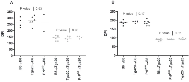
In a first set of experiment, we injected intraperitoneally 100ml of a ME-7 inoculum containing 1025 LogLD50. Wild-type B6 inoculated mice died after 252 days (IQR, 237–267 days) post-IP inoculation. As expected, the inoculated Tga20 mice have a shorter incubation period 130 days (IQR, 123–135 days) than the B6. This is explained by the fact that PrPc expression is 5 to 6 times higher in Tga20 mice than B6 wild type animals [2]. The chimeric mice on the B6 background (B6RB6, n = 4; Tga20RB6, n = 6 and the Prp0/0RB6, n = 2) presented the same incubation period (261 days; IQR, 232–295 days/278 days; IQR, 234–315 days/260 days, respectively) as wild-type B6. Similarly the incubation period of the chimeric mice on the Tga20 background were not significantly different than the Tga20 mice, whatever the origin of the BM used for the reconstitution: Tga20RTga20, n = 6, 140 days (IQR, 124–158 days), B6RTga20, n = 7, 144 days (IQR, 116–161 days), and Prp0/0RTga20, n = 4, 150 days (IQR, 139–155 days).
When injected with the lymphotropic RML strain (1024 LogLD50) (Fig. 1b), wild-type B6 inoculated mice died after 201 days (IQR, 192–207 days) post-IP inoculation. Similar incubation times were observed in the chimeric mice B6RB6, n = 5, 188 days (IQR, 174–196 days), in the Tga20RB6 mice, n = 5, 196 days (IQR, 183–196 days), and in the Prp0/0RB6, n = 8, 185 days (IQR, 175–192 days). The Tga20 mice died after 99 days (IQR, 94–106 days) post-IP inoculation, compared to 92 days (IQR, 91–97 days) for the chimeric Tga20RTga20 (n = 8), 91 days (IQR, 87–93 days) for the B6RTga20 mice (n = 8) and 92 days (IQR, 91–94 days) for Prp0/0RTga20 (n = 8). As expected, none of the chimeric mice harboring a Prp0/0genetic background developed a clinical disease, for lack of expression of PrPc protein [1].
From these experiments, we could conclude that the expression level of the PrPcby the hematopoietic cells does not influence the scrapie incubation period. However it has been suggested that, when using high doses of inoculum, the PrPScmight be able to bypass the lymphoreticular system and invade directly the peripheral nervous system [3], therefore the PrPc expression of BM derived cells would have little influence on the incubation time. In agreement with this idea, amplification of infectivity in PrP positive BM derived cells might be necessary in order to achieve neuroinvasion after inoculation with lower doses of prions.
To test this hypothesis we have inoculated intraperitoneally the chimeric mice on the Tga20 background with limiting doses of the lymphotropic RML strain (1027LogLD50). Accumulation of the RML strain in lymphoid organs has been extensively described [9,10], in contrast to the ME-7 strain [11,12]. If RML amplification by BM derived cells is required for neuroinvasion, therefore we would expect to observe a difference in the incubation period between Tga20 mice reconstituted with BM overexpressing PrPc and Tga20 mice reconstituted with BM sampled from Prp0/0mice. As shown in Fig. 2, no difference in incubation period was observed whatever the origin of the BM
Figure 1. PrPcoverexpression by the hematopoietic system does not play a role in neuroinvasion. B6 (black) or Tga20 (grey) mice were lethally irradiated, reconstituted with femoral bone marrow cells from B6 (square), Tga20 (triangle) or Prp0/0(circle) mice and inoculated either with
the ME-7 (1025LogLD50) (A) or RML (1024LogLD50) (B) prion scrapie strains. The incubation periods are expressed as days post-inoculation (DPI). P values were obtained using the Kruskal-Wallis ANOVA test. In panel (A), P value of 0.93 was determined for the reconstituted B6 mice group and of 0.9 for the reconstituted Tga20 group. In panel (B), the P values were of 0.17 and of 0.32 for reconstituted B6 and Tga20 mice groups, respectively. doi:10.1371/journal.pone.0013166.g001
used for reconstitution: B6RTga20, n = 6, 140 days (IQR, 120– 165 days), Tga20RTga20, n = 11, 139 days (IQR, 119–179 days), and Prp0/0RTga20, n = 7, 160 days (IQR, 126–224 days). Since the amount of inoculum was very low, three inoculated mice did not develop clinical disease, while the seven mice that developed scrapie showed longer and more dispersed incubation periods than mice inoculated with high infective doses (Fig. 1). These results demonstrate that the replication of lymphotropic RML strain in BM derived cells does not play a critical role in neuroinvasion.
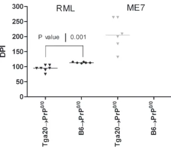
ME-7 infectivity can be detected in the spleen of PrP0/0 mice reconstituted with Tga20 BM
Since RML strain replicates in BM derived cells [13] while ME-7 does not [5], the spleens of PrP0/0chimeric mice were analyzed for the presence of prion infectivity. As expected, PrP0/0chimeric mice did not develop a clinical disease, and therefore have been sacrificed at the end of the experiments (450 days post-inoculation). Because low levels of infectivity could not be detected by western blotting analysis, we performed a bioassay by intracerebral inoculation of Tga20 mice with these spleen extracts. Tga20 mice inoculated with spleen extract from both Tga20RPrp0/0 (n = 7) and B6RPrp0/0 (n = 6) died of scrapie after 95 days (IQR, 75–105 days) and 113 days (IQR, 111–116 days) post-inoculation, respectively (Fig. 3). These results corrob-orate reported data [13]. In contrast, no Tga20 mice infected with a crude extract from the spleen of B6RPrp0/0ME-7 infected mice developed the disease after 450 days post-inoculation (n = 6). This is in agreement with previous data [5]. However, 6/6 Tga20 mice inoculated with the spleen of Tga20RPrp0/0 infected mice have developed a typical clinical disease, with accumulation of PrPScin brain (data not shown). This indicates that ME-7 strain can replicate in the hematopoietic compartment at a low level, but only when BM derived cells overexpress PrPc.
Discussion
Prion diseases are caused by a conformational change in widely expressed PrPcprotein, leading to the formation and accumulation of PrP aggregates. Although prion diseases cause degeneration of the central nervous system, the presence of infectivity can be detected in lymphoid tissues at a very early stage of the disease after peripheral inoculation [14]. Studies have shown that severely immunodeficient mice lacking B and T lymphocytes are resistant to peripheral prion infection, but susceptibility can be restored following BM transplantation [3], [4]. Further studies point out the prominent role of FDCs, which are not BM derived cells, in the initial replication of prion [5,6,7]. Even if infectivity has been demonstrated in lymphoid organs and blood, the role of the hematopoietic compartment still remains unclear. The question is whether prion replication by BM derived cells is involved in neuroinvasion.
Besides differences in scrapie strains used, interpretation of these sophisticated experiments is complicated by variations in the amounts of inoculum, and the genetic background of the mice utilized [15]. The sanitary status might also interfere with interpretation of the results. As a matter of fact, mice infection with specific pathogen and/or opportunistic agents could lead to chronic inflammation that is known to modify prion infection [16]. Similarly the fact that in all the studies published the mice were not on a congenic background could not guarantee a full histocompatibility situation.
In order to avoid possible graft host reaction, our strategy was to create chimeric mice on the B6 background expressing different levels of PrPcin the hematopoietic compartment. In addition we performed embryo transfers to obtain animals devoid of pathogens, to limit inflammatory chronic infection that could interfere with the pathophysiology of the disease [16]. After lethal irradiation, Prp0/0, Tga20 or C57/BL6J congenic mice were
Figure 2. No difference in disease incubation period is observed in Tga20 chimeric mice inoculated with limiting doses of the RML strain (1027LogLD50). Tga20 mice were lethally
irradiated, reconstituted with femoral bone marrow cells from B6 (square), Tga20 (triangle) or Prp0/0(circle) mice and inoculated with the
RML prion scrapie strains. The incubation periods are expressed as days post-inoculation (DPI). At this low dose, only 7/10 mice developed scrapie. A P value of 0.19 was obtained using the Kruskal-Wallis ANOVA test when comparing the three groups of mice.
doi:10.1371/journal.pone.0013166.g002
Figure 3. PrPcoverexpression by the hematopoietic cells favors
prion agent replication in the spleen of reconstituted Prp0/0 mice. Spleen of Prp0/0 mice lethally irradiated reconstituted with
femoral bone marrow cells from Tga20 (inverse triangle) or B6 (circle) mice and inoculated with RML (black) or ME-7 (grey) strains were sampled 450 days post-inoculation and subsequently inoculated to Tga20 mice. Days post-inoculation (DPI) are represented. A P value of 0.001 was obtained using the Mann-Whitney t-test when comparing the Tga20RPrp0/0and B6RPrp0/0mice.
reconstituted with BM from each of the other mice to yield nine different types of chimeric animals. The chimeric mice models used in this study were not designed to assess the role of FDCs. These cells do not derive from hematopoietic precursors, and it has been clearly observed that mice defective for FDCs present a delay in the development of the clinical diseases, showing that the initial replication of infectivity in FDCs is critical for neuroinvasion [5,6,7]. Nevertheless some PrpSc strains such as RML show lymphotropism and their infectivity may involve a contribution of hematopoietic cells [9,10]. In this situation the level of PrP expression by hematopoietic cells should influence the incubation period. Conversely, no effect should be expected when using ME-7, a non lymphotropic strain [11],[12].
PrPc is widely expressed in various types of tissues and cells, including hematopoietic stem cell [17]. Tga20 mice that carry 60 copies a ‘half genomic’ sequence of the prion protein gene, express approximately 5–6 fold higher levels of PrPcin the central nervous system. In these mice, the PrPc overexpression has also been observed both in CD3 positive thymocytes [18] and splenocytes [18,19,20] indicating that Tga20 BM derived cells overexpressed PrPc.
Using high doses of either ME-7 or RML scrapie agents, we observed that the PrP status of the hematopoietic compartment did not modify the incubation time of the disease. These results are congruent with some previous partial data [21,22]. To explain this, it has been proposed that high doses of inoculums may bypass the lymphoreticular system and directly invade the central nervous system via peripheral nerves, with no amplification in lymphoid tissues [3]. However, when chimeric mice were inoculated with limiting doses of RML, we observed that the level of PrPc expression by BM derived cells did not affect the time course of scrapie infection. Therefore it is clear that the replication of prion
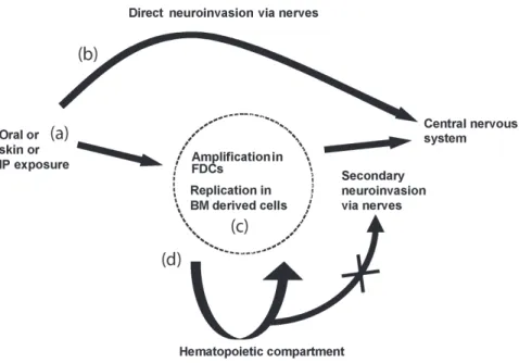
in the hematopoietic compartment has no influence on neuroinva-sion, even when the strain accumulates in lymphoid cells. This could indicate, as previously reported [23], that circulating cells are unlikely to play a role in neuroinvasion. However this does not exclude that cells such as dendritic cells could spread infectivity to other cells or to peripheral nerves, which in turn are involved in neuroinvasion [24]. If BM cells do not play a significant direct role in neuroinvasion, in situations such as contaminated blood products, these cells could transfer infectivity as previously reported [25–26]. As a summary, possible routes of prion neuroinvasion after peripheral exposure have been schematized in Fig. 4.
An unexpected result from this study concerns the paradigm of Brown and Blutter. When inoculated with lymphotropic RML strain, wild-type bone marrow cells transplanted in PrP deficient mice can restore accumulation and replication of prion in spleen, indicating that cells other than FDCs can replicate prion in the secondary lymphoid tissues [9]. By contrast Brown et al. reported a diametrically opposite outcome of similar experiment when reconstituted mice were inoculated with ME-7 strain [5]. We confirm these data; however we have evidenced infectivity in the spleen tissue of chimeric mice reconstituted with BM derived cells sampled from Tga20, when the ME-7 infectivity titer was low. This demonstrates that both ME-7 and RML can replicate in cells derived from hematopoietic compartment, other than FDCs.
Methods Ethics Statement
Animals were housed according with the French Ethical Committee (Decree 87–848) and European Community Directive 86/609/EEC. Experiments were carried out under the supervision
Figure 4. Routes of prion neuroinvasion after peripheral exposure. (a) Natural prion diseases are often acquired via peripheral exposure such as orally, or through skin lesions. How prion reaches its peripheral targets is not known. (b) Direct invasion of the central nervous system might occur with high doses of prion or exposure to neuroinvasive strains. (c) Whereas after exposure to limiting doses of infectivity or less neuroinvasive strains, replication in FDCs in the germinal centers of local lymphoid tissues might be necessary prior to neuroinvasion via closely associated nerve fibers. FDCs are dependent on the presence of B lymphocytes for maturation signals, such as lymphotoxin. (d) Haematogenous spread of infectivity via circulating bone marrow derived cells would not play a role in direct neuroinvasion.
doi:10.1371/journal.pone.0013166.g004
of JYC (agreement nu 38 05 17) in the animal care facilities approved by the Direction des Services Ve´te´rinaires de l’Ise`re (Nu A 38 516 10006). Before surgical procedure and prion inoculation, mice were anesthetized with a mixture injected intra-peritoneally of ketamine hydrochloride (Imalgen 500, Merial, 25 mg/kg body weight) and xylasine (Rompun, Bayer Healthcare, 12.5 mg/kg body weight).
Mice
In order to circumvent tolerance problems after hematopoietic transfer, the speed congenic technology was used to generate mice with the same C57/BL6J genetic background for the two following strains: Prp0/0prion deficient mice [1], and Tga20 mice [2] that were obtained from Pr. Charles Weissmann (Scripps Institute, Florida). The technology consists in using genetic markers throughout the genome to speed up ‘recovery’ of the recipient genome in the backcrossing phase of the construction of a congenic strain as described [27]. Tga20 mice, Prp0/0 and C57/ BL6J, hereafter called B6 (Charles River Laboratories, Lyon), were housed in ventilated cages and maintained under specific pathogen-free conditions. Scrapie inoculated mice were housed in a biosafety laboratory level 3 animal care facility in cages placed in ventilated and negative pressure insulator.
Bone marrow chimeras
Chimeric mice (.120) were reconstituted by injecting into the tail vein 5 to 106106femoral BM cells into lethally irradiated (9.5 Gy) recipient 4 weeks old mice. B6 mice were reconstituted with BM cells from either B6, Tga20 or Prp0/0 mice; Tga20 mice reconstitution was performed with Tga20, B6 or Prp0/0BM cells; and Prp0/0were reconstituted with BM sampled from Tga20, B6 or Prp0/0 mice. The day following reconstitution, mice were treated with ciprofloxacine (0.1 mg/ml in drinking water) for ten days. Successful hematopoietic reconstitution was assessed 2 months after engraftment: CD3, CD4, CD8 lymphocytes total
numbers and PrPcexpression by these cells were determined by flow cytometry analysis (data not shown). Failure led to the death of the mice within two weeks following irradiation.
Source of the scrapie agent and inoculation
The ME-7 and the RML prion strains were maintained by successive inoculations into B6 mice. The scrapie inocula were prepared from brain tissues collected from terminally sick mice. The brain homogenate was prepared in PBS (10% w/v), and the presence of PrPScconfirmed by western blot analysis as described in [28]. More than 100 mice were successfully reconstituted and intraperitoneally inoculated with 100ml of inoculum. Endpoint titrations were performed as described in [10].
Measurement of the incubation period
For ethical reasons, the mice were sacrificed at the onset of the disease rather then waiting for their death. The onset was defined by the clear appearance of at least three of the following neurological symptoms: trembling, prostration, feet clasping when lifted, increased tone of the tail. The incubation period was taken as the time from inoculation to the euthanasia of the mice. Mice were monitored three times a week, beginning two months after inoculation. Tissues were collected, and frozen (280uC) for subsequent western blot analysis, or reinoculation into Tga20 mice. Incubation period data was expressed as median and Inter Quartile Range (IQR). Differences in incubation periods were tested by the Kruskal-Wallis ANOVA test (Fig. 1 & 2) or the Mann-Whitney t-test (Fig. 3). In all comparisons, the level of significance was set at 0.05.
Author Contributions
Conceived and designed the experiments: CL PN MFCD JG JYC. Performed the experiments: CL CLV. Analyzed the data: CL CLV JG JYC. Wrote the paper: CL JG JYC.
References
1. Bueler H, Aguzzi A, Sailer A, Greiner RA, Autenried P, et al. (1993) Mice devoid of PrP are resistant to scrapie. Cell 73: 1339–1347.
2. Fischer M, Rulicke T, Raeber A, Sailer A, Moser M, et al. (1996) Prion protein (PrP) with amino-proximal deletions restoring susceptibility of PrP knockout mice to scrapie. Embo J 15: 1255–1264.
3. Lasmezas CI, Cesbron JY, Deslys JP, Demaimay R, Adjou KT, et al. (1996) Immune system-dependent and -independent replication of the scrapie agent. J Virol 70: 1292–1295.
4. Fraser H, Brown KL, Stewart K, McConnell I, McBride P, et al. (1996) Replication of scrapie in spleens of SCID mice follows reconstitution with wild-type mouse bone marrow. J Gen Virol 77: 1935–1940.
5. Brown KL, Stewart K, Ritchie DL, Mabbott NA, Williams A, et al. (1999) Scrapie replication in lymphoid tissues depends on prion protein- expressing follicular dendritic cells. Nat Med 5: 1308–1312.
6. Thielen C, Antoine N, Melot F, Cesbron JY, Heinen E, et al. (2001) Human FDC express PrPc in vivo and in vitro. Dev Immunol 8: 259–266.
7. Klein MA, Frigg R, Raeber AJ, Flechsig E, Hegyi I, et al. (1998) PrP expression in B lymphocytes is not required for prion neuroinvasion. Nat Med 4: 1429–1433.
8. van Nierop K, de Groot C (2002) Human follicular dendritic cells: function, origin and development. Semin Immunol 14: 251–257.
9. Blattler T, Brandner S, Raeber AJ, Klein MA, Voigtlander T, et al. (1997) PrP-expressing tissue required for transfer of scrapie infectivity from spleen to brain. Nature 389: 69–73.
10. Kaeser PS, Klein MA, Schwarz P, Aguzzi A (2001) Efficient lymphoreticular prion propagation requires PrP(c) in stromal and hematopoietic cells. J Virol 75: 7097–7106.
11. Race R, Oldstone M, Chesebro B (2000) Entry versus blockade of brain infection following oral or intraperitoneal scrapie administration: role of prion protein expression in peripheral nerves and spleen. J Virol 74: 828–833. 12. Mabbott NA, Williams A, Farquhar CF, Pasparakis M, Kollias G, et al. (2000)
Tumor necrosis factor alpha-deficient, but not interleukin-6-deficient, mice resist peripheral infection with scrapie. J Virol 74: 3338–3344.
13. Aguzzi A (2003) Prions and the immune system: a journey through gut, spleen, and nerves. Adv Immunol 81: 123–171.
14. Mabbott NA, Bruce ME (2001) The immunobiology of TSE diseases. J Gen Virol 82: 2307–2318.
15. Aucouturier P, Carnaud C (2002) The immune system and prion diseases: a relationship of complicity and blindness. J Leukoc Biol 72: 1075–1083. 16. Heikenwalder M, Zeller N, Seeger H, Prinz M, Klohn PC, et al. (2005) Chronic
lymphocytic inflammation specifies the organ tropism of prions. Science 307: 1107–1110.
17. Zhang CC, Steele AD, Lindquist S, Lodish HF (2006) Prion protein is expressed on long-term repopulating hematopoietic stem cells and is important for their self-renewal. Proc Natl Acad Sci U S A 103: 2184–2189.
18. Jouvin-Marche E, Attuil-Audenis V, Aude-Garcia C, Rachidi W, Zabel M, et al. (2006) Overexpression of Cellular Prion Protein Induces an Antioxidant Environment Altering T Cell Development in the Thymus. J Immunol 176: 3490–3497.
19. Glatzel M, Aguzzi A (2000) PrP(C) expression in the peripheral nervous system is a determinant of prion neuroinvasion. J Gen Virol 81: 2813–2821.
20. Zabel M, Greenwood C, Thackray AM, Pulford B, Rens W, et al. (2009) Perturbation of T-cell development by insertional mutation of a PrP transgene. Immunology 127: 226–236.
21. Prinz M, Montrasio F, Klein MA, Schwarz P, Priller J, et al. (2002) Lymph nodal prion replication and neuroinvasion in mice devoid of follicular dendritic cells. Proc Natl Acad Sci U S A 99: 919–924.
22. Raeber AJ, Klein MA, Frigg R, Flechsig E, Aguzzi A, et al. (1999) PrP-dependent association of prions with splenic but not circulating lymphocytes of scrapie-infected mice. Embo J 18: 2702–2706.
23. Raymond CR, Mabbott NA (2007) Assessing the involvement of migratory dendritic cells in the transfer of the scrapie agent from the immune to peripheral nervous systems. J Neuroimmunol 187: 114–125.
24. Aucouturier P, Geissmann F, Damotte D, Saborio GP, Meeker HC, et al. (2001) Infected splenic dendritic cells are sufficient for prion transmission to the CNS in mouse scrapie. J Clin Invest 108: 703–708.
25. Hunter N, Foster J, Chong A, McCutcheon S, Parnham D, et al. (2002) Transmission of prion diseases by blood transfusion. J Gen Virol 83: 2897–2905. 26. Wroe SJ, Pal S, Siddique D, Hyare H, Macfarlane R, et al. (2006) Clinical presentation and pre-mortem diagnosis of variant Creutzfeldt-Jakob disease associated with blood transfusion: a case report. Lancet 368: 2061–2067.
27. Visscher PM (1999) Speed congenics: accelerated genome recovery using genetic markers. Genet Res 74: 81–85.
28. Follet J, Lemaire-Vieille C, Blanquet-Grossard F, Podevin-Dimster V, Lehmann S, et al. (2002) PrP expression and replication by Schwann cells: implications in prion spreading. J Virol 76: 2434–2439.