HAL Id: hal-02154583
https://hal.archives-ouvertes.fr/hal-02154583
Submitted on 26 May 2020
HAL is a multi-disciplinary open access
archive for the deposit and dissemination of
sci-entific research documents, whether they are
pub-lished or not. The documents may come from
teaching and research institutions in France or
abroad, or from public or private research centers.
L’archive ouverte pluridisciplinaire HAL, est
destinée au dépôt et à la diffusion de documents
scientifiques de niveau recherche, publiés ou non,
émanant des établissements d’enseignement et de
recherche français ou étrangers, des laboratoires
publics ou privés.
Copyright
sequence extensions but retains fusogenic function
Aurélie Landemaine, Andres Ramirez-Martinez, Olivier Monestier, Nathalie
Sabin, Pierre-Yves Rescan, Eric N. Olson, Jean-Charles Gabillard
To cite this version:
Aurélie Landemaine, Andres Ramirez-Martinez, Olivier Monestier, Nathalie Sabin, Pierre-Yves
Res-can, et al.. Trout myomaker contains 14 minisatellites and two sequence extensions but retains
fu-sogenic function. Journal of Biological Chemistry, American Society for Biochemistry and Molecular
Biology, 2019, 294 (16), pp.6364-6374. �10.1074/jbc.RA118.006047�. �hal-02154583�
Trout myomaker contains 14 minisatellites and two sequence
extensions but retains fusogenic function
Received for publication, October 2, 2018, and in revised form, February 26, 2019 Published, Papers in Press, February 28, 2019, DOI 10.1074/jbc.RA118.006047 Aurélie Landemaine‡, Andres Ramirez-Martinez§, Olivier Monestier¶, Nathalie Sabin‡, Pierre-Yves Rescan‡, X Eric N. Olson§, andX Jean-Charles Gabillard‡1
From the‡Institut National de la Recherche Agronomique, UR1037 Laboratory of Fish Physiology and Genomics, 35000 Rennes, France, the§Department of Molecular Biology, Hamon Center for Regenerative Science and Medicine, University of Texas Southwestern Medical Center, Dallas, Texas 75390, and¶Institute of Interdisciplinary Research in Human and Molecular Biology, Université Libre de Bruxelles, 1070 Bruxelles, Belgium
Edited by Jeffrey E. Pessin
The formation of new myofibers in vertebrates occurs by myoblast fusion and requires fusogenic activity of the muscle-specific membrane protein myomaker. Here, using in silico (BLAST) genome analyses, we show that the myomaker gene from trout includes 14 minisatellites, indicating that it has an unusual structure compared with those of other animal species. We found that the trout myomaker gene encodes a 434 –amino acid (aa) protein, in accordance with its apparent molecular mass (⬃40 kDa) observed by immunoblotting. The first half of the trout myomaker protein (1–220 aa) is similar to the 221-aa mouse myomaker protein, whereas the second half (222–234 aa) does not correspond to any known motifs and arises from two protein extensions. The first extension (⬃70 aa) apparently appeared with the radiation of the bony fish clade Euteleostei, whereas the second extension (up to 236 aa) is restricted to the superorder Protacanthopterygii (containing salmonids and pike) and corresponds to the insertion of minisatellites having a length of 30 nucleotides. According to gene expression analyses, trout myomaker expression is consistently associated with the formation of new myofibers during embryonic development, postlarval growth, and muscle regeneration. Using cell-mixing experiments, we observed that trout myomaker has retained the ability to drive the fusion of mouse fibroblasts with C2C12 myo-blasts. Our work reveals that trout myomaker has fusogenic function despite containing two protein extensions.
Skeletal muscle is largely composed of myofibers: multinu-cleated cells whose formation depends on fusion of progenitor cells known as myoblasts. Myoblasts proliferate, differentiate into myocytes, fuse to form multinucleated myotubes, and finally mature into functional myofibers. The fusion process is a
critical step in the formation and regeneration of muscle. In mammals, some proteins involved in myoblast fusion have been identified, but the complete molecular mechanisms that coor-dinate this process are not completely understood. Nephrin, a cell surface protein, has been shown to be essential for myocyte
fusion in mice and normal muscle development in zebrafish (1).
The protein Kirrel, the homolog of the Drosophila Kirre pro-tein, is also necessary for proper fusion of myocytes in zebrafish
(2), although its function in mammals has not yet been
con-firmed and remains a subject of debate (3). In zebrafish, a
recep-tor ligand pair (Jam-b/Jam-c) has been reported to be involved
in myocyte fusion (4).
Recently, the muscle-specific micropeptide myomixer has
been shown to be essential for myoblast fusion in mice (5–7)
and zebrafish (8). Another muscle-specific transmembrane
protein of 221 aa,2called myomaker, was found to be necessary
for myocyte fusion during mouse embryonic development (9)
and muscle regeneration (10). In humans, the loss of myomaker
activity can lead to disease (11). In vitro, mouse myomaker
drives heterologous fusion between fibroblasts and myoblasts,
but not between fibroblasts (9). However, when myomaker and
myomixer are ectopically overexpressed together, they are
suf-ficient to drive fusion between fibroblasts (5–7). A structure–
function analysis demonstrated that the two last cysteines of the C-terminal end of myomaker are necessary for its fusogenic
function (12, 13).
In adult mouse muscle, myomaker is not expressed except in response to injury, when it is up-regulated to promote
regener-ation (10). In zebrafish, the 221-aa myomaker protein shows
high similarities with murine myomaker and is necessary for
myocyte fusion during embryonic development (14, 15), and it
also promotes the heterologous fusion between mouse
fibro-blasts and myofibro-blasts (12). As shown in our previous study,
myo-maker expression in the zebrafish myotome is no longer
detected just before hatching (14). However, no data are
avail-able on myomaker characteristics and function in nonmodel species.
In the present study, we characterized the trout myomaker gene, which encodes an unexpectedly longer 434-aa protein. This work was supported by National Institutes of Health Grants AR-067294,
HL-130253, DK-099653, and HD-087351. This study was also supported by French National Research Agency Grant ANR-12-JSV7– 0001-01, by Brit-tany region funds (to A. L.), and by Robert A. Welch Foundation Grant 1-0025 (to E. N. O.). The authors declare that they have no conflicts of inter-est with the contents of this article. The content is solely the responsibility of the authors and does not necessarily represent the official views of the National Institutes of Health.
The nucleotide sequence(s) reported in this paper has been submitted to the DDBJ/GenBankTM/EBI Data Bank with accession number(s) KY563699.
This article containsFigs. S1–S4.
1To whom correspondence should be addressed. E-mail: Jean-Charles.
2The abbreviations used are: aa, amino acid(s); nt, nucleotide(s); dpf, day(s) postfertilization; qRT, quantitative real-time; WGD, whole genome dupli-cation; DM, differentiation medium.
cro
ARTICLE
6364
J. Biol. Chem. (2019) 294(16) 6364 –6374at INRA Institut National de la Recherche Agronomique on June 4, 2019
http://www.jbc.org/
Whole-mount in situ hybridization and quantitative real-time PCR analyses revealed that myomaker is expressed not only in hyperplasic zones of embryonic myotome but also in postlarval myotomal muscle. Our results clearly show that myomaker up-regulation was associated with myotube formation during mus-cle regeneration and the in vitro fusion of trout myocytes. Fur-thermore, the 14 tandem repeats (minisatellites) in the coding region of the trout myomaker gene do not disrupt its fusogenic capacity.
Results
Identification of the trout myomaker gene
We performed a BLAST search in the trout genome (17)
using the sequence of zebrafish myomaker protein (NP_ 001002088) to identify the trout myomaker gene, and we found a single gene (GSONMG00014531001) in scaffold_482 that
contained six exons encoding a protein of 434 aa (Fig. 1).
Although the number of exons was similar to the zebrafish gene, the length of the trout myomaker protein was twice as long as the zebrafish and the mouse orthologs, which only
com-prise 220 and 221 aa, respectively (9, 14). As shown in the
pro-tein sequence alignment, the first half (1–220 aa) of the trout myomaker protein sequence was well-conserved, sharing 88 and 71% identity with the zebrafish and mouse myomaker pro-teins, respectively. An analysis of the amino acid sequence showed that the two cysteines essential for myomaker fuso-genic function were also present in the trout myomaker protein at positions 219 and 220. The second half of the protein (221– 434 aa) was encoded by the sixth exon and exhibited no
homo-logy with the zebrafish or mouse myomaker protein. We ampli-fied exons 5 and 6 from total trout cDNA and sequenced the PCR product to confirm our in silico results. The sequencing results confirmed the splicing site of the sixth exon of the myomaker cDNA, leading to an ORF encoding 434 aa. More-over, using the sequence identified in the trout genome (GSONMG00014531001), we performed BLAST searches in the trout expressed sequence tag database (NCBI) and the
Phy-loFish database (16) that allowed us to identify a transcript of
2029 nt (GenBankTM accession number KY563699) that
included exons 1– 6. Furthermore, using a specific antibody against trout myomaker, we confirmed that the molecular mass
of trout myomaker (⬃40 kDa) is double that of mouse
myo-maker by Western blotting (Fig. 2andFig. S4). The in silico
analysis revealed the presence of three E-boxes (CANNTG) in the myomaker promoter.
According to the results of the synteny analysis, the trout myomaker gene is located in the FAM163b–Adamtsl2–
Tmem8c–TCC16 –Slc2a8locus (Fig. 3) in scaffold_482. Inter-estingly, a synteny conservation of this locus was observed within a region of chromosome 2 of the zebrafish genome and in the equivalent chromosomal region of the mouse genome (Chr 2). A whole genome duplication event occurred in sal-monid genome, leading to the duplication of some genes in the trout genome. Indeed, we were also able to identify another myomaker syntenic group in scaffold_2354 of the trout genome (Fig. 3). Nevertheless, whereas complete copies of the
FAM163b, Adamtsl2, and Slc2a8 genes were identified in this scaffold, only a partial sequence homologous to trout
myo-Figure 1. Structure of the trout myomaker gene. A, exon size (nt) is indicated above each exon, and intron size (nt) is indicated between exons. B, multiple
alignment between the protein sequences of myomaker. Red underlines indicate minisatellites. Accession numbers are as follows: O. mykiss, ARM20036; D. rerio, NP_001002088.1; Gallus gallus, AJZ77002.1; and Mus musculus, NP_079652.1. The alignment was made from the complete protein sequences using ClustalW multiple alignment tool.
at INRA Institut National de la Recherche Agronomique on June 4, 2019
http://www.jbc.org/
maker was identified between Adamtsl2 and TCC16. This sequence contained several deletions and stop codons in the ORF, thus coding for an additional but nonfunctional
myo-makergene (data not shown).
The trout myomaker gene contains 14 minisatellites in its coding region
Trout myomaker protein is 214 aa longer than the zebrafish orthologs because of a long C-terminal extension. A BLAST
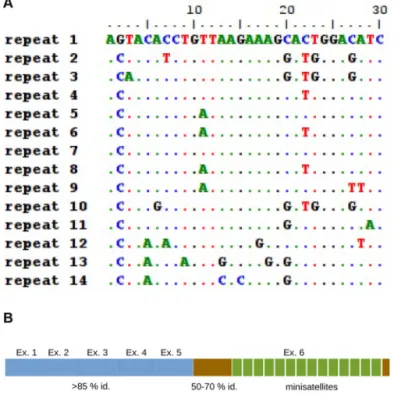
analysis of this protein extension revealed no homology with known motifs or other proteins. Surprisingly, a thorough anal-ysis of the sequence encoding this extension revealed the pres-ence of 14 tandem repeats of 30 nt coding amino acids 265– 424 (Fig. 1). The sequences of these 14 repeats are very well-con-served with each other, with sequence identities ranging from
70 to 96% (Fig. 4A). These tandem repeats are therefore
mini-satellites, as defined in a previous study (18). We performed
protein alignments and a phylogenetic analysis of myomaker proteins from several species to determine whether these
mini-satellites were widespread in teleost fish (Fig. 5). All tetrapod
sequences were found to encode a myomaker protein of 220 – 221 aa. A protein of the same length (221 aa) was observed in ancestral nonteleost fish, such as the spotted gar (Lepisosteus
oculatus), and teleosts that belong to the Otocephala lineage, such as zebrafish (Danio rerio), cave fish (Astyanax mexicanus), and herring (Clupea harengus). In sharp contrast, all teleosts examined that belonged to Euteleostei had a myomaker protein containing more than 220 aa. More specifically, in all consid-ered Neoteleostei species, myomaker consisted of 283–289 aa,
and the first 220 aa were highly similar (⬎85% identity) to the
zebrafish myomaker protein. Within the Neoteleostei species,
the extension of 63– 69 aa was well-conserved (⬃70% identity)
but showed no homology with zebrafish or mouse myomaker. Species belonging to the Protacanthopterygii lineage, such as rainbow trout (Oncorhynchus mykiss), Atlantic salmon (Salmo
salar), and pike (Esox lucius) contained a myomaker gene encoding a protein with more than 434 aa. After sequence alignment, we discovered that all these species contained an insertion of minisatellites within the first extension of 63– 69 aa
specific to the Neoteleostei (Fig. 4B). Although the number of
Figure 2. Myomaker protein has an apparent molecular mass of 40 – 42 kDa. A, Western blotting of trout myocytes extract with a custom trout
myo-maker antibody against the minisatellites. The antibody revealed one major band corresponding to a protein of⬃40 kDa. B, Cos7 cells were transfected with the full-length (WT) or truncated (dRE) myomaker cDNA, and Western blotting was performed 48 h later. A band of similar apparent mass (⬃40 kDa) was observed with the full-length, and no band was observed with the trun-cated form of myomaker (aa 1–219) that did not contain the epitope.
Figure 3. Myomaker gene is present in only one functional copy.
Con-served synteny around myomaker locus was obCon-served in mouse (M. muscu-lus), zebrafish (D. rerio), and rainbow trout (O. mykiss). The Genomicus software program ( http://www.genomicus.biologie.ens.fr/genomicus-trout-01.01/cgi-bin/search.pl;please note that the JBC is not responsible for the long-term archiving and maintenance of this site or any other third party hosted site) (37) was used to identify syntenic genes that were located near the myomaker gene. The cross indicates a pseudogene.
Figure 4. The sequence of the minisatellites are conserved. A, multiple
alignment of the 14 minisatellites present in the trout myomaker protein. B, structure of trout myomaker protein. The mouse homologous part of the protein is represented in blue. The amino acids arising from the first extension are represented in brown, and the minisatellites are in green.
Insertion of minisatellites in trout myomaker protein
at INRA Institut National de la Recherche Agronomique on June 4, 2019
http://www.jbc.org/
minisatellites varied by species (14 for trout, 17 for salmon, and 15 for pike), the minisatellite sequences were highly conserved
(⬎70%) among these species. In contrast, only two
minisatel-lites remain in the nonfunctional copy of myomaker.
Myomaker is expressed in embryonic and postlarval myotomal trout muscle
We performed whole-mount in situ hybridization to exam-ine myomaker expression during embryonic myogenesis. The myomaker transcript was not detected during the early stage of somitogenesis (13 dpf, data not shown) but was readily detected
at 17 dpf in all somites (Fig. 6A) when multinucleated fibers
begin to form (19). Transverse sections (Fig. 6B) through the
somites at 17 dpf showed that myomaker was expressed in the deep myotome, with stronger expression observed within the dorsal and ventral domains of the myotome. In contrast, the
myomakertranscript was not detected in the undifferentiated myogenic dermomyotome-like epithelium surrounding the primary myotome.
In addition, we measured myomaker expression in white muscle from 4-, 8-, and 18-month-old fish weighing 15, 150,
and 1500 g, respectively (Fig. 6D). Interestingly, at all three
stages, myomaker expression was readily detected in trout mus-cle samples, although its expression decreased as body weight increased. We analyzed trout myomaker expression in several tissues by qRT-PCR to determine whether myomaker
expres-sion was restricted to muscle. As shown inFig. 6C, the
myo-makergene was only expressed in white and red muscle and was expressed at similar levels between both muscle types. In line with this observation, Western blotting analysis revealed the
presence of myomaker protein only in myocyte extract and not
in other tissues (Fig. S4).
Myomaker is strongly up-regulated during the regeneration of trout muscle
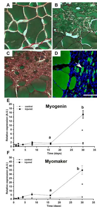
In vertebrates, the formation of new muscle fibers occurs during both embryogenesis and muscle regeneration. We stud-ied the kinetic of muscle regeneration in adult trout following mechanical muscle injury to determine whether myomaker is up-regulated during regeneration of trout muscle. The
histo-logical analysis (Fig. 7A) showed that 16 days after muscle injury
(Fig. 7B), a large number of myofibers was degraded, with many immune cells infiltrating into the injury site. After 30 days, all the injured fibers disappeared and were replaced with
connec-tive tissue containing small (⬍20m) round cells labeled in
green(Fig. 7C). Immunocytofluorescence staining revealed that these cells expressed myosin and that their nuclei was often
centrally positioned (Fig. 7D). These observations point out the
presence of newly formed muscle fibers on day 30, showing that muscle regeneration had occurred. Importantly, uninjured
control muscle did not contain small nascent myofibers (Fig.
7A), indicating that myofiber formation had ceased at this
stage, consistent with the results reported by Rescan et al. (20).
According to the qRT-PCR analysis, myogenin and myomaker gene expression in muscle did not change until day 16
postin-jury (Fig. 7,Eand F). In contrast, 30 days after injury, a sharp
increase in both myomaker and myogenin expression was observed in the injured muscle. Indeed, myogenin and
myo-makerwere expressed at 10- and 15-fold higher levels,
respec-Figure 5. Phylogenetic analysis of myomaker in tetrapods and teleosts. The phylogenetic tree was constructed from a multiple alignment of the complete
protein sequences using the neighbor-joining method. The numbers at the tree nodes represent percentage of bootstrap values after 1000 replicates. Full scientific names of species and respective accession numbers are detailed inFig. S3. The red star represents a whole genome duplication (3R or 4R). The numbers between brackets indicate the number of amino acids encoded by myomaker in each species.
at INRA Institut National de la Recherche Agronomique on June 4, 2019
http://www.jbc.org/
tively, in injured muscle compared with control muscle (Fig. 7,
Eand F).
Myomaker is up-regulated during myotube formation in vitro
After extracting trout satellite cells from white muscle, we induced the differentiation and fusion of trout satellite cells in
vitro (22). Quantitative PCR analysis showed an increase (2-fold) in myomaker expression soon after satellite cell
differ-entiation was induced (Fig. 8A). By performing
immunofluores-cence staining with an anti-myosin antibody, we quantified the
number of small myotubes (2⬍ nuclei ⱕ 4) and large myotubes
(5 ⱕ nuclei) during differentiation (Fig. 8B). Small myotubes
began to form 1 day after the induction of differentiation and were strongly increased up to day 3 of differentiation, whereas large myotubes appeared on day 2. These results showed that the maximum level of myocyte fusion occurred on days 2 and 3 and correlate with highest myomaker expression.
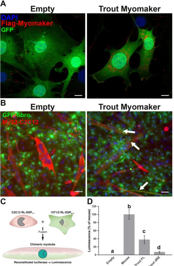
Trout myomaker drives heterologous cell fusion
We performed cell-mixing experiments using myoblasts (C2C12) and fibroblasts expressing GFP infected with a vector
Figure 6. Muscle-specific expression of myomaker starts in the embryo and persists after hatching in trout. A and B, analysis of myomaker
expres-sion during trout embryonic development by in situ hybridization. Embryos were analyzed at stage 20. The scale bars correspond to 500m (A) and 50 m (B). C and D, qRT-PCR analysis of myomaker expression in several tissues of trout (150 g; C) and in muscle of trout of different weight (D). A black star indicates the deep myotome where primary myogenesis takes place, and black arrows indicate dorsal and ventral domains of the myotome where stratified hyperplasia takes place. The red arrow indicates the dermomyo-tome-like epithelium. Total RNA was extracted from three different fish (n⫽ 3). The qRT-PCR results are presented as a ratio of myomaker expression and eF1a expression, and the bars represent the standard error. The letters (a– d) in D indicate the significant differences between means (p⬍ 0.05; Kruskal– Wallis rank test followed by the post hoc Dunn test). W., white; R., red.
Figure 7. Myomaker is induced during trout muscle regeneration. A–D,
histological analysis during muscle regeneration in rainbow trout. Muscle sections (10m) were stained with Sirius Red (connective tissue), Fast Green (muscle fibers), and hematoxylin (nuclei). A–D, uninjured white muscle (A) and white muscle at days 16 (B) and 30 (C and D) after muscle injury. Im-munocytofluorescence detection of myosin heavy chain (MyHC) was per-formed at 30 days (D). A white arrow indicates newly per-formed myofibers. The scale corresponds to 100m. E and F, gene expression profile of myogenin (E) and myomaker (F) during muscle regeneration in rainbow trout. The expres-sion of myogenin and myomaker were normalized with eF1a expresexpres-sion, and bars represent the standard error. The letters (a and b) in E and F indicate the significant differences between means within the same treatment (control or injured). The asterisk indicates significant differences between treatments at a given time. Statistical significance (p⬍ 0.05) was determined using the Kruskal–Wallis rank test followed by the post hoc Dunn test.
Insertion of minisatellites in trout myomaker protein
at INRA Institut National de la Recherche Agronomique on June 4, 2019
http://www.jbc.org/
expressing trout myomaker cDNA to determine whether trout myomaker is able to drive cell fusion. We first showed that the infection of fibroblasts with trout myomaker construct actually resulted in the production of trout myomaker protein using
immunofluorecence staining (Fig. 9A). After 5 days of
co-cul-ture, we failed to observe any fusion between C2C12 and the
GFP-empty fibroblasts (Fig. 9B). In contrast, GFP-myomaker–
infected fibroblasts were able to fuse with C2C12 myoblasts. We implemented a dual split luciferase assay previously used to monitor cell– cell fusion to accurately quantify the fusion
between fibroblasts and myoblasts (21). In this assay, luciferase
activity is only reconstituted when fusion between fibroblasts
and myoblasts occurs (Fig. 9C). Fibroblasts were infected with
full-length or truncated trout myomaker along with mouse
myomixer to increase the basal fusion level (5). This assay
con-firms that the full-length myomaker protein is able to drive the fusion of fibroblasts with myoblasts. Surprisingly, deletion of trout myomaker C terminus resulted in a significant reduction in cell fusion. Together, our results confirmed that trout myo-maker drove heterologous cell fusion, although to a lesser
extent than mouse myomaker (Fig. 9D).
Discussion
Given the unique structure of the trout myomaker protein, we aimed in this study to determine whether the expression and function of myomaker are conserved in this nonmodel fish. Based on sequence alignments and phylogenetic and syntenic analyses, we identified the unique functional myomaker gene in the trout genome. We also identified a myomaker pseudogene containing long deletions in the coding region. This pseudo-gene probably results from the salmonid-specific whole
genome duplication (WGD) that occurred⬃96 ⫾ 5 million
years ago (17). After the WGD, the salmonid genome
under-went a process of gene pseudogenization that resulted in the
loss of half of the duplicated genes in the trout genome (17).
Therefore, we hypothesize that the second identified
myo-makergene originated from the WGD and became a pseudo-gene through deletions and mutations.
The trout myomaker protein consists of 434 aa with an apparent molecular mass of 40 kDa and is nearly twice the size of the mouse and zebrafish myomaker proteins. As shown in the sequence alignment, the first 220 aa of trout myomaker are 71– 88% similar to the mouse and zebrafish orthologs. Impor-tantly, trout myomaker contains the two conserved cysteines
essential for mouse myomaker function (12). Furthermore, the
hydrophobicity analysis (data not shown) strongly suggested the presence of seven transmembrane domains, similar to the
mouse myomaker protein (9). Together, these results highlight
the strong evolutionary conservation of the first section of the trout myomaker protein.
The additional amino acid stretch in trout myomaker mainly consists of 14 tandem repeats of 30 nucleotides in the coding region. A tandem repeat is a short sequence (unit) that is
repeated several times in a head-to-tail orientation (18).
Repeats with units less than 9 nucleotides in length are known as microsatellites, and those with 10 nucleotides or more are known as minisatellites. Therefore, the identified tandem repeats in trout myomaker correspond to minisatellites. Tan-dem repeats are preferentially located in noncoding regions, but when tandem repeats are present in protein coding regions, they most commonly occur in multiples of three nucleotides to
avoid frameshifts (18). Accordingly, the trout myomaker
mini-satellites consist of 30 nucleotides that preserve the ORF of
myomaker. An analysis of the human genome indicates that tandem repeats are present in only 17% of the genes and that 14
repeated minisatellites (⬎30 nt) in the ORF are very rare (18).
The phylogenetic analysis and sequence alignments showed that the minisatellites detected in trout myomaker are also pres-ent and conserved in other Protacanthopterygii species such as salmon (S. salar) and pike (E. lucius). For the minisatellites to be present through the Protacanthopterygii group, they must have appeared before the salmonid-specific WGD. Tandem
repeats are unstable (18), and thus the minisatellites in
myo-makermust have undergone duplications and deletions at dif-ferent rates in salmonids, leading to differing numbers of mini-satellites in this group. The phylogenetic analysis also revealed that teleost myomaker sequences were classified into three groups according to myomaker protein length. Teleost belong-ing to Otocephala contain a myomaker sequence encodbelong-ing 220 –221 aa, as observed in all tetrapods. The Protacanthop-terygii contain minisatellites in the myomaker gene that are translated into a protein of 434 – 456 aa. Surprisingly, teleost of the third group (Neoteleostei) do not contain minisatellites, and the length of the myomaker protein ranges from 283 to 289 aa. The first 220 aa of that myomaker sequences are highly similar to mouse myomaker, whereas residues 221–289 have no homology with any known protein motif. Interestingly, this
sequence of⬃70 aa is well-conserved (⬃70% identity) in
Neo-teleostei and is also present in Protacanthopterygii, in addition to the minisatellites. This phylogenetic analysis allowed us to determine the evolutionary history of the myomaker gene. The ancestral myomaker gene consisted of 220 –221 aa and evolved by maintaining the same sized protein in tetrapods and some teleost fish (Otocephala). With the appearance of Euteleostei, a
Figure 8. Myomaker expression is induced during trout satellite cell dif-ferentiation. A, analysis of myomaker gene expression during satellite cell
differentiation. The cells were cultivated in proliferative medium (PM) and then in differentiation medium for 1, 2, and 3 days (DM1, DM2, and DM3). The qRT-PCR results are presented as a ratio of myomaker and eF1a expression, and bars represent the standard error. B, myocyte fusion quantification dur-ing the satellite cell differentiation. The cells were cultivated in proliferative medium (PM) and then in differentiation medium for 1, 2, and 3 days (DM1, DM2, and DM3). The number of myotubes with 2– 4 nuclei or with⬎5 nuclei was determined by immunocytofluorescence analysis of MyHC. Different let-ters (a– c or A and B) indicate significant differences between means. Statisti-cal significance (p⬍ 0.05) was determined using the Kruskal–Wallis rank test followed by the post hoc Dunn test.
at INRA Institut National de la Recherche Agronomique on June 4, 2019
http://www.jbc.org/
Figure 9. Trout myomaker is fusogenic in vitro. A, expression of trout myomaker in 10T1/2 mouse GFP fibroblasts after infection with trout Myomaker vector
or empty vector. A strong red signal was observed only in cells infected with the trout myomaker vector. B, cell-mixing experiments with GFP-fibroblast and myoblasts (C2C12). The cultures were differentiated for 4 days and then immunostained with myosin antibody as a marker for myotubes. White arrows indicate fusion between fibroblasts and myoblasts (orange labeling). Scale bar corresponds to 50m. C, schematic representation of the dual split luciferase assays. The assay measures reconstituted luciferase activity from fusion between myoblasts expressing pRluc8155–156-DSP1–7 (RL-DSP1) and fibroblasts expressing pRluc8155–156-DSP8 –11 (RL-DSP2) and infected with myomaker construct. Myotube formation was then induced for 5 days in DM, and luminescence was measured. D, quantification of fusion by luciferase assay. Fusion activity of mouse full-length (Trout FL) and truncated (Trout dRE) myomaker was quantified using a split luciferase assay system. Different letters (a– d) indicate significant differences between means. Statistical significance (p⬍ 0.05) was determined using the Kruskal–Wallis rank test followed by the post hoc Dunn test. DAPI, 4⬘,6⬘-diamino-2-phenylindole.
Insertion of minisatellites in trout myomaker protein
at INRA Institut National de la Recherche Agronomique on June 4, 2019
http://www.jbc.org/
first extension (60 –70 aa) of the myomaker protein occurred that has been conserved to the present day. Later, minisatellites appeared in this extension in Protacanthopterygii, further lengthening the protein.
Given the unique evolution of trout myomaker gene se-quence, we examined whether its expression pattern was differ-ent from that of the mouse or zebrafish. Using whole-mount in
situhybridization, we observed that myomaker was expressed
in the myotome at the end of somitogenesis (stage 20) when
myoblasts differentiate and start to fuse (19, 23). This result is
reminiscent of our previous observation in zebrafish (14),
which showed that myomaker expression was induced at 20 h postfertilization, at the inception of embryonic myocyte fusion
(24). Consistent with this observation, the trout
dermomyo-tome, a somitic external epithelium that contains
undifferenti-ated muscle progenitors, did not express myomaker (25).
Fur-thermore, vibratome sectioning of stage 20 trout embryos revealed that myomaker was expressed in the deep myotome, which is formed during the primary wave of myogenesis. Inter-estingly, myomaker was expressed at the highest level in the dorsal and ventral domains of the myotome, indicating that it is particularly associated with the secondary wave of myogenesis
known as stratified hyperplasia (19, 27).
In contrast to mammals, salmonids have undergone an addi-tional third wave of fiber formation (mosaic hyperplasia) that is responsible for the large increase in the muscle mass of larvae and juveniles, as well as the sustained muscle growth in adults
(19). Based on our results, myomaker is expressed in the muscle
of fry, juvenile, and, to a lesser extent, mature fish. Thus,
myo-makerexpression persists in growing fish, in contrast to adult mice where myomaker expression in muscle is only observed
during regeneration (9, 10). The persistence of myomaker
expression in trout white muscle is associated with the persis-tence of new fiber formation from mosaic hyperplasia. Accord-ingly, the lowest expression of myomaker was observed in
mature fish, when hyperplasia is reduced (20, 28).
Because myomaker expression decreases as fish mature, we wondered whether its expression in aged trout was reinduced during muscle regeneration. Few data on muscle regeneration in fish are available, but some studies have successfully used mechanical injury to induce muscle regeneration in zebrafish
(29), sea bream (29), trout (20), and salmon (30). Our
histolog-ical and immunocytofluorescence analyses clearly indicated that mechanical muscle injury induced the formation of new myofibers on day 30. At the molecular level, the appearance of new fibers coincided with the peak of myogenin expression, thus confirming the resumption of myogenesis at this time,
consistent with the findings reported by Rescan et al. (20). As
expected, the expression of myomaker was also up-regulated at day 30. The parallel expression of myogenin and myomaker sug-gested that myogenin directly regulates myomaker expression,
as reported in mice (10). In keeping with this, an analysis of the
trout proximal promoter revealed the presence of several E-boxes (CANNTG) that are known to be binding sites for myogenic transcription factors such as MyoD and Myogenin
(26). Our results from the muscle regeneration experiment
indicated that myomaker up-regulation is associated with the appearance of new myofibers when myocyte fusion occurs.
Accordingly, trout satellite cell cultures exhibited increased
myomaker expression when the fusion of trout myocytes occurred. Based on these results, myomaker appears essential for fiber formation during muscle regeneration in trout and in
mice (9, 10).
Because the coding sequence of trout myomaker contains 14 minisatellites, we examined whether the protein conserved its fusogenic function. Although tandem repeats are generally located in noncoding regions, some tandem repeats in coding
regions alter protein activity and lead to disease (18). We tested
the fusogenic activity of trout myomaker using heterologous
fibroblast–myoblast fusion experiments in mouse cells (9).
Trout myomaker was sufficient to induce fusion of mouse fibroblasts with myoblasts, although at lower efficiency than with the mouse orthologs. Furthermore, deletion of the ex-panded C terminus impaired myomaker activity, which sug-gests that minisatellites are required for full trout myomaker activity. Thus, despite the presence of 14 minisatellites, trout myomaker has preserved its fusogenic function, similar to
mouse and zebrafish orthologs (12).
In conclusion, we identified the unique myomaker gene in the trout genome and discovered 14 minisatellites of 30 nucle-otides in length at the end of the coding region. Surprisingly, this long insertion did not abolish the fusogenic activity of myo-maker. Furthermore, the formation of new fibers was con-stantly accompanied by an up-regulation of myomaker showing that this gene should be considered a marker of muscle hyperplasia.
Experimental procedures
Animals and experimental design
The experiments were performed in accordance with legis-lation governing the ethical treatment of animals (décret no. 2001-464, May 29, 2001) and the Institut National de la Recher-che Agronomique PEIMA (Pisciculture Expe´rimentale INRA des Monts d’Arre´e) Institutional Animal Care and Use
Com-mittee (B29⫻777-02), which specifically approved this study.
Investigators were certified by the French government to con-duct animal experiments (agreement no. 35-47). The fish facil-ity was approved by the Ministère de l’Enseignement Supérieur et de la Recherche (authorization no. A352386).
Muscle regeneration experiment
Muscle regeneration experiments were performed at the Institut National de la Recherche Agronomique facility PEIMA (Sizun, Britany, France). Rainbow trout (O. mykiss) with a mean
weight of 1530⫾ 279 g were anesthetized with MS-222 (50
ml/liter). Using a sterile 1.2-mm needle, muscle injuries were done on the left side, posterior to the dorsal fin and above the lateral line. Prior to sampling of muscle tissue, fish were sacri-ficed with an overdose of MS-222. Muscle sampling was per-formed at 0, 1, 2, 4, 8, 16, and 30 days postinjury using a sterile scalpel. White muscle was collected from the site of injury, and noninjured muscle tissue from the opposite side of the fish was used as control. During the experiment, all fish remained alive, and no infection was observed. The samples were stored in liquid nitrogen until RNA extraction or fixed with Carnoy’s solution (6:3:1 absolute ethanol, chloroform, and acetic acid)
at INRA Institut National de la Recherche Agronomique on June 4, 2019
http://www.jbc.org/
for 24 h at 4 °C, dehydrated with 95% alcohol and alcohol/bu-tanol (50/50), and then embedded in paraffin. Transverse
mus-cle sections (10m) were cut using a microtome (Microm HM
355; Microm Microtech, Francheville, France), stained with Sirius Red and 0.1% Fast Green in saturated picric acid, and counterstained with hematoxylin. This staining marks the mus-cle fibers in green, the connective tissue in red, and the numus-clei in
black.
Trout satellite cell culture
Satellite cells from trout white muscle (5–10 g) were cultured
as previously described (22, 33). Briefly, after several enzymatic
digestions and cell filtration steps, the cells were seeded on glass
coverslips at a density of 160,000 cells/cm2and incubated for 40
min. The cells were cultured in F10 medium (nutrient mixture Ham’s F10, Sigma, N6635) supplemented with 10% fetal bovine serum to stimulate cell proliferation. The medium was changed to Dulbecco’s modified Eagle’s medium (Sigma, D7777) con-taining 2% fetal bovine serum to stimulate cell differentiation.
The cells on glass coverslips were briefly washed twice with PBS and fixed with 4% paraformaldehyde for 10 min. For per-meabilization, the cells were incubated with 0.1% Triton X-100 in PBS for 3 min. After three washes, the cells were incubated with 3% BSA and 0.1% Tween 20 in PBS (PBST) for 1 h. The cells were incubated with the primary anti-myosin antibody (catalog no. MF20; Hybridoma Bank) diluted in blocking buffer for 3 h. The secondary antibody (catalog no. A11001, Molecular Probes) was diluted in PBST and applied for 1 h. The cells were mounted with Mowiol 4-88 (catalog no. 475904, Calbiochem)
containing 4⬘,6⬘-diamino-2-phenylindole (10g/ml). The cells
were photographed using a Nikon digital camera coupled to a Nikon 90i microscope.
Phylogenetic analysis
The amino acid sequences were aligned using Clustal X
soft-ware (31). A phylogenetic tree was generated using the
se-quences of vertebrates myomaker proteins listed inFig. S1. The
phylogenetic tree was created using the neighbor-joining
method with MEGA 7 software (32). The robustness of the
nodes of the phylogenetic tree was tested using bootstrapping methods.
RNA extraction, cDNA synthesis, and quantitative PCR analyses
Total RNA was extracted from cell cultures or from 100 mg of muscle using TRI reagent (Sigma–Aldrich, catalog no. T9424). Extracted RNA was quantified by measuring the absor-bance at 260 nm (NanoDrop ND-1000 spectrophotometer),
and 0.5 g of total RNA was used for reverse transcription
(Applied Biosystems kit, catalog no. 4368813). Trout
myo-maker primers (forward, 5
⬘-AATCACTGTCAAATGGTTAC-AGA-3⬘; and reverse, 5⬘-GTAGTCCCACTCCTCGAAGT-3⬘)
were designed at exon– exon boundaries to avoid genomic DNA amplification. The sequences amplified were tested for
secondary structure formation using mFOLD (34). The
ampli-fication conditions were optimized before the expression
anal-ysis. Quantitative PCR analyses were performed with 5l of
cDNA using a real-time PCR kit that contained a SYBR威 Green
fluorophore (Applied Biosystems), according to the
manufa-cturer’s instructions, with a final concentration of 300 nMof
each primer. The amplification was performed using the fol-lowing cycle: 40 cycles of 95 °C for 3 s and 60 °C for 30 s. The relative abundance of target cDNAs within the sample set was calculated from a serial dilution (1:1–1:256) (standard curve) of
a cDNA pool using StepOneTMsoftware V2.0.2 (Applied
Bio-systems). Subsequently, real-time PCR data were normalized
using elongation factor-1␣ (eF1␣) gene expression as
previ-ously detailed (35).
Whole-mount in situ hybridization
Embryos were fixed with 4% paraformaldehyde overnight
and stored in methanol at⫺20 °C until use. Digoxigenin
anti-sense RNA probes were synthesized from PCR-amplified tem-plates using appropriate RNA polymerases. Whole-mount in
situhybridization was performed using standard protocols (36)
with an INSITU PRO VS automated instrument (INTAVIS AG). For the histological examination of sections, the samples were embedded in 2.5% gelatin and 2% agar in distilled water.
Blocks were sectioned at 35m on a Leica vibratome. Images of
the sections were obtained using a Nikon 90i microscope.
Western blotting
After 3 days of culture in differentiation medium, the cells were washed with cold PBS, and proteins were extracted with radioimmune precipitation assay buffer supplemented with 5
mM NaF,1 mM NaVO4, and a protease inhibitor mixture
(Roche). The samples were subjected to 12% SDS-PAGE and Western blot analysis. The membranes were saturated with 5%
nonfat milk in 25 mMTBST and subsequently incubated with a
rabbit antibody against the trout myomaker protein overnight. This antibody (GenScript, Piscataway, NJ) was produced using a synthetic peptide (CTTPDKKALDINTTPPVKK) located in the tandem repeats of trout myomaker. After several washes, the membranes were incubated with a horseradish peroxidase– conjugated secondary antibody (1/15,000) (Jackson Immu-noresearch) for 1 h. Immunoreactive bands were visualized using enhanced chemiluminescence, and images were obtained with an image acquisition system (Fusion FX7, Vil-bert Lourmat).
Amplification, cloning, and sequencing of myomaker sequences
Reverse transcription (Applied Biosystems kit, catalog no.
4368813) was performed with 10g of total RNA extracted
from trout embryos in a total volume of 100l. After a 10-fold
dilution, we performed PCR (Promega GoTaq, catalog no.
M7122) with primers (forward, 5
⬘-TGGGACTACGCCTATG-TCCACA-3⬘; and reverse,
5⬘-CCCATCCTTTCTTAACAGG-CGTA-3⬘ ) that amplified exons 5 and 6. A single band of 595 bp
was obtained, purified, and sequenced (Eurofins).
We first produced a synthetic gBlocks DNA fragment
(Inte-grated DNA Technologies, Coralville, IA) of the 5⬘ part of the
cDNA (1–561 nt) and inserted a FLAG tag (GATTACAAGG-ATGACGACGATAAG) into exon 2 to clone the full-length
cDNA (12). The second part of the cDNA (562–1348 nt) was
obtained by PCR using the following primers: 5
⬘-GGGACTA-Insertion of minisatellites in trout myomaker protein
at INRA Institut National de la Recherche Agronomique on June 4, 2019
http://www.jbc.org/
CGCCTATGTCCACA-3⬘ and
5⬘-TCACTTCCACCCATTC-TGTTCTTTG-3⬘. Then the two DNA fragments were ligated
and inserted into the pGEM vector. The full-length myomaker cDNA was then sequenced to validate the cDNA insert. We also produced a truncated form of myomaker (from 1 to 219 aa) that did not contain the repetitive elements, by conventional PCR
with the following primers: 5
⬘-GAGATCTAGAGAATTCCG-CCACC-3 and 5
⬘-GAGAGAATTCTCATGTGCAGCA-GAG-3. Untagged constructs for all trout plasmids were gener-ated by mutagenesis (200519, QuikChange Agilent) with the
primers 5
⬘-CTCTCCATCATATGTTTCATGAAGTATGA-GATCCTGGAGTAC-3 and 5
⬘-GTACTCCAGGATCTCAT-ACTTCATGAAACATATGATGGAGAG-3. The vectors con-taining the mouse myomaker cDNA and myomixer cDNA were
prepared as previously reported (5, 9).
In vitro fusion assay
For retroviral infection, Platinum-E cells (Cell Biolabs,
cata-log no. RV-101) were transfected with 10g of plasmid DNA
using FuGENE 6 (Promega, catalog no. E2692) in a 10-cm cell culture dish. Two days after transfection, viral medium was
filtered through a 0.45-m cellulose syringe filter and mixed
with Polybrene (Sigma) at a final concentration of 6g/ml.
Then 10T1/2 mouse fibroblast cells that had been plated on the day before infection were incubated with the viral medium for 24 h before the cells were mixed. C2C12 mouse myoblasts were mixed with virus-infected 10T1/2 fibroblasts at equal amounts
of 3⫻ 105cells of each type and plated on a 35-mm dish. Twelve
hours after plating, the cells were switched to myoblast differ-entiation medium (Dulbecco’s modified Eagle’s medium sup-plemented with 2% horse serum and 1% penicillin/streptomy-cin) and incubated for 4 days with a medium change on day 2 of differentiation.
Immunocytochemistry was performed by fixing cells with 4% paraformaldehyde in PBS, permeabilization with 0.2% Triton X-100 in PBS, blocking with 3% BSA in PBS, incubation with the primary antibody for 2 h, and incubation with Alexa Fluor– conjugated secondary antibodies for 1 h. The anti-mouse M2 flag antibody (Sigma) and myosin antibody (MY32; Sigma) were used as primary antibodies at 1:500 and 1:200 dilutions, respec-tively. The nuclei were stained with Hoechst (Invitrogen). These cultures were visualized under a Zeiss LSM 800 confocal microscope. ImageJ software was used to merge images.
Dual split luciferase assays
We developed a dual split luciferase/GFP assay to accurately quantify the fusion efficiency. The reporter system consists of a pair of chimeras (RL-DSP1 and RL-DSP2) encoding the N- or C-terminal portions of a fusion protein of Renilla luciferase and
GFP protein (24). The original pRluc8155-156-DSP1–7
(RL-DSP1) and pRluc8155-156-DSP8 –11 (RL-DSP2) sequences were kindly provided by Zene Matsuda (Institute of Medical Science, University of Tokyo, Tokyo, Japan) and subcloned into pMXs-puro using conventional PCR. Initially, two different cell populations expressing either RL-DSP1 or RL-DSP2, which are catalytically inactive, were generated. When both populations are mixed and fusion occurs, each fragment of the reporter protein spontaneously interacts. Thus, in chimeric myotubes,
the reporter becomes active, and luciferase activity is used as a surrogate for fusion efficiency. Luciferase readings were per-formed after 5 days of culture in differentiation medium using a CLARIOstar microplate reader (BMG Labtech) and the cell-permeable ViviRen substrate (Promega). Medium was replaced
with 50l of 60 MViviRen, and the cells were incubated for 12
min at room temperature before measuring the luciferase activity.
We first validated whether luciferase readings were able to quantitatively measure fusion. We initially generated a calibra-tion curve using C2C12 myoblasts and expected luciferase activity to increase linearly with the amount of cells expressing the reporters. For this experiment, we generated three stable C2C12 cell lines expressing one component of the split system (C2C12-RL-DSP1 and C2C12-RL-DSP2) or only the puromy-cin resistance cassette (C2C12-Empty). Those populations were then mixed as follows: the amount of C2C12-RL-DSP2 was kept constant at 9,000 cells/well, whereas the amount of C2C12-RL-DSP1 was gradually increased up to 9,000 cells. Myotube formation was then induced for 5 days in differentia-tion medium (DM), and luminescence was measured. The luciferase signal was linearly proportional to the amount of
C2C12-DSP1 cells throughout the range of cells used (Fig. S1).
Then we generated an equivalent calibration curve for heterol-ogous myoblast–fibroblast fusion by mixing C2C12-RL-DSP2 cells with increasing amounts of 10T1/2-RL-DSP1 fibroblasts infected with either the empty vector (no fusion) or mouse myomaker plasmid. As expected, we also observed a high
line-arity within the range of cells used (Fig. S2). For luciferase assays
comparing mouse and trout myomaker, we used untagged myomaker constructs and co-expressed mouse myomixer to increase fusion basal levels.
Statistical analyses
The data were analyzed using the nonparametric Kruskal– Wallis rank test followed by the post hoc Dunn test. All analyses were performed using the R statistical package (3.5.1 version).
Author contributions—A. L. data curation; A. L., A. R.-M., O. M., and E. N. O. formal analysis; A. L., A. R.-M., O. M., and N. S. meth-odology; A. R.-M. and J.-C. G. investigation; A. R.-M., P.-Y. R., E. N. O., and J.-C. G. writing-review and editing; P.-Y. R., E. N. O., and J.-C. G. conceptualization; E. N. O. and J.-C. G. validation; J.-C. G. supervision; J.-C. G. funding acquisition; J.-C. G. writing-original draft; J.-C. G. project administration.
Acknowledgments—We particularly thank F. Borel and C. Duret for trout rearing and egg production and the fish facility PEIMA (Pisci-culture Expe´rimentale INRA des Monts d’Arre´e) for muscle regener-ation experiments. We also thank Dr. Li for assistance with dual split luciferase assays.
References
1. Sohn, R. L., Huang, P., Kawahara, G., Mitchell, M., Guyon, J., Kalluri, R., Kunkel, L. M., and Gussoni, E. (2009) A role for nephrin, a renal protein, in vertebrate skeletal muscle cell fusion. Proc. Natl. Acad. Sci. U.S.A. 106, 9274 –9279CrossRef Medline
2. Srinivas, B. P., Woo, J., Leong, W. Y., and Roy, S. (2007) A conserved molecular pathway mediates myoblast fusion in insects and vertebrates. Nat. Genet. 39,781–786CrossRef Medline
at INRA Institut National de la Recherche Agronomique on June 4, 2019
http://www.jbc.org/
3. Durcan, P. J., Al-Shanti, N., and Stewart, C. E. (2013) Identification and characterization of novel Kirrel isoform during myogenesis. Physiol. Rep.
1,e00044CrossRef Medline
4. Powell, G. T., and Wright, G. J. (2011) Jamb and jamc are essential for vertebrate myocyte fusion. PLoS Biol. 9, e1001216CrossRef Medline
5. Bi, P., Ramirez-Martinez, A., Li, H., Cannavino, J., McAnally, J. R., Shelton, J. M., Sánchez-Ortiz, E., Bassel-Duby, R., and Olson, E. N. (2017) Control of muscle formation by the fusogenic micropeptide myomixer. Science
356,323–327CrossRef Medline
6. Quinn, M. E., Goh, Q., Kurosaka, M., Gamage, D. G., Petrany, M. J., Prasad, V., and Millay, D. P. (2017) Myomerger induces fusion of non-fusogenic cells and is required for skeletal muscle development. Nat. Commun. 8, 15665CrossRef Medline
7. Bi, P., McAnally, J. R., Shelton, J. M., Sánchez-Ortiz, E., Bassel-Duby, R., and Olson, E. N. (2018) Fusogenic micropeptide Myomixer is essential for satellite cell fusion and muscle regeneration. Proc. Natl. Acad. Sci. U.S.A.
115,3864 –3869CrossRef Medline
8. Shi, J., Bi, P., Pei, J., Li, H., Grishin, N. V., Bassel-Duby, R., Chen, E. H., and Olson, E. N. (2017) Requirement of the fusogenic micropeptide myomixer for muscle formation in zebrafish. Proc. Natl. Acad. Sci. U.S.A. 114, 11950 –11955CrossRef Medline
9. Millay, D. P., O’Rourke, J. R., Sutherland, L. B., Bezprozvannaya, S., Shel-ton, J. M., Bassel-Duby, R., and Olson, E. N. (2013) Myomaker is a mem-brane activator of myoblast fusion and muscle formation. Nature 499, 301–305CrossRef Medline
10. Millay, D. P., Sutherland, L. B., Bassel-Duby, R., and Olson, E. N. (2014) Myomaker is essential for muscle regeneration. Genes Dev. 28, 1641–1646
CrossRef Medline
11. Di Gioia, S. A., Connors, S., Matsunami, N., Cannavino, J., Rose, M. F., Gilette, N. M., Artoni, P., de Macena Sobreira, N. L, Chan, W.-M., Webb, B. D., Robson, C. D., Cheng, L., Van Ryzin, C., Ramirez-Martinez, A., Mohassel, P., et al. (2017) A defect in myoblast fusion underlies Carey–Fineman–Ziter syndrome. Nat. Commun. 8, 16077 CrossRef Medline
12. Millay, D. P., Gamage, D. G., Quinn, M. E., Min, Y.-L., Mitani, Y., Bassel-Duby, R., and Olson, E. N. (2016) Structure–function analysis of myo-maker domains required for myoblast fusion. Proc. Natl. Acad. Sci. U.S.A.
113,2116 –2121CrossRef Medline
13. Gamage, D. G., Leikina, E., Quinn, M. E., Ratinov, A., Chernomordik, L. V., and Millay, D. P. (2017) Insights into the localization and function of myomaker during myoblast fusion. J. Biol. Chem. 292, 17272–17289
CrossRef Medline
14. Landemaine, A., Rescan, P.-Y., and Gabillard, J.-C. (2014) Myomaker me-diates fusion of fast myocytes in zebrafish embryos. Biochem. Biophys. Res. Commun. 451,480 – 484CrossRef Medline
15. Zhang, W., and Roy, S. (2017) Myomaker is required for the fusion of fast-twitch myocytes in the zebrafish embryo. Dev. Biol. 423, 24 –33
CrossRef Medline
16. Pasquier, J., Cabau, C., Nguyen, T., Jouanno, E., Severac, D., Braasch, I., Journot, L., Pontarotti, P., Klopp, C., Postlethwait, J. H., Guiguen, Y., and Bobe, J. (2016) Gene evolution and gene expression after whole genome duplication in fish: the PhyloFish database. BMC Genomics 17, 368
CrossRef Medline
17. Berthelot, C., Brunet, F., Chalopin, D., Juanchich, A., Bernard, M., Noel, B., Bento, P., Da Silva, C., Labadie, K., Alberti, A., Aury, J.-M., Louis, A., Dehais, P., Bardou, P., Montfort, J., et al. (2014) The rainbow trout genome provides novel insights into evolution after whole-genome duplication in vertebrates. Nat. Commun. 5, 3657CrossRef Medline
18. Gemayel, R., Vinces, M. D., Legendre, M., and Verstrepen, K. J. (2010) Variable tandem repeats accelerate evolution of coding and regulatory sequences. Annu. Rev. Genet. 44, 445– 477CrossRef Medline
19. Steinbacher, P., Haslett, J. R., Obermayer, A., Marschallinger, J., Bauer, H. C., Sänger, A. M., Stoiber, W. (2007) MyoD and myogenin expression during myogenic phases in brown trout: a precocious onset of mosaic hyperplasia is a prerequisite for fast somatic growth. Dev. Dyn. 236, 1106 –1114CrossRef Medline
20. Rescan, P.-Y., Rallière, C., Lebret, V., and Fretaud, M. (2015) Analysis of muscle fibre input dynamics using a myog:GFP transgenic trout model. J. Exp. Biol. 218,1137–1142CrossRef Medline
21. Ishikawa, H., Meng, F., Kondo, N., Iwamoto, A., and Matsuda, Z. (2012) Generation of a dual-functional split-reporter protein for monitoring membrane fusion using self-associating split GFP. Protein Eng. Des. Sel.
25,813– 820CrossRef Medline
22. Gabillard, J. C., Sabin, N., and Paboeuf, G. (2010) In vitro characterization of proliferation and differentiation of trout satellite cells, Cell Tissue Res.
342,471– 477CrossRef Medline
23. Barresi, M. J., D’Angelo, J. A., Hernández, L. P., and Devoto, S. H. (2001) Distinct mechanisms regulate slow-muscle development. Curr. Biol. 11, 1432–1438CrossRef Medline
24. Moore, C. A., Parkin, C. A., Bidet, Y., and Ingham, P. W. (2007) A role for the myoblast city homologues Dock1 and Dock5 and the adaptor proteins Crk and Crk-like in zebrafish myoblast fusion. Development 134, 3145–3153CrossRef Medline
25. Dumont, E., Rallière, C., and Rescan, P.-Y. (2008) Identification of novel genes including Dermo-1, a marker of dermal differentiation, expressed in trout somitic external cells. J. Exp. Biol. 211, 1163–1168CrossRef Medline
26. French, B. A., Chow, K. L., Olson, E. N., and Schwartz, R. J. (1991) Het-erodimers of myogenic helix–loop– helix regulatory factors and E12 bind a complex element governing myogenic induction of the avian cardiac ␣-actin promoter. Mol. Cell Biol. 11, 2439–2450CrossRef Medline
27. Rescan, P.-Y. (2008) New insights into skeletal muscle development and growth in teleost fishes. J. Exp. Zool. B Mol. Dev. Evol. 310, 541–548
Medline
28. Weatherley, A. H., Gill, H. S., and Rogers, S. C. (1979) Growth dynamics of mosaic muscle fibres in fingerling rainbow trout (Salmo gairdneri) in re-lation to somatic growth rate. Can. Zool. J. 57, 2385–2392CrossRef
29. Rowlerson, A., Radaelli, G., Mascarello, F., and Veggetti, A. (1997) Regen-eration of skeletal muscle in two teleost fish: Sparus aurata and Brachy-danio rerio. Cell Tissue Res. 289, 311–322CrossRef Medline
30. Ingerslev, H. C., Lunder, T., and Nielsen, M. E. (2010) Inflammatory and regenerative responses in salmonids following mechanical tissue damage and natural infection. Fish Shellfish Immunol. 29, 440 – 450Medline
31. Thompson, J. D., Gibson, T. J., Plewniak, F., Jeanmougin, F., and Higgins, D. G. (1997) The CLUSTAL_X windows interface: flexible strategies for multiple sequence alignment aided by quality analysis tools. Nucleic Acids Res. 25,4876 – 4882CrossRef Medline
32. Kumar, S., Stecher, G., and Tamura, K. (2016) MEGA7: Molecular Evolu-tionary Genetics Analysis Version 7.0 for bigger datasets. Mol. Biol. Evol.
33,1870 –1874CrossRef Medline
33. Froehlich, J. M., Seiliez, I., Gabillard, J-C., and Biga, P. R. (2014) Prepara-tion of primary myogenic precursor cell/myoblast cultures from basal vertebrate lineages. J. Vis. Exp. 86, e51354CrossRef Medline
34. Zuker, M. (2003) Mfold web server for nucleic acid folding and hybridiza-tion predichybridiza-tion. Nucleic Acids Res. 31, 3406 –3415CrossRef Medline
35. Gabillard, J. C., Kamangar, B. B., and Montserrat, N. (2006) Coordinated regulation of the GH/IGF system genes during refeeding in rainbow trout (Oncorhynchus mykiss). J. Endocrinol. 191, 15–24CrossRef Medline
36. Thisse, C., and Thisse, B.,. (2008) High-resolution in situ hybridization to whole-mount zebrafish embryos. Nat. Protoc. 3, 59 – 69CrossRef Medline
37. Muffato, M., Louis, A., Poisnel, C.-E., and Roest Crollius, H. (2010) Genomicus: a database and a browser to study gene synteny in modern and ancestral genomes. Bioinformatics 26, 1119 –1121CrossRef Medline
Insertion of minisatellites in trout myomaker protein
at INRA Institut National de la Recherche Agronomique on June 4, 2019
http://www.jbc.org/
Pierre-Yves Rescan, Eric N. Olson and Jean-Charles Gabillard
Aurélie Landemaine, Andres Ramirez-Martinez, Olivier Monestier, Nathalie Sabin,
retains fusogenic function
doi: 10.1074/jbc.RA118.006047 originally published online February 28, 2019 2019, 294:6364-6374.
J. Biol. Chem.
10.1074/jbc.RA118.006047 Access the most updated version of this article at doi:
Alerts:
When a correction for this article is posted •
When this article is cited •
to choose from all of JBC's e-mail alerts Click here
http://www.jbc.org/content/294/16/6364.full.html#ref-list-1
This article cites 37 references, 13 of which can be accessed free at
at INRA Institut National de la Recherche Agronomique on June 4, 2019
http://www.jbc.org/