HAL Id: pasteur-00849830
https://hal-pasteur.archives-ouvertes.fr/pasteur-00849830
Submitted on 5 Jan 2017HAL is a multi-disciplinary open access archive for the deposit and dissemination of sci-entific research documents, whether they are pub-lished or not. The documents may come from teaching and research institutions in France or abroad, or from public or private research centers.
L’archive ouverte pluridisciplinaire HAL, est destinée au dépôt et à la diffusion de documents scientifiques de niveau recherche, publiés ou non, émanant des établissements d’enseignement et de recherche français ou étrangers, des laboratoires publics ou privés.
Distributed under a Creative Commons Attribution - NonCommercial - ShareAlike| 4.0 International License
Synergy of the antibiotic colistin with echinocandin
antifungals in Candida species.
Ute Zeidler, Marie-Elisabeth Bougnoux, Alexandru Lupan, Olivier Helynck,
Antonia Doyen, Zacarias Garcia, Natacha Sertour, Cécile Clavaud, Hélène
Munier-Lehmann, Cosmin Saveanu, et al.
To cite this version:
Ute Zeidler, Marie-Elisabeth Bougnoux, Alexandru Lupan, Olivier Helynck, Antonia Doyen, et al.. Synergy of the antibiotic colistin with echinocandin antifungals in Candida species.. Jour-nal of Antimicrobial Chemotherapy, Oxford University Press (OUP), 2013, 68 (6), pp.1285-96. �10.1093/jac/dks538�. �pasteur-00849830�
Synergy of the antibiotic colistin with echinocandin antifungals in Candida species.
Ute Zeidler1,2,†, Marie-Elisabeth Bougnoux1,2,3, Alexandru Lupan4,5,†, Olivier Helynck4,5,
Antonia Doyen6,7, Zacarias Garcia8, Natacha Sertour1,2, Cécile Clavaud9,†, Hélène
Munier-Lehmann4,5,§, Cosmin Saveanu6,7,§ and Christophe d´Enfert1,2,*
1Institut Pasteur, Unité Biologie et Pathogénicité Fongiques, Département Génomes et
Génétique, F-75015 Paris, France
2INRA, USC 2019, F-75015 Paris, France
3Service de Microbiologie, Hôpital Necker Enfants Malades,Université Paris-Descartes,
F-75015 Paris, France
4Institut Pasteur, Unité de Chimie et Biocatalyse, Département de Biologie Structurale et
Chimie, F-75015 Paris, France
5CNRS, UMR 3523, F-75015 Paris, France
6Institut Pasteur, Unité de Génétique des Interactions Macromoléculaires, Département
Génomes et Génétique, F-75015 Paris, France
7CNRS, UMR3525, F-75015 Paris, France
8Institut Pasteur, Unité Dynamiques des Réponses Immunes, Département d’Immunologie,
F-75015 Paris, France
9Institut Pasteur, Unité des Aspergillus, Département Parasitologie et Mycologie, F-75015
Paris, France
§These authors contributed equally to this work.
Christophe d’Enfert
Unité Biologie et Pathogénicité Fongiques – INRA USC 2019 Institut Pasteur
25-28, rue du Docteur Roux 75724 Paris Cedex 15 France Tel: (+33) 140613257
Fax: (+33) 145688938
Email: christophe.denfert@pasteur.fr
†Present addresses : Ute Zeidler, Sandoz, Austria; Alexandru Lupan, Babes Bolyai University, Chemistry Department, Faculty of Chemistry, Cluj Napoca, Romania; Cécile Clavaud,
L’Oréal, France
Synopsis
Objectives: Candida albicans is the most prevalent fungal pathogen of humans, causing a
wide range of infections from harmless superficial to severe systemic infections. Improvement of the antifungal arsenal is needed since existing antifungals can be associated with limited efficacy, toxicity and antifungal resistance. In this respect, we aimed to identify compounds to identify compounds acting synergistically with echinocandin antifungals and that could contribute to a faster reduction of the fungal burden.
Methods: 38,758 compounds were tested for their ability to act synergistically with
aminocandin, a β--glucan synthase inhibitor of the echinocandin family of antifungals. The synergy between echinocandins and an identified hit was studied with chemogenomic screens and test of individual Saccharomyces cerevisiae and C. albicans mutant strains.
Results: We found that colistin, an antibiotic that targets membranes in Gram negative
bacteria, is synergistic with drugs of the echinocandin family on all Candida species tested. The combination of colistin and aminocandin led to faster and increased permeabilization of
C. albicans cells than either colistin or aminocandin alone. Echinocandin sensitivity was a
pre-requisite to be able to observe the synergy. A large-scale screen for genes involved in natural resistance of yeast cells to low doses of the drugs, alone or in combination, identified efficient sphingolipid and chitin biosynthesis as necessary to protect S. cerevisiae and C.
albicans cells against the antifungal combination.
Conclusions: These results suggest that echinocandin-mediated weakening of the cell wall
facilitates colistin targeting of fungal membranes that in turn reinforces the antifungal activity of echinocandins.
Introduction
Invasive fungal infections (IFI) have emerged as a major cause of infectious complications in hospitalized patients with serious underlying diseases, especially those undergoing treatment of haematological malignancies or hospitalized in ICUs.1,2 Recent and comprehensive
epidemiological surveys have reported that invasive candidiasis accounts for approximately 75% of all IFI3,4, and Candida species are now recurrently found among the five leading
causes of nosocomial bloodstream infections.5,6 Invasive candidiasis remains associated with a
significant mortality, in the range of 40%5,7, even in patients receiving antifungals of the
echinocandin class. However, recent studies suggest that early antifungal treatment and the use of echinocandins might be optimal for patient survival and therapeutic success1,8-10. In this
respect, it can be postulated that compounds that enhance the efficacy of echinocandins c ould contribute to a faster reduction of the fungal burden and have the potential to provide a breakthrough in the management of invasive candidiasis.
The main objective of the present study was to identify compounds acting synergistically with echinocandins, ie compounds that when combined with echinocandins would have a greater antifungal effect than the sum of the effects of the two individual compounds.11 We have screened a collection of 38,758 compounds in order to identify those that would increase the antifungal activity of aminocandin (IP960; HMR3270; NXL201)12, a non-marketed echinocandin to which we had easy access. We found that the antibiotic colistin showed synergism with aminocandin and another echinocandin, caspofungin13 in all Candida species tested. Colistin (polymyxin E) is a member of the polymyxin family of antibiotics that show efficacy towards Gram-negative bacteria.14 Polymyxins are cationic cyclic heptapeptides with a hydrophobic tail that interacts with the bacterial cytoplasmic membrane, therefore changing its permeability and triggering cell death.14 Notably, weak antifungal activity of colistin and polymyxin B has been previously reported.15,16 Moreover, polymyxin B was shown to
synergize with polyenes and azole antifungals and it has been proposed that azole-induced changes in the ergosterol content of fungal membranes might enhance the pore-forming activity of polymyxins.17-19 In contrast to azoles and polyenes that impact fungal membranes, echinocandins are lipopeptides that target cell wall biosynthesis through non-competitive inhibition of β-1,3-glucan synthase, the product of the FKS1 gene in C. albicans.20 Moreover,
echinocandins are proposed to act at the outer face of the plasma membrane21 making it unlikely that their synergy with colistin has the same basis as that observed with azoles. Hence, we have further investigated the synergy between echinocandins and colistin with chemogenomic screens and test of individual mutant strains. Our data suggest that echinocandin-mediated alteration of the cell wall might facilitate colistin access to and perturbation of fungal membranes that in turn facilitate echinocandin activity.
Materials and methods
Strains and media
Clinical or reference isolates of C. albicans (SC531422), clinical isolates of Candida
lusitaniae (NEM_7104301), Candida parapsilosis (NEM_7103546), Candida glabrata
(NEM_7118432), Candida tropicalis (NEM_7114236), Candida krusei (ATCC6258; LGC Standards, Molsheim, France), Yarrowia lipolytica (NEM_7115511) and laboratory isolates of
S. cerevisiae (BY4741, BY474323) were used in this study. Fluconazole- and
caspofungin-resistant strains used in this study are listed in Table 1 and were obtained from D. Perlin (PHRI, New Jersey, USA) and D. Sanglard (CHUV, Lausanne, Switzerland). Strains were routinely grown at 30 °C in Yeast Peptone Dextrose medium (YPD; 1% yeast extract, 2% peptone, 2% glucose), or Synthetic Dextrose minimal medium (SD; 0.67% yeast nitrogen base without amino acids (Difco), 2% glucose) and 2% agar for solid media. RPMI 1640 glutamax medium (Invitrogen) buffered with 50 mM Hepes (4-(2-hydroxyethyl)-1-piperazineethanesulfonic acid), pH 7 was used for screening compounds that synergize with aminocandin and for chequerboard assays. Alternatively RPMI 1640 glutamax medium buffered with 0.165 M MOPS (3-(N-morpholino)propanesulfonic acid) was used. Aminocandin (a kind gift from Dr. Dominique Lebeller), caspofungin (MSD), colistin sulfate Aldrich, Saint-Quentin Fallavier, France), colistin sodium methanesulfonate (Sigma-Aldrich), hygromycin B (Sigma-(Sigma-Aldrich), nourseothricin (Werner BioAgents, Jena, Germany) and 5-fluorocytosine (Sigma-Aldrich) were dissolved in H2O. Fluconazole (Sigma-Aldrich)
and amphotericin B (Sigma-Aldrich) were dissolved in DMSO.
Screening of a chemical library for synergy with aminocandin
A large-scale screen for antifungal synergy between a sub-inhibitory concentration of aminocandin and a collection of 38,758 chemicals obtained from Prestwick, ChemDiv and the
French “Chimiothèque Nationale”24 was performed in 96-well plates using a Tecan Freedom EVO 200 platform. C. albicans wild-type cells of strain SC5314 were grown in the presence of 0.0156 mg/L aminocandin and 50 µM (on average) of each tested compound (dissolved in dimethyl sulfoxide, DMSO) in 100µl RPMI medium, starting from an OD600 of 0.01. Columns
1 and 12 were used for controls: DMSO was used as a negative control to define growth of the cells without compound; 10 mg/L of aminocandin was used as a positive control to kill all cells. Cells were incubated for 24 h at 30°C and cell viability was measured using a resazurin assay25 through addition of resazurin-sodium salt to a final concentration of 100 mg/L. Plates were incubated for 2 h at room temperature (RT). While resazurin is naturally blue, its oxidation is associated with a change to red colour. To measure the colour change a dual wavelength measurement was performed (reference wavelength 604 nm, measurement wavelength 570 nm) using a Tecan Safire2 microplate reader. For each plate the Z´factor26 was calculated and was above 0.55 (Supplemental Figure 1A). The data were normalized using the following formula: % viability = 100 x (sample value - average value of positive controls) / (average of negative controls - average of positive controls). Active compounds were defined as compounds causing <1% viability. Chemical families were defined using clustering methods based on similarity concept using the Tanimoto coefficient and the K-modes clustering algorithm27-29. The best compounds were ordered from the different libraries and validated using minimum inhibitory concentration (MIC) assays.
MIC and chequerboard microdilution assays
In order to define the antifungal activity of a compound, MIC assays were performed in RPMI 1640 glutamax medium buffered with 50 mM Hepes. Compounds were diluted two fold in the concentration range 0.1 mg/L - 50 mg/L and 1.3x10⁵ cells.mL-1 of the C. albicans wild-type
strain SC5314 were added. The MIC assays were incubated for 24 h at 30°C and viability of the cells was measured using the resazurin assay as described above.
In order to assess the synergy between two compounds, checkerboad microdilution assays were performed in RPMI 1640 glutamax medium using 96-well plates.30 The tested concentrations were 0.01-1 mg/L for aminocandin and caspofungin, 0.02-1.25 mg/L for fluconazole, 0.4-2.5 mg/L for amphotericin B, 0.008-0.5 mg/L for 5-fluorocytosine, 8-500 mg/L for hygromycin B, 0.08-5 mg/L for nourseothricin and 0.4-25 mg/L for colistin. The starting inoculum was 2.5x10³ cells/mL. Chequerboards were incubated for 24 h at 30°C and
viability of the cells was measured using the resazurin assay as described above. The Fractional Inhibitory Concentration index (FICI) was calculated by using the formula: MIC of drug A tested in combination/MIC of drug A alone + MIC of drug B tested in combination/MIC of drug B alone and compounds were qualified as synergistic when the FICI was below or equal to 0.5.11
Fitness assay with barcoded S. cerevisiae knock-out collections
Fitness assays were performed with a homozygous and a heterozygous S. cerevisiae knock-out collection: 4885 S. cerevisiae mutants from the systematic deletion collection of Giaever
et al. (strain BY474131) were grown in individual wells in 96 deep-well plates at 30°C for 2
days in YPD medium, pooled and aliquots were stored at -80°C. A diploid heterozygous mutant collection (Invitrogen 95401.H4Pool; Life Technologies, Saint Aubin, France) consisting of 5936 mutants was grown for 15 generations in YPD, pooled and aliquots were stored at -80°C.
For each chemogenomics experiment, aliquots were thawed and cells were allowed to recover in YPD medium for 8 h at 30°C, under shaking. Pools of 3x10⁶ cells of the homozygous
generations at 30°C under the following conditions: With (1) no compound, (2) 0.00125 mg/L aminocandin, (3) 20 mg/L colistin and (4) the combination of 0.00125 mg/L aminocandin and 5 mg/L colistin. The pool of heterozygous mutants was grown with (1) no compound, (2) 0.002 mg/L aminocandin, (3) 30 mg/L colistin and (4) the combination of 0.002 mg/L aminocandin and 10 mg/L colistin.
Subsequently, 7x10⁷ cells were recovered by centrifugation, washed once with cold sterile
water and stored at -80°C until processing. The growth of individual strains in the different cultures was determined by amplifying, labeling and hybridizing the barcodes on custom barcode microarrays (Agilent G2509F - AMADID N°026035; Agilent, Massy, France). Briefly, genomic DNA from the collected cells was extracted with phenol-chloroform by extensive vortexing in the presence of glass beads (425-600 nm size). Primers U1 and KU (see Supplemental Table 1 for primer sequences) were used to amplify the upstream barcodes and primers KD and D1 to amplify the downstream barcodes. 25 cycles of PCR with an annealing temperature of 50°C were used. The resulting PCR products were verified by electrophoresis on an agarose gel and used in a labeling PCR reaction with the Cy3 or Cy5 5'-labeled oligonucleotides U2comp for the upstream tags and D2comp for the downstream tags and unlabeled U1 and D1 as a control. Only 15 cycles of amplification were used in the labeling step. The labeled PCR products were mixed and precipitated in the presence of linear acrylamide and of a mixture of complementary oligonucleotides (U1, D1, U2block, D2block) in four fold molar excess to avoid binding of the fluorescently labeled oligonucleotides to the microarray probes. Hybridization was performed using the DIG Easy Hyb buffer (Roche Applied Science, Meylan, France), at 24°C, overnight, in a rotating Agilent hybridization chamber. The slides were washed in decreasing concentrations of SSPE buffer (10 mM potassium phosphate (pH 7.4), 150 mM NaCl, 0.5 mM EDTA, 0.05% (w/v) Triton X100) down to 0.2 x SSPE, dried and treated immediately with the Agilent Stabilization and Drying
Solution to avoid ozone-induced degradation of the Cy5 fluorophore. Scanning was performed on a Genepix 4200AL scanner (Molecular Devices, Saint Grégoire, France) and the images were analysed using Axon Genepix Pro 7 (Molecular Devices). We filtered the data according to our previous estimates of the reliability of the microarray sig nal.32 Filtered data were normalized using the Loess algorithm (R package marray included in Bioconductor33) separately for signals coming from upstream or downstream barcodes. The average of the values for the upstream barcode and the downstream barcode was calculated. The log2 of the ratio between the signal obtained for a given mutant growing with and without drug was used as an estimate of the drug's effect on the growth rate of the mutant.
Confirmation of selected S. cerevisiae and C. albicans mutants
In order to confirm the data of the fitness screen we selected several strains of the homozygous and the heterozygous deletion mutant collections (Table 1) and retested them singularly. The respective strains were streaked on YPD plates and single colonies were used to isolate genomic DNA of the strains using the Epicentre MasterPure Yeast DNA purification kit (Tebu-bio, Le Perray en Yvelines, France). PCRs were performed using oligonucleotides binding to the up-stream region of the respective gene and on the KANMX4 cassette, which was used to delete the genes. Confirmed strains, i e those harbouring a correctly integrated marker cassette, were inoculated from an overnight culture to an OD600=0.002 in YPD
medium and grown with (1) no compound, (2) 0.00125 mg/L aminocandin, (3) 20 mg/L colistin and (4) the combination of 0.00125 mg/L aminocandin and 5 mg/L colistin over night at 30°C (16 h). Subsequently, the OD600 of the strains was measured to determine the
sensitivity of the respective strains. C. albicans mutant strains (Table 1) were tested using the same procedure except that cells were grown with (1) no compound, (2) 0.00125 mg/L aminocandin, (3) 30 mg/L colistin and (4) the combination of 0.00125 mg/L aminocandin and
20 mg/L colistin.
Analysis of cell permeability using Propidium-Iodide staining
2x10⁵ CFU/mL of the wild-type strain SC5314 were inoculated from an over-night culture in
RPMI and grown for 1 h at 30°C. Subsequently, cells were grown in RPMI with (1) no compound, (2) 0.00125 mg/L aminocandin, (3) 20 mg/L colistin and (4) the combination of 0.00125 mg/L aminocandin and 20 mg/L colistin. After 10, 30, 50 and 70 minutes growth at 30°C, 4x10⁶ cells were harvested, washed with 1x PBS, resuspended in 1x PBS and incubated
for 3 min with 5µg Propidium Iodide (PI; Sigma-Aldrich) in the dark. Subsequently, the cells were washed with 1x PBS, resuspended in 1x PBS and analysed using a Miltenyi Biotec MACS QUANT FACS machine (Miltenyi, Paris, France). Cells treated with 70% ethanol for 1 h were used as positive control while untreated cells were used as negative control. In order to analyse the data and to calculate the percentage of PI-stained cells, the software FlowJo version 7.6.5 (TreeStar Inc, Oregon, USA) was used. The raw data were analysed using the statistical test Chi2-Mantel-Haenszel.
Mouse model of systemic candidiasis
All animal experiments adhered to the EU Directive 86/609 on the approximation of laws, regulations and administrative provisions of Member States regarding the protection of animals used for experimental and other scientific purposes, and to related national regulations. All experiments were performed according to the guidelines of the European Convention For The Protection Of Vertebrate Animals Used For Experimental And Other Scientific Purposes (ETS No. 123). The protocol was approved by Institut Pasteur Health Center Animal Care Committee (Protocol number:10.455). Recovery of organs was performed following euthanasia of animals, and all efforts were made to minimize suffering.
In vivo interaction between colistin and caspofungin was evaluated using a mouse model of
systemic infection. Ten weeks old BALB/c mice (Charles River, L’Arbresle, France) were immuno-supressed with 200mg/kg endoxan (cyclophosphamide; Baxter, Maurepas, France) 4 days before infection and were infected intravenously with 104 colony forming units (CFUs)
per mouse of C. albicans strain SC5314. Treatment with antifungals was performed intraperitoneally in a volume of 200 µl and started 24 hours after infection. For in vivo studies the less toxic prodrug colistin sodium methanesulfonate was used. Colistin sodium methanesulfonate (Sigma-Aldrich) was injected two times per day at a concentration of 40 mg/kg34 while Cancidas™ (caspofungin; MSD) was used at a concentration of 0.3 mg/kg and injected once per day35. Mice were kept in groups of 10 for each treatment gro up. One group of mice was treated with colistin alone, one with caspofungin alone and one with caspofungin and colistin, at the given concentrations. The group of mice not treated received 200 µl of sterile saline i.p. At 3 days postinfection (p.i.), mice were euthanized by CO2 asphyxiation.
Kidneys of each animal were removed, weighed and homogenized in 1ml of sterile 0.15 M NaCl. Serial 10-fold dilutions of the homogenates were plated onto SD agar containing 50 mg/L ticarcillin and 10 mg/L gentamycin and incubated for two days at 30°C. The fungal burden was determined by counting the CFUs and analysed using the software GraphPad Prism5. Data shown in Figure 6 represent pooled results of two independent experiments.
Results
Colistin is synergistic with echinocandins
In order to find compounds acting synergistically with the echinocandin aminocandin12, a high-throughput screen was performed on a total of 38,758 molecules with a large diversity of chemical backbones. The compound collection comprised 16,224 commercial compounds [1,120 obtained from Prestwick Chemical (http://www.prestwickchemical.com/index.php?
pa=26) and 15,104 obtained from ChemDiv (http://chemistryondemand.com/compound-library)] and 22,534 compounds from the French academic chemical library (Chimiothèque Nationale24, http://chimiotheque-nationale.enscm.fr/). Each compound at a final concentration around 50µM was combined with a sub-inhibitory dose of aminocandin (0.0156 mg/L) and growth of C. albicans SC5314 was recorded after 24h at 30°C using a resazurin assay for measuring cell viability. Data were normalized as percentage of viability relative to positive and negative controls. The Z’ factor26, which is a statistical test to define the quality of a large-scale screen was above 0.55 for all plates, indicating the robustness and reliability of the assay (Supplemental Figure 1A). Five hundred and fifty five compounds were identified whose combination with aminocandin resulted in 100% growth inhibition (Supplemental Figure 1B) and were clustered into chemical families. A hundred and fifty six compounds selected within the different families were reevaluated using MIC tests and chequerboard assays. Among these, 98 showed antifungal activity independently of the addition of aminocandin while 58 had no significant antifungal activity on their own but acted synergistically with aminocandin (data not shown).
Colistin sulfate (referred to below as colistin) was chosen for further study since it exhibited the highest synergy with aminocandin in a chequerboard microdilution assay (FICI=0.14 in RPMI-Hepes, FICI=0.25 in RPMI-MOPS; Table 2). Colistin showed antifungal activity towards C. albicans strain SC5314 at concentrations equal or above 60 mg/L (data not shown). Colistin was also tested in combination with different classes of antifungals using chequerboard-assays and the FIC indices were calculated for each combination (Table 2). We found synergy with the other β-1,3-glucan synthase inhibitor caspofungin (FICI=0.26). With both caspofungin and aminocandin, synergy was observed from a concentration of 0.4 mg/L colistin and C. albicans growth was prevented at all echinocandin concentrations tested in combination with a concentration of 3.1 mg/L colistin and above (data not shown). Our data
also indicated synergy of colistin with amphotericin B (FICI=0.27) and fluconazole (FICI=0.47), consistent with previously published data that showed synergy between these antifungals and polymyxin B.17,18 In contrast, colistin did not show synergism with
nourseothricin and hygromycin B that act on protein synthesis (FICI=0.55 and data not shown) and 5-fluorocytosine that acts on DNA and RNA synthesis (FICI=1).
We also tested whether colistin exhibited synergistic activity with aminocandin in other yeast species. We found synergism in all species tested with FICI ranging from 0.07 to 0.28 (Table 2). Colistin also showed weak antifungal activity in C. lusitaniae, C. krusei, C. tropicalis and
Y. lipolytica whereas MICs were equal or above 50 mg/L for the other species (Table 2).
Taken together, these results showed that colistin, an antibiotic in clinical use, slightly increases the antifungal activity of amphotericin B and fluconazole, two drugs that target fungal membranes, and strongly potentiates the antifungal activity of cell wall β-1,3-glucan synthase inhibitors of the echinocandin family in yeasts.
Synergism of colistin and caspofungin in caspofungin- and fluconazole-resistant strains
The rising number of caspofungin- and fluconazole- resistant strains occurring in hospitals is of concern.5 Therefore, we tested whether colistin acted synergistically with caspofungin in C.
albicans caspofungin- and fluconazole-resistant strains (Table 1). While caspofungin-resistant
strains harboured mutations in the FKS1 gene that encodes β-1,3-glucan synthase targeted by echinocandins36, fluconazole-resistant strains had mutations in E R G 11 that encodes cytochrome P450 lanosterol 14-alpha-demethylase targeted by azoles37 and/or TAC1 that encodes a transcription factor regulating the expression of the drug efflux pump-encoding genes CDR1 and CDR2.38 Consistent with results obtained using C. albicans strain SC5314,
we found that colistin acted synergistically with caspofungin or fluconazole in all C. albicans wild-type isolates tested (Table 2 and data not shown). Colistin acted synergistically with
caspofungin in fluconazole-resistant strains but not in caspofungin-resistant strains (Table 2 and data not shown). This suggested that efficacy of caspofungin towards its target was a prerequisite to observe synergy with colistin. Moreover, it indicated that colistin toxicity was not affected by increased expression of drug efflux proteins including Cdr1 and Cdr2.
Genome-wide fitness profiling of S. cerevisiae knock-out mutants reveals a role of
sphingolipid and chitin biosynthesis in tolerance to the aminocandin/colistin
combination
Collections of molecularly-barcoded heterozygous and homozygous knock-out S. cerevisiae mutants are available31. Exposure of pools of both types of mutants to a given compound allows revealing those strains that show reduced or increased sensitivity to this compound. This strategy has been used successfully to obtain insights in the mode-of-action of antifungals39. Hence, we screened a collection of 4885 barcoded S. cerevisiae haploid KO mutants (excluding genes essential for S. cerevisiae growth) and a collection of 5936 barcoded S. cerevisiae heterozygous diploid KO mutants (including essential genes) for sensitivity to colistin, aminocandin, and the combination of aminocandin and colistin. Pools of deletion mutants were grown in YPD without compound, in the presence of 20 mg/L colistin (that led to a decrease in growth rate of about 10% allowing us to test the sensitivity of S. cerevisiae KO mutants to colistin alone), in the presence of 0.00125 mg/L aminocandin and in the presence of the combination of 0.00125 mg/L aminocandin and 5 mg/L colistin. Subsequently, cells treated with the different compounds or combination of compounds were compared to non-treated cells in order to identify those mutants that showed increased or decreased fitness in one of the three conditions tested (Supplemental Tables 2 and 3). Selected mutants identified in this manner were then tested individually in order to confirm the data of the high throughput experiments (Supplemental Figure 2).
Table 3 lists S. cerevisiae genes whose corresponding haploid gene deletion strains were most affected by treatment with colistin [log2(treated/untreated) < -0.7], aminocandin
[log2(treated/untreated) < -0.7] or the combination of the drugs [log2(treated/untreated) <
-1.0]. Enriched functional annotations for haploid mutants showing hypersensitivity to drugs were evaluated using the generic GO Term Finder ( http://go.princeton.edu/cgi-bin/GOTermFinder). “Glycosphingolipid biosynthetic process” was the only term significantly enriched (corrected p value 0.00119) among the 14 strains most affected by colistin treatment. The genes annotated to this term code for Sur1/Csg1 and Csg2, two proteins that act in a complex to catalyse the formation of mannose-inositol-P-ceramide, one of the sphingolipid components that concentrate with ergosterol in lipid rafts of the plasma membrane.40-42 Similarly, the term “Glycosphingolipid biosynthetic process” was enriched when functional annotations for the 20 heterozygous diploid mutants most sensitive to colistin were analysed (p=0.00598). Deletion of one copy of SUR1/CSG1 led to hypersensitivity of the heterozygous diploid strain to colistin as did the deletion of one copy of IPT1, coding for the enzyme that catalyses the next step in this pathway ie synthesis of mannose-(inositol-P)2-ceramide from mannose-inositol-P-mannose-(inositol-P)2-ceramide43, and the deletion of SUR4/ELO3 that encodes an elongase involved in sphingolipid biosynthesis44 (Supplemental Table 3). As shown in Fig. 1 and Table 3, growth of haploid strains with deletions in sphingolipid biosynthesis genes was not altered in the presence of aminocandin. Therefore, the combination of colistin and aminocandin did not aggravate the fitness defect of these strains observed upon treatment with colistin alone even though “Glycosphingolipid biosynthetic process” was among the enriched GO terms (p=0.0085) found upon analysis of the 25 genes whose deletion conferred highest sensitivity to the aminocandin/colistin combination treatment.
We could not find any significant enrichment for a common annotation for the most affected haploid or heterozygous diploid strains from the screen using aminocandin alone. In contrast,
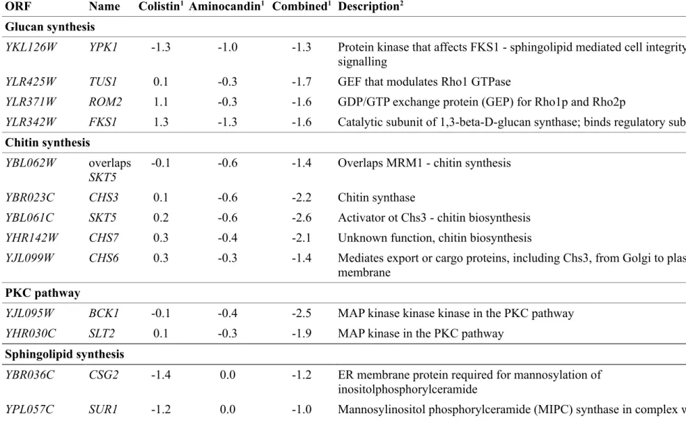
two GO terms were highly enriched among the 25 haploid strains most sensitive to the combination of aminocandin and colistin: “fungal-type cell wall chitin biosynthetic process” (p=1.35x10⁻⁵, 4 genes - CHS3, CHS7, CHS6 and SKT5) and the broader category “cellular cell
wall organization or biogenesis” (p=1.51x10⁻⁵, 9 genes, including chitin biosynthesis genes
and SLT2, YPK1, FKS1, ROM2 and TUS1). As shown in Fig. 1, inactivation of these genes also resulted in increased sensitivity to aminocandin alone but to a lesser extent than the sensitivity observed upon treatment with the combination. The observation that chitin-synthesis mutants with a decreased amount of chitin in their cell wall45-49 have increased sensitivity to aminocandin is consistent with previously published data39 and the role of chitin synthesis in protecting yeast cells against the cell wall alterations resulting from β-1,3-glucan synthase inhibition.50 However, the enhanced reduction in fitness observed with the combination of aminocandin and colistin suggested that combined defects in β-1,3-glucan and chitin synthesis might facilitate colistin antifungal activity. Intriguingly, strains deleted for the
FKS1 and ROM2 genes, the former encoding one of the two catalytic subunits of the S.
cerevisiae β-1,3-glucan synthase and the latter encoding a GDP/GTP exchange protein for the
Rho1 regulatory subunit of β-1,3-glucan synthase51, were less affected than the average population of mutants by colistin treatment while they were hypersensitive to aminocandin and aminocandin/colistin combination treatments (Fig. 1).
Mutations in sphingolipid and chitin biosynthesis result in increased sensitivity to
colistin in C. albicans
Results presented above indicated that defects in sphingolipid biosynthesis increased sensitivity of S. cerevisiae to colistin and the colistin/aminocandin combination while defects in chitin biosynthesis increased sensitivity to this combination and aminocandin alone. In order to test whether this held true in C. albicans, we took advantage of existing C. albicans
knock-out mutants in the MIT1, CSG2, IPT1, CHS3, CHS5 and CHS7 genes that encode orthologues of S. cerevisiae SUR1/CSG1, CSG2, IPT1, CHS3, CHS5, a n d CHS7.52-56 Inactivation of MIT1 and CSG2 did not increase the sensitivity of C. albicans to colistin, aminocandin or their combination (data not shown). In contrast, inactivation of IPT1 resulted in enhanced sensitivity of C. albicans to colistin alone or in combination with aminocandin (Fig. 2A). Furthermore, this increased susceptibility was abolished upon reintroduction of a wild-type allele of IPT1 in the ipt1∆ mutant (Fig. 2A). Data presented in Fig. 2B showed that inactivation of CHS3 increased the sensitivity of C. albicans to the aminocandin/colistin combination while having no significant effect on the susceptibility to the individual drugs. Inactivation of CHS5 enhanced sensitivity of C. albicans to colistin alone or in combination with aminocandin while inactivation of CHS7 had no effect. Taken together, these results indicated that, as observed in S. cerevisiae, efficient sphingolipid and chitin biosynthesis protected C. albicans against the antifungal activity of colistin and the aminocandin/colistin combination, although the impact of inactivating orthologous genes in S. cerevisiae and C.
albicans had somewhat different outcomes.
Colistin treatment increases membrane permeability of aminocandin-treated C. albicans
cells
Colistin might achieve its antifungal activity through targeting of biological membranes, as described for its antibacterial activity.14 In order to test this hypothesis, we assessed the permeability of colistin- and/or aminocandin-treated C. albicans cells using the fluorescent dye propidium-iodide (PI) that enters cells only when the membrane has been permeabilized.57 C. albicans strain SC5314 cells were grown for 1 h at 30°C, treated for different durations with (1) no compound, (2) 0.00125 mg/L aminocandin, (3) 20 mg/L colistin and (4) the combination of 0.00125 mg/L aminocandin and 20 mg/L colistin, and the
proportion of PI-stained cells was determined by flow cytometry. We found that a significantly larger amount of cells became stained with PI when treated with aminocandin and colistin relative to aminocandin alone (P <0.0001; Fig. 3). Untreated cells and cells treated with colistin alone were not stained by the dye. Therefore, these results indicated that colistin treatment increased the permeability of C. albicans cells provided that these had been exposed to aminocandin.
The colistin/caspofungin combination reduces fungal burden in kidneys of mice infected
with C. albicans
We tested whether the synergistic effect of colistin with echinocandins in vitro could be recapitulated during systemic C. albicans infection in mice. We could not identify any condition where the combination between Cancidas™, the clinical formulation of caspofungin, and the less toxic prodrug colistin sodium methanesulfonate allowed improved survival of C. albicans-infected mice (data not shown). However, when we used a combination of 0.3 mg/kgCancidas™ and 40 mg/kg colistin sodium methanesulfonate, we observed a slightly but significantly lower fungal burden in kidneys after 3 days of infection relative to the fungal burden in mice that had been treated with 0.3 mg/kgcaspofungin alone (Fig. 4; Mann Whitney test, p-value <0.05). Taken together, our results indicated that the combination between caspofungin and colistin might have the potential to decrease fungal burden during early stages of infection.
Discussion
Colistin targets the fungal membrane
In this study, we have found that the antibiotic colistin has weak, if any, antifungal activity towards several hemiascomycetous yeasts but that this activity is highly enhanced through
combination with echinocandins. Colistin is a member of the family of polymyxin antibiotics that target bacterial membranes and is currently used for the treatment of Pseudomonas
aeruginosa and Acinetobacter baumannii infections as well as infections caused by
multi-resistant bacteria such as the recently emerged NDM-1 Escherichia coli a n d Klebsiella
pneumoniae.58 Weak antifungal activity of colistin and polymyxin B towards several distantly
related fungal species has already been reported15,16,18,34 and it has been proposed that they act
at the fungal membrane. Indeed, colistin could trigger rapid efflux of ATP from germlings of
Mucorales and alterations of membrane structure.34 Moreover, polymyxin B showed synergy
with membrane-targeting antifungals such as azoles and polyenes17,18, an observation that we
have extended to colistin in this study. Our results are consistent with this antifungal mode of action. Indeed, we could show that under conditions where the antifungal activity towards C.
albicans could be manifested, e g in combination with the β-1,3-glucan synthase inhibitor
aminocandin, C. albicans cells became significantly more permeable to the membrane impermeable fluorescent dye propidium-iodide57, than when treated with aminocandin alone. Furthermore, fitness profiling experiments performed using S. cerevisiae haploid and heterozygous diploid knock-out mutants showed that alteration of sphingolipid biosynthesis results in increased sensitivity to colistin. Similarly, a C. albicans ipt1∆∆ mutant defective for one of the enzymes of sphingolipid biosynthesis showed increased sensitivity to colistin and the aminocandin/colistin combination. Sphingolipids are essential components of the eukaryotic plasma membrane, concentrating together with sterols at so-called lipid rafts where they play a variety of functions.42 Notably, S. cerevisiae csg1, csg2 and ipt1 and C. albicans
ipt1 mutants that are impaired in the latest steps of mannose-inositol-phosphoceramide
(MIPC) and mannose-(inositol phosphate)2-ceramide (M(IP)2C) biosynthesis show increased sensitivity to azoles and amphotericin B.39,53 Synergism of colistin with these antifungals and
mutations that alter sphingolipid biosynthesis are therefore consistent with its targeting of fungal membranes and possibly lipid rafts within these membranes.
Synergy of colistin with echinocandins: a rationale?
We have shown that colistin is synergistic with echinocandins. Analysis of the topology of S.
cerevisiae β-1,3-glucan synthase and the location of aminoacids whose changes render the
enzyme resistant to echinocandins suggest that these drugs interact with regions of the β-1,3-glucan synthase that are exposed to the external milieu or embedded in the outer leaflet of the plasma membrane.21 Therefore, synergy between colistin and echinocandins is unlikely to result from colistin-mediated increased influx of echinocandins into cells. Here, we would like to propose that echinocandin-mediated weakening of the cell wall facilitates colistin targeting of fungal membranes that might in turn reinforce the antifungal activity of echinocandins. As mentioned above, combination of aminocandin with colistin rendered C.
albicans cells highly permeable to propidium iodide while colistin-treated cells appeared
impermeable to this compound. This suggests that echinocandin treatment facilitated access of colistin to membranes and its membrane-targeted antifungal activity, similar to what has been shown for echinocandin-facilitated entry of antimicrobial peptides (AMP).59 Consistently, synergy between colistin and echinocandins was lost in strains harbouring mutations in the FKS1 gene and rendering C. albicans resistant to echinocandins, suggesting that weakening of the cell wall through inhibition of β-glucan synthase was a prerequisite for colistin antifungal activity. Moreover, we have shown that sensitivity to aminocandin of S. cerevisiae and C. albicans mutants with defects in chitin synthesis was markedly aggravated when colistin was added. Chitin synthesis is triggered upon treatment of yeast cells by echinocandins and contributes to the tolerance to these drugs.50,60,61 Our results
aminocandin and the chitin biosynthesis requirement for survival of aminocandin-treated cells. While this might also be explained by the impact of impaired chitin biosynthesis on colistin access to the plasma membrane and its antifungal activity, it is probably not the case, as chitin-defective mutants did not show increased sensitivity to colistin alone. In contrast, S.
cerevisiae mutants with defects in β-1,3-glucan synthesis showed intrinsically high tolerance
to colistin that might reflect adaptative changes in the cell wall in response to constitutive lower β-1,3-glucan levels62, consequently reducing colistin ability to reach membranes.
Mechanisms by which colistin could enhance the activity of echinocandins have not been investigated here. However, it can be proposed that colistin-induced perturbation of the plasma membrane might impair β-glucan synthase activity, rendering it more susceptible to echinocandins, or might facilitate membrane insertion of echinocandins. Yet, it should be noted that not all membrane-targeting drugs are synergistic with echinocandins. For instance, azoles and polyenes are not generally regarded as synergistic with echinocandins in
Candida spp. although some examples of synergy have been reported.63-65 As mentioned
above, synergy between cationic AMP and echinocandins has been observed.59,66 Polymyxins
are cationic cyclic heptapeptides with a hydrophobic tail suggesting that they share properties with other AMP that could specifically impact fungal membranes and β-glucan synthase activity. Interestingly, we did not observe any synergy between colistin and several cell wall-targeting drugs (Congo red, calcofluor white, nykkomycin Z; data not shown) suggesting that there is specificity in the colistin/echinocandin synergy.
Synergism of colistin and echinocandins in diverse fungal species and in a mouse model of
systemic candidiasis
In this study, we have observed in vitro synergy between colistin and echinocandins in several pathogenic yeasts, namely C. albicans, C. glabrata, C. tropicalis, C. parapsilosis, and C.
krusei, as well as in fluconazole-resistant C. albicans strains. In contrast, no synergy could be
observed between aminocandin and colistin in A. fumigatus (data not shown). Using a mouse model, we have shown that the colistin/caspofungin combination could possibly reduce fungal burden at early stages of systemic candidiasis relative caspofungin alone. However, this observation did not translate into an improvement of survival of animals suggesting that further demonstration of the efficacy of this drug combination in the treatment of systemic candidiasis will probably require optimizing delivery routes and evaluating pharmacodynamic interactions of caspofungin and colistin sodium methanesulfonate. Moreover, we believe that our results suggest that more emphasis should be put on the search and development of compounds that specifically target fungal membranes and could be synergistic with echinocandins.
Acknowledgements
We thank Neil Gow and Carol Munro (University of Aberdeen), David Perlin (University of New Jersey), Dominique Sanglard (Centre Hospitalier Universitaire Vaudois) and Rajendra Prasad (Jawaharlal Nehru University) for providing strains. We thank Bruno Didier and Marcel Hibert (Faculté de Pharmacie Strasbourg), Aurélien Lesnard and Sylvain Rault (CERM, Caen), Florence Mahuteau-Betzer and Daniel Dauzonne (Institut Curie, Paris), Francoise Gueritte and Olivier Pamlard (ICSN, Gif-sur-Yvette) and Yves Janin (Institut Pasteur, Paris) for providing some of the selected compounds. We thank Jean-Paul Latgé for constant interest in this study.
Funding
This work was supported by a grant from the “Conseil Régional d'Ile-de-France” (Chemical Library Project, grants n° I 06-222/R and I 09-1739/ R) to H. M.-L., a grant (Programme
Fungi) from Institut Carnot – Pasteur Maladies Infectieuses to H.M.-L. and C.d’E. and a grant from “Agence Nationale de la Recherche” (ANR-08-JCJC-0019-01, GENO-GIM project) to C.S. U.Z. and C.C were the recipients of post-doctoral grants in the framework of the Programme Fungi. A.L. was the recipient of a post-doctoral fellowship from the "Conseil Régional d'Ile-de-France".
Transparency declaration
M.-E.B. has received grant for research from GILEAD and lecture fees from Astellas. All other authors: none to declare.
References
1. Pfaller MA, Diekema DJ. Epidemiology of invasive mycoses in North America. Crit
Rev Microbiol 2010; 36: 1-53.
2. Perlroth J, Choi B, Spellberg B. Nosocomial fungal infections: epidemiology, diagnosis, and treatment. Med Mycol 2007; 45: 321-46.
3. Azie N, Neofytos D, Pfaller M et al. The PATH (Prospective Antifungal Therapy) Alliance(R) registry and invasive fungal infections: update 2012. Diag Microbiol Infec Dis 2012; 73: 293-300.
4. Horn DL, Fishman JA, Steinbach WJ et al. Presentation of the PATH Alliance registry for prospective data collection and analysis of the epidemiology, therapy, and outcomes of invasive fungal infections. Diag Microbiol Infec Dis 2007; 59: 407-14.
5. Pfaller MA, Diekema DJ. Epidemiology of invasive candidiasis: a persistent public health problem. Clin Microbiol Rev 2007; 20: 133-63.
6. Gudlaugsson O, Gillespie S, Lee K et al. Attributable mortality of nosocomial candidemia, revisited. Clin Infect Dis 2003; 37: 1172-7.
7. Lass-Florl C. The changing face of epidemiology of invasive fungal disease in Europe.
Mycoses 2009; 52: 197-205.
8. Garey KW, Rege M, Pai MP et al. Time to initiation of fluconazole therapy impacts mortality in patients with candidemia: a multi-institutional study. Clin Infect Dis 2006; 43: 25-31.
9. Morrell M, Fraser VJ, Kollef MH. Delaying the empiric treatment of Candida
bloodstream infection until positive blood culture results are obtained: a potential risk factor for hospital mortality. Antimicrob Agents Chemother 2005; 49: 3640-5.
10. Andes DR, Safdar N, Baddley JW et al. Impact of treatment strategy on outcomes in patients with candidemia and other forms of invasive candidiasis: a patient-level quantitative review of randomized trials. Clin Infect Dis 2012; 54: 1110-22.
11. Hall MJ, Middleton RF, Westmacott D. The fractional inhibitory concentration (FIC) index as a measure of synergy. J Antimicrob Chemother 1983; 11: 427-33.
12. Pasqualotto AC, Denning DW. New and emerging treatments for fungal infections. J
Antimicrob Chemother 2008; 61 Suppl 1: i19-30.
13. Groll AH, Walsh TJ. Caspofungin: pharmacology, safety and therapeutic potential in superficial and invasive fungal infections. Expert Opin Investig Drugs 2001; 10: 1545-58. 14. Newton BA. The properties and mode of action of the polymyxins. Bacteriol Rev 1956; 20: 14-27.
15. Nicholls MW. Polymyxin sensitivity of Candida tropicalis. J Med Microbiol 1970; 3: 529-38.
16. Schwartz SN, Medoff G, Kobayashi GS et al. Antifungal properties of polymyxin B and its potentiation of tetracycline as an antifungal agent. Antimicrob Agents Chemother 1972;
17. Moneib NA. In-vitro activity of commonly used antifungal agents in the presence of rifampin, polymyxin B and norfloxacin against Candida albicans. J Chemother 1995; 7: 525-9.
18. Zhai B, Zhou H, Yang L et al. Polymyxin B, in combination with fluconazole, exerts a potent fungicidal effect. J Antimicrob Chemother 2010; 65: 931-8.
19. Ogita A, Konishi Y, Borjihan B et al. Synergistic fungicidal activities of polymyxin B and ionophores, and their dependence on direct disruptive action of polymyxin B on fungal vacuole. J Antibiot (Tokyo) 2009; 62: 81-7.
20. Denning DW. Echinocandin antifungal drugs. Lancet 2003; 362: 1142-51.
21. Johnson ME, Edlind TD. Topological and Mutational Analysis of Saccharomyces
cerevisiae Fks1. Eukaryot Cell 2012; 11: 952-60.
22. Gillum AM, Tsay EY, Kirsch DR. Isolation of the Candida albicans gene for
orotidine-5'-phosphate decarboxylase by complementation of S. cerevisiae ura3 and E. coli
pyrF mutations. Mol Gen Genet 1984; 198: 179-82.
23. Brachmann CB, Davies A, Cost GJ et al. Designer deletion strains derived from
Saccharomyces cerevisiae S288C: a useful set of strains and plasmids for PCR-mediated gene
disruption and other applications. Yeast 1998; 14: 115-32.
24. Hibert MF. French/European academic compound library initiative. Drug Discov
Today 2009; 14: 723-5.
25. Tiballi RN, He X, Zarins LT et al. Use of a colorimetric system for yeast susceptibility testing. J Clin Microbiol 1995; 33: 915-7.
26. Zhang JH, Chung TD, Oldenburg KR. A simple statistical parameter for use in evaluation and validation of high throughput screening assays. J Biomol Screen 1999; 4: 67-73.
27. Chatuverdi A, Green PE, Carroll JD. K-modes clustering. J Classification 2001; 18: 35-56.
28. Huang Z. Extensions to the k-means algorithm for clustering large data sets with categorical values. Data Min Knowl Discov 1998; 2: 283-304.
29. Willett P. Similarity and clustering in chemical information systems. Letchworth, Hertfordshire, England: Research Studies Press Ltd., 1987.
30. Scott EM, Tariq VN, McCrory RM. Demonstration of synergy with fluconazole and either ibuprofen, sodium salicylate, or propylparaben against Candida albicans in vitro.
Antimicrob Agents Chemother 1995; 39: 2610-4.
31. Giaever G, Chu AM, Ni L et al. Functional profiling of the Saccharomyces cerevisiae genome. Nature 2002; 418: 387-91.
32. Peyroche G, Saveanu C, Dauplais M et al. Sodium selenide toxicity is mediated by O2-dependent DNA breaks. PLoS ONE 2012; 7: e36343.
33. Gentleman RC, Carey VJ, Bates DM et al. Bioconductor: open software development for computational biology and bioinformatics. Genome Biol 2004; 5: R80.
34. Ben-Ami R, Lewis RE, Tarrand J et al. Antifungal activity of colistin against mucorales species in vitro and in a murine model of Rhizopus oryzae pulmonary infection.
Antimicrob Agents Chemother 2010; 54: 484-90.
35. Louie A, Deziel M, Liu W et al. Pharmacodynamics of caspofungin in a murine model of systemic candidiasis: importance of persistence of caspofungin in tissues to understanding drug activity. Antimic Agents Chemother 2005; 49: 5058-68.
36. Perlin DS. Resistance to echinocandin-class antifungal drugs. Drug Resist Updat 2007; 10: 121-30.
37. Vanden Bossche H, Koymans L, Moereels H. P450 inhibitors of use in medical treatment: focus on mechanisms of action. Pharmacol Ther 1995; 67: 79-100.
38. Coste AT, Karababa M, Ischer F et al. TAC1, transcriptional activator of CDR genes, is a new transcription factor involved in the regulation of Candida albicans ABC transporters
CDR1 and CDR2. Eukaryot Cell 2004; 3: 1639-52.
39. Hillenmeyer ME, Fung E, Wildenhain J et al. The chemical genomic portrait of yeast: uncovering a phenotype for all genes. Science 2008; 320: 362-5.
40. Uemura S, Kihara A, Iwaki S et al. Regulation of the transport and protein levels of the inositol phosphorylceramide mannosyltransferases Csg1 and Csh1 by the Ca2+-binding protein Csg2. J Biol Chem 2007; 282: 8613-21.
41. Uemura S, Kihara A, Inokuchi J et al. Csg1p and newly identified Csh1p function in mannosylinositol phosphorylceramide synthesis by interacting with Csg2p. J Biol Chem 2003;
278: 45049-55.
42. Dickson RC. Thematic review series: sphingolipids. New insights into sphingolipid metabolism and function in budding yeast. J Lipid Res 2008; 49: 909-21.
43. Dickson RC, Nagiec EE, Wells GB et al. Synthesis of
mannose-(inositol-P)2-ceramide, the major sphingolipid in Saccharomyces cerevisiae, requires the IPT1 (YDR072c) gene. J Biol Chem 1997; 272: 29620-5.
44. Oh CS, Toke DA, Mandala S et al. ELO2 and ELO3, homologues of the
Saccharomyces cerevisiae ELO1 gene, function in fatty acid elongation and are required for
sphingolipid formation. J Biol Chem 1997; 272: 17376-84.
45. Cid VJ, Duran A, del Rey F et al. Molecular basis of cell integrity and morphogenesis in Saccharomyces cerevisiae. Microbiol Rev 1995; 59: 345-86.
46. Ziman M, Chuang JS, Tsung M et al. Chs6p-dependent anterograde transport of Chs3p from the chitosome to the plasma membrane in Saccharomyces cerevisiae. Mol Biol
47. Trilla JA, Duran A, Roncero C. Chs7p, a new protein involved in the control of protein export from the endoplasmic reticulum that is specifically engaged in the regulation of chitin synthesis in Saccharomyces cerevisiae. J Cell Biol 1999; 145: 1153-63.
48. DeMarini DJ, Adams AE, Fares H et al. A septin-based hierarchy of proteins required for localized deposition of chitin in the Saccharomyces cerevisiae cell wall. J Cell Biol 1997;
139: 75-93.
49. Lam KK, Davey M, Sun B et al. Palmitoylation by the DHHC protein Pfa4 regulates the ER exit of Chs3. J Cell Biol 2006; 174: 19-25.
50. Walker LA, Munro CA, de Bruijn I et al. Stimulation of chitin synthesis rescues
Candida albicans from echinocandins. PLoS Pathog 2008; 4: e1000040.
51. Ozaki K, Tanaka K, Imamura H et al. Rom1p and Rom2p are GDP/GTP exchange
proteins (GEPs) for the Rho1p small GTP binding protein in Saccharomyces cerevisiae. The
EMBO journal 1996; 15: 2196-207.
52. Mille C, Janbon G, Delplace F et al. Inactivation of CaMIT1 inhibits Candida
albicans phospholipomannan beta-mannosylation, reduces virulence, and alters cell wall
protein beta-mannosylation. J Biol Chem 2004; 279: 47952-60.
53. Prasad T, Saini P, Gaur NA et al. Functional analysis of CaIPT1, a sphingolipid biosynthetic gene involved in multidrug resistance and morphogenesis of Candida albicans.
Antimicrob Agents Chemother 2005; 49: 3442-52.
54. Noble SM, French S, Kohn LA et al. Systematic screens of a Candida albicans
homozygous deletion library decouple morphogenetic switching and pathogenicity. Nat Genet 2010; 42: 590-8.
55. Sanz M, Carrano L, Jimenez C et al. Candida albicans strains deficient in CHS7, a key regulator of chitin synthase III, exhibit morphogenetic alterations and attenuated virulence. Microbiology 2005; 151: 2623-36.
56. Bulawa CE, Miller DW, Henry LK et al. Attenuated virulence of chitin-deficient mutants of Candida albicans. Proc Natl Acad Sci 1995; 92: 10570-4.
57. Deere D, Shen J, Vesey G et al. Flow cytometry and cell sorting for yeast viability assessment and cell selection. Yeast 1998; 14: 147-60.
58. Li J, Nation RL, Turnidge JD et al. Colistin: the re-emerging antibiotic for multidrug-resistant Gram-negative bacterial infections. Lancet Infect Dis 2006; 6: 589-601.
59. Harris MR, Coote PJ. Combination of caspofungin or anidulafungin with antimicrobial peptides results in potent synergistic killing of Candida albicans and Candida glabrata in vitro. Int J Antimic Ag 2010; 35: 347-56.
60. Markovich S, Yekutiel A, Shalit I et al. Genomic approach to identification of mutations affecting caspofungin susceptibility in Saccharomyces cerevisiae. Antimicrob
Agents Chemother 2004; 48: 3871-6.
61. Lee KK, Maccallum DM, Jacobsen MD et al. Elevated cell wall chitin in Candida
albicans confers echinocandin resistance in vivo. Antimicrob Agents Chemother 2010.
62. Lesage G, Sdicu AM, Menard P et al. Analysis of beta-1,3-glucan assembly in
Saccharomyces cerevisiae using a synthetic interaction network and altered sensitivity to
caspofungin. Genetics 2004; 167: 35-49.
63. Kiraz N, Dag I, Yamac M et al. Synergistic activities of three triazoles with caspofungin against Candida glabrata isolates determined by time-kill, Etest, and disk diffusion methods. Antimicrob Agents Chemother 2010; 54: 2244-7.
64. Oliveira ER, Fothergill AW, Kirkpatrick WR et al. In vitro interaction of posaconazole and caspofungin against clinical isolates of Candida glabrata. Antimicrob Agents Chemother 2005; 49: 3544-5.
65. Roling EE, Klepser ME, Wasson A et al. Antifungal activities of fluconazole, caspofungin (MK0991), and anidulafungin (LY 303366) alone and in combination against
Candida spp. and Crytococcus neoformans via time-kill methods. Diagn Microbiol Infect Dis 2002; 43: 13-7.
66. Rossignol T, Kelly B, Dobson C et al. Endocytosis-mediated vacuolar accumulation of the human ApoE apolipoprotein-derived ApoEdpL-W antimicrobial peptide contributes to is antifungal activity in Candida albicans. Antimicrob Agents Chemother 2011; 55: 4670-81. 67. Balashov SV, Park S, Perlin DS. Assessing resistance to the echinocandin antifungal drug caspofungin in Candida albicans by profiling mutations in FKS1. Antimicrob Agents
Chemother 2006; 50: 2058-63.
68. Karababa M, Coste AT, Rognon B et al. Comparison of gene expression profiles of
Candida albicans azole-resistant clinical isolates and laboratory strains exposed to drugs
inducing multidrug transporters. Antimicrob Agents Chemother 2004; 48: 3064-79. 69. Fonzi WA, Irwin MY. Isogenic strain construction and gene mapping in Candida
albicans. Genetics 1993; 134: 717-28.
70. Noble SM, Johnson AD. Strains and strategies for large-scale gene deletion studies of the diploid human fungal pathogen Candida albicans. Eukaryot Cell 2005; 4: 298-309.
71. Cherry JM, Hong EL, Amundsen C et al. Saccharomyces Genome Database: the
genomics resource of budding yeast. Nucl Acids Res 2012; 40: D700-5.
Legends to Figures
Figure 1: Colistin affects sphingholipid biosynthesis deficient strains and increases the
sensitivity of chitin synthesis mutants to aminocandin. A pool of S. cerevisiae haploid
deletion strains was treated with colistin (20 mg/L), aminocandin (0.00125 mg/L) or the combination of 0.00125 mg/L aminocandin and 5 mg/L colistin and grown for 11 generations in liquid medium. Relative growth rates, estimated using barcode-specific microarrays, were
normalized and represented as log2 ratios of the signal obtained with treated versus untreated culture. Mutant strain behaviour in the different conditions is illustrated for genes that belong to functional categories that were found to be significantly enriched (sphingolipid biosynthesis, green; cell wall organization and biogenesis, blue and chitin synthesis, red, see text for details).
Figure 2: Efficient sphingolipid or chitin biosynthesis protects C. albicans cells of the
combined antifungal activity of aminocandin and colistin. C. albicans strains with
mutations in the IPT1 gene involved in sphingolipid biosynthesis (deltaIPT1, TPIPT1-4; deltaIPT1+IPT1, TPIPT1-4; Table 1) and in the CHS3, CHS5 and CHS7 genes involved in chitin biosynthesis (CACB3B-5, ∆CHS5_ARGplu, ∆CHS3_ARGplus, respectively; Table 1) and their parent strains (CAI4 and SN250_ARGplus; Table 1) were grown in the absence of drugs (untreated) or in the presence of aminocandin alone (AMC; 0.00125 mg/L), colistin alone (COL; 30 mg/L), and the combination of aminocandin (0.00125 mg/L) and colistin (20 mg/L) (AMC + COL). Optical density was recorded after 16 h growth and is represented relative to the optical density obtained for the untreated wild-type cells. Data are mean of 2 independent experiments, with bars indicating the range of values.
Figure 3: Colistin increases permeability of echinocandin-treated cells
Cells were left untreated or treated with colistin alone (COL, 20 mg/L), aminocandin alone (AMC, 0.00125 mg/L) or with the combination of both drugs (AMC + COL) and the proportion of propidium-iodide stained cells was measured by flow cytometry. The statistical test Chi2-Mantel-Haenszel was used to analyse the difference between cells treated with aminocandin alone and with the combination. At all time points the difference was significant (p < 0.0001).
Figure 4: The colistin/caspofungin combination reduces fungal burden in kidneys of C.
albicans infected mice. Immuno-supressed female BALB/c mice were infected
intravenously with 104 CFU/mouse of C. albicans strain SC5314. One group of mice was not
treated, one was treated with colistin alone (40 mg/kg), one with caspofungin alone (0.3 mg/kg) and one with caspofungin and colistin at the given concentrations. Mice were killed three days after infection and fungal burden in kidneys of mice was evaluated. * P<0.05. Note that only 7 mice untreated or treated with colistin alone were still alive on day 3 after infection, therefore the CFUs of only 7 kidneys are represented in the graph.
Supplemental Figure 1:
High-throughput screening for compounds acting synergistically with aminocandin. (A)
Evaluation of the test using the calculated Z'-factor value plotted for each plate. (B) Distribution of the number of compounds as a function of the percentage of viability as defined in the material and methods section.
Supplemental Figure 2:
Confirmation of S. cerevisiae mutants exhibiting sensitivity towards the treatment in a
fitness assay. Heterozygous- and homozygous mutants were grown for 16 h at 30°C without
compound (-Amc -CS), in presence of aminocandin only (+Amc -CS), colistin alone (-Amc +CS) and the combination of both compounds (+Amc +CS).
Table 1: Yeast strains used in this study
Species Strain Type of strain Genotype Reference
C. albicans DPL1000 WT, parent of 20S, 22S and 36S D. Perlin
C. albicans 20S Caspofungin-resistant, Fks1S645F FKS1C1934T/FKS1C1934T 67
C. albicans 22S Caspofungin-resistant, Fks1S645Y FKS1C1934A/FKS1C1934A 67
C. albicans 36S Caspofungin-resistant,Fks1S645P FKS1T1933C/FKS1T1933C 67
C. albicans M70 WT, parent of C42
C. albicans C42 Caspfungin-resistant, Fks1F641S FKS1/FKS1
C. albicans DSY294 Fluconazole-susceptible clinical
strain
TAC1-3/TAC1-4 ERG11-3/ERG11-4
68
C. albicans DSY296 Fluconazole-resistant TAC1-5/TAC1-5 ERG11-5/ERG11-5 68
C. albicans DSY3987 Fluconazole-resistant ura3∆::FRT/ura3∆::FRT TAC1-5/TAC1-5
ERG11-5/ERG11-5 RP10::CIp10
D. Sanglard
C. albicans DSY3988 Fluconazole-resistant ura3∆::FRT/ ura3∆::FRT tac1-5∆::hisG/tac1-5∆::hisG
ERG11-5/ERG11-5 RP10::CIp10 D. Sanglard
C. albicans CAI4 ∆ura3::λimm434/∆ura3::λimm434 69
C. albicans TPIPT1-4 ∆ura3::λimm434/∆ura3::λimm434
∆ipt1::hisG/∆ipt1::hisG
53
C. albicans TPIPT1-5 ∆ura3::λimm434/∆ura3::λimm434
∆ipt1::hisG/∆ipt1::hisG::IPT1-URA3
53
C. albicans CACB3B-5 ∆ura3:: λimm434/1∆ura3:: λimm434
∆chs3-2::hisG/∆chs3-3::hisG_URA3_hisG_URA3_hisG
56
C. albicans SN152 arg4∆/arg4∆ leu2∆/leu2∆ his1∆/his1∆
URA3/ura3∆::λimm434 IRO1/iro1∆::λimm434
70
C. albicans SN250_ARGplus his1Δ/his1Δ leu2Δ::CdHIS1/leu2Δ::CmLEU2
arg4Δ::CaARG4/arg4Δ URA3/ura3Δ::imm434 IRO1/iro1Δ::imm434
54
C. albicans ΔCHS5_ARGplus SN152 chs5∆::CmLEU2 /chs5∆::CdHIS1
arg4Δ::CaARG4
54
C. albicans ΔCHS7_ARGplus SN152 chs7∆::CmLEU2 /chs7∆::CdHIS1
arg4Δ::CaARG4
54
C. albicans ∆CSG2_ARGplus SN152 csg2∆::CmLEU2 /csg2∆::CdHIS1
arg4Δ::CaARG4
54
S. cerevisiae BY4741 MATa his3∆1 leu2∆0 met15∆0 ura3∆0 23
S. cerevisiae BY4743 MATa his3∆1 leu2∆0 met15∆0 ura3∆0/ MATα his3∆1
leu2∆0 met15∆0 ura3∆0
23
S. cerevisiae sur∆ BY4741 sur1∆KanMX4 31
S. cerevisiae sur1∆ het BY4743 sur1∆KanMX4/SUR1 Invitrogen
S. cerevisiae csg2∆ BY4741 csg2∆KanMX4 31
S. cerevisiae gef1∆ BY4741 gef1∆KanMX4 31
S. cerevisiae erg6∆ BY4741 erg6∆KanMX4 31
S. cerevisiae vps74∆ BY4741 vps74∆KanMX4 31
S. cerevisiae trp1∆ BY4741 trp1∆KanMX4 31
S. cerevisiae chs3∆ BY4741 chs3∆KanMX4 31
S. cerevisiae chs5∆ BY4741 chs5∆KanMX4 31
S. cerevisiae chs6∆ BY4741 chs6∆KanMX4 31
S. cerevisiae chs7∆ BY4741 chs7∆KanMX4 31
S. cerevisiae skt5∆ BY4741 skt5∆KanMX4 31
S. cerevisiae ipt1∆het BY4743 ipt1∆KanMX4/IPT1 Invitrogen