Computational Analysis, Design, and
Experimental Validation of Antibody Binding
Affinity Improvements Beyond In Vivo
Maturation
by
Shaun Matthew Lippow
Bachelor of Science in Chemical Engineering
University of Wisconsin - Madison, 2001
Submitted to the Department of Chemical Engineering
in partial fulfillment of the requirements for the degree of
Doctor of Philosophy in Chemical Engineering
at the
MASSACHUSETTS INSTITUTE OF TECHNOLOGY
June 2007
c
° Massachusetts Institute of Technology 2007. All rights reserved.
Author . . . .
Department of Chemical Engineering
February 9, 2007
Certified by . . . .
Bruce Tidor
Professor of Biological Engineering and Computer Science
Thesis Supervisor
Certified by . . . .
K. Dane Wittrup
J. R. Mares Professor of Chemical Engineering and Biological
Engineering
Thesis Supervisor
Accepted by . . . .
William M. Deen
Chairman, Department Committee on Graduate Students
Computational Analysis, Design, and Experimental
Validation of Antibody Binding Affinity Improvements
Beyond In Vivo Maturation
by
Shaun Matthew Lippow
Submitted to the Department of Chemical Engineering on February 9, 2007, in partial fulfillment of the
requirements for the degree of
Doctor of Philosophy in Chemical Engineering
Abstract
This thesis presents novel methods for the analysis and design of high-affinity protein interactions using a combination of high-resolution structural data and physics-based molecular models. First, computational analysis was used to investigate the molec-ular basis for the affinity improvement of over 1000-fold of the fluorescein-binding antibody variant 4M5.3, engineered previously from the antibody 4-4-20 using di-rected evolution. Electrostatic calculations revealed mechanistic hypotheses for the role of four mutations in a portion of the improvement, subsequently validated by separate biochemical experiments. Next, methods were developed to computation-ally redesign protein interactions in order to rationcomputation-ally improve binding affinity. In the anti-lysozyme model antibody D1.3, modest binding improvements were achieved, with the results indicating potentially increased sucesss using predictions that empha-size electrostatics, as well as the need to address the over-prediction of large amino acids. New methods, taking advantage of the computed electrostatics of binding, yielded robust and significant improvements for both model and therapeutic antibod-ies. The antibody D44.1 was improved 140-fold to 30 pM, and the FDA-approved antibody cetuximab (Erbitux) was improved 10-fold to 52 pM, with an experimental success rate of greater than 60% for single mutations designed to remove under-satisfied polar groups or improve misbalanced electrostatic interactions. Finally, a physics-based improvement to the calculation of the nonpolar component of solva-tion free energy was implemented and parameterized to address the over-predicsolva-tion of large amino acids. These results demonstrate novel computational capabilities and indicate their applicability for enhancing and accelerating development of reagents and therapeutics.
Thesis Supervisor: Bruce Tidor
Thesis Supervisor: K. Dane Wittrup
Acknowledgments
I am indebted to by advisors for their support, ideas, and cooperation throughout the entire thesis process. I thank Dane, especially for his outlook on projects, acute research sense, and career advice from early on, and Bruce, especially for keeping an eye on the 30,000 foot view, emphasizing the rigorous way to do good science, and guiding me with effective scientific presentation.
I am grateful for the useful and timely advice from both Bernhardt Trout and Doug Lauffenburger. Their insights proved invaluable in shaping this work and hopefully increasing its impact.
This work would also not have been possible without the help of many past and present members of both the Tidor and Wittrup labs. I would like to thank everyone that I had the privilege of overlapping with: a phenomenal group of people that gave freely of their time to help and teach me, and to share with me their own research.
In particular, I thank: Karl Hanf for helping in the very beginning and laying part of the protein design groundwork; David Green for insightful research opinions and technical help in particular with continuum electrostatics; Michael Altman for exceed-ingly unselfish assistance in all areas of computational execution, as well as tailoring development of his dead-end elimination and A* software for my work; Alessandro Senes for contributions to the protein side chain rotamer libraries; Stefan Zajic for patience and skill in teaching me new molecular biology techniques; Wai Lau and Daˇsa Lipovˇsek for additional wet lab instruction; Brian Joughin for valuable research discussions; and David, Michael, Brian, Mala Radhakrishnan, Kathryn Armstrong, Bracken King, and Dave Huggins for feedback on my protein design software.
I thank my family for years of love and support. This wouldn’t have happened without them (don’t worry, you don’t have to read the whole thing).
Contents
1 Introduction 11
2 Computational analysis of a high-affinity mutant antibody 16
2.1 Introduction . . . 17
2.2 Results . . . 18
2.2.1 Contribution of electrostatics to binding . . . 18
2.2.2 Effect of minimization on electrostatic contributions . . . 19
2.3 Discussion . . . 24
2.4 Methods . . . 28
2.4.1 Preparation of protein structures . . . 28
2.4.2 Electrostatic calculations . . . 29
3 Development of computational methods for the design of improved protein binding affinity 32 3.1 Introduction . . . 33
3.2 Methods . . . 33
3.2.1 Structure preparation . . . 34
3.2.2 Search space . . . 35
3.2.3 Energy function and model . . . 35
3.2.4 Initial conformational search . . . 37
3.2.5 Reevaluation of electrostatics . . . 39
3.2.6 Prediction of binding and folding . . . 40
3.2.8 Binding affinity measurements . . . 41
3.3 Results . . . 43
3.4 Discussion . . . 50
4 Computational design of antibody improvement beyond in vivo mat-uration 54 4.1 Introduction . . . 55 4.2 Results . . . 55 4.3 Discussion . . . 63 4.4 Methods . . . 65 4.4.1 Structure preparation . . . 65 4.4.2 Design of mutations . . . 66
4.4.3 Yeast surface display constructs . . . 66
4.4.4 Measurement of binding affinity . . . 67
4.4.5 Measurement of binding kinetics . . . 67
5 Development of an improved nonpolar solvation model 70 5.1 Introduction . . . 71
5.2 Methods . . . 74
5.2.1 Continuum van der Waals model . . . 74
5.2.2 Numerical solution . . . 75
5.2.3 Cavitation model . . . 76
5.2.4 Fit to alkane experimental data . . . 76
5.2.5 Protein test systems . . . 77
5.3 Results . . . 77
5.4 Discussion . . . 83
6 General conclusions 86
A Evolution of an interloop disulfide bond in high-affinity fibronectin type III antibody mimics: Molecular convergence with
A.1 Introduction . . . 91
A.2 Results . . . 91
A.3 Discussion . . . 95
List of Figures
1.1 Comparison of binding affinities determined by yeast surface display
and other methods . . . 12
2.1 Diagram of electrostatic component differences . . . 23
2.2 Molecular detail of mutated side chains . . . 25
2.3 Computational parameterization of fluorescein . . . 30
3.1 D1.3 CDR positions . . . 36
3.2 Yeast surface display schematic . . . 41
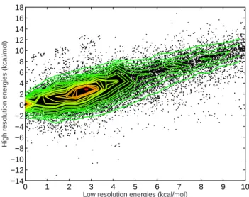
3.3 Correlation of low- and high-resolution free energies within a protein sequence . . . 43
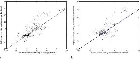
3.4 Reevaluation of relative binding energies with the high-resolution free energy function . . . 44
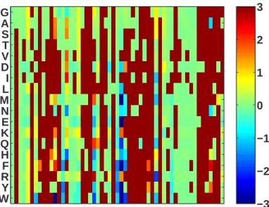
3.5 Design of single mutations in D1.3 . . . 45
3.6 Designs arranged by type of position . . . 46
3.7 Proliferation of large amino acids . . . 46
3.8 Examples of D1.3 single mutation designs . . . 47
3.9 Representative experimental binding affinity curves for D1.3 mutations 48 3.10 Number and difficulty of single and double mutation designs . . . 50
4.1 Predicted structures for D44.1 mutations . . . 57
4.2 Predicted structure for D44.1 cooperative double mutation . . . 60
4.3 Designed high-affinity mutations in D44.1 . . . 61
4.5 Comparison of calculated and experimental binding energetics . . . . 64
5.1 Stepwise solvation process . . . 73
5.2 Convergence of continuum van der Waals numerical solution . . . 78
5.3 Convergence of relative binding energies . . . 79
5.4 Parameterization of nonpolar solvation models to alkane data . . . . 80
5.5 Effect of continuum van der Waals on D1.3 single mutation designs . 81
5.6 Effect of CVDW for small-to-big mutations . . . 82
5.7 Continuum van der Waals effect in four systems. . . 84
A.1 Modeling of disulfide bonds between cysteines in BC and FG loops . 92
List of Tables
2.1 Computed total electrostatic contributions to binding . . . 18
2.2 Difference in electrostatic energy components . . . 20
2.3 Electrostatic impact at the mutated side-chain positions . . . 21
2.4 Total electrostatic contributions after minimization . . . 21
2.5 Components most affected by minimization . . . 22
2.6 Net electrostatic impact of the mutated side-chain components after minimization . . . 22
3.1 Predicted and experimental D1.3 single mutation binding affinities . . 49
4.1 Predicted and experimental D44.1 single mutation binding affinities . 56 4.2 Predicted and experimental D44.1 combination mutations . . . 57
4.3 Characterization of D44.1 quadruple mutant . . . 58
4.4 Second round design in D44.1 quadruple mutant . . . 59
4.5 Double mutant cycle added to D44.1 quadruple mutant . . . 59
4.6 Predicted and experimental cetuximab binding affinities . . . 62
4.7 Sequence details for new D44.1 surface display plasmid . . . 68
Chapter 1
Introduction
Antibodies are used extensively in diagnostics and as therapeutic agents. Achieving high-affinity binding of antibodies to their antigens is important for expanding detec-tion limits, extending dissociadetec-tion half-times, decreasing drug dosages, and increasing drug efficacy. However, antibodies produced from an in vivo immune response gen-erally exhibit affinities in the 10 nM to 100 pM range [1], often necessitating further engineering. Directed evolution can be used to engineer molecular properties, but computational design holds the promise of far greater exploration of sequence space than possible experimentally, enabling rapid and inexpensive antibody improvement. The improvement of protein–ligand interactions using different molecular display and directed evolution methods has been reviewed recently by Levin and Weiss [2]. Platforms for display include phage, bacteria, yeast, ribosome, and mRNA, each with their respective advantages and disadvantages. These engineering strategies require significantly more time than would be required to directly express and vali-date computationally-designed variants. In this work we take advantage of the yeast-display system for rapid and accurate characterization of rationally-designed antibody variants. Equilibrium binding affinities can be measured for 1–20 variants within 1–2 weeks time, and the yeast-display method yields measured binding affinities in quan-titative agreement with measurements from other, off-yeast methods (Figure 1.1).
This thesis presents the development of computational protein design methods for the rational selection of mutations to improve binding affinity. Computational design depends critically on two capabilities: accurate energetic evaluation and thorough conformational search. Previous work has addressed many problems related to the design of improved protein–protein binding affinity, such as the design of stable pro-tein folds [14–18], binding pockets for peptides and small molecules [19–22], altered
1.E-13 1.E-12 1.E-11 1.E-10 1.E-09 1.E-08 1.E-07 1.E-06
1.E-13 1.E-12 1.E-11 1.E-10 1.E-09 1.E-08 1.E-07 1.E-06
K
d other(M)
K
d Y S D(M
)
System and yeast-display reference(s) Off-yeast method
◦ scFvs binding to hen egg-white Fluorescence quenching [3]
lysozyme (HEL) [4]
• 10Fn3-based antibody mimics binding Equilibrium competition with
to HEL [5] purified antibody mimics [5]
+ Neutralizing scFvs binding to Surface plasmon resonance [6]
botulinum neurotoxin [6]
N scFvs binding to fluorescein [7] Fluorescence quenching [8]
¤ scFv binding to carcinoembryonic Mammalian cell-displayed
antigen (CEA) [9] CEA with soluble scFv [10]
■ scFv binding to xeroderma pigmentosum- Surface plasmon resonance [11]
complementing protein group A [11]
¦ scFvs binding to p53 peptides [11] Surface plasmon resonance [11]
∗ scFv binding to epidermal growth Surface plasmon resonance [11]
factor (EGF) [11]
× scFv binding to heparin-binding EGF [11] Surface plasmon resonance [11]
◆ Soluble T-cell receptors binding to Surface plasmon resonance [12]
staphylococcal enterotoxin C3 [12]
4 Soluble T-cell receptor binding to Surface plasmon resonance [13]
toxic shock syndrome toxin-1 [13]
Figure 1.1: Comparison of binding affinities determined by yeast surface display (Kd YSD) and other methods (Kd other). scFv, single-chain antibody fragment.
protein–protein specificity [23–29], and altered enzymatic activity [30–35]. The design of improved antigen-binding affinity has met with limited success, however [36–39]. Challenges for protein–protein affinity design include conformational change upon binding, water molecules trapped between binding partners, polar and charged side chains, and the trade-off of protein–solvent with protein–protein interactions from the unbound to bound state. Fine energy discrimination for redesign from nanomolar to picomolar affinities is a particular challenge.
A robust design strategy should both produce a significant fraction of designs that are successful when tested experimentally and yield substantial improvements across multiple systems. Though there are potentially many mutations that confer improved binding affinity for a particular interaction, calculations need only identify a subset to be successful [40, 41]. Our approach utilizes thorough optimization tech-niques that exhaustively rank-order the best solutions in a discretized search space. Although some of these solutions are expected to be improved designs, others will be unsuccessful but may be useful in learning about deficiencies in the energy functions, search procedures, or other methodology.
The present work
In Chapter 2, we use computational tools to analyze a high-affinity variant of a fluorescein-binding antibody. The motivation for the work was to gain insight into the molecular mechanisms for the over 1000-fold binding affinity improvement. Pre-vious biochemical and structural analyses had not elucidated the energetic role of any of the 14 mutations accumulated by the four rounds of directed evolution [8]. At the same time, we were motivated by an interest in applying similar compu-tational tools for the rational design of affinity-enhancing mutations. Though the 14-mutation high-affinity variant represents just one of possibly many pathways for affinity maturation, if the calculations could explain or recapitulate even a subset of the mutations selected, the potential applicability of similar computational methods for design would be partially validated. Here we show that rigorous analysis
us-ing the Poisson–Boltzmann continuum electrostatic framework alone results in novel hypotheses for molecular mechanisms for four of the mutations involved [42], with subsequent, separate mutational analyses supporting our conclusions [7].
Chapter 3 introduces a two-stage hierarchical design procedure to address the lim-itations of energy function accuracy and conformational search thoroughness. Single mutations for improving the binding affinity of an anti-lysozyme model antibody were computationally designed and experimentally assayed. While the affinity improve-ments were marginal and the success rate was low, the systematic design approach facilitated two important conclusions. First, additional emphasis on the electrostatic term of the binding free energy may improve the predictive accuracy of the methods. Second, an energy function improvement was needed to address the over-prediction of unintuitive mutations to larger amino acids.
In Chapter 4, the hypothesis that prediction accuracy would improve using a method with greater attention to calculated electrostatics was explored. Moreover, several new antibody systems were used to investigate the dependence on system versus the change in methods, as our goal is the development of computational meth-ods that are robust for both high experimental success rates and transferability to different proteins. Overall, we find significant improvements for both model and thera-peutic antibodies, including 140-fold improvement to 30 pM and 10-fold improvement to 52 pM, respectively, validating the methods and indicating their applicability for enhancing and accelerating the developement of reagents and therapeutics.
Despite successful affinity maturation in Chapter 4 using computational protein design, deficiencies in the methods clearly remain and limit their further extension to more ambitious design challenges. The finding in Chapter 3 that predictions for improving affinity are dominated by mutations to larger amino acids indicated a fun-damental problem in the underlying energy function. Future computational endeavors addressing problems involving the simultaneous design of greater numbers of protein positions would like by limited by this accuracy issue.
Chapter 5 investigates a new model [43] for the nonpolar component of the sol-vation free energy in order to address a hypothesized imbalance in the calculation
of protein–protein and protein–water interactions. The model is implemented and parameterized, and then analyzed for its effect on the design of single mutations for improved binding affinity. The new nonpolar model attenuates as expected the overall prediction of larger side chains, however, it does not greatly affect the few largest-magnitude unintuitive designs.
This thesis contributes novel and robust methods for the improvement of pro-tein binding affinity using computationally-predicted side-chain mutations. We have advanced the field of computational protein design in two directions: one toward engineering, and one toward basic science. We developed prediction methods that focus on calculated improved electrostatic free energy of binding and yield significant affinity improvements in model and therapeutic antibodies. Also, we investigated an improved, physics-based calculation of the nonpolar component of solvation free energy to address a fundamental tendency of current design methods to favor muta-tion to larger amino acids. Our improvements to current design capabilities should enhance and accelerate the development of protein reagents and therapeutics.
Chapter 2
Computational analysis of a
high-affinity mutant antibody
1
Abstract
Computational analysis was used to study the molecular basis for affinity improve-ment in an ultra-high-affinity single-chain antibody. Previous biochemical and struc-tural work resulted in little insight for the 14 mutations used for over 1000-fold im-provement. Here, electrostatic calculations reveal several mechanistic hypotheses for the role of four mutations in a portion of the energetic improvement. Subsequent biochemical experiments validated many of these hypotheses, supporting the role for computation in molecular analysis. Overall, the binding affinity improvement ap-pears to be the sum of many small changes. This work indicates potentially novel computational capabilities for using electrostatic calculations to design high-affinity protein interactions.
1Portions of this chapter have been previously published as:
Midelfort, K. S., Hernandez, H. H., Lippow, S. M., Tidor, B., Drennan, C. L. & Wittrup, K. D. Substantial energetic improvement with minimal structural perturbation in a high affinity mutant antibody. J. Mol. Biol. 343:685-701 (2004).
2.1
Introduction
Expanding the fundamental understanding of high-affinity molecular interactions is important for improving the ability to engineer molecules with enhanced affinity. Analysis of protein variants with diverse binding affinities for a shared target can reveal insights into the mechanistic, molecular basis of improved protein interactions. Although there has been much study of the transition from micromolar to nanomolar affinity [44–49], there are few examples of studies of improvement from nanomolar to picomolar and higher affinity.
Boder and co-workers previously engineered the 4M5.3 variant of the 4-4-20 single-chain antibody to bind its hapten, fluorescein, with over 1000-fold improved binding affinity from nanomolar to femtomolar [8]. Subsequently, Midelfort and co-workers compared the two antibodies using thermodynamic, kinetic, structural, and theoreti-cal analyses [42]. A summary of their experimental findings is below, followed by the details of this theoretical contribution.
Thermodynamics of the 4-4-20 and 4M5.3 interactions were studied by direct equi-librium titration, equiequi-librium competition titration, and isothermal titration calorime-try. Improvement in the change in enthalpy of binding, −4.0 (±0.1) kcal/mol, is the majority of the −4.5 (±0.1) kcal/mol free energy difference between 4-4-20 and 4M5.3. Stopped-flow fluorescence binding kinetics demonstrate the equivalence of Kd
and koff/kon, consistent with a two-state transition from the unbound to bound states.
The kinetic data provide no evidence for an encounter complex.
The structure of 4M5.3 in complex with fluorescein was solved to 1.5 ˚A resolution by molecular replacement using the previously reported 4-4-20 structure [50]. Overall, there are no large differences between the two structures. The RSMD over backbone atoms is 0.60 ˚A. Of the 14 mutations in 4M5.3, three are in the first contact shell, four in the second, and three in the third. The final four side chains are further away and solvent-exposed. 4-4-20 buries 25 ˚A2 more polar surface area and there is a negligible
difference in shape complementarity.
Table 2.1: Computed total electrostatic contributions to binding.a
scFv Fluorescein Net
Desolvation Desolvation Interaction Binding
4-4-20 14.36 14.80 −19.97 9.20
4M5.3 14.68 16.64 −25.62 5.70
4M5.3 − 4-4-20 0.32 1.84 −5.65 −3.50
aAll values in kcal/mol.
explanations for the over three orders of magnitude binding improvement of 4M5.3. With the crystal structure of the 4M5.3/fluorescein complex solved in collaboration with the Drennan lab, a collaboration was started between the Wittrup and Tidor labs to use computation to investigate the pair of antibodies.
2.2
Results
2.2.1
Contribution of electrostatics to binding
The role of improved electrostatic interactions was investigated through continuum electrostatic binding calculations on the two prepared, unminimized crystal struc-tures. The overall electrostatic contribution to binding computed by this model is summarized in Table 2.1. An overall improvement of −3.5 kcal/mol for 4M5.3 relative to 4-4-20 was computed due to improved interactions with fluorescein (−5.7 kcal/mol) despite increased desolvation penalty for the fluorescein (+1.8 kcal/mol). Interest-ingly, the antibody desolvation cost was very similar for both complexes and in neither case was the full cost of desolvating the binding partners recovered in intermolecular interactions.
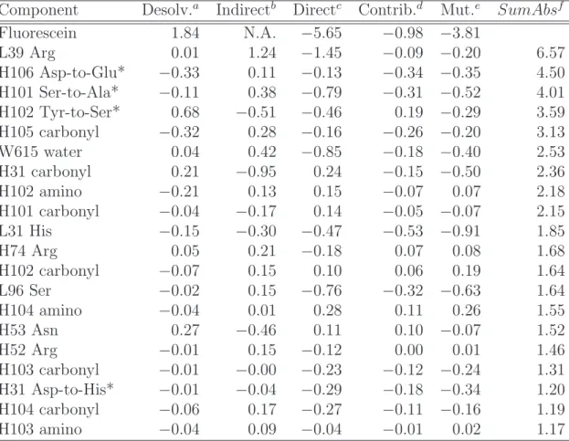
To further understand the computed difference in electrostatics, the −3.5 kcal/mol total electrostatic affinity difference was dissected into individual components. A separate component was defined for each backbone amino (Cα–NH), each backbone carbonyl (C=O), and each side chain group beyond Cα. The energetics for each component were divided into desolvation (changed interactions with solvent due to binding), indirect interactions (changed intramolecular interactions due to binding),
and direct interactions (changed intermolecular interactions between binding partners in the bound state). The components provide additive contributions to the computed difference in binding affinity. This type of dissection is possible in a strictly additive manner because of the superposition properties of the linearized Poisson–Boltzmann equation. After breaking the 4-4-20 and 4M5.3 energetics into their respective com-ponents, all corresponding components between the two antibody variants were dif-ferenced, so that non-zero values indicate components of potential interest. Table 2.2 shows the difference of component analyses between the two crystal structures. Three of the top four components and four of the top 20 components, ranked by the metric
SumAbs (Equation 2.1), are 4M5.3 side chain mutations. SumAbsi = |Desolvi4M5.3− Desolv4−4−20i | +
X
j6=i
|Inter4M5.3i−j − Interi−j4−4−20| (2.1)
SumAbs is a measure of how different each component is between the two
analy-ses. DesolvX
i represents the desolvation penalty for component i in complex X, and InterX
i−j represents the interaction free energy between the atoms in component i and
the atoms in component j in complex X.
The direct role of the 14 mutations together was calculated by summing to-gether the components involving the 14 mutated side chains, revealing a total of
−1.1 kcal/mol (Table 2.3). Four side chains dominate this value: H31
Asp-to-His, H101 Ser-to-Ala, H102 Tyr-to-Ser, and H106 Asp-to-Glu. The remaining
−2.4 kcal/mol in computed electrostatic improvement is the result of secondary
ef-fects from the mutated residues (e.g. altering the binding conformation of fluorescein in the site). Three components dominate this −2.4 kcal/mol: L31 His, L39 Arg, and L96 Ser, each of which exhibit slightly shortened hydrogen-bond distances in 4M5.3 relative to 4-4-20.
2.2.2
Effect of minimization on electrostatic contributions
Because the results of this analysis produced significant free energy differences that were the sum of quite small values from individual changed interactions, further
calcu-Table 2.2: Difference in electrostatic energy components.
Component Desolv.a Indirectb Directc Contrib.d Mut.e SumAbsf
Fluorescein 1.84 N.A. −5.65 −0.98 −3.81 L39 Arg 0.01 1.24 −1.45 −0.09 −0.20 6.57 H106 Asp-to-Glu* −0.33 0.11 −0.13 −0.34 −0.35 4.50 H101 Ser-to-Ala* −0.11 0.38 −0.79 −0.31 −0.52 4.01 H102 Tyr-to-Ser* 0.68 −0.51 −0.46 0.19 −0.29 3.59 H105 carbonyl −0.32 0.28 −0.16 −0.26 −0.20 3.13 W615 water 0.04 0.42 −0.85 −0.18 −0.40 2.53 H31 carbonyl 0.21 −0.95 0.24 −0.15 −0.50 2.36 H102 amino −0.21 0.13 0.15 −0.07 0.07 2.18 H101 carbonyl −0.04 −0.17 0.14 −0.05 −0.07 2.15 L31 His −0.15 −0.30 −0.47 −0.53 −0.91 1.85 H74 Arg 0.05 0.21 −0.18 0.07 0.08 1.68 H102 carbonyl −0.07 0.15 0.10 0.06 0.19 1.64 L96 Ser −0.02 0.15 −0.76 −0.32 −0.63 1.64 H104 amino −0.04 0.01 0.28 0.11 0.26 1.55 H53 Asn 0.27 −0.46 0.11 0.10 −0.07 1.52 H52 Arg −0.01 0.15 −0.12 0.00 0.01 1.46 H103 carbonyl −0.01 −0.00 −0.23 −0.12 −0.24 1.31 H31 Asp-to-His* −0.01 −0.04 −0.29 −0.18 −0.34 1.20 H104 carbonyl −0.06 0.17 −0.27 −0.11 −0.16 1.19 H103 amino −0.04 0.09 −0.04 −0.01 0.02 1.17
All values are the differences between analyses on 4M5.3 and 4-4-20, in kcal/mol. Negative (positive) values correspond to a contribution toward improvement (reduc-tion) for 4M5.3 binding relative to 4-4-20.
*Site of 4M5.3 mutation
aDesolvation penalty.
bSum of indirect interactions with all other scFv components. cDirect interaction between scFv component(s) and fluorescein. dContribution = desolvation + (1/2)indirect + (1/2)direct. eMutation = desolvation + indirect + direct.
fSumbAbs, sum of the absolute value of desolvation and all interaction terms as
Table 2.3: Electrostatic impact at the mutated side-chain positions. Position Desolv.a Ind. inb Ind. outc Directd Total
H1 Glu-to-Gly 0.00 −0.01 0.03 −0.06 −0.03 H31 Asp-to-His −0.01 0.03 −0.09 −0.29 −0.37 H51 Ile-to-Phe 0.00 0.00 0.02 −0.03 −0.02 H101 Ser-to-Ala −0.11 −0.20 0.78 −0.79 −0.31 H102 Tyr-to-Ser 0.68 0.03 −0.57 −0.46 −0.32 H106 Asp-to-Glu −0.33 −0.20 0.50 −0.13 −0.15 H108 Trp-to-Leu 0.00 0.03 −0.13 0.18 0.08 Total 0.23 −0.32 0.54 −1.58 −1.13
All values are the differences between analyses on 4M5.3 and 4-4-20, in kcal/mol. For the mutations L60 Phe-to-Val, L81 Ser-to-Asn, H16 Arg-to-Gly, H17 Pro-to-Ala, H24 Ala-to-Thr, H30 Ser-to-Gly, and H93 Met-to-Thr, each of the individual five electrostatic values (in columns in Table 2.2) are ≤ 0.01 in magnitude, and these rows are left out of the Table. Negative (positive) values indicate an improvement (reduction) for 4M5.3.
aDesolvation penalty.
bHalf sum of indirect interactions with the 13 other mutated side-chain components. cSum of indirect interactions with all but the 13 other mutated side-chain
compo-nents.
dDirect interaction with fluorescein.
Table 2.4: Total electrostatic contributions after minimization.a,b
scFv Fluorescein Net
Desolvation Desolvation Interaction Binding
4-4-20 14.39 (+0.03) 14.28 (−0.52) −21.57 (−1.60) 7.11 (−2.09)
4M5.3 14.66 (−0.02) 16.62 (−0.02) −25.45 (+0.17) 5.84 (+0.14)
Difference 0.27 (−0.05) 2.34 (+0.50) −3.87 (+1.78) −1.27 (+2.23)
aAll values in kcal/mol.
Table 2.5: Components most affected by minimization. Specific interaction Change in strength (kcal/mol) L39 Arg-fluorescein +0.58
L96 Ser-fluorescein +0.55 L31 His-fluorescein +0.53
Table 2.6: Net electrostatic impact of the mutated side-chain components after min-imization
Desolv. Ind. in Ind. out Direct Total Totala 0.29 −0.29 0.27 −1.42 −1.14 aCompared to before minimization in Table 2.3.
lations were performed to investigate sensitivities to precise crystal structure atomic locations. The electrostatic component analyses were repeated on 4-4-20 and 4M5.3 crystal structures each subjected to a constrained minimization. Table 2.4 summa-rizes the effects of minimization on electrostatic calculations. The net calculated 4M5.3 improvement decreased from −3.50 kcal/mol down to −1.27 kcal/mol, and the biggest change is the interaction between 4-4-20 and fluorescein computed to be more favorable after minimization than before.
Analysis of the effects of minimization on the component analyses reveals that three interactions are responsible for most of the change (Table 2.5). L31 His, L39 Arg, and L96 Ser, the same three residues identified above as having shortened hydro-gen bond lengths and a large computed role in the 4M5.3 improvement, are no longer calculated to be as different between 4-4-20 and 4M5.3 when using the minimized structures. The small changes in the hydrogen-bond distances due to the minimiza-tion result in relatively large changes in the interacminimiza-tion differences. On the other hand, the contribution of the 14 mutated side chain components after minimization is predominantly unchanged, totaling −1.1 kcal/mol (Table 2.6). Thus, the calcu-lations suggest that the subtle structural differences between the crystal structures (“secondary effects”) appear to coalesce during minimization and may not be real, but the direct computed effects appear robust to small structural changes.
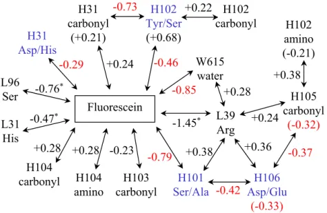
L39 Arg H105 carbonyl (-0.32) H106 Asp/Glu (-0.33) H102 amino (-0.21) H101 Ser/Ala H103 carbonyl W615 water H102 Tyr/Ser (+0.68) H31 carbonyl (+0.21) Fluorescein H102 carbonyl H31 Asp/His L96 Ser L31 His H104 amino -0.73 +0.24 -0.46 -0.85 -1.45* -0.79 -0.23 +0.28 -0.47* -0.76* -0.29 +0.38 -0.42 +0.24 -0.37 +0.28 +0.22 +0.38 +0.36 H104 carbonyl +0.28
Figure 2.1: Diagram of all major (> 0.2 kcal/mol) electrostatic interaction and desol-vation penalty differences (4M5.3 − 4-4-20) among components in Table 2.2. Arrows connecting to fluorescein correspond to changes in direct interactions, whereas ar-rows connecting scFv components correspond to changes in indirected interactions, and changes in desolvation penalties are shown in parentheses. The three intera-tion differences marked with * are those which dramatically decrease (become more positive) with minimization. All other values < −0.25 kcal/mol are shown in red. Mutated side chains (four) are labeled in blue.
A diagram of the calculated differences in electrostatic components of binding between 4-4-20 and 4M5.3 is presented in Figure 2.1. Besides the three interaction differences that were shown to be highly sensitive to minimization, seven out of the nine significant energetic changes (values in red) are associated with one or more of the four mutated residues: H31, H101, H102, or H106. The H31 and H102 side chains are uncoupled from other side chains, whereas H101 and H106 are strongly coupled to each other.
Four of the 14 mutations account for the majority of the electrostatic differences in Table 2.3: H31 Asp-to-His, H101 Ser-to-Ala, H102 Tyr-to-Ser, and H106 Asp-to-Glu. Reexamination of the 4-4-20 and 4M5.3 structures with the electrostatic results in mind reveals several mechanistic hypotheses for the 4M5.3 improved binding affinity. These mechanisms are outlined in the Discussion section.
2.3
Discussion
Electrostatic components of the binding free energy were calculated using crystal structure snapshots and the framework of a rigid binding model with continuum sol-vent. Calculations on the two crystal structures revealed −3.5 kcal/mol in favor of 4M5.3, though subsequent analysis shows that hydrogen bonds from the non-mutated residues L39 Arg, L96 Ser, and L31 His with fluorescein account for much of the
−3.5 kcal/mol improvement and are highly sensitive to precise atomic locations. It is
possible, but still uncertain, that mutations in 4M5.3 are responsible for small struc-tural changes in conserved residues. At the same time, however, four of the mutated residues dominated −1.1 kcal/mol of the binding affinity improvement, regardless of whether the structures used for analysis had been subjected to the constrained mini-mization. Several specific mechanisms for affinity improvement are revealed through these four residues.
Overall, improvements are found in all three terms of the electrostatic binding free energy: direct interactions (e.g. a hydrogen bond between the scFv and fluorescein), indirect interactions (e.g. a hydrogen bond within the scFv that is buried upon binding and hence strengthened due to loss of solvent-screening), and desolvation (i.e. the penalty paid by a polar group for leaving its unbound, aqueous environment and entering a buried environment upon binding). Binding affinity improvements can be the result of either the introduction/strengthening of a favorable interaction, the removal/weakening of an unfavorable interaction, or the reduction of a desolvation penalty.
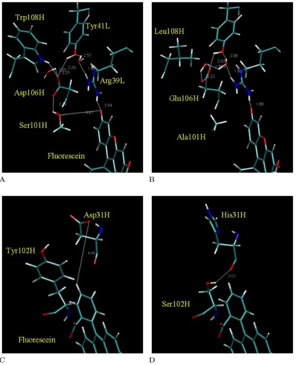
Figures 2.2A and 2.2B show the local environment of the mutated sites H101, H106, and H108. H108 was not indicated by the electrostatic analysis for binding free energy, although the position is clearly part of an intramolecular hydrogen-bonding network that includes H101 and H106. Integrating the quantitative calculations in Table 2.3 with qualitative structure analysis reveals the following mechanisms for enhanced affinity. The H101 serine to alanine mutation eliminates the unsatisfied hy-droxyl oxygen, removing a small desolvation penalty, yet loses the favorable indirect
A B
C D
Figure 2.2: Molecular detail of mutated side chains, showing the two regions of the binding site that include the four residues identified by the electrostatic analysis. (A-B) Region of H101 and H106 together with neighboring hydrogen-bonding residues L39, L41, and H108; (A) 4-4-20, and (B) 4M5.3. (C-D) Region of H31 and H102 with fluorescein; (C) 4-4-20, and (D) 4M5.3.
interaction from the H106 aspartate hydrogen bond (mutated to glutamate), for a net favorable effect. This effect is captured in the context of the other 13 mutations, but this analysis does not preclude the possibility of H101 serine to alanine also being a beneficial single mutation (although some interaction with the mutation at H106 is likely). The H106 aspartate to glutamate mutation reduces the residue desolvation penalty by burying the negative charge further into the protein. More notably, the H101 and H106 mutations appear to mutually maintain a satisfied intramolecular hydrogen-bonding network and either mutation alone might disrupt antibody stabil-ity. The differences in calculated component energies shown in Figure 2.1 support possible cooperativity between positions H101 and H106. Likewise, the H108 tryp-tophan to leucine mutation in the bordering region with two bound water molecules may be coupled to the H101 and H106 mutations via hydrogen bonds.
Figures 2.2C and 2.2D show the local environments of H31 and H102. The muta-tion at H102 from tyrosine to serine creates a new intramolecular hydrogen bond to the backbone carbonyl at residue H31. This hydrogen bond is buried and strength-ened upon binding, as shown by the −0.73 kcal/mol term in Figure 2.1. Though the hydrogen bond is to a mutated position (H31), it is independent of the side chain, making this a distinct mechanism from the actual side chain mutation at H31. The aspartate to histidine mutation at H31 removes a negative charge, which although solvent-exposed, was within 7 ˚A of the −2 charged fluorescein. This mutation is calculated to improve the direct interaction by eliminating a long-range electrostatic repulsion (−0.3 kcal/mol).
The electrostatic calculations indicate 4 of the 14 mutations for a role in the affinity improvement. Mechanisms discovered include the removal of an unsatisfied hydrogen-bonding group (H101 Ser-to-Ala), reduction of desolvation penalty (H106 Asp-to-Glu), creation of an intramolecular hydrogen bond for an indirect effect (H102 Tyr-to-Ser), and removal of long-range charge repulsion (H31 Asp-to-His). Other hypothesized mechanisms include the maintenance of a well-ordered intramolecular hydrogen-bonding network, though calculations of antibody stability were not per-formed.
The 4-4-20 and 4M5.3 structures used for computational analysis were prepared to minimize bias from inherent crystal structure differences; however, a few differences remain between the structures used for computational analysis and the single-chain antibodies used in the binding experiments. The N-terminal residue of the heavy chain in 4-4-20 is a glutamate in all experiments but the crystal structure contains an aspartate at this position [50]. This discrepancy is most likely minimal since both side chains are charged −1, solvent exposed, and about 20 ˚A from the fluorescein. Second, the fluorescein used in all binding experiments was biotinylated at the 5 carbon posi-tion, pointing out of the binding site, whereas both crystal structures were obtained with neat fluorescein. A concern is whether the 4M5.3 mutations enhance binding affinity through interaction with atoms only present in the biotinylation linker. Two of the mutated residues, H31 Asp-to-His and H102 Tyr-to-Ser are within at least 5 ˚A of the beginning of the thiourea linker with hydrogen-bonding capabilities. Previous experiments to indicate the extent of the importance of the linker in the binding interaction were not successful.
The experimental and computational results do not definitely elucidate the mech-anism for the full 1,800-fold affinity improvement of 4M5.3. The binding improvement appears to be a result of a variety of many interactions and the sum of many small changes. Nevertheless, the electrostatic calculations reveal several mechanisms that may account for part of the improvement.
Subsequent work by Midelfort and Wittrup investigated the individual effects of seven of the 14 4M5.3 mutations, confirming many of the electrostatics-based hypothe-ses [7]. The single mutations H31 Asp-to-His, H101 Ser-to-Ala, and H102 Tyr-to-Ser are each improved as expected, with H31 and H102 energetically independent as ex-pected. The mutations at H101, H106, and H108 show interactions, or non-additivity, in their effects on ligand affinity, consistent with the postulated role of intramolecular hydrogen bonding.
Two key conclusions are drawn from the complementary experimental and com-putational work. First, calculations can be used to generate hypotheses for struc-tural mechanisms not found by biochemical techniques. Second, consistency of the
electrostatic analysis with energetics of improved binding support the use of electro-statics as an engineering design tool. This work indicates that it may be realistic for structure-based calculations to correctly identify mutations that improve bind-ing affinity. Moreover, large improvements in affinity may be designed usbind-ing many smaller, yet additive mutations.
2.4
Methods
2.4.1
Preparation of protein structures
The 1.85 ˚A 4-4-20 Fab structure (1FLR [50]) and the 1.5 ˚A structure of 4M5.3 (1X9Q [42]) were used as the basis for molecular modeling, continuum electrostatic calcu-lations, and theoretical analysis. The two structures were prepared in parallel to minimize differences that might bias comparative analysis. Only residues with corre-sponding crystallographic data in both structures were used, prompting the removal of the 4-4-20 CL and CH1 domains (residues after L112 and after H117) and a few
N-and C-terminal residues corresponding to the flexible linker (4-4-20 VL112 and 4M5.3
VL0, VH-2, VH-1, VH0), all of which are solvent exposed and at least 15 ˚A from
the fluorescein binding pocket. Six water molecules were retained in each structure (4-4-20: 606, 608, 615, 618, 676, 689; 4M5.3: 6, 3, 1, 11, 26, 47); these six solvent molecules make corresponding interactions in the two structures. All other water molecules were removed from the structure files and were modeled implicitly. Side chain titration states, single conformations for all multiple-occupancy residues, and the crystallographic carbon/nitrogen/oxygen uncertainties in asparagine, glutamine, and histidine side chains were resolved based on examination of side chain local envi-ronments. The fluorescein was modeled in its net charge −2 state reflecting the pH of 8 in the binding experiments, and this choice is supported by the local environments of the fluorescein protonatable sites in each structure.
Molecular mechanics was used to prepare the protein structures. The HBUILD facility [51] in the program CHARMM [52] was used to build all hydrogen atoms onto
each structure. The CHARMm22 all-atom parameter set [53] with the CHARMM-adapted TIP3P water model was used for molecular mechanics calculations, assigning appropriate general atom types for the fluorescein (Figure 2.3). Fluorescein partial atomic charges (Figure 2.3) were obtained as described by Green and Tidor [54] by first using the program GAUSSIAN98 [55] with restricted Hartree–Fock and the 6-31G* basis set to optimize the geometry of fluorescein starting from that in the 4-4-20 structure, and subsequently fitting the electrostatic potential using the restrained-fitting methods (RESP) of Bayly et al [56].
A second set of 4-4-20 and 4M5.3 structures for comparison were prepared by subjecting each to a harmonically restrained minimization to convergence with 10 kcal/mol/˚A2 force constants on every atom in the system, no non-bonded cut-offs,
and a 4r distance-dependent dielectric constant. The root-mean-square deviation for structures before and after minimization were 0.093 ˚A (0.094 ˚A) and 0.053 ˚A (0.053 ˚A) for 4-4-20 and 4M5.3, respectively, taken over all atoms (taken over side chain atoms only).
2.4.2
Electrostatic calculations
Electrostatic contributions of individual functional groups to the binding free energy were computed using a rigid binding model with continuum solvent following pre-vious work [57]. A locally modified version of the DELPHI program [58–61] was used to solve the linearized Poisson–Boltzmann equation with finite-difference meth-ods. PARSE parameters [62] were used for atomic radii and partial atomic charges of protein. Fluorescein partial atomic charges were obtained from the RESP fitting procedure described above, and radii were assigned based on the PARSE convention. PARSE parameters do not include aliphatic hydrogen atoms and thus the appropri-ate hydrogen atoms were removed from the system. Fluorescein does not contain any aliphatic hydrogens. The dielectric constant was assigned to 4 for protein, ligand, and explicit water, and 80 for the implicit solvent regions. A salt concentration of 0.145 M was used with a 2.0-˚A Stern layer and a molecular surface generated with a 1.4-˚A probe sphere. Each molecule was oriented to minimize the volume of the bounding
C13 C2 C12 O2 C4 C9 C10 C5 C6 C7 C8 O3 O1 C14 C15 C16 C17 C18 C19 HC8 HC7 HC13 HC12 HC15 HC16 HC17 HC18 HC5 HC2 C20 O5 O4 A
Atom CHARMm22 Partial Atomic Atom CHARMm22 Partial Atomic Name Atom Type Charge Radius Name Atom Type Charge Radius
C1 C6R 0.69369 1.7 C19 C6R 0.15207 1.7 C2 C6R -0.56851 1.7 C20 C 0.68940 1.7 C3 CR66 0.30935 1.7 O1 OC -0.72934 1.4 C4 CR66 0.30921 1.7 O2 O6R -0.27339 1.4 C5 C6R -0.56775 1.7 O3 OC -0.72934 1.4 C6 C6R 0.69370 1.7 O4 OC -0.68458 1.4 C7 C6R -0.39479 1.7 O5 OC -0.78647 1.4 C8 C6R -0.19359 1.7 HC2 HA 0.15375 1.0 C9 CR66 0.02389 1.7 HC5 HA 0.15338 1.0 C10 C6RP 0.03161 1.7 HC7 HA 0.12383 1.0 C11 CR66 0.02436 1.7 HC8 HA 0.18721 1.0 C12 C6R -0.19481 1.7 HC12 HA 0.18765 1.0 C13 C6R -0.39421 1.7 HC13 HA 0.12377 1.0 C14 C6RP -0.09826 1.7 HC15 HA 0.10480 1.0 C15 C6R -0.19170 1.7 HC16 HA 0.10643 1.0 C16 C6R -0.09936 1.7 HC17 HA 0.12460 1.0 C17 C6R -0.24327 1.7 HC18 HA 0.14604 1.0 C18 C6R -0.18937 1.7 B
Figure 2.3: Computational parameterization of fluorescein. The appropriate general atom types from the CHARMm22 all-atom parameter set were used for molecular mechanics calculations. The partial atomic charge distribution used in both molec-ular mechanics and continuum electrostatics calculations was obtained by quantum-mechanical geometry minimization followed by restrained fitting of the electrostatic potential. Radii for continuum electrostatic calculations were assigned following PARSE convention.
cube. A focusing procedure was used that includes a low grid spacing using 23% fill and Debye–H¨uckel boundary conditions followed sequentially by higher resolution cal-culations first at 92% and then at 184% fill centered on the specific functional group of interest. Ten translations relative to the grid were performed and averages were used. The standard error of the mean was on the order of 0.001 kcal/mol, much less than the 0.1-1.0 kcal/mol range of differences identified between 4-4-20 and 4M5.3 components. A 129 × 129 × 129 grid was used, resulting in final grid spacings of 4.49 and 4.40 grid units per ˚A for 4-4-20 and 4M5.3, respectively. Convergence of free energies with respect to grid resolution (data not shown) indicate that a difference of 0.09 grid units per ˚A results in about 0.01 kcal/mol change in net binding free en-ergy. Therefore, the comparison between 4-4-20 and 4M5.3 electrostatic calculations on slightly different grids remains valid. For binding calculations, all explicit water molecules remained associated with the antibody in the unbound state. Overall elec-trostatic contributions to binding were dissected into component contributions from individual chemical groups and physical sources based on the work of Hendsch and Tidor [57].
Chapter 3
Development of computational
methods for the design of
improved protein binding affinity
Abstract
The development of computational methods for the redesign of high-affinity protein interactions is an important problem that is both fundamental to the advancement of protein design and directly applicable to solving pressing biotechnology needs. Here we have developed structure-based computational methods for the redesign of proteins with improved binding affinity for their protein or small-molecule targets. These methods were applied to the redesign of the model anti-lysozyme antibody D1.3. The results of design calculations to improve binding affinity were unintu-itively dominated by mutations to larger amino acids. Predictions of antibody single mutations to improve binding were tested experimentally, yielding a low success-rate and only marginal improvements. Nevertheless, the results validated predictions based on improved binding electrostatics, suggesting an altered design procedure that emphasizes electrostatic predictions (Chapter 4). The results also validated ini-tial energy-function concerns, leading to a subsequent investigation of an improved nonpolar solvation model (Chapter 5).
3.1
Introduction
A fundamental challenge in computational protein design is balancing the accurate evaluation of atomic interactions and the efficient search of a combinatorially-complex space. Early progress in the field addressed the redesign of hydrophobic protein cores in which the physical interactions could be well-approximated by straight-forward pairwise-additive van der Waals interactions, allowing the adoption and development of dead-end elimination algorithms for finding the global minimum energy conforma-tion (GMEC) in a rotamerized discrete side chain space [14, 16, 17, 63–67]. As more complex problems were addressed, such as binding affinity and specificity, enzymatic activity, and de novo structures, increasingly complex energy functions were intro-duced to more accurately evaluate interactions [15, 20–24, 26–29, 68–82]. However, the constraint of a pairwise-additive energy function has limited the development of accurate electrostatics in protein design [83–87].
A two-stage hierarchical design procedure was used to overcome the combined limitations of energy function accuracy and conformational search thoroughness [88]. In the first stage, approximations are made such that the problem is amenable to a suite of algorithms adept at pruning large combinatorial search spaces. In the second stage, with conformational space greatly reduced, low-energy structures are enumer-ated using more accurate, yet more computationally-demanding models. Together, these steps allow us to examine problems of significant conformational complexity without sacrificing our choice of energy function for final evaluation.
3.2
Methods
Conformational search is initially simplified by assuming a rigid protein backbone and allowing only discrete side chain rotamers. The physics-based energy function is pairwise-decomposable, permitting application of dead-end elimination and A* search algorithms. For each protein sequence, we find its global minimum energy conforma-tion (GMEC), and if this energy is within a specified energy cut-off of the wild-type
GMEC energy, then a continued list of lowest-energy structures are found for that sequence. Second, we reevaluate the lowest-energy structures of each sequence using more accurate, yet more more computationally-demanding models, such as Poisson– Boltzmann continuum electrostatics, unbound state side chain conformation search, and minimization. Structures are reranked based on these latter calculations. Binding energy is initially predicted from the bound state conformation and a rigid binding model. The unbound state search is used to approximate flexible binding and esti-mate a deformation penalty which offsets binding. Changes to protein fold stability are approximated from the energetic difference between the folded state and isolated model compounds.
3.2.1
Structure preparation
The crystal structure of the complex between the Fv fragment (light chain “L”, heavy chain “H”, and lysozyme “C”) of the antibody D1.3 and hen egg-white lysozyme was obtained from the Protein Data Bank (1VFB [89]). Most crystallographic water molecules were removed, except for 19 that bridge the binding interface or are buried away from bulk solvent. For calculations of two-state rigid binding, water molecules were either assigned to the antibody (water numbers 145, 149, 150, 173, 174, 177, 178, 179, 180, 223, 257, 748) or to lysozyme (water numbers 152, 155, 169, 181, 200, 222, 228). Titration states, multiple occupancies, and asparagine, glutamine, and histidine carbon/nitrogen/oxygen crystallographic uncertainties were resolved based on optimization of hydrogen-bonding in the side-chain local environments. All side chains remained in their default protonation state, with the histidine tautomers as follows. Proton on N²: positions L90 and H86; proton on Nδ: C15 and L30. The terminal dihedral angle of the histidine, asparagine, or glutamine side chains at the following positions were rotated 180◦: H56, H77, L37, L90, C19, C37, C59,
C93. Hydrogen-atom positions were assigned using the HBUILD facility [51] in the computer program package CHARMM [52] with the PARAM22 all-atom parameter set [90] and the CHARMM-adapted TIP3P water model.
3.2.2
Search space
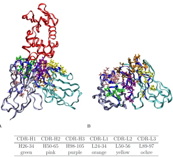
The Kabat definition for antibody complementarity determining region (CDR) posi-tions was used to select antibody sites for potential computational mutation. This presented 61 positions spanning the six contiguous stretches in primary sequence, as shown in Figure 3.1. The proline at position L95 was left as wild type in all designs. For the design of single mutants, each of these residues was individually mutated to 17 other amino acids (proline and cysteine excluded). For the design of double mutations, the only pairs of positions considered were those with at least one pair of side-chain atoms within 4.75 ˚A (non-hydrogen atoms beyond Cβ, or the Hα1 atom for glycine). For each independent design, the one or two mutated positions were given rotameric degrees of freedom, as were side chains at nearby positions (same definition of nearby as used above). The two neutral tautomers and the protonated form of histidine were allowed.
The rotamer library was based on the backbone-independent May 2002 library from Dunbrack [91, 92], expanded by ± 10◦ in both χ
1 and χ2. Prior to expansion,
three histidine rotamers were added for an unsampled ring flip (64.8, -13.8; -172.8, 9.7; -68.1, -11.3), and two asparagine rotamers were added to increase sampling of the final dihedral angle rotation (-169.3, -155.7; -170.8, 159.8). Hydroxyls of serine, threonine, and tyrosine were sampled every 30◦. The library contained 4,025 side-chain rotamers.
A novel water library allowed for conformational freedom of crystallographic water molecules. The oxygen atom location was fixed and the hydrogen atoms were placed to create 60 symmetric water molecule rotations. A 61stwater rotamer allowed for the
water to no longer exist in the structure. In addition, each crystallographic wild-type rotamer was added in a position-specific manner to the library, using the complete cartesian representation of the side chain, rather than just the dihedral angles.
3.2.3
Energy function and model
The energy function for initial search (named “low-resolution”) was the CHARMM PARAM22 all-atom parameter set [90] with no cut-offs for non-bonded interactions
A B
CDR-H1 CDR-H2 CDR-H3 CDR-L1 CDR-L2 CDR-L3 H26-34 H50-65 H98-105 L24-34 L50-56 L89-97
green pink purple orange yellow ochre
Figure 3.1: D1.3 CDR positions. The protein backbones are depicted by ribbons (red: lysozyme; iceblue: antibody heavy chain; cyan: antibody light chain; blue: CDRs). The D1.3 wild type side chains at the CDR positions are color-coded. (A) Antibody/lysozyme complex. (B) Antibody-only, looking down on interface in (A).
and a 4r distance-dependent dielectric constant. All energy terms were used (bond, angle, Urey-Bradley, dihedral, improper, Lennard-Jones, and electrostatic). The ob-jective function was the difference between the bound state energy and a sum of isolated, model compounds. Each model compound conformation was the lowest en-ergy of all side-chain rotamers with the local single amino acid backbone with an acetylated N-terminus and an N-methylamide C-terminus. The model compounds are important for canceling intrinsic side-chain energies such as ring strain (especially in PARAM22) when comparing the state energy of different protein sequences; how-ever, the model compounds do not affect actual binding free energy predictions. In
the procedure, which enumerates all possible amino acid sequences during design, the model compounds become irrelevant; when using approximate methods to order sequences in a design with non-enumerable sequence space, the model compounds are important for sequence-to-sequence comparison.
This low-resolution energy and objective function satisfies pairwise-additivity (Equation 3.1) and is readily used in dead-end elimination algorithms.
Etotal = Econst+ p X i=1 Eselfi + p X i=1 p X j>i Epairi,j (3.1)
Each summation is over the conformational search positions, p. Eselfi is the
inter-action between the residue and the fixed positions, subtracting off the energy of the isolated, model compound for that amino-acid type. The self interaction term includes intra-rotamer molecular mechanics covalent bonding terms (bonds, angles, etc.) and bonding terms to the backbone atoms of −1 or +1 positions if those positions are fixed. Epairi,j is the nonbonded interaction between two positions, plus bonding terms
only if the two positions are neighbors in primary sequence. Even though molecular mechanics covalent bonding terms span more than two atoms, e.g. four-atom dihedral angles, each energy term is always a function of the conformation of no more than two positions (made possible by a three-atom long backbone repeat unit), thereby maintaining pairwise additivity.
3.2.4
Initial conformational search
Each protein sequence was explicitly considered, for example, all 1,080 single muta-tions from 60 CDR posimuta-tions with 18 amino acids each. Side chain rotamers that clashed, either with backbone atoms or with the side chains of positions not given conformational flexibility, were eliminated from the search space. The dead-end elim-ination and A* algorithms were used to find the global minimum energy conformation (GMEC) of each sequence and up to 10 kcal/mol of lowest-energy structures [93–99]. To select the single conformation of each sequence to be used for binding energy calculation, low-energy structures were reevaluated and reranked according to more
accurate energy functions. However, because of the extra 10◦ torsional sampling in
the rotamer library, the strictly next-lowest-energy structures from the GMEC are usually not qualitatively different. We used the low-resolution energy function to rank-order structures that only differ by 10◦ dihedral angle rotations; we used a
high-resolution energy function to reevaluate different low-energy side chain conformations that may, for example, exhibit different burial and trade-off between electrostatic in-teraction and desolvation. The expansion of χ1 and χ2 by ± 10◦ generally created
families of nine rotamers (a “fleximer” [100]); we reevaluated the 30 lowest-energy structures within 10 kcal/mol that contain at least one rotamer from a new rotamer family (contain at least one new fleximer).
An in-house implementation of dead-end elimination and A* (DEE/A*) was at the core of the conformation search [101]. A novel procedure was developed to balance the speed, memory, and disk issues. A fundamental difficulty was that the input to DEE/A* is an energy cutoff from the GMEC specifying the energetic range of struc-tures to be output, rather than upfront specifying the desired number of strucstruc-tures. Furthermore, even if one could specify the number of lowest-energy structures, we actually had to look through an a priori unknown number structures to find the 30 structures of unique fleximers. Our solution was to progressively increase the energy cutoff from near-zero to 10 kcal/mol. For each energy cutoff, the lowest-energy struc-tures were output from DEE/A*. To avoid memory limitations, iterative deepening A* was used, but as a consequence, the structures were not output in order of energy. As each structure was output, it was determined if the structure contained at least one new fleximer as compared to already kept structures. If so, the structure was kept. If not, the structure only replaced the already-kept same-fleximer structure if the new structure was of lower energy. Once all structures within the energy cutoff were output, if 30 unique structures had not been obtained, then the energy cut-off was heuristically increased according to how many more unique structures were needed. The progression was stopped once 30 unique structures were obtained or the energy cutoff reached 10 kcal/mol. By evaluating structures as they were output from DEE/A*, it was not required to have a large amount of disk space for storage of all
lowest-energy structures.
Each execution of DEE/A* requires parameters specifying the schedule of different elimination routines. One can think of the different schedules as falling somewhere on a single axis of increasing/decreasing complexity. Increased scheduling complexity is necessary to solve more difficult conformational search problems, yet yields much less efficient solving of less difficult problems as compared to the time it would take for a less complex schedule. Our solution was to progressively increase the scheduling complexity. After a first attempt with a less-complex schedule, if DEE/A* did not complete within a specified amount of time, the process was aborted and a higher level of scheduling complexity was started with a longer allowed execution time.
3.2.5
Reevaluation of electrostatics
Structures were reevaluated using Poisson–Boltzmann continuum electrostatics as a substitute for the distance-dependent electrostatic energy from the molecular me-chanics force field. The PARSE parameters were used for partial atomic charges and radii [62]. A locally modified version of the DELPHI computer program was used to solve the linearized Poisson–Boltzmann equation [58–61]. A dielectric constant of 4 was used for protein and explicit water, and 80 for implicit solvent regions. Ionic strength was set to match experiments at 0.145 M, modeled with a 2.0-˚A Stern layer and a molecular surface generated with a 1.4-˚A probe sphere. Calculations were per-formed with two-step focusing from 23% to 92% molecular fill of the grid. All initial calculations used one translation of a 129 × 129 × 129 grid; interesting sequences were reevaluated with 10 translations of a 161 × 161 × 161 grid. The nonpolar com-ponent of the solvation free energy was added as a solvent-accessible surface area (SASA) term of 5 cal/mol/˚A2 [62], calculated using the analytical surface area
rou-tine in the program CHARMM; the constant intercept of this linear model cancels for two-component binding and referencing energies to wild type. In total, the typical “high-resolution” energy function for rigid binding was comprised of van der Waals, electrostatic, and nonpolar terms. Folding energies include additional molecular me-chanics covalent bonding terms for conformational change. The nonpolar term for
binding relative to wild type is typically less than 0.2 kcal/mol in magnitude and rarely as large as 0.5 kcal/mol.
3.2.6
Prediction of binding and folding
A standard calculation yielded prediction of binding affinity relative to wild type, using the predicted bound-state structure and assuming rigid binding, as well as prediction of folding stability relative to wild type, using the same structure and sub-tracting off energies for isolated, model compounds. The sensitivity of a prediction to the conformation of neighboring side chains was mitigated by matching neighboring side-chain conformations between mutant and wild type where possible. Inclusion of rotamers from the crystal structure for each position was critical; the wild type was given the benefit of the doubt for predicting relative binding free energies. In gen-eral, mutations predicted to destabilize the antibody by more than 3 kcal/mol were disregarded. In addition, we modeled conformational change upon binding by ap-proximating the lower-energy unbound state with a second side-chain conformational search. The same positions from the bound-state design were allowed conformational relaxation, again with a fixed backbone but without the binding partner present. Binding free energy was the difference between the bound and unbound states. Fi-nally, the structures for select sequences were subjected to energy-minimization of the designed positions and reevaluation of binding. All calculations for single mutations across 60 positions finished within 24 hours on a 100-CPU cluster.
3.2.7
Prediction of cooperative mutations
Double mutations predicted to be favorably cooperative were distinguished by en-suring that predicted favorability was greater than the predicted energetics for any mutation subset. Double mutation energetics were required to exceed each single mutation as well as the computed sum of each single mutation. Within the design of 182 = 324 sequences, the 18−1 = 17 single mutations were used for computing
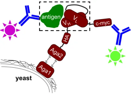
Ag
a1
antigen
S S S Syeast
V
H c-myc HAAg
a2
V
LFigure 3.2: Yeast surface display schematic. The dashed box highlights the interaction of interest, the single-chain variable fragment antibody (scFv) with its antigen.
0.2 kcal/mol to avoid marginal positives, e.g. a double mutation at −0.7 kcal/mol was not considered cooperative if the two single mutations were −0.3 kcal/mol each, but would be titled cooperative if the double mutation exceeded −0.8 kcal/mol.
3.2.8
Binding affinity measurements
The single-chain format of D1.3 [4] was displayed on the surface of yeast [102, 103]. A schematic is shown in Figure 3.2. Antibody variants were constructed using site-directed mutagenesis (Stratagene, La Jolla, CA) with oligonucleotide primers from MWG Biotech (High Point, NC). Sequences were confirmed with forward and reverse sequencing (MIT CCR Biopolymers Laboratory).
The yeast strain EBY-100 was transformed with each surface-display plasmid and binding affinities were measured as previously described [104]. Briefly, each titra-tion used 10–16 tubes with equal amounts of antibody-displaying cells and varying lysozyme concentration. Experiments were carried out at 25◦C in phosphate-buffered
saline (PBS), pH 7.3–7.5, 0.145 M salt (0.167 M ionic strength). Three or more hours elapsed as binding approached equilibrium. Cells were then pelleted and washed ice-cold, and incubated with secondary reagents for detection of antibody–lysozyme
complexes.
Binding to lysozyme (Sigma Aldrich, St. Louis, MO) was detected through sec-ondary labeling with biotinylated rabbit polyclonal anti-lysozyme antibodies (Re-search Diagnostics, now Fitzgerald Industries, Concord, MA) and tertiary labeling with streptavidin-phycoerythrin (Invitrogen, Eugene, OR). Polyclonal antibodies that bound yeast nonspecifically were removed prior to use. Though binding was detected of D1.3 to biotinylated lysozyme, neat lysozyme was used for all titrations to avoid any effects of biotinylated lysines near the binding interface.
The equilibrium dissociation constant (Kd) was fit using the mean fluorescence of
only the fraction of cells that display antibody (MF Udisp) as a function of antigen
concentration ([Ag]). The data were fit to Equation 3.2 as a function of the three free parameters by minimizing the sum of the squared residuals using the Solver tool in the program Excel.
MF Udisp= MF Umin+
MF Urange[Ag]
[Ag] + Kd
(3.2)
MF Udisp was determined by subtracting the autofluorescence of the nondisplaying
cells from the total fluorescence. Data from high concentrations of lysozyme where the displaying fraction was distinctly separated form the nondisplaying fraction were used to measure the nondisplaying fraction (fnon) and its mean fluorescence (MF Unon),
where both values were assumed to be constant within tubes of a titration because the displaying cells were the same. MF Udispwas then determined using Equation 3.3.
MF Udisp =
MF Utot− fnonMF Unon
1 − fnon
(3.3)
Data fitting using MF Utot directly instead of MF Udisp yields an identical Kd value,
but MF Umin and MF Urange values change. Reported Kd values use the average and
standard deviation from independent experiments, performed with different inocula-tions of yeast cells and typically on different days.