HAL Id: inserm-03131982
https://www.hal.inserm.fr/inserm-03131982
Submitted on 4 Feb 2021HAL is a multi-disciplinary open access
archive for the deposit and dissemination of sci-entific research documents, whether they are pub-lished or not. The documents may come from teaching and research institutions in France or abroad, or from public or private research centers.
L’archive ouverte pluridisciplinaire HAL, est destinée au dépôt et à la diffusion de documents scientifiques de niveau recherche, publiés ou non, émanant des établissements d’enseignement et de recherche français ou étrangers, des laboratoires publics ou privés.
Transport-dependent and independent functions of
KCC2 at excitatory synapses
Quentin Chevy, Clémence Simonnet, Sana Al Awabdh, Sabine Lévi, Jean
Christophe Poncer
To cite this version:
Quentin Chevy, Clémence Simonnet, Sana Al Awabdh, Sabine Lévi, Jean Christophe Poncer. Transport-dependent and independent functions of KCC2 at excitatory synapses. Neuronal Chloride Transporters in Health and Disease, Elsevier, pp.133-158, 2020, �10.1016/B978-0-12-815318-5.00007-8�. �inserm-03131982�
Chapter 7
Transport-dependent and independent functions of KCC2 at excitatory synapses
Quentin Chevy° Clémence Simonnet, Sana Al Awabdh, Sabine Lévi,
Jean Christophe Poncer*
Institut du Fer à Moulin, INSERM, Sorbonne Université, Paris, France * corresponding author
° present address: Cold Spring Harbor Laboratory, New York, USA
[NON PRINT ITEMS]
Abstract: This chapter explores non-canonical functions of the neuronal chloride/potassium
KCC2 transporter at glutamatergic, excitatory synapses. The first section describes KCC2
expression and membrane dynamics in cortical neurons to show that KCC2 is enriched in
dendritic spines that host excitatory synapses. Then, it reviews KCC2 protein binding
partners, with a specific focus on those that may contribute to the specific confinement and
role of KCC2 in dendritic spines. With this background, the next section describes how KCC2
contributes to both dendritic spine morphology and excitatory synaptic function and plasticity.
The chapter ends with a discussion on the implications of the multiple functions of KCC2, in
particular with respect to the pathology.
Key Words: Synaptic plasticity; LTP; Dendritic spines; Actin cytoskeleton; Cation chloride
1. Introduction
Mature neurons maintain intracellular chloride concentrations way below those observed in
other cell types. This ensures that activation of the chloride-permeable GABA and glycine
receptors generates hyperpolarizing, inhibitory potentials. The cation-chloride co-transporters
(CCC) KCC2 and NKCC1 are secondary active transporters that play a prominent role in
regulating transmembrane chloride gradients, as discussed in previous chapters. Although
their existence and function have been documented since the early 1970’s, Payne and
collaborators cloned the KCC2 gene (later termed Slc12a5) from the rat brain only in 1996
(Payne, Stevenson, & Donaldson, 1996). Three years later, KCC2 was identified as
responsible for the early postnatal shift in the polarity of GABA transmission in the rat
hippocampus (Rivera et al., 1999). Since then, more than 600 original publications and 80
reviews have explored KCC2 expression, function, regulation and involvement of the
pathology. For obvious reasons, most of those have focused on KCC2 in the context of
inhibitory neurotransmission. However, an ever-growing body of evidence indicates that
KCC2 function extends beyond the mere control of transmembrane chloride gradients and
GABA transmission. First, the pattern of KCC2 expression reveals specific clustering in
dendritic spines, which harbor most glutamatergic synapses. Second, KCC2 genetic ablation
or knockdown have revealed much more complex phenotypes than previously expected,
including alterations in dendritic spine maturation and morphology as well as excitatory
synaptic function. In 2015, Blaesse and Schmidt coined this heterogeneity of KCC2 functions
with the term moonlighting protein, which appeared in the late 1990’s to designate the
In this chapter, we review experimental data supporting the notion that KCC2 is indeed
multifunctional and that, through interactions with a variety of molecular partners, it
participates in several biological processes, which only partly involve its ion-transport
function. We suggest that, considering the variety of pathological conditions involving
down-regulation of KCC2 expression (Kahle et al., 2008; Kaila, Price, Payne, Puskarjov, & Voipio,
2014), fully understanding the various processes and molecular interactions KCC2 is engaged
in will help predicting and designing most efficient and specific therapeutic strategies.
2. KCC2 expression in the vicinity of excitatory synapses
The Slc12a5 gene encodes two KCC2 protein isoforms, KCC2a and KCC2b. that are
expressed under the control of distinct promoters (Markkanen et al., 2014; Uvarov et al.,
2007; Uvarov et al., 2009). KCC2a defers from KCC2b by a unique 40-amino acid sequence
in its N-terminal domain, containing a putative regulatory domain by the SPS1-related
proline/alanine-rich kinase (SPAK)(Uvarov et al., 2007). Using antibodies raised against
subtype specific epitopes, KCC2b was shown to be the most prominently expressed isoform
in the adult forebrain, whereas KCC2a expression is higher in neonates and remains relatively
constant during development (Markkanen et al., 2014). Remarkably, whereas full KCC2 KO
mice die at birth due to severe motor and respiratory deficits (Hubner et al., 2001),
isoform-selective KO show less severe phenotypes. Thus, KCC2b KO mice survive for 2-3 weeks and
then die due to generalized seizures (Woo et al., 2002) whereas KCC2a KO show altered
breathing behavior, most importantly at early postnatal stages (Dubois et al., 2018;
Markkanen et al., 2014). Therefore, although both isoforms form functional transporters
(Markkanen et al., 2017; Uvarov et al., 2007), whether they fulfill distinct or partially
overlapping functions remains unclear. In the rest of this chapter, KCC2 implicitly refers to
In heterologous systems, KCC2 forms both homo- and hetero-oligomeres with other
cation-chloride co-transporters (Simard et al., 2007). Dimers of the two isoforms, KCC2a and
KCC2b, are also formed both in vivo and in heterologous cells (Uvarov et al., 2009).
Oligomerization appears to rely mainly on disulfide bonds, as treatment with reducing agents
mostly yields monomeric forms (Agez et al., 2017; Blaesse et al., 2006) and may involve
KCC2 carboxy-terminal domain (Agez et al., 2017), as also demonstrated for KCC1 (Casula
et al., 2001). KCC2 oligomerization and function were suggested to be correlated, based in
part on their parallel developmental profile in the rat lateral superior olive (Blaesse et al.,
2006). In addition, lipid rafts may also influence KCC2 oligomerization and clustering, even
though the impact of KCC2 accumulation in lipid rafts on its transport function remain
debated (Hartmann et al., 2009; Watanabe, Wake, Moorhouse, & Nabekura, 2009).
At the subcellular level, KCC2 is present throughout the somato-dendritic membrane of most
cortical neurons including GABAergic interneurons (Gulyas, Sik, Payne, Kaila, & Freund,
2001), but is virtually excluded from their axon, including their axon initial segment (Baldi,
Varga, & Tamas, 2010; Szabadics et al., 2006). In the hippocampus, high-resolution
pre-embedding KCC2 immunolocalization revealed cell-type specific differences in the
somatic/dendritic expression levels (Baldi et al., 2010). While granule cells show more
intense membrane expression in dendritic than somatic regions, KCC2 seems more evenly
distributed in CA1 pyramidal cells. Most strikingly, as initially reported by Guylas and
collaborators (Gulyas et al., 2001), KCC2 expression is often observed in dendritic spines,
and is more abundant near excitatory than inhibitory synapses (Baldi et al., 2010). Confocal
imaging of hippocampal neurons immunostained for KCC2 also revealed a higher intensity of
KCC2 cluster immunofluorescence in dendritic spines than on the adjacent shafts (Gauvain et
al., 2011)(Fig. 1A). Although these clusters were mostly found near the postsynaptic
primarily aggregates perisynaptically. These intriguing observations raise several important
questions, in particular regarding the specific targeting and aggregation in dendritic spines, as
well as the possible role of a K/Cl co-transporter near excitatory synapses.
(Figure 1 near here)
Figure 1. Molecular determinants of KCC2 aggregation near synapses. A,
Confocal images of a hippocampal neuron immunostained for KCC2. Right,
magnification of boxed area in the image shown on the left. Note high-intensity
fluorescent KCC2 clusters in dendritic spines. Scale, 5µm. B, Reconstructed
trajectories of recombinant KCC2 molecules tracked with quantum dots (white),
overlaid with fluorescent micrographs showing Homer1c-GFP (green) and
gephyrin-mRFP (red) identifying glutamatergic and GABAergic synapses,
respectively. KCC2 molecules show less constrained trajectories in the
extrasynaptic membrane. Scale, 1 µm. C, Schematic representation of KCC2
diffusion in the neuronal plasma membrane. Near excitatory synapses, interaction
with submembrane actin via 4.1N hinders KCC2 lateral diffusion. Similarly,
KCC2 diffusion is constrained near inhibitory synapses, through so far
unidentified molecular interactions, possibly with GABA receptors and/or
scaffolding molecules such as gephyrin. Extrasynaptic KCC2 on the other hand
are more mobile, as indicated by the larger arrow.
Credits. Adapted from (Gauvain et al., 2011) (A) and (Chamma et al., 2013) (B)
with permission.
KCC2 aggregation in dendritic spines is somewhat reminiscent of postsynaptic receptor
(Choquet & Triller, 2013). Single particle tracking experiments using photostable fluorescent
nanocrystals called quantum dots (Bannai, Levi, Schweizer, Dahan, & Triller, 2006) have
revealed that the somewhat static picture of the postsynaptic element with tightly anchored
receptors was oversimplified. Thus, receptors shown near-Brownian diffusion within the
plasma membrane and only get temporarily trapped at synapses by high-affinity interaction
with scaffolding molecules, such that synaptic and extrasynaptic receptors undergo a
continuous and dynamic exchange, a phenomenon termed diffusion-trapping (Choquet &
Triller, 2003). Such phenomenon represents a general mechanism allowing molecular
heterogeneity within the plasma membrane, based on reversible interactions of
transmembrane molecules with membrane or sub-membrane molecules. Using similar
approaches as those used to study postsynaptic receptors dynamics and synaptic trapping,
Chamma et al. have explored KCC2 membrane dynamics in cultured hippocampal neurons
(Chamma et al., 2013). Recombinant KCC2 showed diffusion properties comparable to
postsynaptic receptors with greater diffusion coefficients and confinement domains in the
extrasynaptic membrane than near excitatory and inhibitory synapses. Importantly, the
dwell-time of KCC2 molecules near excitatory synapses was longer than near inhibitory synapses,
suggestive of distinct molecular constraints over KCC2 diffusion at these synapses (Fig. 1B).
This raised the question of the molecular identity of the scaffolding molecules that hinder
KCC2 diffusion at excitatory vs. inhibitory synapses.
As we discuss below (Section 3, see also Chapter 8 and Chapter 12), KCC2 interacts with a
variety of molecular partners that may accumulate near synapses. In particular, dendritic
spines that harbor glutamatergic synapses are highly enriched in actin and actin-binding
proteins (Cingolani & Goda, 2008) which act as scaffolding molecules for numerous ion
transport proteins (ion channels, transporters, exchangers…)(Denker & Barber, 2002). Thus,
dwell-time near excitatory synapses (Chamma et al., 2013). This effect likely involves KCC2
interaction with submembrane actin scaffold via interaction of its carboxy-terminal domain
with the 4.1 Ezrin Radixin Moesin (FERM)-domain protein 4.1N, an actin/spectrin-binding
protein also enriched in dendritic spines and that was shown to interact with KCC2 (Li et al.,
2007). Thus, overexpression of KCC2 carboxy-terminal domain or 4.1N knockdown by RNA
interference both mimicked the effect of latrunculin. Importantly, the dwell-time of KCC2
near inhibitory synapses was unaffected by these manipulations. This suggests that distinct
molecular interactions are at play to confine KCC2 near inhibitory vs. excitatory synapses
(Fig. 1C). Interactors responsible for trapping KCC2 near GABAergic synapses remain to be
identified.
3. KCC2 interacts with synaptic and perisynaptic proteins
Molecular interactions are not just governing the subcellular distribution of transmembrane
proteins but also modulate their function. Most of them are engaged in macro-molecular
complexes with intricate physical and functional interactions. Full understanding of the
function and regulation of individual transmembrane proteins therefore relies on a
comprehensive identification of the molecular interactions they engage in. Thus, several
recent studies have revealed a number of somewhat unexpected molecular interactions
involving KCC2 (Table 1). Those shed new light on its functions and regulation in neurons.
Yeast two-hybrid screening was first used to identify KCC2 binding partners. This approach
revealed KCC2 interactions that are likely important for local regulation of KCC2 function.
For instance, KCC2 was shown to interact with the brain-type creatine kinase (CKB) (Inoue,
Ueno, & Fukuda, 2004), a neuronal ATP-generating enzyme acting to increase KCC2
Yamada, Ueno, & Fukuda, 2006). Interestingly, the alpha2 subunit of the Na+/K+ ATPase was
also found to interact with KCC2 (Ikeda et al., 2004). The Na+/K+ ATPase establishes ion
gradients across the plasma membrane used for KCC2-mediated ion transport. Therefore, a
functional complex comprising KCC2, CKB and the Na+/K+ ATPase may constitute an
ion-transport metabolon (Kaila et al., 2014), in which ATPase-dependent potassium import would
be promoted by local ATP production by CKB which would in turn support secondary active
chloride export by KCC2, via a locally generated potassium gradient.
Other interactions identified by immunoprecipitation assays suggested new mechanisms for
KCC2 regulation, further extending an already vast repertoire of regulatory posttranslational
modifications ((Chamma, Chevy, Poncer, & Levi, 2012; Kaila et al., 2014; Medina et al.,
2014) for review; See also Chapter 11). For instance, the clathrin-binding adaptor protein-2
(AP-2), Ras-associated binding protein 11b (Rab11b) and the actin-associated protein 4.1N
were shown to be critical for KCC2 endocytosis, recycling and clustering, respectively
(Chamma et al., 2013; Li et al., 2007; Roussa et al., 2016; Zhao et al., 2008). Perhaps more
intriguing is the discovery of KCC2 interaction with the glutamatergic, kainate receptor
subunit 2 (GluK2; (Mahadevan et al., 2014; Pressey et al., 2017)) and its auxiliary subunit
neuropilin and tolloid-like 2 (Neto2; (Ivakine et al., 2013)). These interactions were shown to
promote KCC2 function primarily by enhancing its recycling to the plasma membrane,
leading to enhanced membrane expression and function (Pressey et al., 2017). Interestingly,
kainate receptors also interact with 4.1N (Copits & Swanson, 2013) that in turn interacts with
the AMPA receptor subunit GluA1 (Lin et al., 2009). Collectively, these data support the
view that KCC2 may be part of a macromolecular complex comprising postsynaptic
glutamate receptors and elements of the submembrane cytoskeleton. This complex may both
stabilize KCC2 in specific subcellular membrane compartments as well as locally regulate
(Table 1 near here)
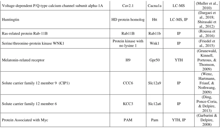
Table 1.
Protein name Alt. name Gene Interaction
detection References
Proteins enriched at excitatory synapses
Band 4.1-like protein 1 4.1N Epb41l1 IP
(Chamma et al., 2013; Li et al., 2007)
Rho guanine nucleotide exchange factor 7 Beta-Pix Arhgef7 IP
(Chevy et al., 2015; Llano et al., 2015)
Cofilin-1 p18 Cfl1 LC-MS (Mahadevan
et al., 2017)
Metabotropic glutamate receptor 1 mGluR1 Grm1 LC-MS (Kato et al.,
2012)
Metabotropic glutamate receptor 5 mGluR5 Grm5 LC-MS (Farr et al.,
2004)
Glutamate receptor ionotropic, kainate 2 GluK2 Grik2 LC-MS, IP
(Mahadevan et al., 2017; Pressey et al.,
2017)
Neuropilin and tolloid-like protein 2 Neto2 Neto2 GST/LC-MS, IP
(Ivakine et al., 2013; Mahadevan et al., 2017) Voltage-dependent R-type calcium channel subunit alpha-1E Cav2.3 Cacna1e LC-MS (Muller et al.,
2010)
Amyloid-beta A4 protein APP App IP (M. Chen et
al., 2017)
Neuroligin-1 Neuroligin-1 Nlgn1 IP (Loh et al.,
2016)
Proteins enriched at inhibitory synapses
Gamma-aminobutyric acid receptor subunit alpha-1 GABAAR1 Gabra1 IP (Y. Huang et
al., 2012)
Gamma-aminobutyric acid receptor subunit beta-1 GABABR1 Gabrb1 LC-MS, IP (Wright et
al., 2017)
Neuroligin-2 Neuroligin-2 Nlgn2 IP
(Sun, Zhang, & Chen,
2013)
Proteins enriched at synapses
Protein kinase C and casein kinase substrate in neurons 1 SdpI Pacsin1 LC-MS, IP (Mahadevan et al., 2017) Serine/threonine-protein phosphatase PP1-alpha catalytic subunit PP-1A Ppp1ca LC-MS (Mahadevan et al., 2017)
Adaptor protein complex AP-2 Alpha2-adaptin Ap2 LC-MS, IP
(Mahadevan et al., 2017;
Zhao et al., 2008)
Non-synaptic proteins
STE20/SPS1-related proline-alanine-rich protein kinase PSTK1 Stk39 IP (Friedel et
al., 2015) Sodium/potassium-transporting ATPase subunit alpha-2 Na(+)/K(+) ATPase
alpha-2 subunit Atp1a2 LC-MS, IP
(Ikeda et al., 2004; Mahadevan et al., 2017)
Creatine kinase B-type CPK-B Ckb LC-MS,YTH, IP
(Inoue et al., 2004; Inoue et al., 2006; Mahadevan et al., 2017)
Voltage-dependent P/Q-type calcium channel subunit alpha-1A Cav2.1 Cacna1a LC-MS (Muller et al., 2010)
Huntingtin HD protein homolog Htt LC-MS, IP
(Dargaei et al., 2018; Shirasaki et
al., 2012)
Ras-related protein Rab-11B Rab11B Rab11b IP (Roussa et
al., 2016)
Serine/threonine-protein kinase WNK1 Protein kinase with
no lysine 1 Wnk1 IP
(Friedel et al., 2015)
Melatonin-related receptor H9 Gpr50 YTH
(Grunewald, Kinnell, Porteous, &
Thomson, 2009)
Solute carrier family 12 member 9 (CIP1) CCC6 Slc12a9 IP
(Wenz, Hartmann,
Friauf, & Nothwang,
2009)
Solute carrier family 12 member 6 KCC3 Slc12a6 IP
(Ding, Ponce-Coria,
& Delpire, 2013)
Protein Associated with Myc PAM Pam YTH, IP
(Garbarini & Delpire,
2008)
Table 1. KCC2 molecular interactions in cortical neurons. The table shows
putative KCC2 interactors, as identified in yeast two-hybrid (YTH), GST pulldown
(GST), immunoprecipitation (IP) or liquid chromatography and mass
spectrometry (LC-MS). Note that a large proportion of putative interactors are
enriched at or near excitatory synapses. The impact of putative or validated
KCC2 interactors on KCC2 function, membrane expression or traffic has not
always been assessed and is therefore not indicated.
Functional proteomic analysis of KCC2 recently further extended the list of putative KCC2
interactors (Mahadevan et al., 2017). It is remarkable that most identified or putative KCC2
partners are expressed at or near excitatory and, to a lesser extent, inhibitory synapses (Table
1), where KCC2 clusters are most abundant as discussed previously (Baldi et al., 2010;
Chamma et al., 2013). Thus, KCC2 candidate interactors predominantly include ion-transport
cytoskeleton-related proteins and proteins involved in receptor trafficking (Mahadevan et al., 2017). This
study also identified the protein kinase C and casein kinase II substrate in neurons (PACSIN1)
as a new interactor that acts as a negative regulator of KCC2 expression and function, through
mechanisms that remain to be determined. Interestingly, PACSIN1 is known to interact with
the PKC-interacting proteins PICK1 and regulate activity-dependent trafficking of the AMPA
receptor (Anggono et al., 2013), further supporting the tight interaction of KCC2 with
glutamatergic signaling.
In conclusion, KCC2 appears to interact with a wealth of synaptic and perisynaptic proteins
and notably proteins related to glutamatergic rather than GABAergic signaling (Table 1).
These interactions have most often been explored from the point of view of KCC2 function,
expression and regulation. However, KCC2 expression undergoes rapid and multifactorial up-
and down-regulation both under physiological and pathological conditions (Chamma et al.,
2012; Kaila et al., 2014; Medina et al., 2014). It seems unlikely that changes in KCC2
expression at the plasma membrane may not reciprocally affect the expression and/or function
of its molecular partners. In particular, KCC2 clustering in dendritic spines and interaction
with several synaptic and persisynaptic proteins raise the question of its role in dendritic spine
physiology and excitatory synaptic function.
4. KCC2 activity and the regulation of dendritic spine volume
As described above, although KCC2 is expressed throughout the somato-dendritic membrane
of most CNS neurons, it specifically aggregates in dendritic spines, near glutamatergic
synapses through specific interactions with scaffolding molecules (Chamma et al., 2013;
K/Cl co-transport is one of the major mechanisms of cell volume regulation in mammalian
cells (Hoffmann, Lambert, & Pedersen, 2009; Zeuthen, 2010). Since CNS neurons are mostly
devoid of dedicated water channels such as aquaporins (Amiry-Moghaddam & Ottersen,
2003; Badaut, Lasbennes, Magistretti, & Regli, 2002; Kahle et al., 2015), the reported ability
of CCCs to transport water alongside the ion fluxes (Hamann, Herrera-Perez, Bundgaard,
Alvarez-Leefmans, & Zeuthen, 2005; Zeuthen, 1991, 2010) may be particularly important to
deal with osmotic challenges. In neurons, such osmotic challenges may prominently result
from intracellular influx of Na+ and Cl- ions associated with synaptic activity. Thus, whereas
synaptic activation of postsynaptic AMPARs may yield Na+ transients that could lead to local
concentration up to 10 mM that rapidly get cleared through spine neck diffusion, NMDAR
activation leads to way larger and longer-lasting transients (Miyazaki & Ross, 2017).
Therefore, repetitive activation of postsynaptic receptors is likely to generate significant, local
osmotic challenges that could be partly compensated by KCC2 activity (Gulyas et al., 2001).
The first demonstration for a role of KCC2 in water fluxes and neuronal volume regulation
was provided in a study using digital holographic microscopy, a noninvasive optical imaging
technique to monitor transmembrane water fluxes as detected by the phase signal (Jourdain et
al., 2011). Blocking KCC2 with furosemide reduced phase shifts induced by glutamate
application onto cultured cortical neurons. Since then, several experimental observations
supported a specific role of KCC2 in spine volume regulation. For example, chronic KCC2
suppression in mature hippocampal neurons using RNA interference leads to a prominent
increase in spine head volume (Chevy et al., 2015; Gauvain et al., 2011). This effect likely
reflects the loss of KCC2 transport function as it is mimicked by chronic application of an
antagonist of the transporter (Gauvain et al., 2011) but not by overexpression of its
carboxy-terminal tail, acting to prevent KCC2 interaction with molecular partners involved in actin
also recently associated with increased spine volume (Heubl et al., 2017). In these
experiments, phosphorylation-dependent dispersion of KCC2 resulted in increased spine head
volume in just 30 minutes. Even though the precise timing of this effect should be further
characterized, these data suggest that loss of KCC2 activity could rapidly control spine
volume. Remarkably, these effects were not associated with changes in spine length or density
or in the proportion of filopodia-like structures (Gauvain et al., 2011). This contrasts with the
genetic ablation of KCC2, which compromises the formation and maturation of dendritic
spines in immature hippocampal neurons, through a mechanism involving its interaction with
actin cytoskeleton (Li et al., 2007) (See Chapter XX). Therefore, whereas KCC2 interaction
with actin is required for spinogenesis during development, KCC2 expression and function in
mature neurons appear to be predominantly required to control spine head volume, not spine
maintenance or structure.
Increased spine volume is associated with long term potentiation (LTP) at a variety of
glutamatergic synapses in the CNS (Bosch & Hayashi, 2012). Such increase is extremely
rapid and usually reaches its maximum during or immediately after the conditioning stimulus
and then decays over minutes to stabilize at above-control values, involving actin
cytoskeleton rearrangements (Bosch et al., 2014; Kopec, Real, Kessels, & Malinow, 2007;
Murakoshi, Wang, & Yasuda, 2011). Although the initial increase in spine head volume may
also reflect massive protein translocation into the spine head, as suggested using FRET-based
probes for several scaffolding molecules (Bosch et al., 2014), the bi-phasic time-course of
these structural changes is hard to explain solely by cytoskeleton remodeling. Alternatively, a
tempting hypothesis would be that intense synaptic activity and postsynaptic ion influx during
serve to restore osmotic pressure through ion and water export. This hypothesis however
currently lacks experimental validation.
5. KCC2-actin interaction hinders protein diffusion in dendritic spines
Many integral membrane proteins serve as cytoskeleton anchors to the plasma membrane.
These include adhesion molecules as well as ion channels, pumps, cotransporters and
exchangers (Denker & Barber, 2002). Such interactions are critical for the stability and
maintenance of the shape of subcellular compartments. The role of ion transport proteins in
cytoskeleton anchoring to plasma membrane was first illustrated in erythrocytes by the anion
exchanger 1 (AE1), which binds to spectrin/actin via ankyrin and the FERM domain
containing adaptor protein 4.1R, thereby contributing to the typical shape and viscoelastic
properties of these cells (Bennett & Baines, 2001; Jons & Drenckhahn, 1992).
As discussed above, KCC2 interacts through its carboxy-terminal domain with 4.1N, a
member of the 4.1 family (Li et al., 2007). This interaction is at least partly responsible for
confining KCC2 near glutamatergic synapses onto dendritic spines (Chamma et al., 2013).
Conversely, through this interaction, KCC2 is expected to anchor spine actin cytoskeleton to
the plasma membrane. Given the prominent role of actin dynamics in spine morphogenesis
(Tada & Sheng, 2006), it is therefore not totally surprising that cortical KCC2-/- neurons fail
to develop mature, functional spines when cultured in vitro (Li et al., 2007)(but see (Seja et
al., 2012)) whereas KCC2 precocious expression increases dendritic spinogenesis (Fiumelli et
al., 2013).
However, actin dynamics are not only involved in spinogenesis but also influence synaptic
function and plasticity, through modulation of AMPA receptor lateral diffusion. As
AMPA receptors are confined within the postsynaptic density via interactions with
scaffolding molecules and cytoskeleton (Choquet & Triller, 2003; Sheng & Hoogenraad,
2007). They also continuously exchange between synaptic, perisynaptic, and extrasynaptic
pools by lateral diffusion, which acts to maintain a steady state level of functional receptors at
synapses (Opazo & Choquet, 2011). Altering spine actin cytoskeleton therefore influences
AMPAR diffusion and anchoring, and thereby affects the synaptic pool of receptors and
synaptic efficacy (Kerr & Blanpied, 2012; Rust et al., 2010). Actin membrane-anchoring
proteins located near glutamatergic synapses are therefore predicted to influence AMPAR
diffusion within dendritic spines.
(Figure 2 near here)
Figure 2. KCC2 hinders protein diffusion in dendritic spines. A, Left,
Immunostaining of GFP and GluA1 in dendritic sections of hippocampal neurons
expressing non-target (shNT) or KCC2-directed shRNA. KCC2 knockdown leads
to reduced GluA1 immunofluorescence in dendritic spines (arrowheads). Scale, 1
µm. Right, quantification of the normalized cluster intensity of GluA1
immunofluorescence in dendritic spines. B, image sequences of quantum
dot-labeled, mobile GluA1 (arrowheads) in dendritic spines of neurons expressing
non target vs. KCC2-directed shRNA as in A. Quantum dot images are shown in
green while spine membrane is outlined in red. Explored area over 24s is shown
on maximum intensity projections on the right panels. Note the larger explored
area of GluA1 and its escape from the spine head in the absence of KCC2. Scale, 1 μm. C, Schematic representation of structural changes in dendritic spines upon KCC2 suppression. Under control conditions, KCC2 interaction with
submembrane actin scaffold hinders the diffusion of the mobile (persisynaptic)
NCAM180. KCC2 knockdown reduces this diffusion constraint, leading to
enhanced diffusion of perisynaptic AMPA receptors. Continuous exchange
between synaptic and persisynaptic pools then leads to the progressive partial
depletion of the former. Note that diffusion of the less mobile (synaptic) fraction of
AMPA receptors and of membrane-bound NCAM is unaffected. KCC2
suppression also leads to an increase in spine head volume as discussed in section
4.
Credits. Adapted from (Gauvain et al., 2011) with permission.
This hypothesis was tested in experiments where KCC2 expression was suppressed in
hippocampal neurons with RNA interference (Gauvain et al., 2011). Single particle tracking
of AMPA receptors containing the GluA1 subunit revealed enhanced lateral diffusion of the
mobile (likely non-synaptic) pool of receptors, with no detectable effect on the immobile
(likely synaptic) fraction (Tardin, Cognet, Bats, Lounis, & Choquet, 2003)(Fig. 2B). Notably,
this effect was observed in dendritic spines but not on dendritic shafts. It was not specific to
AMPA receptors, as lateral diffusion of the cell adhesion molecule NCAM was also increased
in absence of KCC2. More specifically, this effect was observed for the transmembrane
isoform NCAM 180, bearing a short intracellular domain that interacts with actin cytoskeleton
through β-spectrin, but not the membrane-anchored NCAM 120 isoform that lacks an intracellular domain (Buttner & Horstkorte, 2010). These results support a model in which
KCC2 is an element of a molecular complex acting as a barrier for the lateral diffusion of
non-synaptic, transmembrane proteins within dendritic spines (Fig. 2). Functionally, enhanced
lateral diffusion of perisynaptic AMPA receptors was associated with a reduced
immunostaining of the receptors in spines and reduced synaptic efficacy at glutamatergic
with reduced KCC2 expression. Importantly, these effects were mimicked by preventing
KCC2 interaction with intracellular partners using its carboxy-terminal domain as dominant
negative, but not by a KCC2 antagonist. This suggests KCC2, like other transmembrane
ion-transport proteins (Denker & Barber, 2002), acts as an anchor to submembrane actin
cytoskeleton that contributes to confine persisynaptic AMPA receptors within dendritic
spines, independent of its ion transport function.
In conclusion, KCC2 interacts with submembrane actin cytoskeleton and thereby contributes
to a molecular barrier hindering the lateral diffusion of transmembrane proteins within
dendritic spines. Disrupting this barrier promotes AMPA receptor lateral diffusion and likely
depletes a perisynaptic reserve pool of receptors and, subsequently, the synaptic pool, leading
to reduced efficacy of glutamatergic synapses (Fig. 2C). This phenomenon may be
particularly relevant in the pathology but also for the physiological regulation of excitatory
transmission. KCC2 clusters are rapidly dispersed upon sustained NMDA receptor activation
(Chamma et al., 2013; Lee, Deeb, Walker, Davies, & Moss, 2011). Subsequent enhancement
of lateral diffusion and depletion of the synaptic and perisynaptic pools may then contribute to
NMDA receptor-induced plasticity of glutamatergic synapses.
6. KCC2-dependent control of actin dynamics and long term potentiation at glutamatergic synapses
The relation between the KCC2 cotransporter and actin extends beyond the mere organization
of a molecular barrier for the lateral diffusion of transmembrane proteins. KCC2 knockdown
or precocious expression was shown to influence the dynamics of actin polymerization in
several cell types. Although KCC2 is largely considered a neuron-specific transporter, it
appears to be expressed in several human cancer cell lines (Wei et al., 2011). In some of
stress fiber organization. Thus, suppressing KCC2 resulted in increased filamentous actin (f-actin) content, suggestive of enhanced actin polymerization while overexpressing KCC2 led
to the opposite phenotype. Conversely, neuron-specific overexpression of KCC2 in mouse
embryos resulted in aberrant f-actin distribution with reduced density at the adherens
junctions lining the neural tube (Horn, Ringstedt, Blaesse, Kaila, & Herlenius, 2010). In both
studies, the observed phenotype was independent of the transporter function of KCC2 as it
was mimicked by recombinant, Cl- transport-deficient mutant KCC2. These results suggested
that KCC2 may not just interact with actin cytoskeleton but also somehow control actin
polymerization.
Dynamic actin remodeling in neurons is controlled by small GTPases of the Rho family that
includes Rho, Rac1, and Cdc42 (Cingolani & Goda, 2008). In dendritic spines, which are
enriched in filamentous actin (f-actin), local Rac1 activation involves the synaptic anchoring
by the G-protein-coupled receptor kinase-interacting protein 1 (GIT1) of its partner βPIX (H.
Zhang, Webb, Asmussen, & Horwitz, 2003), a guanine nucleotide exchange factor (GEF)
which facilitates the exchange of GDP for GTP. Activated Rac1 may then bind to several
effector proteins including the p21 activated kinase (PAK), which in turn activates LIM
kinase. This ultimately leads to inhibition of the actin-severing protein cofilin through
phosphorylation of its Ser3 residue (Yang et al., 1998)(Fig. 3).
Increased dendritic spine actin polymerization upon KCC2 knockdown was observed in
hippocampal neurons (Llano et al. 2015, Chevy et al. 2015). This effect likely involves a
direct interaction between KCC2 and βPIX, as demonstrated in immunoprecipitation assays,
even though this interaction was not identified in recent functional proteomic analysis
(Mahadevan et al., 2017). Thus, KCC2 genetic ablation or knockdown by RNA interference
leads to reduced actin turnover and accumulation of f-actin in dendritic spines.
spine necks but did not reveal specific patterns of f-actin accumulation within spine heads
(Chevy et al., 2015). This effect was associated with mobilization of βPIX in dendritic spines,
a specific increase in Rac1 but not Rho-A activity as well as enhanced cofilin phosphorylation
(Chevy et al., 2015; Llano et al., 2015). These observations are consistent with activation of
the Rac1-PAK1-LIMK pathway and inhibition of cofilin. Importantly, again, these effects
where independent of ion transport by KCC2 as they were mimicked by a dominant negative
peptide that inhibits KCC2 interaction with intracellular partners (Chevy et al., 2015) and
rescued by the expression of a transport-deficient recombinant KCC2 (Llano et al., 2015).
(Figure 3 near here)
Figure 3. KCC2 interaction with βPIX controls spine actin dynamics and gates
LTP at excitatory synapses. A, Viral-based chronic KCC2 suppression by RNA
interference. Confocal micrograph showing viral expression (as detected by GFP
fluorescence, green) in a rat hippocampal slice with DAPI staining (blue).
Stimulation and recording electrodes are shown. Scale, 200 µm. B, Summary
graph from experiment as in A, showing LTP of the fEPSP recorded in st.
moleculare upon high frequency stimulation (HFS) of perforant path afferents.
KCC2 knockdown (shKCC2) precluded LTP expression at this synapse. C,
schematic representation of the molecular cascade involved in this effect. Upon
KCC2 suppression, βPIX relocates at the postsynaptic density and interacts with
GIT1 to specifically activate Rac1, leading to enhanced PAK1 and LIMK activity.
This in turn inhibits cofilin leading to enhanced f-actin content in dendritic spines.
f-actin is non-permissive for activity-driven AMPA receptor delivery, thereby
precluding LTP expression. Preventing cofilin inhibition is sufficient to restore
AMPA receptor exocytosis in KCC2 knockdown neurons.
How KCC2 precisely regulates Rac1 activity through βPIX remains to be fully elucidated but
may involve molecular trapping of βPIX by KCC2. As discussed above (section 2), KCC2
appears to be excluded from the glutamatergic postsynaptic density. KCC2 might therefore
contribute to sequestrate βPIX away from the postsynaptic density, where GIT1 acts as a
postsynaptic scaffold for multiprotein signaling complex with Rac1 (H. Zhang et al., 2003).
Overexpressed KCC2 carboxy-terminal domain may then compete with KCC2-βPIX interaction and thereby favor βPIX binding to GIT1 within the postsynaptic density, similar to KCC2 knockdown. Although more evidence is needed to support this hypothesis, including
PSD purification assays and super-resolution imaging, it is remarkable that KCC2 knockdown
increases the clustering of βPIX but not GIT1 in dendritic spines (Chevy et al., 2015).
Tight regulation of actin dynamics is critical to the structural and functional changes involved
in long term plasticity of glutamatergic synapses (Bosch & Hayashi, 2012). Long term
potentiation is known to rely on a concomitant increase of both the number of postsynaptic
AMPA receptors (Hayashi et al., 2000; Herring & Nicoll, 2016; Malinow, Mainen, &
Hayashi, 2000; Poncer, 2003; Rumpel, LeDoux, Zador, & Malinow, 2005) and the volume of
dendritic spines that host glutamatergic synapses (Bosch et al., 2014; L. Y. Chen, Rex, Casale,
Gall, & Lynch, 2007; Fortin et al., 2010; Gu et al., 2010; Kopec et al., 2007; Okamoto, Bosch,
& Hayashi, 2009; Park et al., 2006; Sala & Segal, 2014). The latter is associated with a
persistent increase in actin polymerization within the potentiated dendritic spine (Okamoto,
Nagai, Miyawaki, & Hayashi, 2004). However, an initial and transient actin depolymerization
is required for both activity-driven AMPA receptor exocytosis and increase in spine volume
(Gu et al., 2010; Ouyang et al., 2005). This reflects the transient activation (i.e.,
dephosphorylation) of the actin-severing enzyme cofilin at the time of LTP induction (Gu et
receptor membrane insertion by promoting vesicle exocytosis that may otherwise be hindered
by cortical actin, as described in adrenal chromaffin cells (Gasman et al., 1999). Alternatively,
partial cytoskeleton disassembly may be required to release secretory vesicles and promote
their traffic to the plasma membrane.
KCC2 interaction with the Rac-PAK-LIMK pathway regulating cofilin activity is then
expected to impact functional and structural plasticity at excitatory synapses. This hypothesis
was first tested by knocking-down KCC2 in dentate gyrus granule cells where LTP of
entorhinal afferents is mediated through postsynaptic AMPA receptor traffic (Poncer &
Malinow, 2001). LTP at these synapses was almost fully abolished upon KCC2 knockdown
(Chevy et al., 2015) (Fig. 3A-B). Suppression of KCC2 expression by RNA interference in
primary hippocampal culture similarly precluded chemically-induced LTP (cLTP). Thus, both
structural (i.e. change in dendritic spine volume) and functional (i.e. addition of glutamatergic
receptors) cLTP was abolished in KCC2 knockdown neurons. This effect was acting
downstream of CaMKII activation, since overexpression of a constitutively active enzyme
also failed to induce LTP in neurons with suppressed KCC2 expression. In fact, LTP
hindrance upon KCC2 suppression directly and specifically relied on dysregulation of actin
dynamics, as inhibitors of either PAK1 or LIMK fully rescued structural and functional LTP
expression (Chevy et al., 2015). Importantly, all these experiments were performed in the
presence of the GABAA receptor antagonist bicuculline. Therefore, LTP hindrance was
independent of any change in GABA signaling induced upon KCC2 suppression in
hippocampal neurons (Pellegrino et al., 2011). A recent study, however, suggested that altered
GABA signaling upon KCC2 downregulation may compromise synapse specificity of LTP in
the ageing hippocampus, through mechanisms that remain to be clarified (Ferando, Faas, &
In conclusion, direct interaction of the KCC2 co-transporter with both actin-associated
proteins (such as 4.1N) and proteins controlling actin dynamics (such as βPIX) turns out to
strongly impact the structure, function and plasticity of glutamatergic synapses (Fig. 3C). This
may be important in physiological settings. As discussed above, KCC2 clusters are rapidly
dispersed upon NMDA receptor activation and subsequent Ca2+ influx, partly through Ser940
dephosphorylation and calpain-induced cleavage (Chamma et al., 2013; Lee et al., 2011;
Puskarjov, Ahmad, Kaila, & Blaesse, 2012). Ca2+-dependent KCC2 down-regulation has been
shown to underlie some forms of activity-dependent plasticity of GABA signaling in cortical
neurons (Fiumelli, Cancedda, & Poo, 2005; Woodin, Ganguly, & Poo, 2003). However, as
discussed above, activity-induced dispersion of KCC2 clusters within dendritic spines may
also rapidly affect spine actin cytoskeleton and polymerization. Such changes are likely to
influence further induction of synaptic plasticity at recently active synapses. It is then
tempting to suggest that KCC2 may then act as a metaplastic switch (Abraham, 2008) for
glutamatergic synapses, acting to adjust synaptic plasticity to prior activity due to changes in
actin polymerization. Thus, NMDA receptor activation has been shown to inhibit subsequent
LTP induction in CA1 hippocampal neurons (Y. Y. Huang, Colino, Selig, & Malenka, 1992).
The mechanisms underlying synaptic metaplasticty remain elusive (Abraham, 2008; Hulme,
Jones, & Abraham, 2013), but PP1- and calpain-mediated KCC2 clearance and subsequent
actin remodeling may represent a molecular substrate deserving further experimental
investigation.
7. Conclusions
As discussed in this and the preceding chapters, KCC2 clearly appears to fulfill more than just
chloride gradients to regulation of dendritic spine volume, spine actin membrane anchoring
and dynamics.
KCC2 belongs to the CCC family, which itself is part of the family of the solute carriers with
its 52 distinct subgroups (Hediger et al., 2004). Phylogenetic analysis of the CCC family
shows that KCC proteins have emerged from a common ancestor gene through three main
gene duplication events (Hartmann, Tesch, Nothwang, & Bininda-Emonds, 2014). As
discussed by Blaesse and Schmidt (Blaesse & Schmidt, 2015), multi-functional proteins such
as KCC2 may represent an intermediate stage of evolution, in which several functions have
not yet led to gene duplication and specialization that may be required to optimize each
individual function. The term function, however, should be considered with caution, as it is
perhaps questionable whether the relationship between KCC2 and actin cytoskeleton and its
functional impact represents an actual function of KCC2 or whether they primarily represent
the consequence of molecular interactions and subcellular membrane anchoring required for
fulfilling its original, ion- (and water-) transport function. In this context, it is worth noting
that a variety of ion transport proteins are also used to anchor and regulate cytoskeleton in
many different cells types (Denker & Barber, 2002).
Finally, in addition to the aforementioned activity-induced changes at the posttranslational
level, KCC2 expression is down-regulated in a variety of pathological conditions ranging
from epilepsy, neuropathic pain, spasticity and stroke to schizophrenia, autism-spectrum
disorders and Alzheimer’s disease (reviewed in (Kahle et al., 2008; Kaila et al., 2014; Moore, Kelley, Brandon, Deeb, & Moss, 2017); see also Chapter 14-23). Reduced KCC2 expression
and activity are often assumed to contribute to pathological network activities underlying
these conditions by reducing the efficacy of GABAergic transmission. Drugs acting to
compensate for KCC2 suppression therefore hold great therapeutic potential. For instance,
KCC2-lacking neurons and thereby normalize neuronal activity (Ben-Ari, 2017). However,
this approach would only compensate ion-transport related KCC2 functions. In this context, it
is interesting to note that the first KCC2 polymorphism associated with idiopathic epilepsy
compromised both ion transport and dendritic spine morphogenesis (Puskarjov et al., 2014).
How ion-transport independent functions actually contribute to the pathology remains to be
fully explored. We suggest however that pharmacological strategies aiming to enhance KCC2
expression or promote its membrane stability (Gagnon et al., 2013; J. Zhang, Karimy, Delpire,
& Kahle, 2017) rather than solely rescue neuronal chloride homeostasis may more fully
compensate synaptic and neuronal deficits induced by KCC2 down-regulation in neurological
and psychiatric disorders.
References
Abraham, W. C. (2008). Metaplasticity: tuning synapses and networks for plasticity. Nat Rev
Neurosci, 9(5), 387. doi:10.1038/nrn2356
Agez, M., Schultz, P., Medina, I., Baker, D. J., Burnham, M. P., Cardarelli, R. A., . . .
Jawhari, A. (2017). Molecular architecture of potassium chloride co-transporter
KCC2. Sci Rep, 7(1), 16452. doi:10.1038/s41598-017-15739-1
Amiry-Moghaddam, M., & Ottersen, O. P. (2003). The molecular basis of water transport in
the brain. Nat Rev Neurosci, 4(12), 991-1001. doi:10.1038/nrn1252
Anggono, V., Koc-Schmitz, Y., Widagdo, J., Kormann, J., Quan, A., Chen, C. M., . . .
Huganir, R. L. (2013). PICK1 interacts with PACSIN to regulate AMPA receptor
internalization and cerebellar long-term depression. Proc Natl Acad Sci U S A,
Badaut, J., Lasbennes, F., Magistretti, P. J., & Regli, L. (2002). Aquaporins in brain:
distribution, physiology, and pathophysiology. J Cereb Blood Flow Metab, 22(4),
367-378. doi:10.1097/00004647-200204000-00001
Baldi, R., Varga, C., & Tamas, G. (2010). Differential distribution of KCC2 along the
axo-somato-dendritic axis of hippocampal principal cells. Eur J Neurosci, 32(8),
1319-1325. doi:10.1111/j.1460-9568.2010.07361.x
Bannai, H., Levi, S., Schweizer, C., Dahan, M., & Triller, A. (2006). Imaging the lateral
diffusion of membrane molecules with quantum dots. Nat Protoc, 1(6), 2628-2634.
doi:10.1038/nprot.2006.429
Ben-Ari, Y. (2017). NKCC1 Chloride Importer Antagonists Attenuate Many Neurological
and Psychiatric Disorders. Trends Neurosci, 40(9), 536-554.
doi:10.1016/j.tins.2017.07.001
Bennett, V., & Baines, A. J. (2001). Spectrin and ankyrin-based pathways: metazoan
inventions for integrating cells into tissues. Physiol Rev, 81(3), 1353-1392.
doi:10.1152/physrev.2001.81.3.1353
Blaesse, P., Guillemin, I., Schindler, J., Schweizer, M., Delpire, E., Khiroug, L., . . .
Nothwang, H. G. (2006). Oligomerization of KCC2 correlates with development of
inhibitory neurotransmission. J Neurosci, 26(41), 10407-10419.
doi:10.1523/jneurosci.3257-06.2006
Blaesse, P., & Schmidt, T. (2015). K-Cl cotransporter KCC2--a moonlighting protein in
excitatory and inhibitory synapse development and function. Pflugers Arch, 467(4),
615-624. doi:10.1007/s00424-014-1547-6
Bosch, M., Castro, J., Saneyoshi, T., Matsuno, H., Sur, M., & Hayashi, Y. (2014). Structural
and molecular remodeling of dendritic spine substructures during long-term
Bosch, M., & Hayashi, Y. (2012). Structural plasticity of dendritic spines. Curr Opin
Neurobiol, 22(3), 383-388. doi:10.1016/j.conb.2011.09.002
Buttner, B., & Horstkorte, R. (2010). Intracelluar ligands of NCAM. Adv Exp Med Biol, 663,
55-66. doi:10.1007/978-1-4419-1170-4_3
Casula, S., Shmukler, B. E., Wilhelm, S., Stuart-Tilley, A. K., Su, W., Chernova, M. N., . . .
Alper, S. L. (2001). A dominant negative mutant of the KCC1 K-Cl cotransporter:
both N- and C-terminal cytoplasmic domains are required for K-Cl cotransport
activity. J Biol Chem, 276(45), 41870-41878. doi:10.1074/jbc.M107155200
Chamma, I., Chevy, Q., Poncer, J. C., & Levi, S. (2012). Role of the neuronal K-Cl
co-transporter KCC2 in inhibitory and excitatory neurotransmission. Front Cell Neurosci,
6, 5. doi:10.3389/fncel.2012.00005
Chamma, I., Heubl, M., Chevy, Q., Renner, M., Moutkine, I., Eugene, E., . . . Levi, S. (2013).
Activity-dependent regulation of the K/Cl transporter KCC2 membrane diffusion,
clustering, and function in hippocampal neurons. J Neurosci, 33(39), 15488-15503.
doi:10.1523/JNEUROSCI.5889-12.2013
Chen, L. Y., Rex, C. S., Casale, M. S., Gall, C. M., & Lynch, G. (2007). Changes in synaptic
morphology accompany actin signaling during LTP. J Neurosci, 27(20), 5363-5372.
doi:10.1523/JNEUROSCI.0164-07.2007
Chen, M., Wang, J., Jiang, J., Zheng, X., Justice, N. J., Wang, K., . . . Yang, L. (2017). APP
modulates KCC2 expression and function in hippocampal GABAergic inhibition.
Elife, 6. doi:10.7554/eLife.20142
Chevy, Q., Heubl, M., Goutierre, M., Backer, S., Moutkine, I., Eugene, E., . . . Poncer, J. C.
(2015). KCC2 Gates Activity-Driven AMPA Receptor Traffic through Cofilin
Phosphorylation. J Neurosci, 35(48), 15772-15786.
Choquet, D., & Triller, A. (2003). The role of receptor diffusion in the organization of the
postsynaptic membrane. Nat Rev Neurosci, 4(4), 251-265. doi:10.1038/nrn1077
Choquet, D., & Triller, A. (2013). The dynamic synapse. Neuron, 80(3), 691-703.
doi:10.1016/j.neuron.2013.10.013
Cingolani, L. A., & Goda, Y. (2008). Actin in action: the interplay between the actin
cytoskeleton and synaptic efficacy. Nat Rev Neurosci, 9(5), 344-356.
doi:10.1038/nrn2373
Copits, B. A., & Swanson, G. T. (2013). Kainate receptor post-translational modifications
differentially regulate association with 4.1N to control activity-dependent receptor
endocytosis. J Biol Chem, 288(13), 8952-8965. doi:10.1074/jbc.M112.440719
Dargaei, Z., Bang, J. Y., Mahadevan, V., Khademullah, C. S., Bedard, S., Parfitt, G. M., . . .
Woodin, M. A. (2018). Restoring GABAergic inhibition rescues memory deficits in a
Huntington's disease mouse model. Proc Natl Acad Sci U S A, 115(7), E1618-e1626.
doi:10.1073/pnas.1716871115
Denker, S. P., & Barber, D. L. (2002). Ion transport proteins anchor and regulate the
cytoskeleton. Current Opinion in Cell Biology, 14(2), 214-220.
doi:10.1016/s0955-0674(02)00304-6
Ding, J., Ponce-Coria, J., & Delpire, E. (2013). A trafficking-deficient mutant of KCC3
reveals dominant-negative effects on K-Cl cotransport function. PLoS One, 8(4),
e61112. doi:10.1371/journal.pone.0061112
Dubois, C. J., Cardoit, L., Schwarz, V., Markkanen, M., Airaksinen, M. S., Uvarov, P., . . .
Thoby-Brisson, M. (2018). Role of the K(+)-Cl(-) Cotransporter KCC2a Isoform in
Mammalian Respiration at Birth. eNeuro, 5(5). doi:10.1523/ENEURO.0264-18.2018
Farr, C. D., Gafken, P. R., Norbeck, A. D., Doneanu, C. E., Stapels, M. D., Barofsky, D. F., . .
5 protein complexes reveals novel molecular constituents. J Neurochem, 91(2),
438-450. doi:10.1111/j.1471-4159.2004.02735.x
Ferando, I., Faas, G. C., & Mody, I. (2016). Diminished KCC2 confounds synapse specificity
of LTP during senescence. Nat Neurosci. doi:10.1038/nn.4357
Fiumelli, H., Briner, A., Puskarjov, M., Blaesse, P., Belem, B. J., Dayer, A. G., . . . Vutskits,
L. (2013). An ion transport-independent role for the cation-chloride cotransporter
KCC2 in dendritic spinogenesis in vivo. Cereb Cortex, 23(2), 378-388.
doi:10.1093/cercor/bhs027
Fiumelli, H., Cancedda, L., & Poo, M. M. (2005). Modulation of GABAergic transmission by
activity via postsynaptic Ca2+-dependent regulation of KCC2 function. Neuron, 48(5),
773-786. doi:10.1016/j.neuron.2005.10.025
Fortin, D. A., Davare, M. A., Srivastava, T., Brady, J. D., Nygaard, S., Derkach, V. A., &
Soderling, T. R. (2010). Long-term potentiation-dependent spine enlargement requires
synaptic Ca2+-permeable AMPA receptors recruited by CaM-kinase I. J Neurosci,
30(35), 11565-11575. doi:10.1523/JNEUROSCI.1746-10.2010
Friedel, P., Kahle, K. T., Zhang, J., Hertz, N., Pisella, L. I., Buhler, E., . . . Medina, I. (2015).
WNK1-regulated inhibitory phosphorylation of the KCC2 cotransporter maintains the
depolarizing action of GABA in immature neurons. Sci Signal, 8(383), ra65.
doi:10.1126/scisignal.aaa0354
Gagnon, M., Bergeron, M. J., Lavertu, G., Castonguay, A., Tripathy, S., Bonin, R. P., . . . De
Koninck, Y. (2013). Chloride extrusion enhancers as novel therapeutics for
neurological diseases. Nat Med, 19(11), 1524-1528. doi:10.1038/nm.3356
Garbarini, N., & Delpire, E. (2008). The RCC1 domain of protein associated with Myc
(PAM) interacts with and regulates KCC2. Cell Physiol Biochem, 22(1-4), 31-44.
Gauvain, G., Chamma, I., Chevy, Q., Cabezas, C., Irinopoulou, T., Bodrug, N., . . . Poncer, J.
C. (2011). The neuronal K-Cl cotransporter KCC2 influences postsynaptic AMPA
receptor content and lateral diffusion in dendritic spines. Proc Natl Acad Sci U S A,
108(37), 15474-15479. doi:10.1073/pnas.1107893108
Grunewald, E., Kinnell, H. L., Porteous, D. J., & Thomson, P. A. (2009). GPR50 interacts
with neuronal NOGO-A and affects neurite outgrowth. Mol Cell Neurosci, 42(4),
363-371. doi:10.1016/j.mcn.2009.08.007
Gu, J., Lee, C. W., Fan, Y., Komlos, D., Tang, X., Sun, C., . . . Zheng, J. Q. (2010).
ADF/cofilin-mediated actin dynamics regulate AMPA receptor trafficking during
synaptic plasticity. Nat Neurosci, 13(10), 1208-1215. doi:10.1038/nn.2634
Gulyas, A. I., Sik, A., Payne, J. A., Kaila, K., & Freund, T. F. (2001). The KCl cotransporter,
KCC2, is highly expressed in the vicinity of excitatory synapses in the rat
hippocampus. Eur J Neurosci, 13(12), 2205-2217.
Hamann, S., Herrera-Perez, J. J., Bundgaard, M., Alvarez-Leefmans, F. J., & Zeuthen, T.
(2005). Water permeability of Na+-K+-2Cl- cotransporters in mammalian epithelial
cells. J Physiol, 568(Pt 1), 123-135. doi:10.1113/jphysiol.2005.093526
Hartmann, A. M., Blaesse, P., Kranz, T., Wenz, M., Schindler, J., Kaila, K., . . . Nothwang, H.
G. (2009). Opposite effect of membrane raft perturbation on transport activity of
KCC2 and NKCC1. J Neurochem, 111(2), 321-331.
doi:10.1111/j.1471-4159.2009.06343.x
Hartmann, A. M., Tesch, D., Nothwang, H. G., & Bininda-Emonds, O. R. (2014). Evolution
of the cation chloride cotransporter family: ancient origins, gene losses, and
subfunctionalization through duplication. Mol Biol Evol, 31(2), 434-447.
Hayashi, Y., Shi, S. H., Esteban, J. A., Piccini, A., Poncer, J. C., & Malinow, R. (2000).
Driving AMPA receptors into synapses by LTP and CaMKII: requirement for GluR1
and PDZ domain interaction. Science, 287(5461), 2262-2267.
Hediger, M. A., Romero, M. F., Peng, J. B., Rolfs, A., Takanaga, H., & Bruford, E. A. (2004).
The ABCs of solute carriers: physiological, pathological and therapeutic implications
of human membrane transport proteinsIntroduction. Pflugers Arch, 447(5), 465-468.
doi:10.1007/s00424-003-1192-y
Herring, B. E., & Nicoll, R. A. (2016). Long-Term Potentiation: From CaMKII to AMPA
Receptor Trafficking. Annu Rev Physiol, 78, 351-365.
doi:10.1146/annurev-physiol-021014-071753
Heubl, M., Zhang, J., Pressey, J. C., Al Awabdh, S., Renner, M., Gomez-Castro, F., . . . Lévi,
S. (2017). GABAA receptor dependent synaptic inhibition rapidly tunes KCC2
activity via the Cl−-sensitive WNK1 kinase. Nat Commun, 8(1), 1776.
doi:10.1038/s41467-017-01749-0
Hoffmann, E. K., Lambert, I. H., & Pedersen, S. F. (2009). Physiology of cell volume
regulation in vertebrates. Physiol Rev, 89(1), 193-277.
doi:10.1152/physrev.00037.2007
Horn, Z., Ringstedt, T., Blaesse, P., Kaila, K., & Herlenius, E. (2010). Premature expression
of KCC2 in embryonic mice perturbs neural development by an ion
transport-independent mechanism. Eur J Neurosci, 31(12), 2142-2155.
doi:10.1111/j.1460-9568.2010.07258.x
Huang, Y., Ko, H., Cheung, Z. H., Yung, K. K., Yao, T., Wang, J. J., . . . Yung, W. H. (2012).
Dual actions of brain-derived neurotrophic factor on GABAergic transmission in
cerebellar Purkinje neurons. Exp Neurol, 233(2), 791-798.
Huang, Y. Y., Colino, A., Selig, D. K., & Malenka, R. C. (1992). The influence of prior
synaptic activity on the induction of long-term potentiation. Science, 255(5045),
730-733.
Hubner, C. A., Stein, V., Hermans-Borgmeyer, I., Meyer, T., Ballanyi, K., & Jentsch, T. J.
(2001). Disruption of KCC2 reveals an essential role of K-Cl cotransport already in
early synaptic inhibition. Neuron, 30(2), 515-524.
Hulme, S. R., Jones, O. D., & Abraham, W. C. (2013). Emerging roles of metaplasticity in
behaviour and disease. Trends Neurosci, 36(6), 353-362.
doi:10.1016/j.tins.2013.03.007
Ikeda, K., Onimaru, H., Yamada, J., Inoue, K., Ueno, S., Onaka, T., . . . Kawakami, K.
(2004). Malfunction of respiratory-related neuronal activity in Na+, K+-ATPase
alpha2 subunit-deficient mice is attributable to abnormal Cl- homeostasis in brainstem
neurons. J Neurosci, 24(47), 10693-10701. doi:10.1523/jneurosci.2909-04.2004
Inoue, K., Ueno, S., & Fukuda, A. (2004). Interaction of neuron-specific K+-Cl-
cotransporter, KCC2, with brain-type creatine kinase. FEBS Lett, 564(1-2), 131-135.
doi:10.1016/s0014-5793(04)00328-x
Inoue, K., Yamada, J., Ueno, S., & Fukuda, A. (2006). Brain-type creatine kinase activates
neuron-specific K+-Cl- co-transporter KCC2. J Neurochem, 96(2), 598-608.
doi:10.1111/j.1471-4159.2005.03560.x
Ivakine, E. A., Acton, B. A., Mahadevan, V., Ormond, J., Tang, M., Pressey, J. C., . . .
McInnes, R. R. (2013). Neto2 is a KCC2 interacting protein required for neuronal Cl-
regulation in hippocampal neurons. Proc Natl Acad Sci U S A, 110(9), 3561-3566.
doi:10.1073/pnas.1212907110
Jons, T., & Drenckhahn, D. (1992). Identification of the binding interface involved in linkage
of cytoskeletal protein 4.1 to the erythrocyte anion exchanger. EMBO J, 11(8),
2863-2867.
Jourdain, P., Pavillon, N., Moratal, C., Boss, D., Rappaz, B., Depeursinge, C., . . . Magistretti,
P. J. (2011). Determination of transmembrane water fluxes in neurons elicited by
glutamate ionotropic receptors and by the cotransporters KCC2 and NKCC1: a digital
holographic microscopy study. J Neurosci, 31(33), 11846-11854.
doi:10.1523/JNEUROSCI.0286-11.2011
Kahle, K. T., Khanna, A. R., Alper, S. L., Adragna, N. C., Lauf, P. K., Sun, D., & Delpire, E.
(2015). K-Cl cotransporters, cell volume homeostasis, and neurological disease.
Trends Mol Med, 21(8), 513-523. doi:10.1016/j.molmed.2015.05.008
Kahle, K. T., Staley, K. J., Nahed, B. V., Gamba, G., Hebert, S. C., Lifton, R. P., & Mount, D.
B. (2008). Roles of the cation-chloride cotransporters in neurological disease. Nat Clin
Pract Neurol, 4(9), 490-503. doi:10.1038/ncpneuro0883
Kaila, K., Price, T. J., Payne, J. A., Puskarjov, M., & Voipio, J. (2014). Cation-chloride
cotransporters in neuronal development, plasticity and disease. Nat Rev Neurosci,
15(10), 637-654. doi:10.1038/nrn3819
Kato, A. S., Knierman, M. D., Siuda, E. R., Isaac, J. T., Nisenbaum, E. S., & Bredt, D. S.
(2012). Glutamate receptor delta2 associates with metabotropic glutamate receptor 1
(mGluR1), protein kinase Cgamma, and canonical transient receptor potential 3 and
regulates mGluR1-mediated synaptic transmission in cerebellar Purkinje neurons. J
Neurosci, 32(44), 15296-15308. doi:10.1523/jneurosci.0705-12.2012
Kerr, J. M., & Blanpied, T. A. (2012). Subsynaptic AMPA receptor distribution is acutely
regulated by actin-driven reorganization of the postsynaptic density. J Neurosci, 32(2),
Kopec, C. D., Real, E., Kessels, H. W., & Malinow, R. (2007). GluR1 links structural and
functional plasticity at excitatory synapses. J Neurosci, 27(50), 13706-13718.
doi:10.1523/JNEUROSCI.3503-07.2007
Lee, H. H., Deeb, T. Z., Walker, J. A., Davies, P. A., & Moss, S. J. (2011). NMDA receptor
activity downregulates KCC2 resulting in depolarizing GABAA receptor-mediated
currents. Nat Neurosci, 14(6), 736-743. doi:10.1038/nn.2806
Li, H., Khirug, S., Cai, C., Ludwig, A., Blaesse, P., Kolikova, J., . . . Rivera, C. (2007). KCC2
interacts with the dendritic cytoskeleton to promote spine development. Neuron, 56(6),
1019-1033. doi:10.1016/j.neuron.2007.10.039
Lin, D. T., Makino, Y., Sharma, K., Hayashi, T., Neve, R., Takamiya, K., & Huganir, R. L.
(2009). Regulation of AMPA receptor extrasynaptic insertion by 4.1N,
phosphorylation and palmitoylation. Nat Neurosci, 12(7), 879-887.
doi:10.1038/nn.2351
Llano, O., Smirnov, S., Soni, S., Golubtsov, A., Guillemin, I., Hotulainen, P., . . . Ludwig, A.
(2015). KCC2 regulates actin dynamics in dendritic spines via interaction with
beta-PIX. J Cell Biol, 209(5), 671-686. doi:10.1083/jcb.201411008
Loh, K. H., Stawski, P. S., Draycott, A. S., Udeshi, N. D., Lehrman, E. K., Wilton, D. K., . . .
Ting, A. Y. (2016). Proteomic Analysis of Unbounded Cellular Compartments:
Synaptic Clefts. Cell, 166(5), 1295-1307.e1221. doi:10.1016/j.cell.2016.07.041
Mahadevan, V., Khademullah, C. S., Dargaei, Z., Chevrier, J., Uvarov, P., Kwan, J., . . .
Woodin, M. A. (2017). Native KCC2 interactome reveals PACSIN1 as a critical
regulator of synaptic inhibition. Elife, 6. doi:10.7554/eLife.28270
Mahadevan, V., Pressey, J. C., Acton, B. A., Uvarov, P., Huang, M. Y., Chevrier, J., . . .