HAL Id: inserm-00968357
https://www.hal.inserm.fr/inserm-00968357
Submitted on 31 Mar 2014
HAL is a multi-disciplinary open access
archive for the deposit and dissemination of
sci-entific research documents, whether they are
pub-lished or not. The documents may come from
teaching and research institutions in France or
abroad, or from public or private research centers.
L’archive ouverte pluridisciplinaire HAL, est
destinée au dépôt et à la diffusion de documents
scientifiques de niveau recherche, publiés ou non,
émanant des établissements d’enseignement et de
recherche français ou étrangers, des laboratoires
publics ou privés.
Etiological heterogeneity in autism spectrum disorders:
role of rare variants
Catalina Betancur, Mary Coleman
To cite this version:
Catalina Betancur, Mary Coleman. Etiological heterogeneity in autism spectrum disorders: role of
rare variants. Joseph D. Buxbaum, Patrick R. Hof. The Neuroscience of Autism Spectrum Disorders,
Academic Press, pp.113-144, 2013. �inserm-00968357�
The Neuroscience of Autism Spectrum Disorders
Edited by Joseph D. Buxbaum & Patrick R. Hof
Academic Press, Oxford
Chapter 2.1
Etiological heterogeneity in autism spectrum disorders: role of rare variants
Catalina Betancur
1,2,3Mary Coleman
41
INSERM, U952, Paris, France
2CNRS, UMR 7224, Paris, France
3UPMC Univ Paris 06, Paris, France
4
Foundation for Autism Research, Sarasota, Florida, USA
Correspondence: Catalina.Betancur@inserm.fr
Abstract
Autism spectrum disorders (ASD) encompass a group of behaviorally defined developmental
disabilities characterized by marked clinical and etiological heterogeneity. There is increasing evidence
that ASD can arise from rare highly penetrant mutations and genomic imbalances. There are at
present over 100 disease genes and 50 recurrent genomic imbalances implicated in the etiology of
ASD. These genes and loci have so far all been causally implicated in intellectual disability, indicating
that these two neurodevelopmental disorders share common genetic bases. Similarly, many genes
involved in epilepsy can also result in ASD. These observations indicate that these genes cause a
continuum of neurodevelopmental disorders that manifest in different ways depending on other
genetic, environmental or stochastic factors. Increased recognition of the etiological heterogeneity of
ASD will expand the number of target genes for neurobiological investigations, reveal functional
pathways and assist the development of novel therapeutic approaches.
Key words: autism; genetic syndrome; intellectual disability; epilepsy; metabolic disorder; mutation;
Introduction
Autism spectrum disorders (ASD) encompass a group of behaviorally defined developmental
disabilities characterized by marked clinical and etiological heterogeneity. ASD can be associated with
intellectual disability (ID) of varying degrees (∼70%), epilepsy (∼30%), dysmorphic features and
congenital malformations (∼20%) (Coleman and Gillberg, 2012). ASD can thus be considered
syndromic (i.e., associated with dysmorphic, neuromuscular, metabolic or other distinctive clinical
features, including structural brain abnormalities) or nonsyndromic, similar to the division of ID into
syndromic and nonsyndromic forms (Gecz et al., 2009).
1The genetic architecture of ASD is highly heterogeneous (Abrahams and Geschwind, 2008;
Betancur, 2011; State, 2010). About 20% of individuals have an identified genetic etiology.
Cytogenetically visible chromosomal aberrations have been reported in ∼5% of cases, involving many
different loci on all chromosomes. The most frequent abnormalities are maternally derived 15q11-‐q13
duplications involving the imprinted Prader-‐Willi/Angelman region, detected in ∼1%. ASD can also be
due to mutations of numerous single genes involved in autosomal dominant, autosomal recessive and
X-‐linked disorders. The most common single gene defect identified in ASD is fragile X syndrome
(FMR1), present in ∼2% of cases (Kielinen et al., 2004) (Chapter 4.5). Other monogenic disorders
described in ASD include tuberous sclerosis (TSC1, TSC2) (Chapter 4.8), Angelman syndrome (UBE3A),
Rett syndrome (MECP2) (Chapter 4.6), and PTEN mutations in patients with macrocephaly and autism
(Chapter 4.8). Rare mutations have been identified in multiple synaptic genes, including NLGN3,
NLGN4X (Jamain et al., 2003), SHANK3 (Durand et al., 2007), and SHANK2 (Berkel et al., 2010; Pinto et
al., 2010) (Chapter 4.7). Recent genome-‐wide microarray studies in large ASD samples have
highlighted the important contribution of rare submicroscopic deletions and duplications, called copy
number variation (CNV), to the etiology of ASD, including de novo events in 5%–10% of cases (Marshall
et al., 2008; Pinto et al., 2010; Sanders et al., 2011; Sebat et al., 2007) (Chapter 2.2). Most recently,
the first whole-‐exome sequencing studies in ASD have shown an increased rate of rare de novo point
mutations and confirmed a high degree of locus heterogeneity (Neale et al., 2012; O'Roak et al., 2011;
O'Roak et al., 2012; Sanders et al., 2012) (Chapter 2.4).
The constantly increasing number of distinct, individually rare genetic causes of ASD and the
substantial contribution of de novo events indicates that the genetic architecture of ASD resembles
that of ID, with hundreds of genetic and genomic disorders involved, each accounting for a very small
fraction of cases. In fact, all the known genetic causes of ASD are also causes of ID, indicating that
these two neurodevelopmental disorders share common genetic bases.
We recently performed an exhaustive review of all the genetic and genomic disorders reported in
subjects with ASD or autistic behavior, and identified 103 disease genes and 44 recurrent genomic
imbalances (Betancur, 2011), and the numbers have continued to grow. These findings are in stark
contrast to a persisting claim among the autism research community that we know very little about
the etiology of autism and that there are only a modest number of autism loci known. Here, rather
than listing all the genetic and genomic disorders involved in ASD, we review what can we learn about
1 Note that the term ‘syndromic’ autism refers to the clinical presentation of the patient and not to the fact that a genetic disorder or syndrome has been identified in the patient. Genetic defects can be associated with syndromic or nonsyndromic clinical presentations. Furthermore, note that the term ‘idiopathic’ autism means that a specific etiology has not been identified in that patient (i.e., unexplained autism); the term ‘idiopathic’ should not be used in lieu of nonsyndromic or isolated autism. Finally, the use of the terms ‘primary’ and ‘secondary’ autism to refer to nonsyndromic and syndromic
the profound etiological heterogeneity underlying ASD.
The most obvious conclusion we can draw is that, when examined from an etiological perspective,
ASD is not a single disease entity but a behavioral manifestation of many hundreds of single gene and
genomic disorders. In addition, it is emerging that de novo variants are an important part of the
architecture of ASD, consistent with purifying selection against deleterious genetic variants of major
effect. One of the most important observations is that there is considerable overlap in high-‐risk genes
and loci for ASD, ID, and epilepsy. Similarly, many of the rare recurrent CNVs identified recently have
been found to confer risk for a broad range of neurologic and psychiatric phenotypes, including not
only ID, ASD, and epilepsy, but also schizophrenia and attention deficit hyperactivity disorder (ADHD).
This highlights how disruption of core neurodevelopmental processes can give rise to a wide range of
clinical manifestations and that greater attention should be placed on the neurobiological processes of
brain development and function rather than on the precise behavioral manifestation. Finally, we show
how some of the genes implicate specific pathways, subcellular organelles, or systems in the
pathophysiology of ASD, which can lead to biological and neurobiological insights into disease
mechanisms.
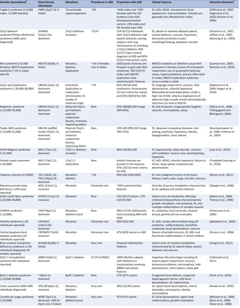
Genetic disorders strongly associated with ASD
Table 1 shows genetic and genomic disorders in which ASD is a common manifestation. For some of
these disorders, ASD is among the clinical hallmarks, including Phelan-‐McDermid syndrome (22q13
deletion syndrome/SHANK3 mutations), maternal 15q11-‐q13 duplications, Rett syndrome (MECP2)
and MECP2 duplication syndrome, fragile X syndrome (FMR1), tuberous sclerosis (TSC1, TSC2),
adenylosuccinate lyase deficiency (ADSL), Timothy syndrome (CACNA1C), cortical dysplasia-‐focal
epilepsy syndrome (CNTNAP2), Smith-‐Lemli-‐Opitz syndrome (DHCR7), Smith-‐Magenis syndrome
(17p11.2 deletion, RAI1 mutations), and Potocki-‐Lupski syndrome (17p11.2 duplication) (see Table 1
for references). Another disorder strongly associated with ASD is the recently described 2q23.1
microdeletion syndrome, caused by haploinsufficiency of the methyl-‐CpG-‐binding domain 5 (MBD5)
gene. An analysis of 65 individuals with deletions or translocations involving MBD5 reported that all
had "autistic-‐like" behaviors (Talkowski et al., 2011). If these findings were confirmed using
standardized diagnostic assessments, this would constitute the first genetic disorder exhibiting fully-‐
penetrant ASD. However, this appears unlikely, given that none of the disorders implicated in ASD to
date are associated with ASD in 100% of cases, reflecting the variable expressivity of many genetic
conditions.
Other disorders with common ASD manifestations are brain creatine deficiency (SLC6A8, GAMT,
GATM), Cornelia de Lange syndrome (NIPBL, SMC1A), CHARGE syndrome (CHD7), Cohen syndrome
(VPS13B), Joubert syndrome and related syndromes (AHI1, NPHP1, CEP290, RPGRIP1L), myotonic
dystrophy type 1 (DMPK), X-‐linked female-‐limited epilepsy and ID (PCDH19), 2q37 deletion syndrome,
Cri du Chat syndrome (5p deletion), Williams syndrome (7q11.23 deletion), 7q11.23 duplication
syndrome, 8p23.1 deletion syndrome, WAGR syndrome (11p13 deletion), Angelman syndrome
(maternal 15q11-‐q13 deletion), 16p11.2 microdeletion, and 22q11 deletion syndrome
(velocardiofacial/DiGeorge syndrome).
In other disorders, ASD appear to be somewhat less frequent but still much higher than in the
general population, such as in PTEN related syndromes, Kleefstra syndrome (9q subtelomeric deletion
syndrome/EHMT1 mutations), Prader-‐Willi syndrome (paternal 15q11-‐q13 deletion), 15q24
microdeletion syndrome, and 16p11.2 microduplication. Finally, certain chromosomal aneuploidies
are associated with an increased risk for ASD, including Down syndrome, Klinefelter syndrome (XXY),
XYY syndrome, and XXYY syndrome.
Note that for most genetic disorders, no reliable estimates of the frequency of ASD among affected
individuals or the frequency of the disorder among patients with ASD are available. Even in disorders
for which such studies have been conducted, the samples are usually quite small and few are
population-‐based. While it is assumed that these genetic syndromes are rare, some could be
underdiagnosed, since only a minority of patients with ASD has been screened for most of these
conditions. Several genetic disorders have been described only recently and their prevalence is
unknown. Furthermore, the methods employed to diagnose ASD in these studies are very variable,
and in some instances no standardized diagnostic assessments were used. Clearly, more data is
needed on the prevalence of specific genetic disorders in ASD, and of ASD in genetic disorders, using
reliable diagnostic assessment tools in large samples. The frequencies cited in Table 1 should serve to
give an idea of the association between ASD and certain genetic disorders but should not be
considered precise. Most of the disorders associated with a high risk for ASD are rare or very rare;
apart from fragile X syndrome (∼2%), only a few account for at most ∼0.5%–1% of ASD cases (Table 1).
Genetic overlap between ASD and intellectual disability
Like ASD, ID is a common and highly heterogeneous neurodevelopmental disorder, affecting 2%–3% of
the population. Like in ASD, chromosomal abnormalities detected with conventional karyotyping
account for about 5% of cases of ID, while novel microarray-‐based methods have a diagnostic yield of
10%-‐15%, underscoring the major role of submicroscopic CNVs as causes of ID. Down syndrome
(trisomy 21) is the most frequent chromosomal cause of ID, and has also been identified as a relatively
frequent cause of autism in several epidemiological studies (Table 1). The most common single-‐gene
defect in male patients with ID is fragile X syndrome, with full mutations identified in 2.6% of patients;
the combined frequency in males and females with ID is 2% (Michelson et al., 2011), like in ASD. In
females
with moderate to severe ID, MECP2 testing is diagnostic in 1.5% (Michelson et al., 2011). At
least 93 genes have been identified that are implicated in X-‐linked ID; 52 are associated with
syndromic ID, while 41 genes have been found to be associated with nonsyndromic ID (Figure 1) (Gecz
et al., 2009; Ropers, 2010). The distinction between syndromic and nonsyndromic ID is not precise,
and many genes, initially identified in syndromic conditions, were later reported in subjects with
nonsyndromic forms. Among the 93 genes involved in X-‐linked ID, 45 have also been implicated in ASD
(Figure 1), demonstrating the profound etiologic overlap between these phenotypes. In addition,
numerous autosomal genes, either due to dominant, usually de novo mutations or to recessive gene
defects, have been implicated in ID (and ASD), but many more remain unidentified.
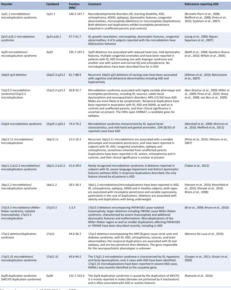
Table 2 shows several recently identified recurrent microdeletions and microduplications reported
in individuals with ID, ASD and other neurodevelopmental or neuropsychiatric disorders (Chapter 2.2).
Some of these novel recurrent CNVs have a recognizable phenotype, such as the 17q21.31
microdeletion syndrome, with a distinctive facial dysmorphism. Others, such as CNVs at 1q21.1,
15q13.3, 16p13.11 and 16p11.2, give rise to less consistent phenotypes (variable expressivity) and
have been identified in cohorts of patients ascertained for ID, epilepsy, ASD, or schizophrenia, blurring
the current nosological boundaries of these disorders. Several of these aberrations show incomplete
penetrance, as demonstrated by their presence in clinically unaffected relatives and in controls. These
CNVs have been studied in very large samples of subjects with various neurocognitive and
controls for some of them, suggesting that they act as risk factors; for other CNVs, particularly those
that appear to be relatively more frequent in controls, the clinical significance is still uncertain (e.g.,
15q13.3 and 16p13.11 duplications).
When reviewing these studies, it is clear that not all ‘intellectual disability genes and loci’ are
necessarily associated with ID. As shown in Box 1, several genetic and genomic disorders have been
reported in individuals with higher function ASD (Asperger syndrome). Similarly, not all genetic defects
involved in the etiology of ID and ASD are identified in individuals presenting with marked dysmorphic
features or other congenital malformations. In fact, many disease genes implicated in ASD can be
associated with nonsyndromic presentations (Box 2).
It should be clear when looking at the genetic and genomic disorders for which ASD is a
manifestation that variable expressivity is the rule rather than the exception, and none will invariably
present with ASD. This point is important to consider from a neurobiological perspective. There is, for
example, an emphasis on studying ASD-‐like behaviors in rodent and primate models of ASD; if
mutations in the underlying genes do not reliably lead to ASD in humans, other intermediate
neurobiological phenotypes are perhaps equally or even more relevant to understanding disease
pathogenesis (Chapter 4).
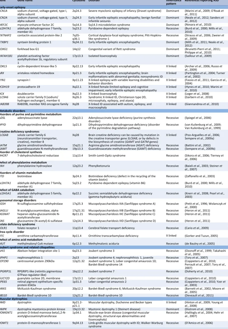
Genetic overlap between ASD and epilepsy
Epilepsies are common and etiologically heterogeneous disorders, affecting up to 3% of the
population. About 30% of children with epilepsy have ASD, and conversely, epilepsy is observed in
about a third of ASD individuals. Many well-‐known genetic disorders share ID, ASD, and epilepsy as
prominent phenotypic features, including fragile X syndrome, tuberous sclerosis, Rett syndrome and
Angelman syndrome. In addition, monogenic forms involving mutations in genes encoding voltage-‐
gated or ligand-‐gated ion channels, referred to as "channelopathies" have been identified in epilepsy,
and increasingly in ASD, such as the neuronal voltage-‐gated sodium channel genes SCN1A and SCN2A
(Table 3). Both genes have been implicated in various forms of epilepsy, including early-‐onset epileptic
encephalopathies. This group of severe epilepsies is characterized by progressive intellectual deficits
or regression, and includes West syndrome (infantile spasms), Dravet syndrome (severe myoclonic
epilepsy of infancy), and Ohtahara syndrome (early infantile epileptic encephalopathy with burst-‐
suppression) (Mastrangelo and Leuzzi, 2012; Paciorkowski et al., 2011). Table 3 shows several genes
involved in early infantile epileptic encephalopathies that can also manifest with ASD (e.g., ARX,
CDKL5, MECP2, MEF2C, FOXG1, STXBP1, and PCDH19).
Like MECP2, mutations in the X-‐linked cyclin-‐dependent kinase-‐like 5 (CDKL5) gene are more
common in girls and are associated with a Rett-‐like phenotype with infantile spasms and ID; several
cases have been described with autism (Table 3). Another X-‐linked gene, protocadherin 19 (PCDH19),
was recently implicated in "epilepsy and mental retardation limited to females", a familial disorder
with an unusual mode of inheritance, since only heterozygous females are affected and transmitting
males are asymptomatic. PCDH19 mutations, mostly occurring de novo, have also been shown to be a
frequent cause of sporadic infantile-‐onset epileptic encephalopathy in females, and have been
reported in females with epilepsy without cognitive impairment (Depienne and Leguern, 2012). ASD or
autistic features appear to be frequent among patients with PCDH19 mutations, with rates varying
between 22%-‐38% (Table 1). Interestingly, a PCDH19 mutation was reported in a female with Asperger
syndrome and normal IQ, with a history of infantile onset seizures (Hynes et al., 2010). The female-‐
limited expression is explained by a phenomenon called cellular interference; random X inactivation in
mutated females leads to tissue mosaicism, with PCDH19-‐positive and PCDH19-‐negative cells, with
altered interactions between the two populations (Depienne and Leguern, 2012). In contrast,
complete absence of the protein, as seen in mutated males, is not deleterious. The only affected male
reported to date was shown to be mosaic for the PCDH19 deletion in skin fibroblasts (Depienne and
Leguern, 2012).
In addition to the genes involved in early onset epilepsy and ASD listed in Table 3, many other
genes implicated in ASD and ID are associated with epilepsy, including those involved in metabolic
disorders (Table 3), Joubert syndrome and related disorders (Table 3), and disorders of the
RAS/mitogen activated protein kinase (MAPK) pathway (Table 4). Moreover, several recently
discovered recurrent CNVs associated with ID and ASD, such as 15q13.3 and 16p13.11 deletions,
increase risk for various forms of epilepsy (Table 2). Large, rare non-‐recurrent CNVs also play a role in
the genetic etiology of epilepsy (Mulley and Mefford, 2011), similar to what has been observed in ID,
ASD and other neuropsychiatric disorders.
The strong association between ASD and epilepsy suggests that they share common mechanisms of
synaptic dysfunction. From the neurobiological perspective, understanding this shared vulnerability is
an important direction and the model of excitatory/inhibitory imbalance, first developed in epilepsy, is
now being considered in forms of ASD (Chapter 3.9).
Metabolic disorders associated with ASD
Several metabolic disorders have been associated with an autistic phenotype (Table 3). Although
inborn errors of metabolism are rare and probably account for a small proportion of individuals with
ASD, their diagnosis is important because some are potentially treatable. Metabolic disorders may be
suspected on the basis of parental consanguinity, affected family members, early seizures, episodic
decompensation, developmental regression, and coarse facial features. However, many recently
described disorders can present as nonsyndromic ID and/or ASD and should therefore be considered
in the etiological diagnosis of ASD (Kayser, 2008).
Phenylketonuria was identified as a relatively common cause of ASD in older studies, but since the
introduction of newborn screening programs and with early dietary intervention, affected children can
now expect to lead relatively normal lives (Baieli et al., 2003). Unfortunately, phenylketonuria is still
identified among patients with ASD in emerging countries without neonatal testing or among subjects
born before these screening programs were started (Steiner et al., 2007).
Cerebral creatine deficiency syndromes may be due to two disorders of creatine synthesis,
arginine:glycine amidinotransferase deficiency (GATM) and guanidinoacetate methyltransferase
deficiency (GAMT), inherited as autosomal recessive traits, or to creatine transporter deficiency
(SLC6A8), an X-‐linked disorder (Longo et al., 2011). All three deficiencies are characterized by ID,
severe speech impairment, epilepsy and autistic behavior (Table 3). Although GATM and GAMT
mutations are very rare, creatine transporter deficiency could account for up to 1% of unexplained ID
in males (Clark et al., 2006). Because the presentation is nonsyndromic and autistic behavior is
common, this condition could be underdiagnosed in populations of lower functioning males with ASD.
Autism may also occur in the context of mitochondrial disorders, resulting either from mutations in
mitochondrial DNA or, more commonly, in nuclear DNA genes encoding mitochondrial-‐targeted
proteins (see Chapter 2.5). Mitochondrial disorders can present with a vast range of symptoms,
severity, age of onset and outcome, with a minimum prevalence estimated at 1:5000.
understanding of the pathophysiology of ASD. At the same time, the frequently indirect nature of this
relationship may make such studies more challenging than, for example, studying how synaptic genes
alter brain functioning. However, because many metabolic disorders are treatable, understanding the
range of metabolic disorders associated with ASD and testing for them can provide immediate clinical
benefits, and allow for genetic counseling.
Other examples of etiological subgroups associated with ASD
Joubert syndrome is a clinically and genetically heterogeneous group of disorders characterized by a
distinctive cerebellar and brainstem malformation, cerebellar ataxia, ID and breathing abnormalities,
sometimes including retinal dystrophy and renal disease. ASD is a relatively frequent finding in
individuals with Joubert syndrome, present in 13%-‐36% of patients (Table 1). Sixteen genes have been
implicated in Joubert syndrome, the majority very recently; thus, it is not surprising that so far only 4
of these genes have been reported to be mutated in subjects with ASD/autistic traits (Table 3).
Joubert syndrome and related disorders arise from ciliary dysfunction and are collectively termed
ciliopathies. Other ciliopathies reported in subjects with ASD include Leber congenital amaurosis and
Bardet-‐Biedl syndrome, both of which exhibit phenotypic overlap with Joubert syndrome (Table 3).
The means by which cilia are involved in neurodevelopmental processes, and by which ciliopathies
lead to neurodevelopmental disorders, are areas of active research. One exciting emerging finding is
that primary (or nonmotile) cilia, found on most neurons and astrocytes, play roles as modulators of
signal transduction during both brain development and homeostasis (Lee and Gleeson, 2011). The
primary cilia can mediate signaling through sonic hedgehog, wingless, planar cell polarity and
fibroblast growth factor pathways.
Another group of disorders that can be associated with ASD is muscular dystrophies (Table 3).
Duchenne and Becker muscular dystrophies are caused by deficient expression of the cytoskeletal
protein dystrophin, coded by the DMD gene on chromosome Xp21.2-‐p21.1. One-‐third of the children
with Duchenne muscular dystrophy and about 12% of those with the Becker type also have ID. A small
subgroup of these boys with both of these disorders also have ASD, with frequencies varying between
3% and 19% (Hinton et al., 2009; Kumagai et al., 2001; Wu et al., 2005). Several maternally-‐inherited
exonic duplications of DMD have been identified in males ascertained for ASD, with no documented
muscle disease (Pagnamenta et al., 2011; Pinto et al., 2010), suggestive of the mild end of the
spectrum of dystrophinopathies seen in Becker muscular dystrophy, with later onset or subclinical
muscle involvement. Another form of muscular dystrophy that includes cases with autistic features is
myotonic dystrophy type 1, also known as Steinert disease, caused by expansion of a CTG
trinucleotide repeat in the 3’-‐untranslated region in the DMPK gene (Table 3). The clinical findings
span a continuum from mild to severe. In a study of 57 children with myotonic dystrophy type 1, 49%
were found to have ASD; the more clinically severe the myotonic dystrophy, the higher the frequency
of children with autistic features (Ekstrom et al., 2008). This may be an underdiagnosed disease entity
in autistic populations (Coleman and Gillberg, 2012).
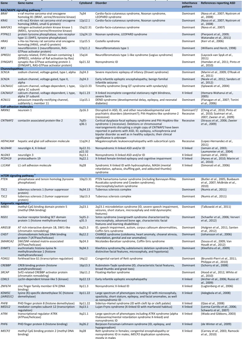
Dysregulation of the RAS/MAPK cascade is the common molecular basis for multiple congenital
anomaly syndromes known as neuro-‐cardio-‐facio-‐cutaneous syndromes and characterized by a
distinctive facial appearance, heart defects, musculocutaneous abnormalities, and ID, including
Noonan syndrome, LEOPARD syndrome (Lentigines, Electrocardiogram abnormalities, Ocular
hypertelorism, Pulmonic valvular stenosis, Abnormalities of genitalia, Retardation of growth, and
Deafness), cardio-‐facio-‐cutaneous syndrome, and Costello syndrome (Table 4) (Samuels et al., 2009).
These overlapping phenotypes can arise from heterozygous mutations in many genes, including
PTPN11, BRAF, RAF1, KRAS, HRAS, MAP2K1, MAP2K2, SOS1 and SHOC2. Neurofibromatosis type I and
neurofibromatosis type I-‐like syndrome, which are caused by loss-‐of-‐function mutations of NF1 and
SPRED1, respectively, can also be included in the same disease entity (Aoki et al., 2008). As shown in
Table 3, all these disorders have been reported in subjects with ASD. In particular, ASD was observed
in 8% of 65 children with Noonan syndrome (Pierpont et al., 2009), as well as in several patients with
cardio-‐facio-‐cutaneous syndrome or Noonan syndrome with BRAF, KRAS or MAP2K1 mutations (Nava
et al., 2007; Nystrom et al., 2008).
Myriad biological pathways
When considering genes involved in autism, neurobiologists usually think about synaptic genes such as
those coding for the postsynaptic cell adhesion molecules neuroligins 3 and 4 (NLGN3, NLGN4X), their
presynaptic partner neurexin 1 (NRXN1), and the postsynaptic scaffolding proteins SHANK2 and
SHANK3 (Betancur et al., 2009). In addition to this pathway (described in Chapter 4.7), further
evidence implicating synaptic dysfunction in the pathogenesis of ASD has come from the study of
genetic disorders with increased rates of ASD, such as fragile X syndrome (FRM1), Rett syndrome
(MECP2), tuberous sclerosis (TSC1 and TSC2) and Angelman syndrome (UBE3A). Rare mutations in
numerous other genes encoding pre-‐ and postsynaptic proteins have also been reported in ID and
ASD, including STXBP1, SYNGAP, as well as the X-‐linked genes AP1S2, ARHGEF6, CASK, GRIA3, FGD1,
IQSEC2, IL1RAPL1, OPHN1, RAB39B, and SYN1 (Figure 1) (for references implicating these genes in
ASD, see Betancur, 2011; for a general review, see van Bokhoven, 2011).
Although the focus on the synaptic pathway in recent years has contributed to our understanding
of the pathophysiology of autism, there are dozens of other non-‐synaptic genes that have been
implicated in ASD and which encompass a wide range of biological functions and cellular processes.
Mechanisms by which such genes disrupt brain and neuronal development and function will provide a
deeper understanding of ASD pathogenesis. Some examples of biological pathways and organelles
recurrently implicated in ASD are highlighted in Tables 3 and 4. In addition to the genes involved in
ciliopathies (Table 3) and the RAS/MAPK signaling pathway (Table 4) mentioned above, Table 4 shows
genes involved in channelopathies, genes coding for cell-‐adhesion molecules and genes implicated in
the protein kinase mammalian target of rapamycin (mTOR) signaling pathway. Hyperactivation of
mTOR as a consequence of loss-‐of-‐function mutations in the genes TSC1, TSC2, and PTEN is
responsible for the development of tuberous sclerosis, PTEN hamartoma-‐tumor syndrome (including
Cowden syndrome, Bannayan-‐Riley-‐Ruvalcaba syndrome, and Proteus syndrome), and
macrocephaly/autism syndrome (for review, see (de Vries, 2010)). Molecularly-‐targeted treatments
using mTOR inhibitors (such as rapamycin) are currently in clinical trials, providing great promise and
hope.
Another emerging pathway involves ASD/ID genes that encode regulators of chromatin structure
and of chromatin-‐mediated transcription (for review, see van Bokhoven and Kramer, 2010). Table 4
shows the genes mutated in ASD involved in epigenetic regulation of neuronal gene expression.
Prominent examples of epigenetic ASD/ID genes include MECP2, CHD7 (CHARGE sydrome), EHMT1
(Kleefstra syndrome), and the recently implicated gene MBD5 (2q23.1 microdeletion syndrome), all
listed in Table 1 as being frequently associated with ASD.
Conclusion
The findings discussed in this review clearly indicate that autism represents the final common pathway
for hundreds of genetic and genomic disorders. Despite the abundant evidence, this etiological
heterogeneity is still not widely recognized by autism researchers, and most studies fail to take it into
account. The genetic overlap and the frequent comorbidity of ASD, ID and epilepsy indicate that the
disruption of essential neurodevelopmental processes can give rise to a wide range of manifestations,
where the final outcome is likely modulated by the genetic background of each individual as well as
other factors including possibly environmental and stochastic factors.
Increased understanding of the
common genetic, molecular, and cellular mechanisms underlying these neurodevelopmental disorders
may provide a framework for novel therapeutic interventions.
Chromosome microarray analysis has revolutionized the molecular diagnostic process in ASD and
other neurodevelopmental conditions and is now recommended as a first-‐line test in the genetic
workup of these children, providing an etiological diagnosis in 10 to 15% of cases. Novel high-‐
throughput whole-‐exome and whole-‐genome sequencing technologies have hugely accelerated the
mutation finding process for Mendelian disorders in the past two years, and hopefully will soon
become a first-‐line approach in the etiological exploration of patients with ASD, replacing targeted
sequencing of candidate disease genes (Chapter 2.4).
Currently the most applicable benefit of genetic testing is family planning. A prospective
longitudinal study of 664 infants with an older biological sibling with ASD found that 18.7% developed
ASD (Ozonoff et al., 2011). Although many of the mutations associated with autism so far identified
are de novo, future siblings are at risk in the cases where the variant is inherited from a parent, such
as in autosomal dominant disorders with variable expressivity inherited from mildly affected parents
(e.g., tuberous sclerosis, PTEN related syndromes, 22q11 deletion syndrome), autosomal recessive
disorders or maternally-‐transmitted X-‐linked disorders (or even a paternally-‐transmitted X-‐linked
disorder, as for PCDH19). Germinal mosaicism in one of the parents can also explain rare instances of
familial recurrence. This mechanism has been implicated in a surprising number of cases of siblings
with ASD carrying apparently de novo mutations, not found in the parents' DNA (e.g., SHANK3
mutations and deletions, NRXN1 deletions, NLGN4X mutation, 16p11.2 deletion, 2q23.1 deletion, Rett
syndrome and tuberous sclerosis) and may remain unrecognized in sporadic cases in small families.
An etiologic diagnosis has important benefits for the patients with ASD and their families. For the
patients, it can help anticipate and manage associated medical and behavioral comorbidities. For the
parents, the benefits include relieving anxiety and uncertainty, limiting further costly or invasive
diagnostic testing, improving understanding of treatment and prognosis, genetic counseling regarding
recurrence risk as well as preventing recurrence through screening for carriers and prenatal testing. A
specific disease diagnosis can be empowering to parents who wish to become involved in more
targeted support and research groups. For the medical and research community, each child who is
accurately diagnosed adds to our presently limited understanding of the pathological cascades which
result in autistic features; undoubtedly new findings will include previously unrecognized disease
mechanisms. For the neurobiologist especially, the myriad genetic findings in ASD offer a rich source
of targets for further study, providing a window into brain and neuronal development and function.
The deeper understanding of these brain and neuronal processes will ultimately lead to better
outcomes in ASD and other neurodevelopmental disorders.
References
Abrahams, B. S., and Geschwind, D. H. (2008). Advances in autism genetics: on the threshold of a new neurobiology. Nat Rev Genet 9, 341-‐355.
Addington, A. M., Gauthier, J., Piton, A., Hamdan, F. F., Raymond, A., Gogtay, N., Miller, R., Tossell, J., Bakalar, J., Germain, G., Gochman, P., Long, R., Rapoport, J. L., and Rouleau, G. A. (2011). A novel frameshift mutation in UPF3B identified in brothers affected with childhood onset schizophrenia and autism spectrum disorders. Mol Psychiatry 16, 238-‐239.
Adegbola, A., Gao, H., Sommer, S., and Browning, M. (2008). A novel mutation in JARID1C/SMCX in a patient with autism spectrum disorder (ASD). Am J Med Genet A 146A, 505-‐511.
Alliman, S., Coppinger, J., Marcadier, J., Thiese, H., Brock, P., Shafer, S., Weaver, C., Asamoah, A., Leppig, K., Dyack, S., Morash, B., Schultz, R., Torchia, B. S., Lamb, A. N., and Bejjani, B. A. (2010). Clinical and molecular characterization of individuals with recurrent genomic disorder at 10q22.3q23.2. Clin Genet 78, 162-‐168. Antshel, K. M., Aneja, A., Strunge, L., Peebles, J., Fremont, W. P., Stallone, K., Abdulsabur, N., Higgins, A. M.,
Shprintzen, R. J., and Kates, W. R. (2007). Autistic spectrum disorders in velo-‐cardio facial syndrome (22q11.2 deletion). J Autism Dev Disord 37, 1776-‐1786.
Aoki, Y., Niihori, T., Narumi, Y., Kure, S., and Matsubara, Y. (2008). The RAS/MAPK syndromes: novel roles of the RAS pathway in human genetic disorders. Hum Mutat 29, 992-‐1006.
Archer, H. L., Evans, J., Edwards, S., Colley, J., Newbury-‐Ecob, R., O'Callaghan, F., Huyton, M., O'Regan, M., Tolmie, J., Sampson, J., Clarke, A., and Osborne, J. (2006). CDKL5 mutations cause infantile spasms, early onset seizures, and severe mental retardation in female patients. J Med Genet 43, 729-‐734.
Assumpcao, F., Santos, R. C., Rosario, M., and Mercadante, M. (1999). Brief report: autism and Aarskog syndrome. J Autism Dev Disord 29, 179-‐181.
Aziz, M., Stathopulu, E., Callias, M., Taylor, C., Turk, J., Oostra, B., Willemsen, R., and Patton, M. (2003). Clinical features of boys with fragile X premutations and intermediate alleles. Am J Med Genet B Neuropsychiatr Genet 121B, 119-‐127.
Baieli, S., Pavone, L., Meli, C., Fiumara, A., and Coleman, M. (2003). Autism and phenylketonuria. J Autism Dev Disord 33, 201-‐204.
Balciuniene, J., Feng, N., Iyadurai, K., Hirsch, B., Charnas, L., Bill, B. R., Easterday, M. C., Staaf, J., Oseth, L., Czapansky-‐Beilman, D., Avramopoulos, D., Thomas, G. H., Borg, A., Valle, D., Schimmenti, L. A., and Selleck, S. B. (2007). Recurrent 10q22-‐q23 deletions: a genomic disorder on 10q associated with cognitive and behavioral abnormalities. Am J Hum Genet 80, 938-‐947.
Ballif, B. C., Theisen, A., Coppinger, J., Gowans, G. C., Hersh, J. H., Madan-‐Khetarpal, S., Schmidt, K. R., Tervo, R., Escobar, L. F., Friedrich, C. A., McDonald, M., Campbell, L., Ming, J. E., Zackai, E. H., Bejjani, B. A., and Shaffer, L. G. (2008). Expanding the clinical phenotype of the 3q29 microdeletion syndrome and characterization of the reciprocal microduplication. Mol Cytogenet 1, 8.
Barnett, S., Reilly, S., Carr, L., Ojo, I., Beales, P. L., and Charman, T. (2002). Behavioural phenotype of Bardet-‐Biedl syndrome. J Med Genet 39, e76.
Battini, R., Leuzzi, V., Carducci, C., Tosetti, M., Bianchi, M. C., Item, C. B., Stockler-‐Ipsiroglu, S., and Cioni, G. (2002). Creatine depletion in a new case with AGAT deficiency: clinical and genetic study in a large pedigree. Mol Genet Metab 77, 326-‐331.
Baynam, G., Goldblatt, J., and Townshend, S. (2006). A case of 3q29 microdeletion with novel features and a review of cytogenetically visible terminal 3q deletions. Clin Dysmorphol 15, 145-‐148.
Ben-‐Shachar, S., Lanpher, B., German, J. R., Qasaymeh, M., Potocki, L., Nagamani, S. C., Franco, L. M., Malphrus, A., Bottenfield, G. W., Spence, J. E., Amato, S., Rousseau, J. A., Moghaddam, B., Skinner, C., Skinner, S. A., Bernes, S., Armstrong, N., Shinawi, M., Stankiewicz, P., Patel, A., Cheung, S. W., Lupski, J. R., Beaudet, A. L., and Sahoo, T. (2009). Microdeletion 15q13.3: a locus with incomplete penetrance for autism, mental retardation, and psychiatric disorders. J Med Genet 46, 382-‐388.
Berkel, S., Marshall, C. R., Weiss, B., Howe, J., Roeth, R., Moog, U., Endris, V., Roberts, W., Szatmari, P., Pinto, D., Bonin, M., Riess, A., Engels, H., Sprengel, R., Scherer, S. W., and Rappold, G. A. (2010). Mutations in the SHANK2 synaptic scaffolding gene in autism spectrum disorder and mental retardation. Nat Genet 42, 489-‐ 491.
Betancur, C. (2011). Etiological heterogeneity in autism spectrum disorders: more than 100 genetic and genomic disorders and still counting. Brain Res 1380, 42-‐77.
Betancur, C., Sakurai, T., and Buxbaum, J. D. (2009). The emerging role of synaptic cell-‐adhesion pathways in the pathogenesis of autism spectrum disorders. Trends Neurosci 32, 402-‐412.