HAL Id: tel-02275821
https://tel.archives-ouvertes.fr/tel-02275821
Submitted on 2 Sep 2019HAL is a multi-disciplinary open access archive for the deposit and dissemination of sci-entific research documents, whether they are pub-lished or not. The documents may come from teaching and research institutions in France or abroad, or from public or private research centers.
L’archive ouverte pluridisciplinaire HAL, est destinée au dépôt et à la diffusion de documents scientifiques de niveau recherche, publiés ou non, émanant des établissements d’enseignement et de recherche français ou étrangers, des laboratoires publics ou privés.
antagonist of host autophagy and promoter of the
infection cycle
Serena Testi
To cite this version:
Serena Testi. The Phytophthora parasitica effector Avh195 : an antagonist of host autophagy and promoter of the infection cycle. Molecular biology. Université Côte d’Azur, 2018. English. �NNT : 2018AZUR4087�. �tel-02275821�
L’effecteur Avh195
de Phytophthora parasitica :
antagoniste de l’autophagie chez l’hôte
et promoteur du processus infectieux.
Serena TESTI
UMR ISA, INRA CNRS UCA – Équipe Interactions Plantes-Oomycètes
Présentée en vue de l’obtention
du grade de docteur en Science de la Vie et de la Santé
d’Université Côte d’Azur
Dirigée par :
Franck Panabières / Harald Keller Soutenue le : 26 Octobre 2018
Devant le jury, composé de :
Elodie Gaulin, Maitre de conférences HDR, Université Toulouse III
Franck Panabières, Directeur de Recherche, UMR Institut Sophia Agrobiotech
Gilles Peltier, Chercheur CEA HDR, CEA Cadarache
Pierre Frendo Professeur, Université Côte d’Azur
L’effecteur Avh195 de Phytophthora parasitica :
antagoniste de l’autophagie chez l’hôte et
promoteur du processus infectieux.
Président du Jury
Pierre Frendo, Professeur, Université Côte d’Azur Rapporteurs
Céline Masclaux-Daubresse, Directeur de Recherche, INRA Versailles-Grignon Elodie Gaulin, Maitre de conférences HDR, Université Toulouse III
Examinateurs
Gilles Peltier, Chercheur CEA HDR, CEA Cadarache Directeurs de thèse
Franck Panabières, Directeur de Recherche, UMR Institut Sophia Agrobiotech Harald Keller, Directeur de Recherche, UMR Institut Sophia Agrobiotech
L’agent pathogène Phytophthora parasitica est un oomycète qui a des effets dévastateurs sur l’agriculture et les écosystèmes naturels. En tant qu'organisme hémi-biotrophe, il infecte les racines des plantes en établissant d'abord un contact intime avec les cellules hôtes (biotrophie) avant de les tuer (nécrotrophie) et de terminer son cycle d'infection. Pour contrôler ces processus, les oomycètes sécrètent des protéines effectrices, qui sont internalisées dans les cellules végétales par un motif de translocation (appelé RxLR-EER) pour manipuler la physiologie et les réponses immunitaires de l'hôte. Les études des échanges moléculaires entre Phytophthora parasitica et la plante qui ont été menées par le laboratoire d'accueil ont permis d'identifier un effecteur RxLR, dénommé Avh195. La séquence en acides aminés de l'effecteur est caractérisée par la présence de cinq motifs AIM (« ATG8 Interacting Motive ») qui indiquent une interaction potentielle avec la protéine centrale de l’autophagie, ATG8. Avh195 co-localise avec la fraction membranaire de l'ATG8, et un système double-hybride en levure permettant la détermination d’interactions entre protéines membranaires, a confirmé une interaction non sélective entre Avh195 et plusieurs isoformes d'ATG8. La caractérisation de la perturbation de l'autophagie dépendante de Avh195 a été réalisée dans l'algue unicellulaire Chlamydomonas reinhardtii après génération de lignées transgéniques surexprimant l'effecteur. Les analyses par cytométrie de flux ont révélé que Avh195 ne modifie pas la physiologie et la « fitness » de l'algue dans des conditions de croissance normales et pendant l'autophagie induite par la rapamycine. La microscopie électronique à transmission a révélé que l'effecteur provoque dans les cellules de l’algue un retard dans le flux autophagique, se traduisant par une réduction de la coalescence et de la clairance des vacuoles et une forte accumulation d'amidon dans les chloroplastes. Cependant, ce phénotype est transitoire et seulement légèrement lié aux modifications de la régulation transcriptionnelle de la machinerie autophagique. L'analyse de la fonction effectrice chez les plantes a montré que Avh195 retarde le développement de la mort cellulaire hypersensible, déclenchée par un éliciteur d’oomycète. Cette activité dépend de trois AIM sur cinq, ce qui renforce encore l’importance de l’interaction Avh195-ATG8 pour la fonction de l’effecteur. La surexpression stable d'Avh195 chez A. thaliana a permis de déterminer que l'effecteur n'altère pas les réponses immunitaires des plantes, mais favorise globalement le développement de l'agent pathogène, accélérant le passage de la biotrophie à la nécrotrophie au cours de l'infection. À notre connaissance, le travail présenté dans cette thèse représente la première preuve qu'un effecteur d’oomycète possède une activité transitoire, ciblant de manière non sélective la protéine ATG8 dans différents organismes photosynthétiques pour ralentir le flux autophagique, favorisant ainsi le mode de vie hémi-biotrophe d'un agent pathogène.
The plant pathogen Phytophthora parasitica is an oomycete with devastating impacts on both agriculture and natural ecosystems. As a hemi-biotrophic organism it infects the roots of plants first establishing an intimate contact with host cells (biotrophy) before killing them (necrotrophy) and completing its infection cycle. To control these processes, oomycetes secrete effector proteins, which are internalized in plant cells by a translocation motif (called RxLR-EER) to manipulate the physiology and the immune responses of the host. Studies of the molecular exchanges between Phytophthora parasitica and the plant that were conducted by the hosting laboratory led to the identification of an RxLR effector, designed to as Avh195. The amino acid sequence of the effector is characterized by the presence of five AIMs (ATG8 interacting motifs), that indicate a potential interaction with the autophagic core protein, ATG8. Avh195 colocalizes with the membrane-bound fraction of ATG8, and a yeast two-hybrid system, which allows to determine interactions between membrane proteins, confirmed a non-selective interaction between Avh195 and several ATG8 isoforms. The characterization of Avh195-dependent autophagy perturbation was carried out in the unicellular alga Chlamydomonas reinhardtii after generation of transgenic lines overexpressing the effector. Analyses by flow cytometry revealed that Avh195 does not modify the physiology and fitness of the alga, both under normal growth conditions and during rapamycin-induced autophagy. Transmission electron microscopy of cells revealed that the effector provokes a delay in the autophagic flux, manifested as a reduced coalescence and clearance of autophagic vacuoles and a strong accumulation of starch in chloroplasts. However, this phenotype was transient and only slightly related to modifications in the transcriptional regulation of the autophagic machinery. The analysis of effector function in planta showed that Avh195 delays the development of hypersensitive cell death, which is triggered by an oomycete elicitor. This cell death-delaying activity is dependent on three out of five AIMs, further consolidating the importance of the Avh195-ATG8 interaction for the function of the effector. The stable overexpression of Avh195 in A.
thaliana allowed to determine that the effector does not impair plant defense responses, but overall
promotes the development of the pathogen, accelerating the switch from biotrophy to necrotrophy during infection. To our knowledge, the work presented in this thesis represents the first evidence for an oomycete effector to possess a transitory activity, which targets in a non-selective manner the protein ATG8 in different organisms from the green lineage to slow down autophagic flux, thus promoting the hemibiotrophic life style of a pathogen.
Je tiens d’abord à remercier les membres du jury pour l’intérêt que vous avez manifesté pour ce travail. Merci à Céline Masclaux-Daubresse, et Elodie Gaulin pour avoir accepté d’évaluer mon travail en tant que rapportrices. Merci à Gilles Peltier pour avoir accepté d’être examinateur et merci à Pierre Frendo pour avoir accepté d’être président du jury.
Merci au financement Labex Signalife (ANR-11-LABX-0028-0). Je remercie aussi toutes les personnes qui ont contribué à la réalisation de ce travail de thèse. Merci à Claire Veneault-Fourrey et Christophe Roux pour la discussion fructueuse lors du Comité de thèse. Merci à Gilles Peltier, Fantao Kong et Pascaline Auroy pour la génération des Chlamydomonas transformées. Merci à Nathalie Zucchini-Pascal pour tous ses efforts sur les cellules HeLa. Merci à Georges de Sousa et Éric Boncompagni pour tous les produits qu’ils m’ont fourni et à Olivier Pierre pour ses précieux conseils sur la microscopie. Merci à Valérie Allasia pour avoir complété une partie importante de ce travail. Merci à Sophie Pagnotta et Julie Cazareth pour leur temps et leur engagement dans la réalisation des expériences de TEM et de cyrtométrie en flux.
Merci Franck et Harald de m’avoir accueilli dans votre laboratoire et équipe. Avoir deux directeurs de thèse c’est parfois redouté mais dans mon cas cela a représenté une valeur ajoutée : j’ai vraiment apprécié pouvoir échanger avec deux scientifiques exceptionnels aux compétences si variés. Merci pour l’énorme aide que vous m’avez donné pour la réussite de cette thèse et merci pour tout ce que vous m’avez appris au cours de ces quatre années. Merci aussi pour tous les bons moments que nous avons passé ensemble lors des congrès. Merci pour la confiance que vous m’avez accordée et merci de m’avoir poussé plusieurs fois à sortir de ma zone de confort.
Merci à toute l’équipe IPO qui m’a accueilli comme une grande famille. Merci à Valérie, Agnès, Jo-Yanne et Naïma (mais aussi Sandrine et Elodie) pour tous les sujets de conversations incroyables abordées au café (et aussi pour les bons chocolats !) : je ne suis pas une grande bavarde mais j’ai vraiment adoré passer du temps avec vous. Merci à Cathy et Éric : ça a été un vrai plaisir d’avoir partagé le laboratoire avec vous pendant ces quatre années. Éric, merci pour les discussions scientifiques (et non) que nous avons eu, pour tous tes conseils et encouragements.
Benoît et Laurent : merci pour toutes vos bêtises, vous m’avez redonné le sourire même dans les journées les plus noires. Merci Benoît, tout simplement parce que c’est toi (et aussi parce que t’as supporté avec patience mon dépaysement lors de mes premiers pas dans le labo). Et merci Laurent pour avoir toujours la réponse à tout, maintenant il te manque juste d’ imparare un po’ di italiano !
Merci à tous les doctorants que j’ai croisée au cours de ces quatre ans. Un remerciement spécial va à Laïla Danila et Martina. Vous êtes des collègues exceptionnelles. Merci pour tous les bons moments que nous avons a passée ensemble au labo, sur la plage ou d’arrière une pinte de bière. J’ai adoré partager cette expérience avec vous et je ne vous oublierai jamais : je vous souhaite tout le meilleur pour votre futur. Marie Line, je ne t’ai pas oublié : tu es ici parmi mes remerciements les plus personnels car tu as été non seulement une encadrante mais aussi une amie et parfois une maman. Tu m’as accompagné depuis le début de cette aventure et nous avons partagés ensemble les joies et les douleurs de ce doctorat (et plein de nuits passées avec les doigts croisées). Je pense qu’il n’y a pas de mots pour exprimer ma gratitude pour tout ce que t’as fait pour moi et tu le sais que je ne t’oublierai jamais. Merci et encore merci pour tout ton aide, ton soutien et ta gentillesse envers moi.
rédaction.
Thanks to Nathaly and Caterina (in order of arrival in my life, not of importance!). It’s been four years we met: we begun this huge journey together and finally we are going to see the light at the end of the tunnel together. Maybe we haven’t had the time to meet as much as we planned but I want you to know how much I loved every single moment (and kilometres) we spent together. I will keep our friendship preciously in my heart and I really wish all the best to both of you, while waiting to go for a walk in Nice when we will be three old grumpy ladies.
Grazie ai miei genitori. Vi voglio bene e ovunque andrò i chilometri non ci terranno mai lontani perché siete sempre nel mio cuore. Grazie per tutti gli sforzi che avete fatto per farmi arrivare dove sono e per avermi sempre supportato in ogni mio sogno.
Grazie Andrea, perché questa tesi è anche merito tuo. Quattro anni fa mi hai incoraggiato ad iniziare un dottorato pur sapendo che questo avrebbe significato vivere lontani. Quattro anni sono interminabili ma tu mi sei sempre stato accanto: sei la mia forza e la ragione che mi spinge ogni giorno a dare sempre il meglio. Grazie per tutto il tuo amore e sostegno. Grazie di esserci. Grazie di credere sempre in me, molto più di quanto sarò mai capace di fare.
Table of Contents
Abbreviations ... 1
Figures and Tables... 3
Oomycetes... 5
General Introduction ... 5
1.1 The species Phytophthora parasitica... 9
1.2 Molecular dialogue between plants and Oomycetes ... 14
Autophagy ... 21
General Introduction ... 21
2.1 A focus on macroautophagy ... 22
2.2 Autophagy in plants ... 28
Objectives of the PhD project ... 35
Results ... 37
3.1 Molecular characterization of Avh195 ... 38
3.2 Avh195 interacts with ATG8 ... 47
3.3 Characterization of Avh195 in Chlamydomonas reinhardtii ... 55
3.4 Role of Avh195 during the host-pathogen interaction ... 83
3.5 Trans-kingdom activity of Avh195 ... 89
Discussion ... 97
Conclusions and Perspectives ... 105
Materials and Methods ... 111
- Bioinformatic analysis ... 111
- Vector construction ... 111
- RNA extractions and gene expression analysis ... 114
- SDS-page and Western blot analysis ... 115
- Recombinant protein production and purification ... 116
- Yeast two-hybrid asssay ... 117
- Arabidopsis thaliana assays ... 118
- Solanum lycopersicum assays ... 120
- Nicotiana species assays ... 121
- Chlamydomonas reinhardtii assays ... 123
- HeLa cells assays ... 126
- Statistical analysis ... 126
- Sequences ... 127
- Growth media ... 128
- Primers ... 131
1 3-MA 3-methyladenine
aa Amino acid
A/Ade Adenine
AIM ATG8 interacting motif
At Arabidopsis thaliana
ATG Autophagy related (gene/protein)
ATP Adenosine triphosphate
AVR Avirulence protein
BRET Bioluminescence Resonance Energy Transfer
C/Cys Cysteine
cAMP Cyclic Adenosine mono-phosphate
cDNA Complementary DNA
CFSE Carboxyfluorescein succinimidyl ester
CMV Cytomegalovirus (promoter)
CQ Chloroquine
Cr Chlamydomonas reinhardtii
CRN Crinkling and necrosis
ct Cycle threshold in qPCR
CVT cytosol-to-vacuole targeting
CVT Cytosol-to-vacuole targeting
D/Asp Aspartic acid
DAPI 4'-6-diamino-2 phenylindole dihydrochloride
DMEM Dulbecco's Modified Eagle Medium
DNA Deoxyribonucleic acid
E/Glu Glutamic acid
ECL Enhanced Chemiluminescence
EHM Extrahaustorial membrane
EHMx Extrahaustorial matrix
ER Endoplasmic reticulum
EST Expressed sequence tags
ET Ethylene
ETI Effector triggered immunity
FSC Forward light scattering
FW Conidiospores per mg fresh weight
G/Gly Glycine
GFP Green fluorescent protein
GTP Guanosine triphosphate
H/His Histidine
Hpa Hyaloperonospora arabidopsidis
hpi Hours post inoculation
HR /HR-PCD Hypersensitive Response ( - programmed cell death)
HRP Horse Radish Peroxidase
HSP Heat shock protein
HT Host-targeting signal
I/Ile Isoleucine
JA Jasmonic acid
kDa Kilo Dalton
L/Leu Leucine
LB Luria-Bertani medium
LC3 Mammalian ortholog of ATG8 proteins
LIR LC3 interacting region
LIRCPs LIR motif-containing proteins
M/Met Methionine
MAPK Mitogen activated protein kinase
Mbp Mega base pairs
mbSUS Mating-based split-ubiquitin system
miRNA Micro RNA
ML Maximum Likelihood
MS Murashige and Skoog medium
Mya Million years ago
NCBI National Center for Biotechnology Information
OA Oxalic acid
2 ORF
p35S Cauliflower Mosaic Virus (CaMV) 35s promoter
PAMPs Pathogen associated molecular patterns
PAS Phagophore assembly site
pb Base pairs
PCD Programmed cell death
PCR Polymerase chain reaction
PE Phosphatidyl ethanolamine
Pi Phytophthora infestans
pI Isoelectric point
PKA cAMP-dependent protein kinase
poli(A) Poli adenylation signal
Pp Phytophthora parasitica
PPRs Pattern recognition receptors
PR Pathogenesis related
Ps Phytophthora sojae
PSSM Position-specific scoring matrix
PtdIns Phosphatidyl inositol
PtdIns3K Phosphatidyl-inositol 3 kinase
PtdIns3P Phosphatidyl inositol 3 phosphate
PTI PAMP triggered immunity
PVDF Polyvinylidene difluoride
QoIs Quinone Outside Respiration Inhibitors
Ra Rapamycin
RFP Red fluorescent protein
RNA Ribonucleic acid
ROS Reactive oxygen species
RT Reverse transcription
S/Ser Serine
SA Salicylic acid
SAR Systemic acquired resistance
SC Synthetic complete medium
SD Standard deviation
SDS Sodium dodecyl sulphate
siRNA Small interfering RNA
SNP Single Nucleotide Polymorphism
Spp. Species
SSC Side light scattering
T35S Cauliflower Mosaic Virus (CaMV) 35s terminator
TAP Tris Acetate Phosphate medium
TBS Tris Buffered Saline
TEM Transmission electron microscopy
TMV Tobacco mosaic virus
TOR/TORC Target of Rapamycin (-complex)
Ub Ubiquitin
UBC Ubiquitin conjugating enzyme
UBL ubiquitin-like
UTR Untranslated region
V/Val Valine
VSP Vacuolar protein sorting
W/Trp Tryptophan
WT Wild type
X Any amino acid in aminoacidic sequences
Y/Tyr Tyrosine
Y2H Yeast two-hybrid
3
Figure 1.1: Symptoms associated with Phytophthora parasitica infection. Figure 1.2: Infectious cycle of Phytophthora parasitica.
Figure 1.3: The RxLR effector family.
Figure 1.4: Overview of RxLR effectors targets in plant cells.
Figure 2.1: Core macroautophagy machinery in Saccharomyces cerevisiae.
Figure 2.2: Core autophagy machinery in Arabidopsis thaliana compared to yeast. Figure 2.3: Autophagy in plants.
Figure 2.4: Role of autophagy in plant-pathogen interaction.
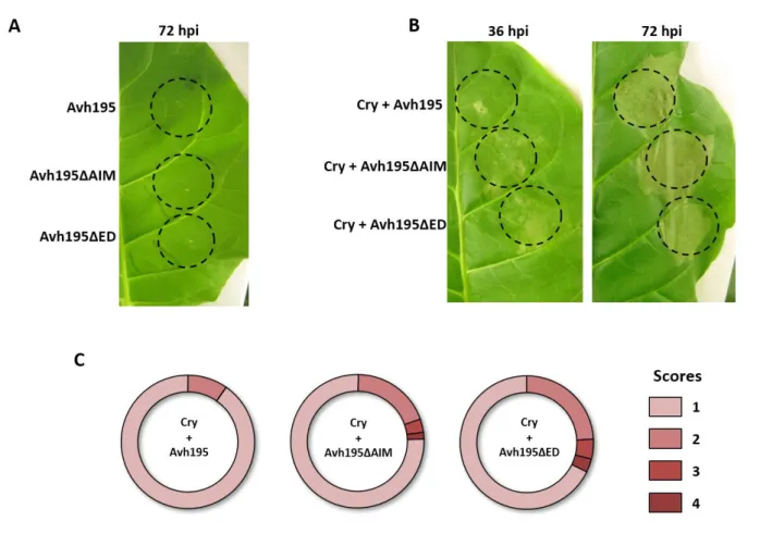
Figure 3.1: Amino acid sequence alignment of Avh195 and PITG_04099. Figure 3.2: Expression profile of Avh195 during the P. parasitica life cycle. Figure 3.3: Schematic representation of the Avh195 protein sequence. Figure 3.4: Avh195 transiently inhibits HR through interaction with ATG8.
Figure 3.5: Relative expression of ATG8 members during Arabidopsis root infection by P. parasitica. Figure 3.6: Phylogenetic relationships of ATG8 sequences from green organisms
Figure 3.7: Avh195 localizes to membranes, whereas ATG8 is both soluble and membrane-associated. Figure 3.8: Avh195 interacts with ATG8 from C. reinhardtii and A. thaliana.
Figure 3.9: The microalga Chlamydomonas reinhardtii.
Figure 3.10: Core autophagy machinery in Chlamydomonas reinhardtii compared to yeast. Figure 3.11: Experimental strategy applied to Chlamydomonas experiments.
Figure 3.12: Characterization of C. reinhardtii transformant cell lines.
Figure 3.13: Flow cytometry analyses of Chlamydomonas reinhardtii WT and transgenic cell lines. Figure 3.14: Heat shock induces lethality in Chlamydomonas transformant strains.
Figure 3.15: Flow cytometry analysis of C. reinhardtii transformant cell lines upon rapamycin treatment. Figure 3.16: Response of Chlamydomonas cells to rapamycin treatment.
Figure 3.17: Subcellular phenotypes of Chlamydomonas cells expressing Avh195, as analyzed by TEM. Figure 3.18: C. reinhardtii Avh195 transformants starch metabolism perturbation.
Figure 3.19: Overexpression of Avh195 in C. reinhardtii only moderately changes the accumulation of autophagy-related gene transcripts and of ATG8 protein.
Figure 3.20: Expression levels of Avh195 in two independent transgenic A. thaliana lines
Figure 3.21: Avh195 promotes infection and changes the timely onset of plant defense responses. Figure 3.22: Expression of Avh195 in A. thaliana changes the timely onset of oomycete life style.
Figure 3.23: Overexpression of Avh195 in A. thaliana promotes susceptibility to biotrophic H. arabidopsidis. Figure 3.24: Exogenous Avh195 is taken up by mammalian cells.
4
Figure 3.26: Exogenous Avh195 increases the accumulation of acidic vesicles. Figure 3.27: Morphological effect of Avh195 transfection.
Supplementary Figure 1: Representative view of Chlamydomonas cells from the wild-type and transgenic lines expressing Avh195, as analyzed by TEM.
Supplementary Figure 2: Accumulation of electron-dense, lysosome-like structures in cells from Avh195-expressing Chlamydomonas lines that were not treated with rapamycin.
5 5
General Introduction
Oomycetes are one of the most economically and ecologically important class of eukaryotic microorganisms playing a fundamental role in nutrient recycling, but also representing a major threat to agriculture and natural ecosystems worldwide.
Historically, Oomycetes were considered as members of the kingdom of Mycota, as an ancient basal lineage of fungi [1]. This misconception was due to the strong similarities that fungi and Oomycetes display, such as the filamentous growth in the form of tip-growing branching hyphae, and their similar ecological role and feeding behavior [2]. However, technical advances in the 20th century
suggested that Oomycetes and Fungi share less than meets the eye: Oomycetes display distinctive features both at a morphological and molecular level, which unite them with brown algae and diatoms among Stramenopiles (Straminipila) [3]. For example, the cell wall of Oomycetes is mainly composed of cellulose and glucans, and the major fungal cell wall compound chitin is only a minor constituent. Oomycetes develop mostly non-septate hyphae, they are auxotrophic for sterols, diploid for the majority of their life cycle, and disseminate mainly asexually with biflagellated zoospores [4]. Molecular phylogenies based on conserved mitochondrial and nuclear DNA sequences and more recent genome-scale phylogenetic studies further confirmed the distant relation of Oomycetes from true fungi [5–7].
It is thought that the common ancestor of the Straminipila lineages was a biflagellate photosynthetic organism that obtained the chloroplasts from endosymbiosis with a red alga. Then Oomycetes underwent repeated loss of plastids and genes for phototropism, leading to the current morphology and lifestyle [3], although some lineages still conserve photosynthesis-related genes [8].
The evolutionary history of Oomycetes is mostly inferred from molecular studies on living species and molecular clock estimates suggest that they diverged from other Straminipila in the first half of the Paleozoic era, up to 430 million years ago [9]. In addition, the existence of preserved oomycete structures in the fossil records from the Carboniferous period (300 million years ago) supports the Palaeozoic origins of this lineage [10]. Fossils also report the ancient parasitic lifestyle of Oomycetes toward plants [11]. In fact, although numerous oomycete species are saprophytic, pathogenicity evolved independently several times in different oomycete lineages [12]. The evolution of this trait
6
appears to be deeply rooted in the oomycete lineage [2], and was certainly facilitated by an ancient horizontal transfer of genes linked to pathogenicity from fungi and bacteria, such as hydrolytic enzymes, toxins, nutrient transporters and effectors [13,14].
Indeed, the attitude toward parasitism is widespread along the phylogenetic branch of Oomycetes. The basal-most lineage of Oomycetes comprises marine organisms which are predominantly parasites of seaweeds, nematodes or crustaceans [2]. The remaining clades can be broadly regrouped in two subclasses: the Saprolegnomycetes and the Peronosporomycetes. Saprolegnian organisms are mainly marine and freshwater saprophytes and parasites of animals such as the members of the genus Saprolegnia [15], with an exception for some species belonging to the Aphanomyces which are plant parasites [16]. Peronosporomycetes is by far the largest order of terrestrial organisms that includes the most studied species of Oomycetes, comprising downy mildews, Albugo, Pythium and Phytophthora and the majority of organisms belonging to those taxa are plant parasites with a wide range of hosts, including crop species, ornamental plants, and native plants and trees [12].
Overall, pathogenic Oomycetes have evolved different lifestyles. Saprophytic species can be facultative necrotrophic pathogens that kill host tissue before feeding on it; such behavior can be found among Pythium and Aphanomyces genera. On the other hand, several lineages evolved a biotrophic lifestyle, characterized by maintaining an intimate interaction with living plant cells. Species belonging to the downy mildews and the Albuginales display obligate biotrophy, requiring metabolic active plant tissues to complete their live cycle and developing highly specific interactions with the host [17]. By contrast, species of the genera Phytophthora and Pythium display an intermediate lifestyle called hemibiotrophy, adopting a two-step infection: the pathogen initially establishes a transient biotrophic relationship with the host, followed by necrotrophy which determines extensive cell death of host as the infection progresses [18,19].
Oomycetes are ubiquitous in natural ecosystems, ranging from marine, freshwater and terrestrial environments and are adapted to the most diverse ecosystems, reaching the cold polar regions, the arid deserts and temperate forests [17]. Unsurprisingly, this global distribution represents a problem when we have to deal with pathogenic Oomycetes that are responsible every year for enormous economic losses and damages in natural and cultured ecosystems. For example,
Saprolegnia and Aphanomyces species are responsible for the decline of natural populations of
7
notorious and devastating Oomycetes are plant pathogens, in particular those belonging to the genus Phytophthora. The late blight epidemics caused in Ireland by Phytophthora infestans in the nineteenth century is probably the most well-known devastating outbreak in cultivated crops, and still represents a threat for potato and tomato crops. Another example, among several others, is the
Phytophthora ramorum epidemics that struck California and southern Oregon in the late 1990s that
led to the destruction of Oak forests. This pathogen is presently spreading across Europe to ornamental plants [21,22].
Control of oomycete diseases relies mainly on the use of fungicides containing molecules with a high specific mode of action. This specificity permitted many Phytophthora species to develop resistance traits. One example is given by the phenylamide Mefenoxam, an inhibitor of RNA synthesis. The emergence of insensitive Phytophthora infestans isolates was documented across Europe, North America and Mexico and was associated with single nucleotide polymorphism (SNP) in the gene encoding the large subunit of RNA polymerase II [23]. Another class of molecules is represented by inhibitors of the respiratory chain called Quinone Outside Respiration Inhibitors (QoIs). Resistant isolates of Phytophthora viticola evolved rapidly in vineyards across Europe and once more the trait was associated with SNP in the sequence of the Cytochrome b gene [24]. The persisting threat by Oomycetes is made worse by the intensification of human displacements and the increase in international trading and climate warming, all factors that favor the introduction and establishment of new pathogens all over the world [25,26].
Is therefore clear that the study of these organisms is crucial for the development of novel strategies for disease management. The ongoing sequencing projects of several Phytophthora species already allowed to improve our understanding of oomycete origins and the molecular basis of the infectious process, and underlined the importance of molecular exchanges between the pathogen and the host [27].
9
1.1
The species Phytophthora parasitica
As described in the previous Section, the most known plant parasitic Oomycetes belong to the genus
Phytophthora ('plant destroyer' in Greek), which includes over 140 described species [28]. In this
work a particular attention will be devoted to Phytophthora parasitica (syn. Phytophthora
nicotianae) which is gaining importance as a pathogen in terms of distribution, host range and
economic impact. The species was first described in 1869 on tobacco plants displaying black skank, and hence the species name nicotianae was coined first. However, this pathogen has currently been isolated all over the world on up to 90 different plant families such as Solanaceae, citrus, horticultural and forest trees, ornamental plants, and medicinal herbs [29,30]. Usually, P. parasitica isolates display preferences for specific hosts, but some of them have a marked host flexibility and can infect numerous hosts, like the model plant Arabidopsis thaliana [31]. This makes the pathogen an ideal model to study the mechanisms regulating host specificity and susceptibility, and the molecular exchanges between the pathogen and the plant.
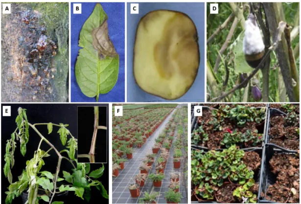
Figure 1.1: Symptoms associated with Phytophthora parasitica infection.
Pictures show various plant hosts and plant organs presenting the disease: (A) Citrus trunk (B) Potato leaf (C) Eggplant fruit (D) Potato tuber (E) Tomato stem. Diseased plantlets of (F) Grevillea lanigera (G) Cyclamen recovered from ornamental plant nursery. Adapted from [21,30,32].
10
1.1.1 P. parasitica life cycle and reproduction
As a soilborne pathogen Phytophthora parasitica mainly attacks the roots of host plants, but it is also able to develop on aerial parts of the plants and to survive outside hosts. The typical life cycle of this oomycete mainly relies on the asexual multinucleated sporangium which ensures long-distance dispersal of the organism according to two strategies: in warm and moisty soils, the sporangium can germinate directly and produce a hypha that starts host colonization. Yet, under most other conditions, the sporangium undergoes sporulation (zoosporangiogenesis). This process happens in specialized hyphae responsible for the asexual reproduction of the organism, in which the production of spores is achieved through rapid cytokinesis leading to the release of single nucleated, wall-less cells called zoospores.
Zoospores are responsible for the infectious cycle (Figure 1.2): they are biflagellated motile cells that swim towards the roots of a potential host by means of a combination of several factors, including chemotaxis, electrotaxis and autotaxis [33]. When contact with the plant occurs, zoospores detach their flagella and adhere to the surface, become cysts through rapid formation of a cell wall and secrete adhesive material. The encasement leads to the formation of a germ tube that emerges close to the plant, growing on the surface towards a suitable penetration site. Here, it develops a specialized structure, the appressorium, a swollen hypha where degrading enzymes enable the penetration of the cuticle and the epidermal layer. Once inside plant tissues, the oomycete starts vegetative growth by forming hyphae that grow and branch intercellularly. During the initial biotrophic phase, hyphae break plant cell walls and develop inside the host cells specialized feeding structures called haustoria. Haustoria are surrounded by the plasma membranes from both the oomycete and the host cell, which provide an intimate contact for nutrient uptake and molecular exchanges with the host [34]. As the infection proceeds, the necrotrophic phase initiates via the release of toxic compounds that kill the host tissue, allowing invasive growth of the pathogen with a rapid increase in biomass, and culminating in the formation of new sporangia, and eventually, in the reiteration of the cycle on a new host [35,36].
11 Figure 1.2: Infectious cycle of Phytophthora parasitica.
A. The infectious cycle of P. parasitica begins with the formation of zoosporangia and the release of biflagellate
zoospores that swim towards plant roots: once the contact takes place the flagella are lost, and the zoospore becomes a cyst.
B. Left panel: Magnification of a root tip from Arabidopsis thaliana being attacked by P. parasitica zoospores. The
zoospores form clumps during encystment at the elongation and differentiation zones. Scale bar represents 100µM. Right panel: confocal laser scanning micrograph of a GFP-expressing zoospore [38] (green fluorescence) penetrating in tomato roots stained with propidium iodide (red staining). The encysted zoospores (Sp) germinate, and the germination tube (Gt) forms an appressorium-like swelling (Sw) to push aside joined epidermal cells (Ec), and to enter a penetration peg (Pp) in between them. The absence of cytoplasmic propidium iodine stain (C) indicates that plant cells are alive. Pictures adapted from [32].
C. At the beginning of the infection (biotrophy) the germinated cyst searches for a suitable site where a specialized
structure, the appressorium, enables the penetration of the oomycete into plant tissues. Here the oomycete starts vegetative growth by forming intercellular hyphae and intracellular specialized feeding structures called haustoria. As the infection proceeds, the necrotrophic phase initiates via the release of toxic compounds that kill the host tissue, allowing invasive growth of the pathogen with a rapid increase in biomass, and culminating in the formation of new sporangia, and eventually, in the reiteration of the cycle on a new host.
12
Phytophthora parasitica can also produce thick walled spores, called Chlamydospores, which are
believed to help the pathogen to survive in a dormant state when the temperatures are too low [37]. If humidity and temperature are not ideal for the infection process Phytophthora can also form haploid gametangia, the antheridia and oogonia, carrying the male and female gametes respectively, which are necessary for sexual reproduction. Phytophthora parasitica is not self-fertile (heterothallic) and requires the interaction between gametes belonging to different mating types. This process leads to the formation of oospores, which may persist in soil for years prior to further formation of sporangia or germ tubes directly. This reproduction strategy does not only allow the organism to overcome adverse environmental conditions but it also constitutes an important source of genetic variation, and potentially of increased virulence [29].
13
1.1.2 P. parasitica pathogenicity
The outcome of oomycete infections depends on plant immune responses that aim at blocking the pathogen, and on the ability of the microorganism to overcome these responses, to adapt, and to exploit the resources of the host. The processes leading to the success or the failure of infection are based on a complex molecular dialog between the plant and the pathogen, and many research efforts were made to understand this dialog. Until recently, most of these efforts were focused on the famous and economically important P. infestans species. However, P. parasitica gains importance as a model, since it represents the soilborne lifestyle of most Phytophthora species (contrary to P. infestans, a foliar pathogen), and due to the diversity of susceptible hosts [30]. For these reasons several resources were developed and are evolving, starting from the generation of a bacterial artificial chromosome library [39] until the public release of the genome sequence [40]. Much interest was also devoted to the profiling of gene expression during different phases of the P.
parasitica life cycle. Expressed sequence tag (EST) libraries were generated from in vitro-growing
hyphae [41], from zoospores and germinating cysts [39,42], as well as from different stages of the plant-pathogen interaction ranging from initial penetration to sporulation [38,39,42,43]. These activities helped to better characterize the molecular program deployed by the pathogen throughout the different stages of the life cycle, especially during the interaction with the plant. More recently, hybridization-based transcriptome experiments allowed refining the description of the events modulating the infection process, both in plant and in the pathogen [44,45].
Emerging from studies on Phytophthora parasitica and other Phytophthora species is that Oomycetes achieve their infection by modulating plant immunity through the deployment of a large repertory of effector proteins. Effectors are molecules, mainly proteins, which are secreted during infection and target plant functions to alter the normal physiology and response of the plant [46]. The genes encoding effector proteins are mostly located in plastic regions of the genome, rich in repeats and transposons that promote duplication events, shuffling, mutagenesis and silencing, thus underlining the importance of effectors in evolution and adaptation of Oomycetes, and to a wider extent, filamentous pathogens [27,47].
14
1.2
Molecular dialogue between plants and Oomycetes
As mentioned above, the success of oomycete infection passes through the manipulation of host defenses, metabolism and functions, overcoming the several layers of defense implemented by plants [48]. The first layer of plant defense is made of passive preformed barriers, such as the cell wall and the cuticle, and of the constitutive accumulation of antimicrobial compounds that allow plants to be protected against most attacks. However, if a pathogen succeeds in overcoming those barriers, plants deploy an innate immune system that controls most of the infection attempts. Detection of the so-called pathogen-associated molecular patterns (PAMPs) is based on the recognition of molecules, mainly proteins, or of activity-related parts of the molecules, that are generally essential for the pathogen’s life cycle. PAMPs are recognized by plants when they are exposed and sensed by specific transmembrane receptors (PPRs) that recognize them. Activation of PRRs leads to the induction of defense responses, involving ion fluxes, oxidative bursts, and the activation of Mitogen-Activated Protein Kinase (MAPK) cascades, which in turn promote transcriptional regulations that aim at blocking further penetration of the pathogen [48,49]. Examples for identified Phytophthora PAMPS are Pep-13 and NPP1. Pep-13 is a highly conserved 13-amino acid fragment within the cell wall glycoprotein GP42 from P. sojae [50,51]. NPP1, necrosis-inducing Phytophthora protein 1, was identified in several Phytophthora species as a cell-wall protein eliciting immune responses in plants [52].
This second layer of plant defense can be overcome by pathogens by means of effector proteins, which can roughly be classified as extracellular (apoplastic) and intracellular effectors. Oomycetes such as Phytophthora secrete different type proteins that either protect the pathogen from host defenses or contribute to the process of invasion. Secreted apoplastic effectors may inhibit hydrolytic enzymes (chitinases, glucanases and proteases) that are released by the plant to block pathogen proliferation. RGD (Arginine–Glycine–Aspartic acid)-containing effectors promote entry and development of the pathogen by interfering with host signaling pathways that regulate adhesion of the plant cell wall to the plasma membrane. Other effectors are cell wall degrading enzymes as well as toxins, used by the oomycete to induce cell death when hemibiotrophic infection switches to necrotrophy [43,53,54]. A particular class of apoplastic effectors from Phytophthora that has been actively studied in the last decades is constituted by the elicitins. Elicitins are structurally conserved proteins that were initially described in Phytophthora spp. as able to elicit hypersensitive (HR) cell death and disease resistance in tobacco [55]. Elicitins are able to bind sterols and other
15
lipids [56]: since Phytophthora spp. are sterol auxotrophs, it was speculated that those molecules may serve as extractors of sterols from the plant membrane to ensure the metabolic needs of the oomycete, and consequently disrupt the membrane integrity leading to cell death [55].
Intracellular oomycete effectors are secreted proteins that carry host-translocation signals for their transport into the plant cell. Additional motifs then allow the relocalisation to specific subcellular compartments. Intracellular oomycete effectors mainly fall into two groups. The first comprises RXLR-effectors, whose typical feature is the presence of a conserved aminoacid signature at the N-terminus of the protein: Arginine, any amino acid, Leucine, Arginine (hence the name RXLR) often followed by a shorter Glutamate-Glutamate-Arginine (-EER) motif [57]. The second class is composed of the so-called Crinklers, named after the cell death phenotype they induce when overexpressed in planta. Similar to RxLR effectors, crinklers have a modular structure comprising conserved aminoacid motifs and seem to target the host nucleus [58].
In some cases, both apoplastic and intracellular effectors may be sensed by plants and activate a further layer of defense, called effector-triggered immunity (ETI). This immunity is provided by resistance (R) proteins, that are able to detect either directly or indirectly effectors proteins (named in this context avirulence proteins; AVRs). The outcome of ETI is more rapid and stronger than PTI, and often leads to a hypersensitive response (HR), a form of localized host cell death to confine the pathogen at the infection site and to prevent further propagation into non-infected plant tissues. This tight interaction of molecules from both the plant and the pathogen is the base of the so called “zig-zag” model postulated by Jones and Dangl [48], according to which the co-evolution of pathogen virulence determinants and plant resistance and immunity factors drives the outcome of plant-pathogen interactions.
16
1.2.1
The RXLR effector family
A study published in 2005 by Rhemany and colleagues compared the sequences of predicted secreted avirulence proteins from several oomycete species, and identified a conserved amino acid motif, namely the RXLR-EER motif [59]. Further searches for candidate effectors in Phytophthora genomes evidenced the presence of an important number of genes that potentially encode for RXLR effector proteins, many of which are part of gene families comprising numerous paralogs with marked positive selection for their C-terminal regions [60].
Proteins belonging to this class share a modular structure, characterized by an N-terminal signal peptide for secretion, the RXLR(-EER) region and a C-terminal effector domain that encodes the functional part of the protein [46,61]. While the N-terminal part of the protein seems to adopt a disordered conformation, crystallographic and in silico analyses of RxLR effectors showed that the C-terminal part of some of them share a conserved three alpha-helix fold, termed WY-domain after their composition in W and Y residues. WY domains probably allow oligomerization of the effectors, or modify their physiochemical properties thus leading to diversification in function [62,63].
The RXLR motif shares some similarities with a host-targeting signal (HT) conserved in proteins from malaria parasites (Plasmodium species), leading to the hypothesis that RXLR functions as a signal that mediates cell entry [59]. Indeed, several experiments show that this motif acts as a signal for host delivery [64,65]. Furthermore, the HT was demonstrated to be functional in Phytophthora to efficiently trigger effector translocation [66]. However, the mechanism used for translocation inside host cells remains unclear and is subject to controversial discussion. Published work showed that the motif is responsible for binding to host Phosphatidyl-Inositol-3-phosphate thus triggering lipid raft-mediated endocytosis in a microbe-independent way [67–69]. Nevertheless, other studies indicate that the motif alone is not sufficient to produce this translocation [53].
17 Figure 1.3: The RxLR effector family.
A. The general structure of RxLR effectors includes a N-terminal domain containing a signal peptide for secretion
(yellow box), followed by a RxLR-EER motif (blue box), which allows the translocation of the effector into the host cell. The effector domain (red box) is localized at the C-terminus of the protein.
B. RxLR effectors are synthetized with a signal peptide for secretion and are assumed to follow the canonical
ER/Golgi secretory pathway. Prior secretion the signal peptide is cleaved, and the mature effector is secreted from Haustoria in the extra-haustorial matrix (EHMx). From here the effector is translocated thorough the extra-haustorial membrane into plant cytosol with a mechanism that is not yet clarified. Adapted from [70].
Due to the great number of putative RxLR effectors revealed by genomic screens, a lot of work still needs to be done to define their activities and functions. Up to now most screens were based on the avirulent activity of a given putative effector, possibly leading to the activation of plant immunity and the HR. More recently different “-omics” approaches showed that the pathogen rather deploys an array of virulence activities that help suppressing plant immune responses to maximize the infection potential. For example, transcriptional profiling of P. sojae during infection of soybean showed that the expression of effectors is finely orchestrated over the different stages of infection. This work evidenced that early expressed effectors are predominantly able to suppress ETI and are followed by a wave of effectors that suppress (PAMP)-triggered immunity (PTI) [71]. In addition, the SNE1 effector from P. infestans, expressed during the early stages of infection, was shown to suppress host plant cell death, but also to counteract the activity of necrosis-inducing proteins released by the pathogen in later stages of the infection, thus providing a regulation for the transition from biotrophy to necrotrophy [72].
To sustain the association with the plant, pathogens have to suppress virtually any step of the immune response machinery. This can rely on the subcellular localization of effector activity in the host. Transient expression screens with effector genes from the oomycete Hyaloperonospora
Signal Peptide RxLR-EER Effector Domain
A B Plant cytosol Haustoria EHM x RxLR effector
18
arabidopsidis showed that RxLR effectors accumulate either in the nucleus, in the membranes or
both in the nucleus and the cytoplasm [73]. In line with this finding, in silico analysis of effector localization predicted that few oomycete RxLR effectors may target chloroplasts or mitochondria, but that a good proportion may target the host’s nucleus [74].
Indeed, some characterized RxLR effectors from Phytophthora species were shown to subvert host defenses by interfering with transcriptional and post-transcriptional regulation. For example, the P.
infestans effector Pi03192 binds to a host transcription factor localized in the endoplasmic reticulum
(ER), thus preventing its relocalisation to the nucleus [75]. Two effectors from P. sojae (PSR1 and PSR2 - Phytophthora suppressors of RNA silencing 1 and 2 respectively) were shown to bind to a host’s RNA helicase involved in processing and accumulation of miRNA and siRNA, thus interfering with RNA silencing and leading to increased plant susceptibility [76].
Other effectors interfere with defense-related phytohormone signaling. The P. infestans effector Pi0431 interferes with the induction of jasmonic acid (JA)- and salicylic acid (SA)-responsive genes [77], and the P. sojae effector Pslsc1 triggers a decrease in the amount of available SA in the host cell [78]. The P. parasitica PSE1 is predominantly expressed during penetration of host roots, and modulates the local auxin content to favor infection, by altering the distribution of auxin efflux carriers [79]. Other oomycete effectors target PTI signaling pathways. For example, a group of effectors from P. infestans (called SFI, Suppressor of early Flg22-induced Immune response) act upstream or downstream of the MAPK signaling cascade, which is activated in response to PAMP perception, interfering with signal transduction events at the plasma membrane or suppressing defense gene upregulation, respectively [80].
19
1.2.2
The Endomembrane system as a target for RxLR effectors
Another important target for RxLR effectors is the endomembrane system of plant cells. In fact, this complex network of membranes, which includes the plasma membrane, membranes of the ER, the Golgi apparatus, vacuoles and endosomes, is essential for the life of a cell and the exchanges within this network are essential for the maintenance of cell homeostasis during development and immune responses.
When pre-existing barriers cannot prevent pathogen ingress, the plants respond to penetration by specifically rearranging the cytoskeleton, organelles, and compartments around a spatially confined area where the invasion occurs. One of the earliest events is a rearrangement of actin microfilaments at the penetration site, together with an aggregation of the ER and Golgi bodies that provide material for secretion, such as callose, phenolic compounds, phytoalexins and PR proteins, to block pathogen entry [81–83].
However, this dramatic reorganization can be targeted and rerouted by the pathogen to facilitate infection. For example, the formation of haustoria for feeding and trafficking of effectors requires important subcellular rearrangements of the host plasma membrane, which accommodates and envelops pathogen haustoria thus forming the so called extrahaustorial membrane (EHM). Formation of EHM is driven by biogenesis of new membranes and the relocalisation of resident proteins that are selectively excluded from this newly formed area [84,85]. These rearrangements seem to be partially due to apposition of host vesicles, which are redirected to the haustorial interface. The pathogen particularly redirects late endosomes, which are emerging as modulators of immune responses by balancing the degradation or recycling of PRRs [86].
Unsurprisingly, several oomycete RxLR effectors were shown to target different processes of the host endomembrane traffic. The well-studied effector AVR3a from P. infestans was initially identified as a cell death suppressor, but it was also shown to reduce internalization of the activated PPR-receptor FLS2 by binding DRP2, a plant GTPase involved in receptor-mediated endocytosis, thus perturbing PTI responses [87]. Effectors can also block plant exocytosis, such as P. infestans AVR1 that was shown to interact with Sec5. This protein is a component of the plant exocyst protein complex, which is required for tethering vesicles and their fusion with the plasma membrane. Binding of AVR1 to Sec5 thus disturbs an essential process in plant immunity [88]. Finally, RxLR effectors were also shown to target processes involving intracellular trafficking of vesicles, in particular autophagy. An example is the PexRD54 effector from P. infestans, which interferes with
20
complex formation between ATG8, a ubiquitin-like protein required for the formation of autophagosomal membranes, and Joka2, an autophagic cargo receptor. This interference leads to the pathogen-driven stimulation of autophagosome formation, which is supposed to favor elimination of plant defense-related compounds [89].
Although the examples for oomycete effectors are limited, research in recent years revealed an important role of endosomes and autophagy-related membrane trafficking in plant-pathogen interactions. The next chapter will focus on the process of autophagy and its role in plant immunity, pointing out how pathogens can manipulate this plant function for successful infection.
Figure 1.4: Overview of RxLR effectors targets in plant cells.
Effector proteins are known to subvert and manipulate plant defenses: among the identified targets are several steps of the PTI responses, starting from receptor internalization (AVR3a [87]) and MAPK signaling cascade (SFI [80]). Other effectors have been shown to deregulate defenses at a transcriptional level by preventing the relocalisation of transcription factors from the ER to the nucleus (Pi03193 [75] or to interfere with miRNA and siRNA-dependent RNA silencing (PSR1 and PSR2 [76]). Other targets are represented by defense hormones: effectors have been shown to deregulate their downstream signaling (Pi0431 [90]), their availability (PsIsc1 [78]) or their redistribution (PSE1 [79]). Finally, effector have been shown to target the endomembrane system and affect plant exocytosis (AVR1 [88]) and plant autophagic machinery (PexRD54 [89]) to prevent release of defense-related compound or to favor their elimination respectively.
21
General Introduction
Autophagy, meaning “self-eating” (from ancient Greek “auto-“, self and “-phagía” eating), describes a complex and highly conserved biological process, which allows eukaryotic cells to degrade and recycle material and to dispose of damaged organelles or toxic compounds. The term was coined by Christian de Duve to describe a mode of protein degradation involving their delivery to lysosomes, which was observed mainly by transmission electron microscopy of mammalian cells [91]. Indeed, during canonical autophagy, portions of the cytoplasm are sequestered within a double-membraned vesicle, which fuses with lysosomes or vacuoles where the engulfed material is degraded to simple molecules such as amino acids.
Since the discovery of autophagy, research focusses on understanding the physiological role and regulation of the process. Initially, progress was hindered by the lack of specific markers allowing to follow the evolution of autophagic flux, both at a biochemical and morphological level [92]. A huge advance in autophagy research was accomplished when the Nobel Prize Yoshinori Ohsumi first described genes regulating this process in Saccharomyces cerevisiae. The use of a simple model system led to the first morphological description of autophagy [93,94]. Few years later, the generation of autophagy-defective mutants allowed the definition of genes composing the core machinery of autophagy [95,96]. At present, we know that autophagy is executed through the coordinated action of more than 30 core Autophagy-related (ATG) proteins, together with other important players such as the members of the Vacuolar Protein Sorting (VPS) protein family [97]. Autophagy likely evolved first in unicellular organisms as a survival mechanism to overcome the scarcity of nutrients, and as a basal function for quality control and clearance of long-lived proteins and organelles. In fact, by digesting portions of the cytoplasm, cells can increase the likelihood of survival in hostile conditions by obtaining nutrients and metabolic precursors when they are not available from the environment. Moreover, autophagy is important to remove superfluous and damaged organelles, pathogens or toxic material, such as misfolded proteins that could endanger cell survival.
After the early works on yeast, autophagy has been identified and described in all higher eukaryotes, pointing out how this process is important and conserved across the tree of life. In multicellular
22
organisms, autophagy is essential during embryonic development and its activity appears to determine lifespan and tissue homeostasis [98]. Work on human cells evidenced the role of autophagy in cell survival and metabolism, and dysfunction of the process leads to pathologic conditions such as neurodegenerative diseases and cancer [99]. In plants, too, autophagy is involved in developmental processes, and in events such as the regulation of programmed cell death (PCD) during immune responses [100].
2.1
A focus on macroautophagy
Due to its fundamental role in cell homeostasis, autophagy seems to be constitutively activated in cells at a basal level. Nonetheless, it can be transiently induced during conditions that require rapid nutrient supply or extensive cell remodeling, such as starvation, abiotic and biotic stress, or switches in physiological programs (development, senescence, cell death) [98].
Autophagy (synonymous for “macroautophagy”) is a coordinated process, in which cellular components are non-selectively engulfed in a double membrane vesicle and delivered to the lytic compartment for degradation. This contrasts to “microautophagy”, which describes the selective direct engulfment of cytoplasm into lysosomes or vacuoles. Under particular conditions, macroautophagy specifically removes whole organelles, such as mitochondria and peroxisomes, regions of the Golgi and the ER, and even plant chloroplasts [101]. Specific proteins might also be delivered to the lytic compartment for degradation via the cytosol-to-vacuole-targeting or CVT in yeast and by chaperone-mediated autophagy in higher eukaryotes [102]. Finally, specific cargoes can be bound by specific adaptor proteins called autophagy receptors and degraded by the macroautophagic machinery via the so-called “Selective autophagy” [103].
During stress responses, autophagy can also mediate programmed cell death. In plants, autophagic components are involved in developmental cell death that is required for the formation of xylem [104]. The role of autophagic cell death in developmental morphogenesis was also described in
Drosophila melanogaster and Caenorhabditis elegans [105]. Other work evidenced a strong
interplay between autophagy and apoptosis since some autophagic components are also hubs for the regulation of caspase-dependent or -independent apoptosis, and of non-apoptotic cell death [106].
23
2.1.1 The autophagic machinery in yeast
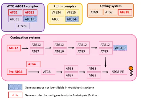
As mentioned before, the earliest studies on autophagy in the 1960s were mainly based on Transmission Electron Microscopy, which allowed the morphological description of events leading to autophagic degradation. Several independent screens of yeast mutants allowed then to describe the molecular actors of autophagy and the identification of 31 ATG genes (autophagy-related genes) that encode the essential machinery of the process [97,107]. Functional analyses of these genes defined five protein groups, which act in a hierarchical way. These protein groups form the ATG1 complex, the Phosphatidyl-Inositol 3-kinase complex, the ATG9 complex, and two conjugation systems involving ATG12 and ATG8 as key players (Figure 2.1).
The following paragraphs describe in detail the molecular autophagic machinery, according to the description for S. cerevisiae.
Figure 2.1: Core macroautophagy machinery in Saccharomyces cerevisiae.
Following autophagy induction, a double-membrane vesicle begins to nucleate and to expand, randomly enclosing cytoplasmic material which can include organelles and aggregated proteins. The formed vesicle then fuses with the cell’s lytic compartment where hydrolases drive the digestion of the enclosed cargo. This process releases aminoacids and lipids that are recycled back to the cytoplasm by specific transporters. Colored boxes present the core functional groups of proteins that participate to autophagosome formation: the ATG1-ATG13 complex, The PtdIns complex, the ATG9 cycling system and the two conjugation systems for ATG12 and ATG8. Adapted from [108].
24
Initiation of autophagy
Autophagy is tightly regulated by the integration of signals coming from the environment as well with signaling pathways regulating cell cycle, metabolism, and programmed cell death. Stimuli coming from the different pathways often converge in a complex including the serine/threonine protein kinase TOR1 (Target Of Rapamycin), which is considered as one of the master regulators of autophagy. As the name of the protein suggests, TOR1 can be inhibited by chemicals such as the bacterial metabolite Rapamycin, isolated in 1965 in Rapa Nui from a soil sample. In S. cerevisiae two isoforms of this protein intervene in the complex: TORC1 integrates extra- and intra-cellular signals coming from nutrient levels, growth factors, and the energy balance, whereas TORC2 is mainly involved in cytoskeleton polarization during cell growth [109]. Under normal conditions, TORC1 phosphorylates several substrates, including the initiation complex of autophagy thus determining a negative regulation of the process. Under unfavorable conditions, TORC1 activity is inhibited thus determining a stop of the cell cycle in G0, and the release of autophagy [110]. In addition to the inhibition of TORC1, autophagy can be activated by alternative signaling pathways such as those involving the cAMP-dependent protein kinase (PKA) pathway [111], or by soluble and membrane-associated receptors to induce the selective degradation of misfolded proteins and organelles [112,113].
ATG1-ATG13 complex and the pre-autophagosomal structure
In yeast, the initiation of autophagosome formation is controlled by a multimeric complex composed of the 5 main proteins, ATG1, ATG13, ATG17, ATG29 and ATG31, which may be supported by specific regulators for targeted degradation of cargos such as ATG11 [114].
The protein ATG1, a serine threonine kinase, and its regulator ATG13, interact constitutively under rich and -deprived conditions via a specific region called the FV-motif [115]. Under nutrient-rich conditions, ATG13 is hyperphosphorylated by multiple kinases including the TORC1 complex [116]. These phosphorylations cause conformational changes that prevent the activation of the ATG1 kinase activity, and the formation of the multimeric complex mentioned above. Upon starvation, dephosphorylation of both ATG1 and ATG13 leads to a stabilization of ATG1, thus enhancing its kinase activity, promoting autophosphorylation, and allowing the interaction with ATG29, ATG31 and ATG17, the latter being specifically recruited in response to starvation [117,118]. The formation of this multimeric complex marks the beginning of nucleation of the Phagophore
25
Assembly Site (PAS). In yeast, this site has been identified as a perivacuolar region [119] where the core machinery proteins will assemble and function either as scaffold, or as regulators for the nucleation of the phagophore, a double membrane-enveloped vesicle that will eventually maturate into an autophagosome [120].
The Phosphatidyl-Inositol 3-kinase (PtdIns3K) Complex
Biogenesis and maturation of the phagophore relies on membrane trafficking converging at the PAS. Membranes involved in this traffic have a unique composition, but their origin remains elusive. However, both the ER and the Golgi were identified as contributors to the nucleation of the phagophore [121]. A characteristic of phagophore membranes is the over-representation of Phosphatidyl-Inositide (PtdIns) and Phosphatidyl-Inositide-3-phosphate (PtdIns3P) [122]. The enrichment of these lipids is enabled by the PtdIns3K complex. The complex is associated to membranes and includes the PtdIns3K VPS34, the Ser/Thr kinase VPS15, and VPS30 (aliases: ATG6, Beclin1), which promotes kinase activity. These three proteins participate in both vacuolar protein sorting (regulating intracellular protein trafficking) and autophagy. To accomplish autophagy, the complex requires an additional factor, ATG14, which supports localization at the PAS. The enzymatic activity of the complex is the phosphorylation of PtdIns, leading to accumulation of PtdIns3P, which is essential for the subsequent steps of phagophore elongation and maturation [122,123].
The Cycling system ATG9 and the ATG2-ATG18 complex
The presence of PtdIns3P at the PAS triggers the recruitment of other protein complexes. One of them is the ATG2-ATG18 complex, composed of ATG2, which associates with the lipid at the PAS, and of ATG18 which in turn recruits and regulates the recycling of another actor in phagophore nucleation, ATG9 [124]. ATG9 is a six-transmembrane domain protein that, unlike other ATG proteins, localizes both to a peripheral reservoir of vesicles located at the PAS and to the close proximity of mitochondria [125]. ATG9 cycles between the two pools, and its transport toward the PAS is supported by ATG11, ATG13 and ATG27. At the PAS, ATG9 self-interacts and tethers upcoming vesicles, thus contributing to membrane supply for the forming phagophore, and facilitating the recruitment of downstream complexes. The regulatory activity of ATG18 together with ATG1-ATG13 and the PtdIns3K complexes relieve the retention of ATG9 at the PAS, thus allowing its shuttling back to the peripheral sites [126].