HAL Id: inserm-00184094
https://www.hal.inserm.fr/inserm-00184094
Submitted on 30 Oct 2007
HAL is a multi-disciplinary open access
archive for the deposit and dissemination of
sci-entific research documents, whether they are
pub-lished or not. The documents may come from
teaching and research institutions in France or
abroad, or from public or private research centers.
L’archive ouverte pluridisciplinaire HAL, est
destinée au dépôt et à la diffusion de documents
scientifiques de niveau recherche, publiés ou non,
émanant des établissements d’enseignement et de
recherche français ou étrangers, des laboratoires
publics ou privés.
A Multiscale Tracking Algorithm for the Coronary
Extraction in MSCT Angiography.
Guanyu Yang, Alexandre Bousse, Christine Toumoulin, Huazhong Shu
To cite this version:
Guanyu Yang, Alexandre Bousse, Christine Toumoulin, Huazhong Shu. A Multiscale Tracking
Algo-rithm for the Coronary Extraction in MSCT Angiography.. Conference proceedings : Annual
Inter-national Conference of the IEEE Engineering in Medicine and Biology Society. IEEE Engineering in
Medicine and Biology Society, 2006, 1, pp.3066-9. �10.1109/IEMBS.2006.260712�. �inserm-00184094�
Abstract – This paper deals with the extraction of the coronary
network on dynamic volume sequences, acquired in Multi-slice Spiral Computed Tomography (MSCT). The proposed approach makes use of a tracking algorithm of the vascular structure, combining a 3D geometric moment operator with a multiscale Hessian filter to estimate the vessel central axis location, its local diameter and orientation.The method performs at the same time, a bifurcation detection to reconstitute the structure of the coronary network. The mean computation time to extract a coronary network is about 3 minutes using a P4-2.4G PC. Preliminary encouraging results are presented on one volume of a sequence.
Key words – Coronary extraction, Multi-scale Hessian filter,
MSCT Angiography, 3-D geometric moment, 3-D tracking
I. INTRODUCTION
The objective is to build a 3-D dynamic model of the heart from patient data acquired in Multi-slice Spiral Computed Tomography (MSCT). This 3-D model is obtained from a dynamic volume sequence and includes cardiac cavities, arteries and veins extracted on each volume. The interest of this model is manifold: (1) evaluate the performances of the segmentation, reconstruction and registration methods involved in the automatic characterization of the pathologies, (2) assess the kinetic properties of the structures in order to evaluate the myocardium viability and perfusion, (3) offer a numerical atlas to the student or clinician for the anatomy learning, (4) provide a virtual surgery training tool.
In our case, we want to simulate the data acquisition from a rotational angiography system to test 3-D reconstruction algorithms of vascular structures. It consists of modeling a continuous rotation of the C-arm system around the 3-D dynamic model and to compute a series of projected images. A wide range of projections in the axial, caudal and cranial angulations can then obtained which will be used for the 3-D reconstruction of the coronary structures.
This paper presents the construction of this 3-D dynamic
model. The acquisition of a dynamic volume sequence leads
to very large data sets (several hundreds of megabytes) from which the structures must be extracted under very strict time constraints to face the clinical requirements. We already dealt with the cardiac cavities in a previous work [1]. The current objective is to extract the vascular structures in order to complete the 3-D model of the heart. Most of the current segmentation methods bring already relevant delineations of
vessels but with too high computation time. They rely on intensity-based methods, generalized cylinder approximations, multiscale and skeletonization schemes, and deformable model approaches applied on successive 2-D slices or on volume data [2].
The challenge is therefore to reach similar performance in few minutes on a standard PC platform, by focusing the extraction process only inside the vascular structure. The present work relies on a geometric moment operator [3] and the response of a multiscale Hessian filter [4] to track a vessel, locally estimate the local diameter and orientation and detect the bifurcation.
The outline of this paper is as follows: section II describes the adopted methodology for the coronary arteries extraction. Section III presents some preliminary results and section IV concludes on the perspectives.
II. METHODOLOGY
The tracking algorithm relies on a local modeling of the vessel by a cylinder in a 3-D homogeneous space. The parameters of this cylinder (location of the center of gravity, and diameter) are estimated using a 3-D geometrical moment operator. Its main advantage is that it provides analytical expressions for the computation of these parameters. A multiscale filter based on eigenvalue analysis of the Hessian matrix is then applied to highlight tubular structures and coping with varying widths. It is also used to locally determine the principal direction of the vessel. A detection of possible bifurcation is performed then in the estimated direction. If one bifurcation is detected, some seed points are extracted which are saved in a list to be further taken as initial
point of the tracking process. The next point Pi+1 on the
centerline is searched for in the estimated direction at the
current point Pi using the moment operator.
The tracking of a branch can be divided into six stages:
a. Interactive selection of a seed point Pi=0;
b. Position refinement of the point Pi
c. Estimation of the local diameter di, vessel intensity Iv
and background Ib;
d. Local direction estimation; e. Bifurcation detection;
f. if a bifurcation is detected then search for some candidate points. These pointswill be stored in the seed point list for further tracking;
else search for the next point Pi+1 in the estimated
direction;
A Multiscale Tracking Algorithm for the Coronary Extraction
in MSCT Angiography
G. Yang
1,2, A. Bousse
1,2, C. Toumoulin
2, H. Shu
11. Laboratory of Image Science and Technology, Southeast University, Nanjing, 210096, P.R. China 2. Laboratoire Traitement du Signal et de l’Image-INSERM, Université de Rennes 1, Rennes, 35042, France
HAL author manuscript inserm-00184094, version 1
HAL author manuscript
g. Go to (b) until stop criterion is satisfied.
The tracking process was performed for every seed point of the list, the latter one being updated during the tracking when a bifurcation was detected. A flowchart of the algorithm is given Fig.1.
2.1 Position refinement of Pi and vessel local diameter
estimation
Starting from the hypothesis that the vessel can be locally
modeled by a cylinder of center of gravity Pi, diameter di and
orientation vector vvessel in the 3-D space, we looked for
estimating the center of gravity of this cylinder so that it corresponds to a point located on the central axis of the vessel. We applied the geometric central moments of order p+q+r to compute this local centroid and estimate the local diameter of the vessel. The coordinates of the local centroid
( , , )x y z inside a spherical window is given by:
) , , ( ) , , ( 000 001 000 010 000 100 M M M M M M z y x = (1)
The distance between the window center and the local centroid allows detecting whether the window is located on the center of a bright structure or not. We applied thus an
iterative process to move the center of the window P'i+1
towards the centroid Pi+1 till this centroid coincides with the
center of this window (P'i+1 =P'i+1), in order to shift it to the
central axis of the vessel (Fig. 2). We then estimated the local
diameter of the vessel from the zero order moment M000, the
local intensity inside (Iv) and outside (Ib) the vessel:
2 / 1 3 / 2 000/4 3 1 2 ⎥ ⎥ ⎦ ⎤ ⎢ ⎢ ⎣ ⎡ ⎟⎟ ⎠ ⎞ ⎜⎜ ⎝ ⎛ − − = − v b v i I I I M R d π (2)
where R is the radius of the spherical window. Optimal parameters were obtained when the size of the spherical window fitted the vessel size. The local diameter was thus estimated using a multi-resolution local moment computation [5].
The intensities Iv and Ib were updated at each step of the
tracking to take into account the heterogeneity of the
intravascular and environmental surroundings [5]. The
background intensity Ib was set to the mean value computed outside the vessel in its normal direction to the currently estimated vessel axis and outside the vessel. In the same way, the vessel intensity Iv was equal to the mean intensity computed along the estimated vessel direction.
2.2 Local vessel direction estimation
A multiscale filter based on eigenvalue analysis of the
Hessian matrix was locally applied at the estimated point Pi to
enhance the tubular structure and estimate the orientation of the vessel. The response of this filter was computed at different scales σ and the eigenvalues were combined into a discriminant function to allow discerning tube-like, blob-like and plate-like structures [4-6].
Let Hσ(x) be the Hessian matrix defined at a given voxel x
at scale σ: ⎥ ⎥ ⎥ ⎦ ⎤ ⎢ ⎢ ⎢ ⎣ ⎡ = ) ( ) ( ) ( ) ( ) ( ) ( ) ( ) ( ) ( ) ( x x x x x x x x x x zz zy zx yz yy yx xz xy xx I I I I I I I I I Hσ (3)
where Iαβ(x) denotes regularized derivatives of the image I(x),
which are obtained by convolving the image using the Gaussian kernel G(x, σ) at scale σ,
( ) 2 2 ( , ) ( ) x x x G I I ∗ ∂ ∂ ∂ = β α σ σ αβ (4) 2 2/2 3 2) 2 ( 1 ) , ( σ πσ σ x x e G = (5)
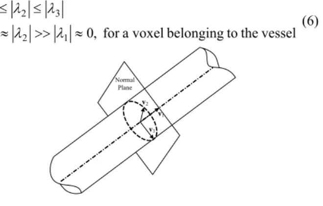
Let λ1, λ2, λ3 and v1, v2, v3 be the eigenvalues and unit
eigenvectors of Hσ(x).
The eigenvector v1 in the direction of the vessel
corresponds to the smallest eigenvalue λ1, while the
eigenvectors v2 and v3 form a base in the orthogonal plane to
Pi
Pi+1
P'i+1
Fig.2: Iterative centering of the spherical window to move its center P'i+1 towards its center of gravity Pi+1
Storage of Points in the List of Seed Points
Search for the Next Point Pi+1
Local Direction Estimation Search Another
Branch Initial Seed Point
Coordinates
End of the Vessel?
Position Refinement and Local Diameter Estimation using Moment Operator Multiscale Hessian Filter Bifurcation? Seed Points List is Empty? Coronary Arteries Centerline Network Construction No Yes No No Yes Yes Extraction Candidate Points
Fig.1: Flowchart of the algorithm
v1 (Fig. 3). The relations between the eigenvalues are thus the following: vessel the to belonging a voxel for , 0 1 2 3 3 2 1 ≈ >> ≈ ≤ ≤ λ λ λ λ λ λ (6)
Frangi et al. [6] introduced two geometric ratios RA, RB in
their vessel likeliness function V(x, σ) :
(
2 2)
2 2(
2 2)
2 3 / 2 / 2 / 2 0, if 0 or 0 ( , ) 1 RA RB 1 S c , else V e α e β e λ λ σ = ⎨⎧⎪ − > − > − − − ⎪⎩ x (7) with∑
= = = = 3 1 3 2 3 2 1 , , j j A B R S R λ λ λ λ λ λ (8)The first ratio RA was used to distinguish between a
plate-like and a tube-plate-like structure. The second one, RB, addressed
the deviation from a blob-like structure and S was a measure of second order structureness, which was used to reduce the response of the background voxels. α, β, c control the
sensitivity of the filter to deviations in RA, RB and S. The
response of the filter is expected to be maximum at a scale σ that approximates the radius of the vessel. The maximum value among the set of response computed at different scales was given by:
) , ( max ) ( max min σ σ σ σ x x V V ≤ ≤ = (9)
The vessel direction vvessel was given by the eigenvector
v1 at the scale σ that provided the optimal response of the filter.
2.3 Incremental displacement along the vessel
Coronary arteries may be very tortuous and highly curved.
We applied thus a small incremental displacement Lstep along
the vessel to ensure a better accuracy of the extraction and avoid a jump into the cardiac cavities. The displacement magnitude was controlled by:
⎪⎩ ⎪ ⎨ ⎧− ≤ − ≤ = + + else , 0 . 2 8 . 1 if , 1 1 i step i vessel i vessel i step i step L L L v v (10)
However, sometimes when reaching the extremity of the vessel or when the vessel is too close to the cardiac cavity, instability occurs inducing some oscillations between the
previous and current points Pi and Pi+1. In that case, we
increased the incremental displacement to possibly pursue the tracking process.
2.4 Stopping criteria
The multi-scale response V(x) of the filter assures a maximum is found. When this maximum is less than a
threshold Thresvessel, the tracking process is stopped. We
initialized then the tracking again by either tacking a new point in the seed point list or interactively pointing a new one on a new branch.
2.5 Bifurcation detection
The local shape, at a bifurcation level, looks like a
plate-like structure and can be discriminated using the parameter RA.
When RA is larger than a threshold Thresbifur, a branch search
process is set, which consisted of:
a. Performing a 3D region growing inside a rectangular box B, whose undersurface was centered on the point
Pi perpendicularly to the vessel direction;
b. Applying the multiscale Hessian filter on the surfaces of the rectangle, excepted on the undersurface, to possibly detect two or three tube-like structures and extract then potential seed points.
If the search process find more than 2 candidate seed points, these seed points are stored in the seed point list for further tracking.
III. RESULTS
3.1 Data Collection
Dynamic volume sequences were acquired on a sub second spiral 16-slice CT scanner GE (LightSpeed 16). Retrospective gating, which allowed optimal gating, was used. The images acquired on several cardiac cycles were reconstructed at every 10% of the R-R interval. Each sequence included thus 10 volumes. The slice thickness was 0.625 mm,
the pixel size 0.488mm and the size of the volumes was
512*512*320.
3.2 Parameter setting
Parameters of the method were set, after a learning stage making use of a greedy algorithm, to the values given Table 1. These parameters were applied for the extraction of the coronaries on all the volumes of each sequence.
Parameter Description
α, β, c Parameters set to 0.5, 0.5 and 0.25 respectively (σmin,σmax) Scale, ranges from 1 to 4
Thresvessel Stop criterion, normally select 50
Thresbifur Bifurcation criterion, normally between 0.18 and 0.20
Lstep Incremental displacement, normally equals to 2 (Bw, Bd) Size of the rectangular box B, depends on the local
estimated diameter of the vessel Table 1: Parameters used in the algorithm
3.3 Experimental results
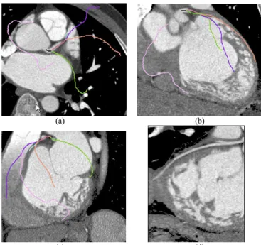
Coronary arteries were extracted on the 10 volumes of
two sequences,focusing on the four main branches: the right
Normal Plane
v1 v2
v3
Fig. 3: Eigenvector approximate the local vessel direction
coronary artery (RCA), the left anterior descending artery (LAD), the circumflex artery (CRX) and the first diagonal artery (DA). Fig.4 illustrates the extraction results for one of the volume of one sequence. All the branches were extracted from only 2 interactively selected seed points. In this volume, most of the branches were correctly extracted. However, some problems may occur when the heart rate varies during the acquisition, inducing thus motion artifacts that lead to detection errors. A way to deal with that problem will be to use the information extracted on the other volumes and perform a motion estimation to recover the branch segment blurred by the motion in the current volume. Fig.5 depicts the
extracted network with estimated local diameter. Fig. 6
provides an estimation of the diameter along the LAD artery
in the perpendicular planes to the vessel.
Fig. 6: Diameter estimation for the CX artery (red dash line) and average diameter (blue solid line)
IV. CONCLUSION
An efficient model-based solution has been proposed for the 3-D tracking of vessels in MSCT volumes. It provides a first approximation of the vascular patterns and allows extracting the structure of the coronary network with a very few initial seed points (between 2 and 4 depending on the complexity of the structure) within 3 minutes on a PC P4-2.4G, 1G RAM. These preliminary results appear promising. Difficulties remain nevertheless in presence of motion artefacts. Further extensions will be to introduce statistical models to improve the coronary extraction as to exploit the other volumes of the sequence to deal with the motion artefacts.
REFERENCES
[1] Yang G., Toumoulin C. Coatrieux J-L., ShuH., Luo L., Boulmier D., " A 3D Static Heart Model from a MSCT Data Set", 27th Annual Conference
of the IEEE Engineering in Medecine and Biology Society (EMBS),
Shanghai, China, Sept. 1-4, N° 1100, 2005.
[2] Kirbas C., Quek F.K.H, “Vessel extraction techniques and algorithms: a survey", IEEE Computer Society, Proceeding of the third IEEE
symposium on Bioinformatics and Bioengineering, 0-7695-1907, 5/03,
2003
[3] Reuzé P, Coatrieux J.L., Luo L.M, Dillenseger J.L., “A 3-D moment based approach for blood vessel detection and quantification in MRA”,
Technology and Health Care, 1, 1993, pp. 181-188.
[4] Pock T.G., “Robust Segmentation of Tubular Structures in 3D Volume Data”, Master thesis, Institut für Maschinelles Sehen und Darstellen Technische Universität, Available at http://www.icg.tu-graz.ac.at/pub/ pubobjects/Master_Thesis.
[5] Bołdak C., Rolland Y., Toumoulin C., “An improved model-based vessel tracking algorithm with application to Computed Tomography Angiography”, Journal of Biocybernetics and Biomédical Engineering, 3(1), 2003, pp.41-64.
[6] Frangi A.F., Niessen W.J., Hoogeveen R.M., Van Walsum T., Viergever M.A.; "Model-based quantification of 3-D Magnetic Resonance Angiographic Images"; IEEE Transaction on Medical Imaging, 18(10),1999, pp. 946–956.
(a) (b)
(c) (d)
Fig. 4: Extraction results for one volume of the sequence. Only 2 seed points was interactively pointed. Slices are displayed in the axial (a), coronal (b) and sagittal (c) planes respectively. The extracted branches are superposed in color on the slices (violet: LAD, green: CX, red: DA and pink: RCA). d) Curvilinear
MPR showing the CX artery
Fig. 5: Extracted 3D coronary tree from two points of view