Directing Single-Molecule Emission with DNA Origami-Assembled
Optical Antennas
Kristina Hübner,
†Mauricio Pilo-Pais,
‡,⊥Florian Selbach,
†Tim Liedl,
‡Philip Tinnefeld,
*
,†Fernando D. Stefani,
*
,§,∥and Guillermo P. Acuna
*
,⊥†Department of Chemistry and Center for NanoScience, Ludwig-Maximilians-Universität München, Butenandtstr. 5-13 Haus E,
81377 München, Germany
‡Faculty of Physics and Center for NanoScience, Ludwig-Maximilians-Universität München, Geschwister-Scholl-Platz 1, 80539
München, Germany
§Centro de Investigaciones en Bionanociencias (CIBION), Consejo Nacional de Investigaciones Científicas y Técnicas
(CONICET), Godoy Cruz 2390, C1425FQD, Ciudad Autónoma de Buenos Aires, Argentina
∥Departamento de Física, Facultad de Ciencias Exactas y Naturales, Universidad de Buenos Aires, Güiraldes 2620, C1428EHA,
Ciudad Autónoma de Buenos Aires, Argentina
⊥Department of Physics, University of Fribourg, Chemin du Musée 3, Fribourg CH-1700, Switzerland
*
S Supporting InformationABSTRACT: We demonstrate the capability of DNA self-assembled optical antennas to direct the emission of an
individualfluorophore, which is free to rotate. DNA origami is
used to fabricate optical antennas composed of two colloidal
gold nanoparticles separated by a predefined gap and to place a
single Cy5 fluorophore near the gap center. Although the
fluorophore is able to rotate, its excitation and far-field emission is mediated by the antenna, with the emission directionality following a dipolar pattern according to the antenna main resonant mode. This work is intended to set out the basis for manipulating the emission pattern of single molecules with self-assembled optical antennas based on colloidal nanoparticles.
KEYWORDS: plasmonics, nanophotonics, metallic nanoparticles, DNA origami, optical antennas
O
ptical antennas (OAs)1 represent the counterparts ofradio- and microwave antennas within the visible spectrum. Essentially, OAs are built from metallic nano-particles (NPs) whose localized surface plasmon resonances
enable the control of lightfields at the nanoscale.2OAs can be
engineered to manipulate the photophysical behavior of single
photon emitters such as organicfluorophores or quantum dots
placed in their vicinity.3Pioneering examples are the“bow tie”
OAs used to demonstrate enhancement of the fluorescence
intensity of organic dyes placed at the hotspot between the
gold elements4 and the monopole, and“Yagi-Uda” OAs used
to demonstrate directionality in the emission of single
emitters.5−7 These examples share the fabrication approach
based on electron beam lithography. Generally, top-down nanofabrication techniques such as e-beam lithography or ion
beam milling offer great geometrical design versatility, but they
also exhibit shortcomings. They are serial, which limits their throughput. The 3D fabrication and organization of (antenna)
elements is difficult and limited to some degree of rotation of
the sample with respect to the beam. There exist limitations to the quality and number of materials that can be used and combined. As a result, attaining OAs made of monocrystalline
elements, as well as combining different materials, is
challenging. Finally, it is virtually impossible to position single photon emitters with controlled stoichiometry in the near-field
of the OAs with nanometer precision.1
The advent of the DNA origami technique8opened up new
pathways for nanophotonics9−16 as colloidal metallic NPs
could be self-assembled in a parallel manner to form OAs. Furthermore, single photon emitters could be positioned in the
near-field of OAs with nanometer precision and stoichiometric
control. Following this approach, the performance of OAs could be revisited at the single-molecule level with higher
geometrical control and more robust statistics,17−20including
their influence on the photophysical behavior of single photon
emitters, such as the electronic transition rates,21
photo-stability,2 2 fluorescence resonance energy transfer
(FRET),23−25surface-enhanced Raman scattering,26−28strong
coupling,29and super-resolution localization.30
http://doc.rero.ch
Published in "Nano Letters 19(9): 6629–6634, 2019"
which should be cited to refer to this work.
DNA origami has turned out to be extremely efficient to organize NPs on nanometric geometries with high degree of
positional and orientational control.31,32 The situation is
different for single photon emitters. Whereas their position
can be controlled well, orienting them over predefined
directions in DNA origami remains an open challenge.33−36
In order to incorporate single fluorophores to DNA origami
structures, they are attached to the backbone or a base of a
short single-stranded DNA, hereafter termed “staple”, which
later binds to its complementary sequence on the DNA
scaffold strand. One fluorophore can be bound to one or two
staples through single or double linkers, respectively.37
Fluorophores integrated into DNA origami structures can
exhibit a variety of behaviors, from free to rotate overfixed in
an undefined orientation to confined in orientation, depending
on their coupling chemistry, molecular structure, charge, and
immediate surrounding environment.23
In this contribution, we investigate the emission directivity
of rotating single fluorophores coupled to OAs. Cy5
fluorophores incorporated to DNA origami structures as
shown in Figure 1A, which are able to rotate, do not present
any directionality in excitation or emission. In contrast, in the presence of a dipolar OA, both their excitation and emission are enhanced and become directional according to the antenna mode.
A schematic of the OA-Cy5 fluorophore system is shown
Figure 1A. A two-layered, rectangular DNA origami sheet with
a size of∼50 nm × 60 nm × 5 nm is used to accommodate
two ultrasmooth spherical Au NPs38with a diameter of 60 nm,
each one at opposite sides of the origami. At approximately the
center of the DNA origami structure, a single Cy5fluorophore
is incorporated at the 3′ end of a DNA single strand (see inset
inFigure 1A). In this way, the single Cy5 molecule is located near the center of the 13 nm gap between the two Au NPs and
is able to rotate when in solution.Figure 1B shows exemplary
TEM images of the dimer OAs illustrating the quality of our structures. The absorption and emission spectra of the Cy5 fluorophore employed together with a numerical simulation of the absorption and scattering cross section of the OA are
included in Figure 1C. The OAs were self-assembled in
solution. For fluorescence measurements, OAs were
immobi-lized on a glass coverslip previously functionaimmobi-lized with BSA-biotin, neutravidin, and biotinylated single-stranded DNA complementary to the single-stranded DNA on the Au NP
surface. As a result, OAs are expected to lieflat with their
inter-particle (main) axis parallel to the substrate surface. All optical
measurements were performed in buffer (see Supporting
Information for sample preparation and measurements de-tails).
Functionalized NPs are mixed in high excess to the DNA origami structure to maximize the yield of dimer OAs. Gel electrophoresis permits to separate the desired structure from unbound NPs and any other unintentionally formed species resulting in a solution containing close to 100% of the target
dimer structure. Figure 1D shows the distinct fluorescence
lifetimes of the reference, monomer, and dimer samples obtained from single-molecule traces (details on the
time-resolved single-moleculefluorescence measurements are given
in the SI). As expected, the interaction between the
fluorophores and the Au NPs reduces the fluorescence lifetime,
with a more pronounced effect for dimer structures.17
The excited state lifetime of the Cy5 in the origami sheet is reduced from 1.7 to 0.6 ns when one Au NP is attached, and to 0.2 ns
(limited by the instrumental response) when the dimer OA is formed. The relatively sharp lifetime distributions and TEM
images reflect the quality of the preparation and the purity of
the samples used in this study.
Two different single-molecule fluorescence measurements
were performed on each of the individual OAs in order to determine the directionality imposed by the OA on the
excitation and on the emission of the singlefluorophores. The
directionality of the emission was determined by wide-field
defocused imaging.39The directionality of the excitation was
probed by monitoring thefluorescence intensity while rotating
Figure 1. (A) Sketch of the OA-Cy5 structure composed of two gold NPs self-assembled onto a rectangular DNA origami. The inset depicts a close-up of the NP gap where the single Cy5fluorophore is incorporated. (B) TEM images of the dimer structures (scale bar is 200 nm). (C) Absorption and emission spectra of Cy5 together with the absorption and scattering cross section of the OA dimer. (D) Fluorescence lifetime histogram of samples containing two NPs (dimers), one NP (monomers), and the reference structure without NPs.
the direction of polarization of a linearly polarized laser used for excitation.
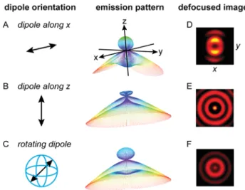
The fluorescence emission of molecules close to a planar
interface has been extensively studied.39,40 The angular
emission pattern depends strongly on the orientation of the molecular emission dipole with respect to the interface. In
Figures2A−C, we include simulations for the emission patterns
of a parallel, perpendicular and free to rotate Cy5 molecule
placed 40 nm above the water−glass interface (on the water
half-space). In all cases molecules emit preferentially into the glass half-space and at angles close to the critical angle of total
internal reflection, but with distinct angular emission patterns.
While the emission pattern of perpendicular molecules has rotational symmetry with respect to the surface normal, the pattern of a parallel molecule has two lobes separated by a gap
along the dipole direction. For afluorophore free to rotate, the
emission pattern corresponds to the isotropic average of dipolar patterns with all possible orientations and has thus
radial symmetry. Defocused imaging is a way to access experimentally the angular emission pattern of single
molecules.39,41Figures 2D−F include the calculated defocused
images of molecules oriented parallel, perpendicular, and freely
rotating when reducing the objective−sample distance by 1 μm
from the focal plane.
Figure 3shows typical defocused images (∼1 μm) of both,
Cy5fluorophores conjugated to the center of the DNA origami
platform with and without OAs. The rotational symmetry of Cy5 samples without OAs demonstrates that the Cy5 fluorophores are able to rotate on a time scale faster than the image acquisition time. Remarkably, the emission patterns of Cy5 molecules change qualitatively when they are coupled to the dimer OAs. In this case, the rotational symmetry is lost, and all detected emission patterns present the two lobes characteristic of an in-plane dipole. Each individual pattern of
an OA-Cy5 structure has a different in-plane orientation. In the
presence of a dipolar OA, a rotatingfluorophore operating at
frequencies below the OA’s resonance2,42 will couple to the
resonant antenna mode when aligned parallel to the antenna’s
main axis.22,43Under this orientation, the emission is expected
to be enhanced and directional with a dipolar pattern.44 In
contrast, for a perpendicular orientation, the fluorophore’s
radiative rate can be significantly suppressed leading to a
negligible emission into the far-field.45−47
This behavior becomes intuitive when picturing the interactions between
the Cy5 and its image charges produced on the NPs48(Figure
3C). A perpendicularly oriented dipole is canceled out by its
image dipole (Figure 3C-I), whereas a parallel oriented dipole
is reinforced (Figure 3C-II). Therefore, despite the fact that
the single Cy5fluorophore is able to rotate, the presence of the
OA will enhance and mediate thefluorophore’s emission when
its orientation is parallel to the main OA axis and suppress it
when perpendicular. This is confirmed by numerical
simulations of the fluorophore’s quantum yield for the two
orientations depicted in Figure 3C (seeFigure S3). For each
defocused pattern inFigure 3B, we extractθemdefined as the
main in-plane emission angle.
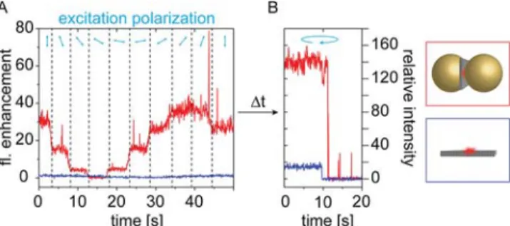
Next, we studied the excitation directionality with the polarization-resolved excitation measurements. For each single
structure, as the ones shown onFigure 3,fluorescent transients
were extracted while rotating the incident light polarization.
Exemplary transients are included in Figure 4A, where
fluorescence enhancement (FE) refers to the fluorescence
intensity normalized to the average fluorescence intensity of
the reference structures (without NPs). The incident light Figure 2. Simulated emission patterns and defocused images of an
emitter located 40 nm above a water (nw= 1.33, z > 0)−glass (ng=
1.5, z < 0) interface (on the water side). The emission wavelength is 670 nm. Defocused images are calculated for the situation where the objective−sample distance has been reduced by 1 μm from the focal plane. (A) Dipolar emitter oriented parallel to the water−glass interface along the x axis. (B) Dipolar emitter oriented perpendicular to the water−glass interface. (C) Isotropic emitter, corresponding to the case of the dipolar emitter that rotates faster than the measurement time.
Figure 3. Defocused images of (A) the Cy5 reference sample and (B) the OA-Cy5 structures. The in-plane emission angle θemis highlighted for a
single OA. (C) Sketch of the image charges induced by afluorophore on the OA elements. The black arrows represent the fluorophore’s emission dipole moment, whereas the red arrows represent the induced dipoles in the NPs.
polarization angle is rotated by 20°, sweeping a range of 180°.
Finally, the fluorophores are photobleached using increased
laser power and circularly polarized light to prove single-step
bleaching behavior (Figure 4B). For the reference structures,
fluorescence transients show negligible variations with the incident polarization angle. This is in line with our previous observation that Cy5 is able to rotate faster than the integration time. In contrast, the transients of OA-Cy5 structures show a clear periodic dependence with the incident polarization angle. As expected for a dipolar dimer OA, the
electricfield enhancement reaches the highest value when the
incident light is polarized along the antenna axis.49
From these measurements, we extracted the polarization
angle of maximum excitationθex, which corresponds to the
in-plane orientation of each OA. We note that for our analysis we considered only OAs that showed a clear cosine square response to the polarization angle and a single-step photo-bleaching, assuring that we probed dipolar OAs with a single fluorophore.
Finally, we combined the results of the independent
measurements displayed in Figures 3B and 4A in order to
study the emission and excitation directionality of each OA.
Figure 5 displays a scatter plot of θem versus θex for 147
randomly oriented dimer OAs with a single Cy5 fluorophore.
The strong correlation between both angles θem and θex
confirms that the OAs impose directionality to the rotating
fluorophores both on excitation and emission according to the
antenna’s main resonant mode.
In summary, using the DNA origami technique we self-assembled dipolar optical antennas made of two closely spaced
gold nanoparticles with a singlefluorescent molecule at their
gap. By means of single-molecule measurements of the emission pattern and the polarization of maximum excitation, we showed that the excitation and emission of single fluorophores that are able to rotate can be made directional
with optical antennas, according to the antenna’s main
resonant mode. These experiments provide a solid ground for more sophisticated photon routing experiments using single
emitters and self-assembled optical antennas.50,51
■
ASSOCIATED CONTENT*
S Supporting InformationThe Supporting Information is available
Detailed information on gold nanoparticle fabrication, DNA origami folding, optical antenna assembly and immobilization, imaging systems, analysis, and numerical
simulations (PDF)
■
AUTHOR INFORMATIONCorresponding Authors
*E-mail:[email protected](P.T.).
*E-mail:[email protected](F.D.S.).
*E-mail:[email protected](G.P.A.).
ORCID Tim Liedl: 0000-0002-0040-0173 Philip Tinnefeld: 0000-0003-4290-7770 Fernando D. Stefani: 0000-0002-3277-7215 Guillermo P. Acuna: 0000-0001-8066-2677 Notes
The authors declare no competingfinancial interest.
■
ACKNOWLEDGMENTSWe would like to thank Mario Raab, Ija Jusuk, Florian Steiner, Jan Vogelsang, and Johann Bohlen for fruitful discussion. This work was supported by the Swiss National Science Foundation through the National Center of Competence in Research Bio-Inspired Materials, with grants PICT2013-0792 and
PICT-2014-0739 of the Agencia Nacional de Promoción Científica y
Tecnológica (ANPCYT, Argentina). P.T. and G.P.A. acknowl-edge support by the DFG (AC 279/2-1 and TI 329/9-1, INST 86/1904-1 FUGG, excellence cluster e-conversion). M.P. and
T.L. thank thefinancial support from the European Research
Council (ERC) through the starting grant 336440 (ORCA) and the DFG through the excellence cluster e-conversion. F.D.S. thanks the support of the Max-Planck-Society and the Alexander von Humboldt Foundation.
■
REFERENCES(1) Novotny, L.; Van Hulst, N. Antennas for Light. Nat. Photonics 2011, 5 (2), 83−90.
(2) Biagioni, P.; Huang, J.-S.; Hecht, B. Nanoantennas for Visible and Infrared Radiation. Rep. Prog. Phys. 2012, 75 (2), 024402.
(3) Koenderink, A. F. Single-Photon Nanoantennas. ACS Photonics 2017, 4, 710−722.
Figure 4. (A) Transients of fluorescence intensity for varying polarization angle of the excitation for the reference (no NP, blue) and OAs (NP dimer, red). The OA intensity is normalized to the average of the reference intensity and thus represents thefluorescence enhancement (FE). During thefirst 50 s, the incident polarization is rotated. (B) Afterwards, the incident light power is increased and its polarization is turned circular in order to bleach thefluorophores to verify a single-step photobleaching.
Figure 5. Scatter plot of the in-plane angles of emission θem and
excitationθexfor 147 individual OAs.
(4) Kinkhabwala, A.; Yu, Z.; Fan, S.; Avlasevich, Y.; Müllen, K.; Moerner, W. E. Large Single-Molecule Fluorescence Enhancements Produced by a Bowtie Nanoantenna. Nat. Photonics 2009, 3 (11), 654−657.
(5) Taminiau, T. H.; Stefani, F. D.; Segerink, F. B.; Van Hulst, N. F. Optical Antennas Direct Single-Molecule Emission. Nat. Photonics 2008, 2 (4), 234−237.
(6) Curto, A. G.; Volpe, G.; Taminiau, T. H.; Kreuzer, M. P.; Quidant, R.; Van Hulst, N. F. Unidirectional Emission of a Quantum Dot Coupled to a Nanoantenna. Science (Washington, DC, U. S.) 2010, 329 (5994), 930−933.
(7) Aouani, H.; Mahboub, O.; Devaux, E.; Rigneault, H.; Ebbesen, T. W.; Wenger, J. Large Molecular Fluorescence Enhancement by a Nanoaperture with Plasmonic Corrugations. Opt. Express 2011, 19 (14), 13056.
(8) Rothemund, P. W. K. Folding DNA to Create Nanoscale Shapes and Patterns. Nature 2006, 440, 297−302.
(9) Kuzyk, A.; Jungmann, R.; Acuna, G. P.; Liu, N. DNA Origami Route for Nanophotonics. ACS Photonics 2018, 5 (4), 1151−1163.
(10) Pilo-Pais, M.; Acuna, G. P.; Tinnefeld, P.; Liedl, T. Sculpting Light by Arranging Optical Components with DNA Nanostructures. MRS Bull. 2017, 42 (12), 936−942.
(11) Liu, N.; Liedl, T. DNA-Assembled Advanced Plasmonic Architectures. Chem. Rev. 2018, 118 (6), 3032−3053.
(12) Samanta, A.; Banerjee, S.; Liu, Y. DNA Nanotechnology for Nanophotonic Applications. Nanoscale 2015, 7 (6), 2210−2220.
(13) Lan, X.; Wang, Q. DNA-Programmed Self-Assembly of Photonic Nanoarchitectures. NPG Asia Mater. 2014, 6 (4), e97−e97. (14) Wang, P.; Meyer, T. A.; Pan, V.; Dutta, P. K.; Ke, Y. The Beauty and Utility of DNA Origami. Chem. 2017, 2 (3), 359−382.
(15) Madsen, M.; Gothelf, K. V. Chemistries for DNA Nano-technology. Chem. Rev. 2019. DOI: 10.1021/acs.chemrev.8b00570.
(16) Gopinath, A.; Miyazono, E.; Faraon, A.; Rothemund, P. W. K. Engineering and Mapping Nanocavity Emission via Precision Placement of DNA Origami. Nature 2016, 535 (7612), 401−405.
(17) Acuna, G. P.; Möller, F. M.; Holzmeister, P.; Beater, S.; Lalkens, B.; Tinnefeld, P. Fluorescence Enhancement at Docking Sites of DNA-Directed Self-Assembled Nanoantennas. Science (Washington, DC, U. S.) 2012, 338 (6106), 506−510.
(18) Prinz, J.; Heck, C.; Ellerik, L.; Merk, V.; Bald, I. DNA Origami Based Au−Ag-Core−Shell Nanoparticle Dimers with Single-Molecule SERS Sensitivity. Nanoscale 2016, 8 (10), 5612−5620.
(19) Simoncelli, S.; Roller, E.-M.; Urban, P.; Schreiber, R.; Turberfield, A. J.; Liedl, T.; Lohmüller, T. Quantitative Single-Molecule Surface-Enhanced Raman Scattering by Optothermal Tuning of DNA Origami-Assembled Plasmonic Nanoantennas. ACS Nano 2016, 10 (11), 9809−9815.
(20) Chikkaraddy, R.; Turek, V. A.; Kongsuwan, N.; Benz, F.; Carnegie, C.; van de Goor, T.; de Nijs, B.; Demetriadou, A.; Hess, O.; Keyser, U. F.; et al. Mapping Nanoscale Hotspots with Single-Molecule Emitters Assembled into Plasmonic Nanocavities Using DNA Origami. Nano Lett. 2018, 18 (1), 405−411.
(21) Holzmeister, P.; Pibiri, E.; Schmied, J. J.; Sen, T.; Acuna, G. P.; Tinnefeld, P. Quantum Yield and Excitation Rate of Single Molecules Close to Metallic Nanostructures. Nat. Commun. 2014, 5 (1), 5356. (22) Kaminska, I.; Vietz, C.; Cuartero-González, Á.; Tinnefeld, P.; Fernández-Domínguez, A. I.; Acuna, G. P. Strong Plasmonic Enhancement of Single Molecule Photostability in Silver Dimer Optical Antennas. Nanophotonics 2018, 7 (3), 643−649.
(23) Aissaoui, N.; Moth-Poulsen, K.; Käll, M.; Johansson, P.; Wilhelmsson, L. M.; Albinsson, B. FRET Enhancement Close to Gold Nanoparticles Positioned in DNA Origami Constructs. Nanoscale 2017, 9 (2), 673−683.
(24) Vietz, C.; Lalkens, B.; Acuna, G. P.; Tinnefeld, P. Synergistic Combination of Unquenching and Plasmonic Fluorescence Enhance-ment in Fluorogenic Nucleic Acid Hybridization Probes. Nano Lett. 2017, 17 (10), 6496−6500.
(25) Bohlen, J.; Cuartero-González, Á.; Pibiri, E.; Ruhlandt, D.; Fernández-Domínguez, A. I.; Tinnefeld, P.; Acuna, G. P.
Plasmon-Assisted Förster Resonance Energy Transfer at the Single-Molecule Level in the Moderate Quenching Regime. Nanoscale 2019, 11 (16), 7674−7681.
(26) Thacker, V. V.; Herrmann, L. O.; Sigle, D. O.; Zhang, T.; Liedl, T.; Baumberg, J. J.; Keyser, U. F. DNA Origami Based Assembly of Gold Nanoparticle Dimers for Surface-Enhanced Raman Scattering. Nat. Commun. 2014, 5 (1), 3448.
(27) Prinz, J.; Schreiber, B.; Olejko, L.; Oertel, J.; Rackwitz, J.; Keller, A.; Bald, I. DNA Origami Substrates for Highly Sensitive Surface-Enhanced Raman Scattering. J. Phys. Chem. Lett. 2013, 4 (23), 4140−4145.
(28) Kühler, P.; Roller, E. M.; Schreiber, R.; Liedl, T.; Lohmüller, T.; Feldmann, J. Plasmonic DNA-Origami Nanoantennas for Surface-Enhanced Raman Spectroscopy. Nano Lett. 2014, 14 (5), 2914−2919. (29) Ojambati, O. S.; Chikkaraddy, R.; Deacon, W. D.; Horton, M.; Kos, D.; Turek, V. A.; Keyser, U. F.; Baumberg, J. J. Quantum Electrodynamics at Room Temperature Coupling a Single Vibrating Molecule with a Plasmonic Nanocavity. Nat. Commun. 2019, 10 (1), 1049.
(30) Raab, M.; Vietz, C.; Stefani, F. D.; Acuna, G. P.; Tinnefeld, P. Shifting Molecular Localization by Plasmonic Coupling in a Single-Molecule Mirage. Nat. Commun. 2017, 8 (1), 13966.
(31) Pal, S.; Deng, Z.; Wang, H.; Zou, S.; Liu, Y.; Yan, H. DNA Directed Self-Assembly of Anisotropic Plasmonic Nanostructures. J. Am. Chem. Soc. 2011, 133 (44), 17606−17609.
(32) Zhan, P.; Wen, T.; Wang, Z.; He, Y.; Shi, J.; Wang, T.; Liu, X.; Lu, G.; Ding, B. DNA Origami Directed Assembly of Gold Bowtie Nanoantennas for Single-Molecule Surface-Enhanced Raman Scatter-ing. Angew. Chem., Int. Ed. 2018, 57 (11), 2846−2850.
(33) de Torres, J.; Mivelle, M.; Moparthi, S. B.; Rigneault, H.; Van Hulst, N. F.; García-Parajó, M. F.; Margeat, E.; Wenger, J. Plasmonic Nanoantennas Enable Forbidden Förster Dipole−Dipole Energy Transfer and Enhance the FRET Efficiency. Nano Lett. 2016, 16 (10), 6222−6230.
(34) Kroutil, O.; Romancová, I.; Šíp, M.; Chval, Z. Cy3 and Cy5 Dyes Terminally Attached to 5′C End of DNA: Structure, Dynamics, and Energetics. J. Phys. Chem. B 2014, 118 (47), 13564−13572.
(35) Boulais, É.; Sawaya, N. P. D.; Veneziano, R.; Andreoni, A.; Banal, J. L.; Kondo, T.; Mandal, S.; Lin, S.; Schlau-Cohen, G. S.; Woodbury, N. W.; et al. Programmed Coherent Coupling in a Synthetic DNA-Based Excitonic Circuit. Nat. Mater. 2018, 17 (2), 159−166.
(36) Gopinath, A.; Thachuk, C.; Mitskovets, A.; Atwater, H. A.; Kirkpatrick, D.; Rothemund, P. W. K. Absolute and Arbitrary Orientation of Single Molecule Shapes. arXiv:1808.04544, 2018.
(37) Stennett, E. M. S.; Ma, N.; van der Vaart, A.; Levitus, M. Photophysical and Dynamical Properties of Doubly Linked Cy3-DNA Constructs. J. Phys. Chem. B 2014, 118 (1), 152−163.
(38) Yoon, J. H.; Selbach, F.; Langolf, L.; Schlücker, S. Ideal Dimers of Gold Nanospheres for Precision Plasmonics: Synthesis and Characterization at the Single-Particle Level for Identification of Higher Order Modes. Small 2018, 14 (4), 1702754.
(39) Böhmer, M.; Enderlein, J. Orientation Imaging of Single Molecules by Wide-Field Epifluorescence Microscopy. J. Opt. Soc. Am. B 2003, 20 (3), 554−559.
(40) Novotny, L.; Hecht, B. Principles of Nano-Optics, 2nd ed.; Cambridge University Press: Cambridge, 2012.
(41) Ghosh, S.; Chizhik, A. M.; Yang, G.; Karedla, N.; Gregor, I.; Oron, D.; Weiss, S.; Enderlein, J.; Chizhik, A. I. Excitation and Emission Transition Dipoles of Type-II Semiconductor Nanorods. Nano Lett. 2019, 19 (3), 1695−1700.
(42) Mertens, H.; Koenderink, A. F.; Polman, A. Plasmon-Enhanced Luminescence near Noble-Metal Nanospheres: Comparison of Exact Theory and an Improved Gersten and Nitzan Model. Phys. Rev. B: Condens. Matter Mater. Phys. 2007, 115123.
(43) Pellegrotti, J. V.; Acuna, G. P.; Puchkova, A.; Holzmeister, P.; Gietl, A.; Lalkens, B.; Stefani, F. D.; Tinnefeld, P. Controlled Reduction of Photobleaching in DNA Origami-Gold Nanoparticle Hybrids. Nano Lett. 2014, 14 (5), 2831−2836.
(44) Rogobete, L.; Kaminski, F.; Agio, M.; Sandoghdar, V. Design of Plasmonic Nanoantennae for Enhancing Spontaneous Emission. Opt. Lett. 2007, 32, 1623.
(45) Blanco, L. A.; García de Abajo, F. J. Spontaneous Emission Enhancement near Nanoparticles. J. Quant. Spectrosc. Radiat. Transfer 2004, 89, 37.
(46) Busson, M. P.; Rolly, B.; Stout, B.; Bonod, N.; Bidault, S. Accelerated Single Photon Emission from Dye Molecule-Driven Nanoantennas Assembled on DNA. Nat. Commun. 2012, 1964.
(47) Liaw, J. W.; Chen, C. S.; Chen, J. H. Enhancement or Quenching Effect of Metallic Nanodimer on Spontaneous Emission. J. Quant. Spectrosc. Radiat. Transfer 2010, 111, 454.
(48) Mock, J. J.; Hill, R. T.; Degiron, A.; Zauscher, S.; Chilkoti, A.; Smith, D. R. Distance-Dependent Plasmon Resonant Coupling between a Gold Nanoparticle and Gold Film. Nano Lett. 2008, 8 (8), 2245−2252.
(49) Puchkova, A.; Vietz, C.; Pibiri, E.; Wünsch, B.; Sanz Paz, M.; Acuna, G. P.; Tinnefeld, P. DNA Origami Nanoantennas with over 5000-Fold Fluorescence Enhancement and Single-Molecule Detection at 25Μm. Nano Lett. 2015, 15 (12), 8354−8359.
(50) Toscano, G.; Raza, S.; Jauho, A.-P.; Mortensen, N. A.; Wubs, M. Modified Field Enhancement in Plasmonic Nanowire Dimers Due to Nonlocal Response. Nat. Commun. 2011, 2 (1), 481.
(51) Yao, K.; Liu, Y. Controlling Electric and Magnetic Resonances for Ultracompact Nanoantennas with Tunable Directionality. ACS Photonics 2016, 3 (6), 953−963.