HAL Id: hal-01367894
https://hal.archives-ouvertes.fr/hal-01367894
Submitted on 17 Sep 2016
HAL is a multi-disciplinary open access archive for the deposit and dissemination of sci-entific research documents, whether they are pub-lished or not. The documents may come from teaching and research institutions in France or abroad, or from public or private research centers.
L’archive ouverte pluridisciplinaire HAL, est destinée au dépôt et à la diffusion de documents scientifiques de niveau recherche, publiés ou non, émanant des établissements d’enseignement et de recherche français ou étrangers, des laboratoires publics ou privés.
Predicting PARP inhibitory activity – A novel quantum
mechanical based model
Clifford Fong
To cite this version:
Clifford Fong. Predicting PARP inhibitory activity – A novel quantum mechanical based model. [Research Report] Eigenenergy, Adelaide, Australia. 2016. �hal-01367894�
Predicting PARP inhibitory activity – A novel quantum mechanical based model Clifford W. Fong
Eigenenergy, Adelaide, South Australia, Australia Email: [email protected]
Abstract
It has been shown that a novel 4 parameter model (IC50=ΔGdesolvation free energy of water +
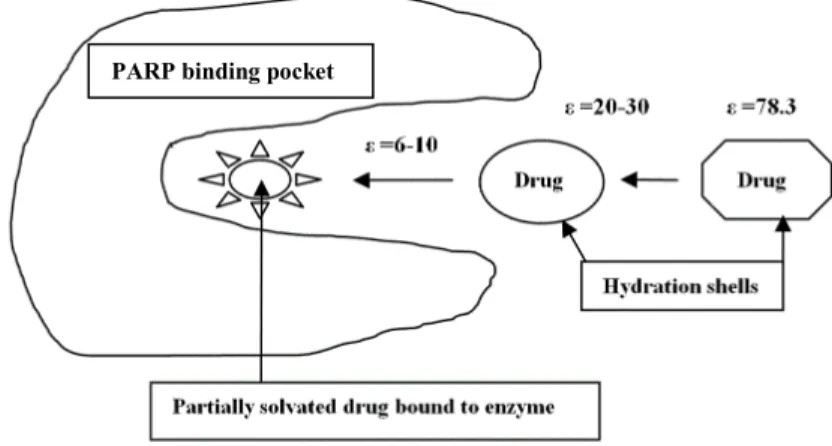
lipophilicity + dipole moment + molecular volume) applies to a broad range of PARP1 and PARP2 inhibitory activity. Similarly to other inhibitors, desolvation plays a critical role in binding activity, a factor usually ignored in quantitative binding models. The model is consistent with a process where the water solvated inhibitor moves from a bulk water environment ε 78.3 to an environment representative of the interior of a protein binding pocket ε 6-10. Correlations have been found between equation 1 and literature docking studies which support this model.
A correlation has been found using a variant of equation 1 using the electron affinity of the inhibitors (along with the desolvation, lipophilicity and dipole moment). This observation may support the notion that PARP inhibitors that show synergism with adjuvant drugs and radiotherapy that are used concomitantly with PARP inhibitors in the clinic may involve redox electron transfer processes.
Keywords: PARP; inhibitor binding; desolvation; electron affinity Abbreviations
ΔGdesolvation free energy of water desolvation, ΔGlipophilicity free energy of lipophilicity or hydrophobicity, ΔGdesolv,CDS free
energy of water desolvation of the cavitation dispersion solvent structure (CDS), ΔGlipo,CDS free energy of lipophilicity or
hydrophobicity for the CDS, DM dipole moment DM, EA electron affinity, ε dielectric constant, R2 multiple correlation coefficient, HOMO highest occupied molecular orbital, F the F test of significance, SEE standards errors for the estimates, SE(ΔGdesolvation) standard errors of ΔGdesolvation, SE(ΔGdesolv,CDS) standard errors of ΔGdesolv,CDS, SE(ΔGlipophilicity), standard
errors of ΔGlipophilicity, SE(Dipole Moment) standard errors for dipole moments, SE (Molecular Volume) standard errors for
molecular volumes as calculated from “t” distribution statistics
Highlights
• Novel model: desolvation energy+lipophilicity+dipole moment+molecular volume (or electron affinity)
• Binding PARP1 and PARP2 inhibitors: desolvation driven • Model consistent with PARP inhibitor docking studies Introduction
The poly(ADP-ribose) polymerase (PARP) superfamily includes at least 17 enzymes involved in several biological processes, including transcriptional regulation, DNA repair, cell cycle regulation, inflammation, hypoxic response, spindle pole function, oncogene-related signalling, and cell death. PARP1 is the most abundant and best characterized protein within this large family. PARP1 is a DNA repair enzyme but has a rolein cell homeostasis at several levels, including transcriptional and epigenetic mechanisms. PARP2 displays almost
overlapping functions with PARP1. PARP3 is involved in DNA repair, mitotic spindle integrity, and telomerase regulation. PARP couples with BRCA. Germline inactivation of BRCA1/2 tumor suppressor genes strongly predisposes to breast and ovarian cancer as well as other malignancies, including pancreatic and prostate cancer. The targeting of tumour suppressor defects using PARP inhibitors has been effective in BRCA-related tumours due to sabotage of functionally interdependent DNA repair routes that ultimately leads to cellular toxicity. PARP inhibitors improve progression-free survival in women with recurrent platinum-sensitive ovarian cancer, particularly by olaparib added to conventional treatment. Olaparib was US FDA approved in 2014 as monotherapy (at 400 mg taken twice per day) for patients with germline BRCA mutated (gBRCAm) advanced ovarian cancer who have been treated with three or more prior lines of chemotherapy. Rucaparib was granted breakthrough therapy designation by the FDA in 2015, for use as monotherapy in patients with BRCA1/2 mutant (germline or somatic) advanced ovarian cancer after at least two prior lines of
platinum-containing therapy. Several other PARP inhibitors are currently undergoing clinical trials.[1-4] (2016) 114, 713–715
DNA is damaged thousands of times during each normal cell cycle, and that damage must be repaired. PARP1 is important for repairing single-strand breaks (‘nicks’ in the DNA). If such nicks remain unrepaired until DNA is replicated before cell division, then the replication itself can cause double strand breaks to form. Drugs that inhibit PARP1 cause multiple double strand breaks to form, and in tumours with BRCA1, BRCA2 or PALB2 mutations, these double strand breaks cannot be efficiently repaired, leading to the death of the cells. Normal cells that don't replicate their DNA as often as cancer cells, and cells that lack any mutated BRCA1 or BRCA2 still have homologous repair operating, which allows them to survive the inhibition of PARP. PARP inhibitors have an additional mode of action involving localizing PARP proteins at sites of DNA damage, which has relevance to their anti-tumour activity. The trapped PARP protein–DNA complexes are highly toxic to cells because they block DNA replication. The trapped PARP–DNA complexes are more toxic to cells than the unrepaired single-strand DNA breaks that accumulate in the absence of PARP activity, indicating that PARP inhibitors act as PARP poisons. It is suggested that there may be two classes of PARP inhibitors, catalytic inhibitors that act mainly to inhibit PARP enzyme activity and do not trap PARP proteins on DNA, and dual inhibitors that both block PARP enzyme activity and act as PARP poisons.[5]
The various classes of PARP inhibitors have been reviewed.[6-9] The various classes include PARP1 and PARP2 specific: isoquinolinone, indazole, phenanthridine,
benzonaphthyridinone. Efforts to discover and optimize new PARP inhibitors have mainly centred on 3D-QSAR studies. 3D-QSAR involve the application of force field calculations requiring three-dimensional structures, e.g. based on protein crystallography or molecule superimposition. It uses computed potentials, e.g. the Lennard-Jones potential, rather than experimental constants and investigates with the overall molecule rather than a single substituent. It examines the steric fields (shape of the molecule), the hydrophobic regions (water-soluble surfaces), and the electrostatic fields. Many such studies have incorporated accompanying computational docking studies.[6] Other studies have used detailed molecular dynamics, principal components analysis and conformational analysis to investigate binding mode of PARP inhibitors.[10]
A detailed analysis of the x-ray structures of the PARP family interaction with inhibitors has identified the common interactions between ligands and the conserved glycine (backbone) and serine (side-chain hydroxyl) residues and the space occupied by various PARP inhibitors
bound to human PARP catalytic domains.[11] It is clear that the hydrogen bonding
interactions, via glycine and serine, are important linkages, but the non-polar or hydrophobic interactions between the inhibitors and the enzyme are also key factors in binding interaction. Clearly the molecular size and hydrophobic areas of the inhibitors, besides the hydrogen bonding interactions, play important roles and must be critical factors in predicting the inhibitory capacity of potential inhibitors.
When a small-molecule binds to a protein, it must displace most of the waters occupying the binding cavity, but rarely are all water molecules displaced. A comprehensive analysis of 392 high-resolution crystal structures of proteins with interacting ligandsshowed that in >84% of the complexes, one or more waters are present and interact with the ligand mediating its interaction with the protein. [21] Some waters can be so strongly bound and conserved among similar proteins they are considered a part of the target structure, altering the binding site structure. Classical examples are HIV-1 proteasewhere stable bridging waters are targeted to increase inhibitor affinitycan play a critical role in HIV-1 resistance.[14,22] Stable waters can also be displaced to improve ligand affinity by the entropy gain resulting from the release of ordered water to the bulk solvent. These strategies have been successfully applied in designing scytalone dehydratase inhibitorsand cyclic urea inhibitors of HIV-1 protease [23,24].Weakly bound waters are more likely to play varying roles depending upon the nature of the bound ligand. The same water can be stabilized by one ligand and displaced by another, as with poly(ADP-ribose) polymerase inhibitors [25,26].
PARP inhibitors have found clinical application as monotherapies (after failure of standard chemotherapies) or combinatorial regimes often with cytotoxic drugs (topoisomerase I and II inhibitors, temozolomide, gemcitabine, Pt drugs etc). Before PARP inhibitors were found to be effective against BRCA-deficient tumours, they were used as chemosensitizers. There is evidence of synergism between certain PARP inhibitors and adjuvant cytotoxic drugs and radiation therapy [2-4]. The basis of this synergism may be because radiotherapy damages cells by causing DNA breaks, and PARP inhibitors disrupt DNA repair mechanisms. Since free radicals are involved in radiation induced damage to DNA, we have previously
suggested that the electron transfer properties of combinatorial drugs such as Pt with adjuvant drugs can lead to synergism between the cytotoxic drugs. [27,28]
This study will use a model (equation 1 or 1a) which has been successfully applied to various transport and drug-protein binding processes. A common model for both processes based on 4 molecular physico-chemical properties of the prospective drugs has been previously applied to passive and facilitated diffusion, and active organic anion transporter drug membrane transport, statins-HMGCoA reductase, some competitive statin-CYP enzyme binding processes, tyrosine kinase inhibitors, HIV-1 protease inhibitors, and cyclin-dependent kinases. Equation 1 also applies to the active competitive transport of these tyrosine kinase inhibitors by the hOCT3, OATP1A2 and OCT1 transporters. [12-15]The model comprises four main properties: (a) desolvation energy in water (b) lipophilicity or hydrophobicity based on the solvation energy in a hydrophobic solvent such as n-octane or n-octanol; lipophilicity being a measure of how well a drug can interact with lipophilic cell membrane bilayers, and hydrophobicity being a measure of non-polar interaction between a drug and the hydrophobic sectors of a protein (c) dipole moment in water, as a measure of the polar
attraction between the drug and its receptor target or cell membrane (d) the molecular
of the target receptor protein or active protein transporter, or how well a drug can diffuse through a cell membrane. The basic model is shown in equation 1.
= desolvation + lipophilicity + !" # $"
+ # !"%&! ' !&$" Or
= desol,CDS + lipophilicity + !" # $"
+ # !"%&! ' !&$"
ΔGdesolvation = ΔGelectrostatic + ΔGCDS where CDS is the cavity dispersion solvent structure. The
CDS involves non-bulk solvent electrostatic contributions to the free energy of hydration. The SMD solvation model is based on ΔGSo = ΔGENP + GCDS where ENP is the electronic
nuclear polarization: the change in the solute free energy due to electrostatic interactions between the solute and the bulk solvent and distortion of the solute’s electronic structure in solution. The solvent is modelled as a dielectric continuum. The CDS represents first solvation shell effects. It involves atomic surface tension (geometry dependent
proportionality constants). The CDS has been parameterized using extensive experimental data sets for optimization, and has the advantage of including a realistic experimentally based hydrogen bonding model. The CDS covers range polarization effects and shorter-range non-electrostatic effects such as cavitation, dispersion, and solvent structural effects (which includes both hydrogen bonding) and exchange repulsion effects.[16] ΔGdesol,CDS can
be substituted for ΔGdesolvation where this non-polar term may be more representative of the
interaction processes being studied, as for example in a ligand-protein binding study within the protein binding pocket where the dielectric constant for that environment is ca. ε=6-10 compared to ε=78.3 for the bulk water environment where the use of ΔGdesolvation is more
appropriate.[13-15]
It has been shown that continuum based hydrophobic non-polar hydration solvation models can fairly accurately model particular protein-protein and ligand-protein complexes, although different protein systems may have unique attractive (van der Waals) properties not precisely transferable to other systems.[17] The hydration hydrophobic effect is primarily due to a decrease in the number of ways that favourable hydrogen bonding can be achieved by solvent water molecules when they are near to a non-hydrogen bonding solute, thereby decreasing the solvent’s entropy. The hydrophobic non-polar hydration free energy ΔGnon-polar can be
considered to comprise of the repulsive ΔGcavity and attractive ΔGvdw terms. The ΔGcavity term
includes the repulsive electrostatic effect of forming a cavity in the solvent whereby the solute interacts with the water solvent, while the ΔGvdw term includes shorter-range
polarization effects and shorter-range non-electrostatic effects such as cavitation, dispersion, and solvent structural effects (which includes both hydrogen bonding) and exchange
repulsion effects.
When a solvated ligand enters a protein binding cavity and begins to bind with the protein, the desolvation processes involving the protein and ligand may involve extensive
rearrangement of water molecules, and possibly some expulsion of water molecules from the cavity. A significant change in solvation entropy should then occur on the side on the ligand and the protein receptor.
We have previously shown that the ΔGdesolv,CDS values may be a close approximation of how
a solvated inhibitor reacts as it approaches and starts to enter the binding pocket (dielectric constant ε=20-30) and leaves the bulk water environment (ε =78.3) and binding starts to occur. [14,15] This scenario has been modelled using more intensive molecular mechanics computations.[18-20]
It has also been shown [13-15] that equation 1 can be modified to model redox processes (eg metabolic oxidation of statins by CYP enzymes) particularly when the molecular volume changes are small. The electron affinity EA or ionization potential IE in water are measures of the ease that drugs can be reduced or oxidised by appropriate species such as enzymes.
," - % . /
= desolvation + lipophilicity + !" # $"
+ 0!"% 122 /
Results
In this study, three sets of PARP inhibitors have been evaluated by applying equation 1 to known experimental IC50 data.[6,10,29] The study of the hexahydrobenzonaphthyridinone
inhibitors [29] has a large and diverse range of inhibitors so the IC50 experimental data is
internally consistent within the experimental error of the experimental conditions. This data is more accurate and internally consistent than the IC50 data used by the other two studies [6,10]
where data was collected from multiple studies, where experimental conditions were
different, so correlations using these data bases are less robust, and data measurements for the notionally same cell line can vary by ten fold. It should be noted that the equations derived in this study are indicative rather than highly robust, since the experimental data sets are too small to be highly robust or internally cross validated. However, the general model does apply to the three internally consistent data sets which cover a wide range of structurally diverse inhibitors, as well as a number of other biological activities [12-15]. The model has been validated against the literature docking scores [10] for 14 PARP-1 inhibitors (as shown below in eq 5a and 5b).
(a) Analysis of hexahydrobenzonaphthyridinone inhibitors [29] The inhibitors analysed are shown in Figure 1. Preliminary analysis showed that inhibitors 4, 7, 20 and 45 were large outliers as neutral species, but fitted the correlation when treated as protonated ions of the amine bases. Protonation of these bases is consistent with similar compounds at the physiological pH (ca 7).
Figure 1. HexahydrobenzonaphthyridinonePARP Inhibitors
(4) Parent R=H (7) R (8) R (9) R (11) R (15) R
Parent (16) R (17) R (18) R (20) R
(29) R (33) R (36) R (39) R (45) R
Figure 1 Footnote: Numbering scheme of hexahydrobenzonaphthyridinone inhibitors refers to numbering scheme in Torrisi 2010 [29].
IC50 values for 15 hexahydrobenzonaphthyridinone inhibitors Eq 1a
3450 = 7. 99 desolvation − 9. ;< lipophilicity − =. ;> ?@ABCD EBFDGH
+ 9. IJ EBCDKLCMN OBCLFD − 63.3
Where R2 = 0.886, SEE = 24.31, SE(ΔGdesolvation) = 1.53, SE(ΔGlipophilicity) = 3.30, SE(Dipole Moment) = 1.35,
SE(Molecular Volume) = 0.18, F=19.50, Significance F = 0.0001
IC50 values for 15 hexahydrobenzonaphthyridinone inhibitors Eq 1b
3450 = −=. ;; desolv,CDS − <. 9; lipophilicity − =. >; ?@ABCD EBFDGH
+ 9. II EBCDKLCMN OBCLFD − 71.6
Where R2 = 0.847, SEE = 28.21, SE(ΔGdesolv,CDS) = 8.79, SE(ΔGlipophilicity) = 1.33, SE(Dipole Moment) = 1.61,
SE(Molecular Volume) = 0.26, F=13.83, Significance F = 0.0004
Analysis of 15 inhibitors as anion radicals incorporating the electron affinity in water: IC50 values for 15 hexahydrobenzonaphthyridinone inhibitors as anion radicals Eq 2a
3450 = −I. >T desolvation+ 7. II lipophilicity+ 7. 9U ?@ABCD EBFDGH
+ 9. IU EBCDKLCMN OBCLFD + 117.4
Where R2 = 0.557, SEE = 47.98, SE(ΔGdesolvation) = 1.05, SE(ΔGlipophilicity) = 2.93, SE(Dipole Moment) = 1.85,
SE(Molecular Volume) = 0.34, F=31.15, Significance F = 0.064
IC50 values for 15 hexahydrobenzonaphthyridinone inhibitors as anion radicals Eq 2b
3450 = +I9. 7T desolv,CDS+ ;. J7 lipophilicity+ I. 9= ?@ABCD EBFDGH
− 9. I= EBCDKLCMN OBCLFD + 102.0
Where R2 = 0.463, SEE = 52.90, SE(ΔGdesolv,CDS) = 15.30, SE(ΔGlipophilicity) = 2.90, SE(Dipole Moment) = 1.92,
SE(Molecular Volume) = 0.49, F=2.15, Significance F = 0.150
Substituting electron affinity for molecular volume in eq 2 and 2a:
IC50 values for 15 hexahydrobenzonaphthyridinone inhibitors as anion radicals Eq 3a
3450 = −>. >; desolvation+ I. 9> lipophilicity+ >. TJ ?@ABCD EBFDGH
Where R2 = 0.748, SEE = 36.20, SE( SE(Electron Affinity) = 91.35, F=7.
IC50 values for 15 hexahydrobenzonaphthyridinone inhibitors
3450 = −>. U< desolv
+ 7I7
Where R2 = 0.704, SEE = 39.20, SE( SE(Electron Affinity) = 110.57, F=5
(b) Analysis of various PARP docking score values FIGURE 2. PARP Inhibitors
(1) (2) (3) (4) (5) (7) (8) (9) (10) (11) (12) (13) (14) (1) ZINC43206370: 2-(4-(Piperidin-3-yl)phenyl) (2) ZINC13860431: 3-(2,4-dioxo-7,8-dihydro (3) ZINC53297239: 2-[2-[4-(1H-indazol-3 (4) ZINC03821234: KU-0058948, or 1-[5-(5) ZINC34031449: 2-(1-propyl-4-piperidinyl) (6) ZINC28467935: 5-hydroxy-N-(3-morpholinopropyl)
, SE(ΔGdesolvation) = 0.85, SE(ΔGlipophilicity) = 1.98, SE(Dipole Moment) = 1.
.42, Significance F = 0.005
hexahydrobenzonaphthyridinone inhibitors as anion radicals
desolv,CDS+ 7. >> lipophilicity+ 9. T7 ?@ABCD
7I7. <U XCDKHNBG YZZ@G@H[ − 332.2
0, SE(ΔGdesolv,CDS) = 10.56, SE(ΔGlipophilicity) = 2.25, SE(Dipole Moment) = 1.
5.93, Significance F = 0.010
PARP-1 inhibitors [10] See Figure 2 for structures
PARP Inhibitors (1) (2) (3) (4) (5) (6) (7) (8) (9) (10) (11) (12) (13) (14) yl)phenyl)-2H-indazole-7-carboxamide dihydro-5H-thiopyrano[3,4-e]pyrimidin-1-yl)-N-[2-oxo-2-(4-pyrimidin-3-yl)piperazin-1-yl]ethylamino]-3H-quinazolin-4-one -[(3,4-dihydro-4-oxo-1-phthalazinyl)methyl]-2-fluorobenzoyl]hexahydro piperidinyl)-1H-benzimidazole-7-carboxamide morpholinopropyl)-11H-indeno[1,2-c]isoquinoline-9-sulfonamide , SE(Dipole Moment) = 1.31, as anion radicals Eq 3b ?@ABCD EBFDGH , SE(Dipole Moment) = 1.14,
See Figure 2 for structures and ref 10 for
-2-ylpiperazin-1 fluorobenzoyl]hexahydro-1H-1,4-dia
(7) ZINC135362325: 8-fluoro-2,3,4,5-tetrahydro-1H-pyrido[4,3-c]isoquinolin-6-one (8) ZINC00150158: Benzoyleneurea
(9) ZINC01590754: 3-Hydroxybenzamide
(10) ZINC02041009: INN or 4-Oxo-2-(beta-chloroethyl)-2,3-dihydrobenzo-1,3-ox (11) ZINC08649468: 4-nitrophenanthridin-6-ol
(12) ZINC1790819: 4-Hydroxyquinazoline
(13) ZINC01512423: 6-keto-5H-phenanthridine-1-carboxylate (14) ZINC00152996: Benzamide
lnIC50 values for 14 PARP-1 inhibitors Eq 4a
! 3450 = −9. I< desolvation− 9. ;> lipophilicity− 9. 9U ?@ABCD EBFDGH
− 9. 9= EBCDKLCMN OBCLFD + 12.2
Where R2 = 0.926, SEE = 0.98, SE(ΔGdesolvation) = 0.05, SE(ΔGlipophilicity) = 0.13, SE(Dipole Moment) = 0.02,
SE(Molecular Volume) = 0.015, F=41.95, Significance F = 0.0000
lnIC50 values for 14 PARP-1inhibitors Eq 4b
! 3450 = +9. =U desolv,CDS+ 9. 9< lipophilicity− 9. 9J ?@ABCD EBFDGH
− 9. 9I; EBCDKLCMN OBCLFD + 10.1
Where R2 = 0.846, SEE = 1.70, SE(ΔGdesolv,CDS) = 0.44, SE(ΔGlipophilicity) = 0.05, SE(Dipole Moment) = 0.5,
SE(Molecular Volume) = 0.008, F=12.22, Significance F = 0.001
Docking Score values for 14 PARP-1 inhibitors Eq 5a ?BK\@G] ^KBND
= +9. 9;; desolvation+ 9. >=; lipophilicity+ 9. 99> ?@ABCD EBFDGH
− 9. 9>; EBCDKLCMN OBCLFD − 2.91
Where R2 = 0.730, SEE = 1.414, SE(ΔGdesolvation) = 0.080, SE(ΔGlipophilicity) = 0.185, SE(Dipole Moment) =
0.038, SE(Molecular Volume) = 0.008, F=5.33, Significance F = 0.017
Docking Score values for 14 PARP-1inhibitors Eq 5b ?BK\@G] ^KBND
= +9. >>T desolvCDS+ 9. 9I; lipophilicity− 9. 997 ?@ABCD EBFDGH
− 9. 9>T EBCDKLCMN OBCLFD + 10.1
Where R2 = 0.692, SEE = 1.44, SE(ΔGdesolv,CDS) = 0.370, SE(ΔGlipophilicity) = 0.050, SE(Dipole Moment) =
0.040, SE(Molecular Volume) = 0.007, F=5.04, Significance F = 0.020
(c) Analysis of various PARP1 and PARP2 inhibitors [6] See Figure 3 for structures. Preliminary analysis found that structure isoquinolone3 consistently was a large outlier from all correlations possible due to different experimental conditions for that inhibitor, and was excluded.
Figure 3. PARP1 and PARP2 inhibitors
Isoquinolinone5 Niraparib Isoquinolinone 5-AIQ Phenanthridine PJ34 Isoquinolinone DPQ
lnIC50 values for 9 PARP-1 inhibitors Eq 6a
! 3450 = −9. J< desolvation− I. 9> lipophilicity− 9. 7I ?@ABCD EBFDGH
− 9. 9T EBCDKLCMN OBCLFD + 12.8
Where R2 = 0.963, SEE = 0.88, SE(ΔGdesolvation) = 0.10, SE(ΔGlipophilicity) = 0.24, SE(Dipole Moment) = 0.07,
SE(Molecular Volume) = 0.01, F=26.31, Significance F = 0.004
lnIC50 values for 8 PARP-1inhibitors Eq 6b Isoquinolone DPQ was a large outlier and
excluded possible due to a specific cause not seen in eq 6a.
! 3450 = +>. =7 desolv,CDS+ 9. 9< lipophilicity− 9. 9U ?@ABCD EBFDGH
− 9. 9U EBCDKLCMN OBCLFD + 10.1
Where R2 = 0.857, SEE = 1.91, SE(ΔGdesolv,CDS) = 0.54, SE(ΔGlipophilicity) = 0.13, SE(Dipole Moment) = 0.16,
SE(Molecular Volume) = 0.02, F=4.52, Significance F = 0.122
lnIC50 values for 9 PARP-2 inhibitors Eq 7a
! 3450 = −9. <7 desolvation− >. J; lipophilicity− 9. 7J ?@ABCD EBFDGH
− 9. >9 EBCDKLCMN OBCLFD + 12.1
Where R2 = 0.875, SEE = 1.58, SE(ΔGdesolvation) = 0.18, SE(ΔGlipophilicity) = 0.43, SE(Dipole Moment) = 0.14,
SE(Molecular Volume) = 0.02, F=7.03, Significance F = 0.042
lnIC50 values for 8 PARP-2inhibitors Eq 7b Isoquinolone DPQ was a large outlier and
excluded possible due to a specific cause not seen in eq 7a.
! 3450 = +>. 77 desolv,CDS − 9. 9T lipophilicity− 9. >; ?@ABCD EBFDGH
− 9. 9J EBCDKLCMN OBCLFD + 9.1
Where R2 = 0.935, SEE = 1.13, SE(ΔGdesolv,CDS) = 0.32, SE(ΔGlipophilicity) = 0.08, SE(Dipole Moment) = 0.10,
SE(Molecular Volume) = 0.01, F=10.87, Significance F = 0.039 Discussion
The results indicate equation 1 applies across a broad range of PARP-1 and PARP-2 inhibitors, and can accurately predict inhibitor activity for a diverse range of structures and ionic species. The correlations with ΔGdesolvation and ΔGdesolv,CDS are consistent with those
found for inhibitors for HIV-1 proteases, cyclin-dependent kinases, tyrosine kinases, statins previously described.[12-15] These observations are consistent with the notions described in Figure 4, showing that ΔGdesolvation describes the bulk water environment and ΔGdesolv,CDS
describes the water environment in the protein binding pocket. The close similarity between eq 4b and 5b, the correlation with the docking scores from Salmas [10] suggest this notion is substantially correct. The docking scores involve consideration of the binding environment in the binding pocket. The difference between eq 5a and 5b represents the change from a bulk water environment (where ΔGdesolvation is dominated by electrostatic effects, ε 78.3) to a
environment more representative of the environment inside the binding pocket ε 6-10. Eq 5b indicates that water desolvation of the inhibitor is the dominant factor determining binding affinity.
Figure 4. Schematic representation of ligand-drug leaving the bulk aqueous environment and docking with the PARP enzyme in the binding pocket (ε is dielectric constant)
The correlations in eq 3a and 3b suggest that redox processes involving electron transfer to PARP inhibitors may be significant in binding interactions with the enzyme as well as interacting with adjuvant drugs used concomitantly with PARP inhibitors in the clinic. Previously observed syngerisms between PARP inhibitors and radiation therapy may involve electron transfer processes. In all cases electron attachment occurred at the –C(=O)-N(H)- moiety of the naphthyridinone ring (with the exception of the parent inhibitor ion 4 where the localised HOMO was located on the C atom of the aromatic ring adjacent to the -C(=O)- group) which is the site where hydrogen bonding between the serine (side chain) and glycine (backbone) residues of the PARP occurs with the inhibitors.[11] A synergism between 5 fluorouracil and oxaliplatin during radiation therapy involving electron transfer has been previously described [28]. So any redox activity would be located at the hydrogen bonding site of the PARP inhibitors at the serine and glycine residue interaction region.
It is noted the correlations for the PARP1 and PARP2 inhibitors shown in eq 6a, 6b, 7a, 7b are very similar, consistent with the close similarity in clinical behaviour of these two classes.[2-5]
The anomalous large outlier behaviour shown by the isoquinolone DPQ in eq 6b and 7b (but not in eq 6a and 7a) appears to be real and may reflect some strong polar interaction by the -O-(CH2)4-N(H+)cyclo(CH2)5 side chain in the binding pocket.
The correlations in eq 1a and 1b appear to show a negative dependency on desolvation in eq 1b, or a positive solvation effect compared to eq 1a, but this apparent anomaly is not
statistically significant, since the SE(ΔGdesolv,CDS) = 8.79 far exceeds the coefficient value
(-4.55).However the large coefficient for lipophilicity -7.05 in eq 1b is significant, whereas the value -0.57 in eq 1a is not significant.
Conclusions
It has been shown that the 4 parameter general equation 1 applies to a broad range of PARP1 and PARP2 inhibitory activity. Similarly to other inhibitors [13-15], desolvation plays a
critical role in binding activity. The model is consistent with a process where the water solvated inhibitor moves from a bulk water environment ε 78.3 to an environment
representative of the interior of a protein binding pocket ε 6-10. Correlations have been found between equation 1 and literature docking studies [10] which support this model.
A correlation has been found using a variant of equation 1 using the electron affinity of the inhibitors (along with the desolvation, lipophilicity and dipole moment). This observation may support the notion that PARP inhibitors that show synergism with adjuvant drugs and radiotherapy that are used concomitantly with PARP inhibitors in the clinic [4] may involve redox electron transfer processes.
Materials and Methods
All calculations were carried out using the Gaussian 09 package at the B3LYP/6-31G**(6d, 7f) level of theory with optimised geometries in water, as this level has been shown to give accurate electrostatic atomic charges, and was used to optimize the IEFPCM/SMD solvent model. With the 6-31G* basis set, the SMD model achieves mean unsigned errors of 0.6 - 1.0 kcal/mol in the solvation free energies of tested neutrals and mean unsigned errors of 4 kcal/mol on average for ions.[16] The 6-31G** basis set has been used to calculate absolute free energies of solvation and compare these data with experimental results for more than 500 neutral and charged compounds. The calculated values were in good agreement with
experimental results across a wide range of compounds.[30,31] Adding diffuse functions to the 6-31G* basis set (ie 6-31+G**) had no significant effect on the solvation energies with a difference of less than 1% observed in solvents, which is within the literature error range for the IEFPCM/SMD solvent model. It is noted that high computational accuracy for each species in different environments is not the focus of this study, but comparative differences between various species is the aim of the study. The use of various literature values for Km,
IC50 to develop the multiple regression equations have much higher uncertainties than the
calculated molecular properties. The statistical analyses include the multiple correlation coefficient R2, the F test of significance, standards errors for the estimates (SEE) and each of the variables SE(ΔGdesolvation), SE(ΔGlipophilicity), SE(Dipole Moment), SE (Molecular
Volume), SE(Molecular Volume), as calculated from “t” distribution statistics. Residual analysis was used to identify outliers.
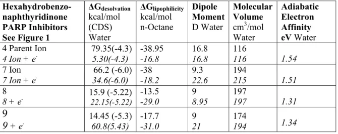
Table Hexahydrobenzo-naphthyridinone PARP Inhibitors See Figure 1 ΔGdesolvation kcal/mol (CDS) Water ΔGlipophilicity kcal/mol n-Octane Dipole Moment D Water Molecular Volume cm3/mol Water Adiabatic Electron Affinity eV Water 4 Parent Ion 4 Ion + e -79.35(-4.3) 5.30(-4.3) -38.95 -16.8 16.8 16.8 116 116 1.54 7 Ion 7 Ion + e -66.2 (-6.0) 34.6(-6.0) -38 -18.2 9.3 22.6 194 215 1.51 8 8 + e -15.9 (-5.22) 22.15(-5.22) -13.5 -29.0 9 8.95 197 197 1.31
9
9
+ e -14.45 (-5.3) 60.8(5.43) -17.7 -31.0 9 21 174 194 1.3411 11 + e -17.75(-5.28) 59.7(-5.28) -14.95 30.8 9.55 22.3 238 229 1.31 15 15 + e -25.6 (-4.85) 64.55(-4.05) -15.4 -30.1 10.4 22.0 242 247 1.42 16 16 + e -16.07 (-.54) 51.1(-5.54) -14.24 -20.3 6.6 21.0 244 204 1.34 17 17 + e -16.1 (-5.15) 51.1(-5.15) -14.1 -28.4 8.8 22.0 255 244 1.37 18 18 + e -16.6 (-5.51) 56.7(-5.51) -14.2 -29.15 9.73 21.8 216 214 1.34 20 Ion 20 Ion + e -87.7 (-8.19) 61.5(-8.19) -47.8 -28.2 42.7 37.5 302 320 1.78 29 29 + e -19.9 (-7.84) 60.2(-7.84) -18.8 -32.75 9.8 31.1 293 341 1.34 33 33 + e -20.8 (-3.98) 60.0(-3.98) -14.6 -29.2 10.1 21.0 186 196 1.34 36 36 + e -22.3 (-2.93) 54.2(-2.93) -14 -15.9 7.9 13.6 192 174 1.34 39 39 + e -14.74 (-4.36) 60.2(-4.36) -12.41 -30.6 9.1 24.2 180 175 1.31 45 Ion 45 Ion + e -78.6 (-4.33) 64.2(-4.33) -42.15 -21.0 33 50.9 237 252 1.39 PARP Inhibitors See Figure 2 (1) ZINC43206370 13.6 (-4.33) -10.1 4.25 136 (2) ZINC13860431 14.1 (-4.66) -7.2 4.4 83 (3) ZINC53297239 10.85 (-.14) -9.4 2.55 110 (4) ZINC03821234 12.6 (-5.35) -9.65 6.4 86 (5) ZINC34031449 10.05 (-.22) -6.73 5.7 91 (6) ZINC28467935 16.1 (-6.19) -11.8 2.9 175 (7) ZINC135362325 8.7 (-7.11) -10.55 10.75 145 (8) ZINC00150158 31.86 (-.66) -23.9 11.8 328 (9) ZINC01590754 66.9 (-4.8) -32.25 35.9 306 (10) ZINC02041009 57.2 (-7.2) -28.9 24.6 311 (11) ZINC08649468 32.8 (-3.93) -20.1 7 269 (12) ZINC1790819 67.55 (-.62) -31 41.3 244 (13) ZINC01512423 74.05 (-.78) -31 37.3 199 (14) ZINC00152996 52.55 (-.78) -22.3 28.85 150 PARP1 and PARP2
Inhibitors See Figure 3 Olaparib 25.7 (-8.96) -18.9 6.8 278 Veliparib 77.3 (-5.93) -39.7 23.6 185 Quinoxaline2 16.3 (-4.13) -12.1 7.8 174 Isoquinolinone3 16.1 (-7.05) -13.5 10.2 200 Isoquinolinone4 11 (-7.64) -11.9 8.3 170 Isoquinolinone5 11 (-7.64) -11.9 8.3 170 Niraparib 76.4 (-7.63) -43.3 39 216
Isoquinolinone
5-AIQ 13.9 (-4.76) -8.8 8.65 99
Phenanthridine PJ34 66.7 (-8.90) -38.3 21.2 197 Isoquinolinone DPQ 69.6 (-6.79) -40.7 27.9 202
References
[1] J.S. Brown, S.B. Kaye, T.A. Yap, PARP inhibitors: the race is on, Br. J. Cancer, 2016, 114, 713.o
[2] B. Lupo, L. Trusolino, Inhibition of poly(ADP-ribosyl)ation in cancer: Old and new paradigms revisited,Biochimica et Biophysica Acta, 2014, 1846, 201.
[3] A. Mangerich, A. Burkle, How to kill tumor cells with inhibitors of
poly(ADP-ribosyl)ation, Int. J. Cancer, 2011, 128, 251.
[4] C. Underhill, M Toulmonde, H. Bonnefoi. A review of PARP inhibitors: from bench to bedside, Annals of Oncology, 2011, 22, 268.
[5] J. Murai, S.Y. Huang, B.B. Das, A. Renaud, Y. Zhang, J.H. Doroshow, S. Takeda, Y. Pommier, Trapping of PARP1 and PARP2 by clinical PARP inhibitors, Cancer Research, 2012, 72, 5588.
[6] J.R. Steffen, R.S. Brody, J.M. Armen, J.D. Pascal, Structural implications for selective targeting of PARPs, Front. Oncol. 2013, 3, 1.
[7] A.K Halder, A. Saha, K.D. Saha, T. Jha, Stepwise development of structure-activity relationship of diverse PARP-1 inhibitors through comparative and validated in silico modeling techniques and molecular dynamics simulation, J. Biomol. Struct. Dyn. 2015, 33, 1756.
[8] E.I. Prokhorov, A.V. Bekker, A.V.Perevoznikov, M.I. Kumskov, I.V. Svitanko, Combining 3D-QSAR and molecular docking for the virtual screening of PARP inhibitors, Mendeleev Comm. 2015, 25, 214.
[9] S. Fatima, R. Bathini, S.K. Sivan, V. Manga, Molecular docking and 3D-QSAR studies on inhibitors of DNA damage signaling enzyme human PARP-1, J. Recept. Signal.
Transduct. Res. 2012, 32, 214.
[10] R.E. Salmas, A. Unlu, M Yurtsever, S.Y. Noskov, S. Durdagi, In silico investigation of PARP-1 catalytic domains in holo and apo states for the design of high-affinity PARP-1 inhibitors, Enzyme Inhib. Med. Chem. 2016, 31, 112.
[11] E.Wahlberg, T. Karlberg, E. Kouznetsova, N. Markova, A. Macchiarulo, A.G. Thorsell, et al, Family-wide chemical profiling and structural analysis of PARP and tankyrase
inhibitors, Nature Biotechnology, 2012, 30, 283.
[12] C.W. Fong, Permeability of the Blood–Brain Barrier: Molecular Mechanism of Transport of Drugs and Physiologically Important Compounds, J. Membr. Biol. 2015, 248, 651.
[13] C.W. Fong, Statins in therapy: Cellular transport, side effects, drug-drug interactions and cytotoxicity - the unrecognized role of lactones, HAL Archives, hal-01338644v2, 2016. [14] C.W. Fong, The effect of desolvation on the binding of inhibitors to HIV-1 protease and cyclin-dependent kinases: Causes of resistance, Bioorg Med Chem Lett June 2016, 26, 3705. DOI: 10.1016/j.bmcl.2016.05.080
[15] C.W. Fong, Drug discovery model using molecular orbital computations: tyrosine kinase inhibitors, HAL Archives , hal-01350862, v.1
[16] A.V. Marenich, C.J. Cramer, D.J. Truhlar, Universal Solvation Model Based on Solute Electron Density and on a Continuum Model of the Solvent Defined by the Bulk Dielectric Constant and Atomic Surface Tensions, J. Phys. Chem B, 2009, 113, 6378.
[17] R.M. Levy, L.Y. Zhang, E. Gallicchio, A.K. Felts, On the Nonpolar Hydration Free Energy of Proteins: Surface Area and Continuum Solvent Models for the Solute-Solvent Interaction Energy, J. Am. Chem. Soc. 2003, 125, 9523.
[18] P.Setny, R.Baron, J.A. McCammon, How Can Hydrophobic Association Be Enthalpy Driven? J. Chem. Theory Comput., 2010, 6, 2866.
[19] J. Mondal, R.A. Friesner, B.J. Berne, Role of Desolvation in Thermodynamics and Kinetics of Ligand Binding to a Kinase, J. Chem. Theory Comput. 2014, 10, 5696.
[20] Y. Shan, E.T. Kim, M.P. Eastwood, R.O. Dror, Seeliger, M.A.; Shaw, D.E. How Does a Drug Molecule Find its Target Binding Site? J. Am. Chem. Soc. 2011, 133, 9181.
[21] Y. Lu, R. Wang, C.Y. Yang, S. Wang, Analysis of ligand-bound water molecules in high-resolution crystal structures of protein-ligand complexes, J. Chem. Inf. Model. 2007, 47, 668.
[22] M. Adachi, T. Ohhara, K. Kurihara, T. Tamada, et al, Structure of HIV-1 protease in complex with potent inhibitor KNI-272 determined by high-resolution X-ray and neutron crystallography, Proc. Natl. Acad. Sci. USA, 2009, 106, 4641.
[23] J.M. Chen, S.L. Xu, Z. Wawrzak, Basarab, G.S. D.B. Jordan, Structure-based design of potent inhibitors of scytalone dehydratase: displacement of a water molecule from the active site, Biochemistry, 1998, 37, 17735..
[24] P.Y. Lam, P.K. Jadhav, C.J. Eyermann, C.N. Hodge, et al, Rational design of potent, bioavailable, nonpeptide cyclic ureas as HIV protease inhibitors, Science, 1994, 263, 380. [25] D. Bellocchi, A. Macchiarulo, G. Costantino, R. Pellicciari, Docking studies on PARP-1 inhibitors: insights into the role of a binding pocket water molecule, Bioorg. Med. Chem.. 2005, 13, 1151.
[26] A.T. Garcia-Sosa, S. Firth-Clark, R.L. Mancera, Including tightly-bound water molecules in de novo drug design. Exemplification through the in silico generation of poly(ADP-ribose)polymerase ligands, J. Chem. Inf. Model. 2005, 45, 624.
[27] Fong, C.W. Molecular mechanisms of cellular transport and resistance of platinum anti-cancer drugs, https://hal.archives-ouvertes.fr/hal-01330118v1
[28] C.W. Fong, Platinum based radiochemotherapies: free radical mechanisms and radiotherapy sensitizers, Free Radicals Biol. Med. 2016, 95, 216-229.
http://dx.doi.org/10.1016/j.freeradbiomed.2016.07.006
[29] C. Torrisi, M. Bisbocci, R. Ingenito, J.M. Ontoria, et al, Discovery and SAR of novel, potent and selective hexahydrobenzonaphthyridinone inhibitors of
poly(ADP-ribose)polymerase-1 (PARP-1), Bioorg. Med. Chem. Lett. 2010, 20, 448.
[30] S. Rayne, K. Forest, Accuracy of computational solvation free energies for neutral and ionic compounds: Dependence on level of theory and solvent model,Nature Proceedings, 2010, http://dx.doi.org/10.1038/npre.2010.4864.1.
[31] R.C. Rizzo, T. Aynechi, D.A. Case, I.D. Kuntz, Estimation of Absolute Free Energies of Hydration Using Continuum Methods: Accuracy of Partial Charge Models and Optimization of Nonpolar Contributions, J. Chem. Theory Comput. 2006, 2, 128.
![Figure 1 Footnote: Numbering scheme of hexahydrobenzonaphthyridinone inhibitors refers to numbering scheme in Torrisi 2010 [29]](https://thumb-eu.123doks.com/thumbv2/123doknet/12768427.360250/7.892.114.706.105.441/figure-footnote-numbering-scheme-hexahydrobenzonaphthyridinone-inhibitors-numbering-torrisi.webp)