HAL Id: hal-02617678
https://hal.inrae.fr/hal-02617678
Submitted on 25 May 2020
HAL is a multi-disciplinary open access archive for the deposit and dissemination of sci-entific research documents, whether they are pub-lished or not. The documents may come from teaching and research institutions in France or abroad, or from public or private research centers.
L’archive ouverte pluridisciplinaire HAL, est destinée au dépôt et à la diffusion de documents scientifiques de niveau recherche, publiés ou non, émanant des établissements d’enseignement et de recherche français ou étrangers, des laboratoires publics ou privés.
lysine 27 methylation
Mareike Moeller, Klaas Schotanus, Jessica Soyer, Janine Haueisen, Kathrin
Happ, Maja Stralucke, Petra Happel, Kristina M. Smith, Lanelle R. Connolly,
Michael Freitag, et al.
To cite this version:
Mareike Moeller, Klaas Schotanus, Jessica Soyer, Janine Haueisen, Kathrin Happ, et al.. Destabiliza-tion of chromosome structure by histone H3 lysine 27 methylaDestabiliza-tion. PLoS Genetics, Public Library of Science, 2019, 15 (4), �10.1371/journal.pgen.1008093�. �hal-02617678�
Destabilization of chromosome structure by
histone H3 lysine 27 methylation
Mareike Mo¨ llerID1,2, Klaas SchotanusID3, Jessica L. SoyerID4, Janine HaueisenID1,2,
Kathrin Happ1, Maja StraluckeID1, Petra Happel5, Kristina M. SmithID6, Lanelle
R. Connolly7, Michael Freitag
ID7, Eva H. StukenbrockID1,2*
1 Environmental Genomics, Christian-Albrechts University, Kiel, Germany, 2 Max Planck Fellow Group Environmental Genomics, Max Planck Institute for Evolutionary Biology, Plo¨n, Germany, 3 Department of Molecular Genetics and Microbiology, Duke University Medical Center, Durham, NC, United States of America, 4 UMR BIOGER, INRA, AgroParisTech, Universite´ Paris-Saclay, Thiverval-Grignon, France, 5 Max Planck Institute for Terrestrial Microbiology, Marburg, Germany, 6 Department of Biology, Oregon State University—Cascades, Bend, OR, United States of America, 7 Department of Biochemistry and Biophysics, Oregon State University, Corvallis, OR, United States of America
*estukenbrock@bot.uni-kiel.de
Abstract
Chromosome and genome stability are important for normal cell function as instability often correlates with disease and dysfunction of DNA repair mechanisms. Many organisms main-tain supernumerary or accessory chromosomes that deviate from standard chromosomes. The pathogenic fungus Zymoseptoria tritici has as many as eight accessory chromosomes, which are highly unstable during meiosis and mitosis, transcriptionally repressed, show enrichment of repetitive elements, and enrichment with heterochromatic histone methylation marks, e.g., trimethylation of H3 lysine 9 or lysine 27 (H3K9me3, H3K27me3). To elucidate the role of heterochromatin on genome stability in Z. tritici, we deleted the genes encoding the methyltransferases responsible for H3K9me3 and H3K27me3, kmt1 and kmt6, respec-tively, and generated a double mutant. We combined experimental evolution and genomic analyses to determine the impact of these deletions on chromosome and genome stability, both in vitro and in planta. We used whole genome sequencing, ChIP-seq, and RNA-seq to compare changes in genome and chromatin structure, and differences in gene expression between mutant and wildtype strains. Analyses of genome and ChIP-seq data in H3K9me3-deficient strains revealed dramatic chromatin reorganization, where H3K27me3 is mostly relocalized into regions that are enriched with H3K9me3 in wild type. Many genome rear-rangements and formation of new chromosomes were found in the absence of H3K9me3, accompanied by activation of transposable elements. In stark contrast, loss of H3K27me3 actually increased the stability of accessory chromosomes under normal growth conditions
in vitro, even without large scale changes in gene activity. We conclude that H3K9me3 is
important for the maintenance of genome stability because it disallows H3K27me3 in regions considered constitutive heterochromatin. In this system, H3K27me3 reduces the overall stability of accessory chromosomes, generating a “metastable” state for these quasi-essential regions of the genome.
a1111111111 a1111111111 a1111111111 a1111111111 a1111111111 OPEN ACCESS
Citation: Mo¨ller M, Schotanus K, Soyer JL,
Haueisen J, Happ K, Stralucke M, et al. (2019) Destabilization of chromosome structure by histone H3 lysine 27 methylation. PLoS Genet 15 (4): e1008093.https://doi.org/10.1371/journal. pgen.1008093
Editor: Hiten D. Madhani, University of California
San Francisco, UNITED STATES
Received: November 10, 2018 Accepted: March 15, 2019 Published: April 22, 2019
Copyright:© 2019 Mo¨ller et al. This is an open access article distributed under the terms of the
Creative Commons Attribution License, which permits unrestricted use, distribution, and reproduction in any medium, provided the original author and source are credited.
Data Availability Statement: Sequencing raw
reads (FASTQ files) of all genomic, ChIP-seq and RNA-seq data are available online at Sequence Read Archive (SRA) under BioProject ID PRJNA494102.
Funding: Research in the lab of EHS on histone
modifications in Zymoseptoria tritici is funded by a fellowship from the Max Planck Society to EHS. Work on H3 lysine 27 methylation in the lab of MF was supported by NSF grants (MCB 1515998 and MCB1818006).The funders had no role in study
Author summary
Genome and chromosome stability are essential to maintain normal cell function and via-bility. However, differences in genome and chromosome structure are frequently found in organisms that undergo rapid adaptation to changing environmental conditions, and in humans are often found in cancer cells. We study genome instability in a fungal patho-gen that exhibits a high degree of patho-genetic diversity. Regions that show extraordinary diver-sity in this pathogen are the transposon-rich accessory chromosomes, which contain few genes that are of unknown benefit to the organism but maintained in the population and thus considered “quasi-essential”. Accessory chromosomes in all fungi studied so far are enriched with markers for heterochromatin, namely trimethylation of H3 lysine 9 and 27 (H3K9me3, H3K27me3). We show that loss of these heterochromatin marks has strong but opposing effects on genome stability. While loss of the transposon-associated mark H3K9me3 destabilizes the entire genome, presence of H3K27me3 favors instability of accessory chromosomes. Our study provides insight into the relationship between chro-matin and genome stability and why some regions are more susceptible to genetic diver-sity than others.
Introduction
Chromatin structure plays an important role in genome organization and gene expression [1–
3]. A well-studied hallmark of epigenetic regulation is the reversible modification of histone tails, which can alter chromatin structure [4]. Chromatin structure determines accessibility of the underlying DNA to regulatory elements, whereby tightly packed DNA, known as hetero-chromatin, is less accessible for DNA binding proteins and usually shows little transcriptional activity [5]. Heterochromatic regions often cluster together and are spatially separated from more transcriptionally active and accessible euchromatic regions [6]. Specific histone modifi-cations are associated with either heterochromatic or euchromatic regions. Some of the most studied histone modifications are histone H3 lysine 9 di- or trimethylation (H3K9me2/3) and H3K27me2/3 as markers for heterochromatin and H3K4me2/3 as markers for euchromatin [7].
H3K9me2/3 is catalyzed by the histone methyltransferase KMT1 (Su[var]3–9) [8,9], in fungi also called Clr4 [10] or DIM-5 [11]. Previous studies demonstrated enrichment of this constitutive heterochromatin mark in repeat-rich regions and a clear link with the control of transposable elements (TE) and genome stability [12–14]. For example, H3K9 methylation has been shown to be involved in suppression of meiotic recombination inArabidopsis thaliana
[15] and the control of DNA methylation inNeurospora crassa [11].
H3K27me2/3, usually associated with “facultative heterochromatin”, is catalyzed by KMT6 (E[Z]) as part of the PRC2 complex [16]. In plants, fungi, and animals, this histone mark is used to generate “transcriptional memory” and is easily reversible when environmental or endogenous stimuli require organismal responses. In many organisms, H3K27 methylation is required for development and cell differentiation [17–23], and aberrant H3K7me3 distribution is prevalent in cancer cells [24–26]. In fungi, H3K27me3 correlates with subtelomeric gene silencing [22,23,27], and has been shown to play a role in development, pathogenicity, and transcriptional regulation of secondary metabolite gene clusters [21,28,29].
H3K27me3 is also a hallmark of accessory chromosomes, which are found in several fungal plant pathogens [28,30,31]. Accessory chromosomes are not essential for survival under all
design, data collection and analysis, decision to publish, or preparation of the manuscript.
Competing interests: The authors have declared
environmental conditions, and thus encode “quasi-essential” genes [32] that can confer selec-tive advantages under some conditions e.g. in a specific host species, resulting in presence or absence of these chromosomes among specific individuals of a given species. They are also characterized by extensive structural rearrangements and length variation [33,34]. In some species (Fusarium oxysporum, Nectria haematococca, Alternaria alternata), accessory
chromo-somes increase virulence [35–38]. However, in the wheat pathogenZymoseptoria tritici, some
accessory chromosomes have been demonstrated to confer reduced fitness and virulencein planta [39], suggesting that there are other stages in the life cycle when they become important. Accessory chromosomes of fungi differ structurally from core chromosomes by higher repeat and lower gene density compared to core chromosomes and show little transcriptional activity [35,40–43]. Transcriptional silencing can be explained by their predominantly heterochro-matic structure, with H3K27me3 enrichment on almost the entire chromosome and H3K9me3 covering repetitive sequences [28,30]. Centromeres and telomeres are important structural components of chromosomes. In plants, centromeres of B chromosomes, equiva-lents to fungal accessory chromosomes, differ from those of A chromosomes [44], but inZ. tri-tici centromeres, telomere repeats, and subtelomeric regions are so far by all measures near
identical on core and accessory chromosomes [31]. Though accessory chromosomes are a fre-quent phenomenon in fungi, little is known about their origin and maintenance. Studies on chromosome stability revealed that accessory chromosomes are highly unstable, both during mitosis [36,45,46] and meiosis [47].
Here, we investigated to what extent the particular histone methylation pattern on acces-sory chromosomes contributes to the structural differences, transcriptional repression and instability. We shed light on the roles of H3K9me3 and H3K27me3 on genome stability in the hemi-biotrophic wheat pathogenZ. tritici that reproduces both asexually and sexually. By
combining experimental evolution with genome, transcriptome and ChIP sequencing, we show that both heterochromatin-associated histone methylation marks contribute signifi-cantly, but in distinct ways, to chromosome stability and integrity. While the presence of H3K27me3 enhances chromosome loss and instability, loss of H3K9me3 promotes chromo-some breakage, segmental duplications as well as the formation of new chromochromo-somes− possi-bly resembling the emergence of accessory chromosomes. Taken together, our findings demonstrate the importance of constitutive heterochromatin for maintaining genome stability and gene silencing as well as an unexpected destabilizing influence of facultative heteromatin on mitotic accessory chromosome transmission. The presence of eight accessory chro-mosomes in the reference isolate IPO323 makesZ. tritici an excellent model to study accessory
chromosome characteristics and dynamics, which relates to general interest in chromosome maintenance in cancer or other aneuploid cell types.
Results
Deletion of histone methyltransferase encoding genes
kmt1 and kmt6 in
Zymoseptoria tritici
To investigate the impact of heterochromatin on fitness, transcription and genome stability in
Z. tritici, we generated mutants of two histone methyltransferases Kmt1 (S. pombe Clr4; N. crassa DIM-5, Fusarium KMT1, H. sapiens SUV39H1) and Kmt6 (N. crassa SET-7; Fusarium
KMT6;H. sapiens EZH2). We identified the Z. tritici genes by BLAST searches with the N. crassa and F. graminearum protein coding sequences as baits. Kmt1 is encoded by kmt1
(Zt_chr_1_01919), and Kmt6 is encoded bykmt6 (Zt_chr_4_00551) [48]. We used Agrobacter-ium tumefaciens-mediated transformation [49] to delete both genes in a derivate of theZ. tri-tici reference isolate IPO323 that lost chromosome 18 during in vitro growth, here called Zt09
[31,40,41]. Correct integration of thehph gene, which confers hygromycin resistance [49], and
kmt1 or kmt6 deletion were verified by PCR and Southern analyses (S1andS2Figs). We gen-erated a double deletion mutant by deleting thekmt1 gene in a kmt6 deletion mutant
back-ground by using resistance to nourseothricin conferred by thenat gene [50] as an additional selection marker. We isolated several independent transformants, including eightΔkmt1, six
Δkmt6 and ten Δkmt1 Δkmt6 double mutants (from here on abbreviated Δk1/k6). For further
studies we selected two or three mutants of each type (S1 Table).Δkmt1 and Δkmt6 single mutants were complemented by re-integrating the previously deleted gene and aneo+
resis-tance marker that can confer G418 resisresis-tance at the native gene loci (S2 Fig).
We performed ChIP-seq on Zt09,Δkmt1 (Zt125-#68, -#80), Δkmt6 (Zt110-#283, -#285, -#365) and the double deletion mutantΔk1/k6 (Zt219-#23, -#116), which verified the absence of H3K9me3 inΔkmt1 and Δk1/k6, and the absence of H3K27me3 in Δkmt6 and Δk1/k6 mutants (S3 Fig), confirming that Kmt1 and Kmt6 are the only histone methyltransferases in
Z. tritici responsible for H3K9 and H3K27 trimethylation, respectively. An overview of the
subsequent experiments and key results are summarized inS1 Fig.
Deletion of
kmt1, but not kmt6, severely impacts in vitro and in planta
growth
To assess if deletion ofkmt1 and kmt6 has an impact on in vitro growth or pathogenicity on
wheat, we performed comparative growth and virulence assays comparing the mutants to the wild type Zt09. To compare growth rates, the reference strain Zt09, deletion and comple-mented strains were grown in liquid YMS cultures and the OD600was measured until cells reached stationary phase. Overall, theΔkmt1 strains and Δk1/k6 double deletion mutants showed significantly reduced growthin vitro (S4 Fig). TheΔkmt6 mutants and both kmt1+ andkmt6+complementation strains showed no significant differences in growth compared to Zt09 (Wilcoxon rank-sum test,p-values: Δkmt1 0.025; Δkmt6 0.42; Δk1/k6 0.005; kmt1+0.28;
kmt6+0.63).
We furthermore assessed the tolerance of theΔkmt1, Δkmt6 and Δk1/k6 mutants to abiotic stressin vitro by testing temperature, cell wall, oxidative, and genotoxic stressors. As observed
in the growth assays, theΔkmt1 and Δk1/k6 double deletion mutants showed overall reduced growth under all tested conditions (S5 Fig), especially under osmotic stress induced by high sorbitol concentrations. TheΔkmt6 mutants showed little differences compared to Zt09; how-ever, elevated temperatures often, but not always, led to increased melanization in theΔkmt6 mutants suggesting involvement of H3K27me3 in the response to temperature stress. This phenotype was reversed in the complementedkmt6+strain (S6 Fig).
To study the effect of the histone methyltransferase deletions on the ability to infect wheat (Triticum aestivum), we inoculated leaves of the susceptible cultivar Obelisk with single cell
cultures ofΔkmt1, Δkmt6, the Δk1/k6 double deletion mutant and Zt09. The infection assays demonstrated significant impact of both H3K27me3 and H3K9me3 on virulence. While the number of pycnidia and necrotic leaf areas only decreased in theΔkmt6 mutants, wheat infec-tion byΔkmt1 and Δk1/k6 mutants resulted in almost no symptoms (S7 Fig). If any symptoms developed, these appeared considerably later than symptoms caused by the reference Zt09 and theΔkmt6 mutants (S7 Fig).
Loss of H3K9me3 allows H3K27me3 to invade repeat-rich regions
We next addressed how the deletion ofkmt1 and kmt6 impacts the distribution of three
his-tone modifications (H3K4me2, H3K9me3, H3K27me3) by ChIP-seq (S2 Table). We previ-ously found that H3K4me2 is associated with gene-rich, transcriptionally active regions on
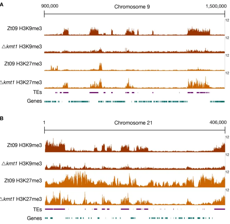
core chromosomes, that constitutive heterochromatin, enriched with H3K9me3, forms almost exclusively on repetitive elements, and that facultative heterochromatin, enriched with H3K27me3, forms nearly on the entire length of all accessory chromosomes and the subtelo-meric regions of core chromosomes [31] (Fig 1).
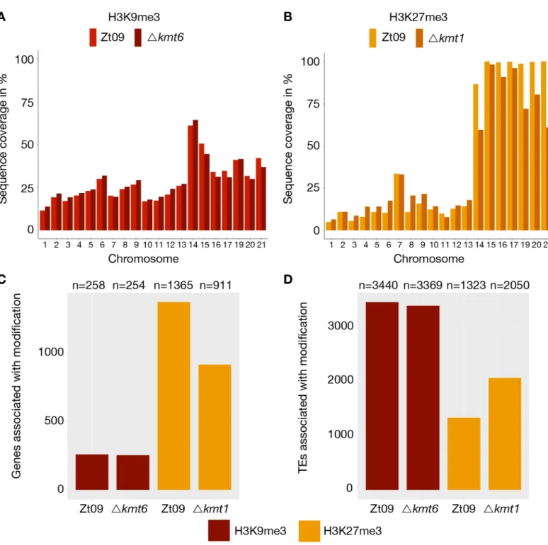
We computed the sequence coverage of each histone modification per chromosome to esti-mate the global effects on chromatin structure. The absence of one histone methylation mark had differential effects on the distribution of the other two methylation marks on core and accessory chromosomes (Fig 1,Table 1). In theΔkmt1 mutants, the amount of sequences enriched with H3K27me3 decreases on the accessory chromosomes when compared to Zt09, representing the opposite trend to the observations made on the core chromosomes, where we observed an increased amount of sequences enriched in H3K27me3 (Fig 1,Table 1). However, this effect varies on different accessory chromosomes (Table 1,Fig 2). The difference in H3K27me3 distribution can be explained by relocation of H3K27me3 to former H3K9me3-as-sociated sequences in theΔkmt1 mutant (Fig 1). While fewer genes are associated with H3K27me3 (Fig 2), more TEs show H3K27me3 enrichment in theΔkmt1 mutant (Fig 2) com-pared to Zt09. These observations reveal that loss of H3K9me3 promotes H3K27me3 reloca-tion to TEs and confers simultaneous loss of H3K27me3 at posireloca-tions with this histone mark in the reference strain. The subtelomeric H3K27me3 enrichment, however, is not affected by this relocation, which explains why we observe opposite effects on core and accessory chromo-somes, as core chromosomes predominantly show H3K27me3 enrichment in subtelomeric regions while accessory chromosomes show overall enrichment with H3K27me3. H3K4me2 increases on both core and accessory chromosomes, with accessory chromosomes showing a considerably higher relative increase compared to H3K4me2 in Zt09 (Table 1).
Conversely, H3K9me3 is not affected by loss of H3K27me3 in theΔkmt6 mutants, and we did not detect relocation of H3K9me3 as well as only minor differences in coverage. H3K4me2 enrichment does increase on accessory chromosomes, but not to the same extent as observed in theΔkmt1 mutants and it slightly decreases on core chromosomes (Table 1), suggesting minor effects ofΔkmt6 on transcriptional activation. In the Δk1/k6 double deletion mutants, where both H3K9me3 and H3K27me3 are not present, we detected an increase in H3K4me2, similar to theΔkmt1 single mutants on core chromosomes and slightly higher on the accessory chromosomes.
In summary, loss of H3K9me3 has a great impact on H3K27me3 distribution, while loss of H3K27me3 has little influence on H3K9me3. Deletion ofkmt1 promotes large scale
relocaliza-tion of other histone modificarelocaliza-tions, suggesting more dramatic effects on genome organizarelocaliza-tion and transcriptional activation than deletion ofkmt6.
H3K27me3 has little effects on transcriptional activation, while loss of
H3K9me3 enhances activation of TEs
In other species, H3K27me3 plays a crucial role in gene regulation, while H3K9me3 is involved in silencing of TEs [13,21,28]. Based on our observations from ChIP-seq data, we hypothesized that the two histone methylation marks have similar effects inZ. tritici. To test this hypothesis
directly, we sequenced transcriptomes of two biological replicates of Zt09 and two indepen-dent transformants of theΔkmt1, Δkmt6, and Δk1/k6 deletion mutants after in vitro growth for 2 days representing exponential growth (S2 Table).
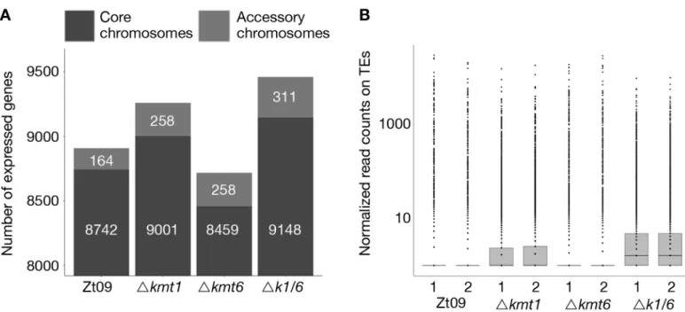
First, we compared the total number of expressed genes. In total, 11,839 genes are anno-tated in the reference isolate [48]. Out of these, 8,906 are expressed (RPKM >2) in Zt09 during
in vitro growth. The number of expressed genes is higher in both the Δkmt1 (9,259) and the
Fig 1. ChIP-seq reveals relocation of H3K27me3 on core (A) and accessory (B) chromosomes inΔkmt1 mutants. By analyzing ChIP-seq data in the Δkmt1 mutants
we found that enrichment of H3K27me3 moves to sequences that are normally enriched with H3K9me3. A region on core chromosome 9 (A) is shown, where H3K27me3 is strongly enriched at former H3K9me3 regions, but depleted from its original positions. On accessory chromosomes (B), here full-length chromosome 21 as an example, there are similar dynamics as observed on core chromosomes. Accessory chromosomes normally show overall enrichment of H3K27me3. In absence of H3K9me3, H3K27me3 concentrates on former H3K9me3 regions, again being depleted from its original position. However, this effect varies between accessory chromosomes (Fig 2). The low amount of background found inΔkmt1 is due to the repetitive nature of the H3K9me3-enriched regions. All shown ChIP-seq tracks are normalized to 1x coverage (coverage indicated on the right) [107].
Table). This is in contrast to previous studies, where deletion ofkmt6 resulted in activation of
otherwise silenced gene clusters and overall transcriptional activation [21,23,27,28]. We focused on differential gene expression between core and accessory chromosomes because genes on accessory chromosomes are silent under most conditions that have been tested. While 80% of genes on core chromosomes are expressed in Zt09, only ~25% of genes located on accessory chromosomes display transcriptional activity. Transcription of genes on acces-sory chromosomes is higher in all mutant strains, ~40–50% (Fig 3A,S3 Table), revealing gene activation on accessory chromosomes specifically upon removal of H3K27me3 or H3K9me3.
We further explored patterns of differential gene expression. Genome wide, 1,365 predicted genes were associated with H3K27me3 and 258 genes with H3K9me3 in Zt09 and the vast majority of these genes shows little transcriptional activity. Interestingly, only a small fraction of genes associated with these histone marks were activated or differentially expressed in the mutants (S4 Table). This indicates that loss of any of these methylation marks is not sufficient for transcriptional activation suggesting additional mechanisms involved in the transcriptional regulation of these genes.
In other fungi, removal of H3K9me3 and especially H3K27me3 was linked to the activation of certain gene classes, in particular secondary metabolite gene clusters [21,28,29]. To assess if genes with a specific function are enriched amongst the activated genes, we performed Gene Ontology (GO) enrichment analysis (topGO, Fisher’s exact test,p-value < 0.01). Consistent
with the higher total number of expressed genes, we found the majority of differentially expressed (DE) genes (DESeq2,Padj< 0.001, log2 fold-change > 2) to be significantly upregu-lated in theΔkmt1 mutant (365 of 477) and in the Δk1/k6 mutant (368 of 477), whereas a majority of DE genes was downregulated in theΔkmt6 mutant (188 of 310) (S5 Table).
We found two GO categories enriched amongst upregulated genes inΔkmt1 and Δk1/k6 mutants: DNA integration (GO:0015074) and RNA-dependent DNA replication
(GO:0006278). Predicted functions assessed by BLAST analyses of the proteins encoded by the upregulated genes in these categories include reverse transcriptases, integrases, recombinases and genes containing transposon- or virus-related domains (S6 Table). Consistent with these findings, we detected an increased number of transcripts originating from annotated TEs in theΔkmt1 and Δk1/k6 mutants, but not in Δkmt6 mutants (Fig 3B). This is in agreement with the strong association of TEs with H3K9me3 [31]. Transposons in subtelomeric regions and on accessory chromosomes show additional H3K27me3 enrichment. Removal of H3K9me3, but not of H3K27me3, appears to be responsible for transposon activation but transcription is
Table 1. Percentage of sequence coverage (significantly enriched regions) of core and accessory chromosomes with H3K4me2, H3K9me3 and H3K27me3 relative to the chromosome length. Minimum and maximum values refer to the chromosomes showing highest or lowest sequence coverage with enrichment of the respective
his-tone modification. H3K4me2 coverage on accessory chromosomes increases in all mutant strains, while there are little differences in the overall coverage with H3K9me3 between Zt09 andΔkmt6. H3K27me3 enrichment increases on core chromosomes and decreases on accessory chromosomes in the Δkmt1 mutant.
Core Accessory
Modification mean Min max mean min max
Zt09 H3K4me2 23.99 17.76 31.26 5.03 1.19 9.44 4kmt1 H3K4me2 25.25 18.51 32.78 9.68 5.67 17.32 4kmt6 H3K4me2 22.74 16.83 29.54 6.22 3.32 10.97 4k1/k6 H3K4me2 25.39 19.11 32.69 12.28 8.47 19.96 Zt09 H3K9me3 20.14 10.84 28.71 41.03 30.78 55.97 4kmt6 H3K9me3 20.30 11.24 28.27 36.59 26.71 55.74 Zt09 H3K27me3 9.74 3.74 31.72 92.53 67.07 99.20 4kmt1 H3K27me3 14.99 6.08 32.77 77.55 56.44 96.82 https://doi.org/10.1371/journal.pgen.1008093.t001
further enhanced when both, H3K27me3 and H3K9me3 are removed in theΔk1/k6 mutant (Fig 3B;S7 Table).
Fig 2. Genome-wide distribution of H3K9me3 and H3K27me3 in Zt09 and mutants. (A) and (B) display the percentage of sequence coverage of core and accessory
chromosomes with H3K9me3 and H3K27me3 relative to the chromosome length. While there were little differences in the overall coverage with H3K9me3 between Zt09 andΔkmt6 (A), H3K27me3 enrichment was increased on core chromosomes and decreased on accessory chromosomes in the Δkmt1 mutant (B). Chromosome 7 displayed a higher H3K27me3 coverage compared to the other core chromosomes as the right arm showed characteristics of an accessory chromosome [31]. (C and D) Genes (C) and TEs (D) associated with H3K9me3 or H3K27me3 in Zt09 and mutant strains. While there was almost no difference in terms of H3K9me3-associated genes or TEs in theΔkmt6 mutants, H3K27me3 relocated from genes to TEs in the Δkmt1 mutants.
Amongst the genes upregulated in theΔkmt6 mutant no GO categories were enriched but based on the previous finding of secondary metabolite activation, we further investigated pos-sible roles of H3K9me3 and H3K27me3 in secondary metabolite gene regulation. Therefore, we identified putative secondary metabolite clusters in theZ. tritici reference genome using
antiSMASH (antibiotics & Secondary Metabolite Analysis SHell) [51]. We found a total of 27 secondary metabolite clusters, all located on core chromosomes, and merged the identified genes with the existing gene annotation (S8 Table). Except for the activation of one putative cluster on chromosome 7 in theΔk1/k6 mutant, we did not identify any differential expression of genes in secondary metabolite clusters. Based on these findings, we conclude that, unlike in other fungi [21,29], H3K9me3 and H3K27me3 are not involved in transcriptional regulation of secondary metabolites inZ. tritici under the tested conditions.
Taken together, removal of these histone modifications has little consequences for the expression of the vast majority of associated genes. As expected from its localization, loss of H3K9me3 increases expression of TEs while absence of H3K27me3 by itself has very little impact on transcriptional activation, thus suggesting that in this organism H3K27me3 does not delineate stereotypical “facultative heterochromatin” as removal of H3K27me3 does not activate gene expression.
Loss of H3K27me3 drastically reduces the loss of accessory chromosomes
Chromosome landmarks, namely centromeric and pericentric regions, telomere repeats and subtelomeric regions are similar on core and accessory chromosomes inZ. tritici [31]. Acces-sory chromosomes are enriched with TEs but share the same TE families as core chromosomes [48]. Nevertheless, accessory chromosomes ofZ. tritici are highly unstable, both during
Fig 3. Gene (A) and transposon (B) expression increases in absence of H3K9me3, while loss of H3K27me3 alone decreases the number of expressed genes and does not impact transposon activity. (A) We compared the number of expressed genes in Zt09 and mutant strains. While in all mutants the number of expressed genes
increased on accessory chromosomes, surprisingly loss of H3K27me3 alone in theΔkmt6 mutants resulted in a reduction of genes expressed on core chromosomes and only a small increase in numbers of genes expressed on accessory chromosomes. (B) Loss of H3K9me3, but not H3K27me3 alone, increased the number of transcripts originating from TEs. In absence of both marks (Δk1/k6), the number further increased, likely because H3K27me3 moves to TEs in the Δkmt1 single mutant, facilitating silencing.
meiosis and vegetative growthin vitro and in planta [46,47]. The most striking feature that sets these chromosomes apart is almost chromosome-wide enrichment with H3K27me3 and, as a consequence of the higher TE content, increased enrichment with H3K9me3 [31]. To test whether loss of these modifications affects genome and chromosome stability inZ. tritici, we
conducted two different long-term growth or “lab evolution” experiments to study genome stability and to detect dynamics of accessory chromosome losses in strains deficient for two important chromatin marks (S8 Fig).
To assess whether the specific histone methylation pattern on accessory chromosomes con-tributes to instability of accessory chromosomes, we performed a short-termin vitro growth
experiment over four weeks, representing ~80 asexual generations. Zt09,Δkmt6, Δkmt1 and a Δk1/k6 double deletion mutant, as well as the complemented strains kmt1+
andkmt6+were used as progenitors in the experiment. The presence of all accessory chromosomes in the pro-genitor strains was verified by PCR at the beginning of the experiment. Each strain was grown in three replicate cultures and ~4% of the cell population was transferred to fresh medium every three to four days. After four weeks of growth, we plated dilutions of each culture to obtain single colonies that were subsequently screened by a PCR assay for the presence of accessory chromosomes (Table 2).
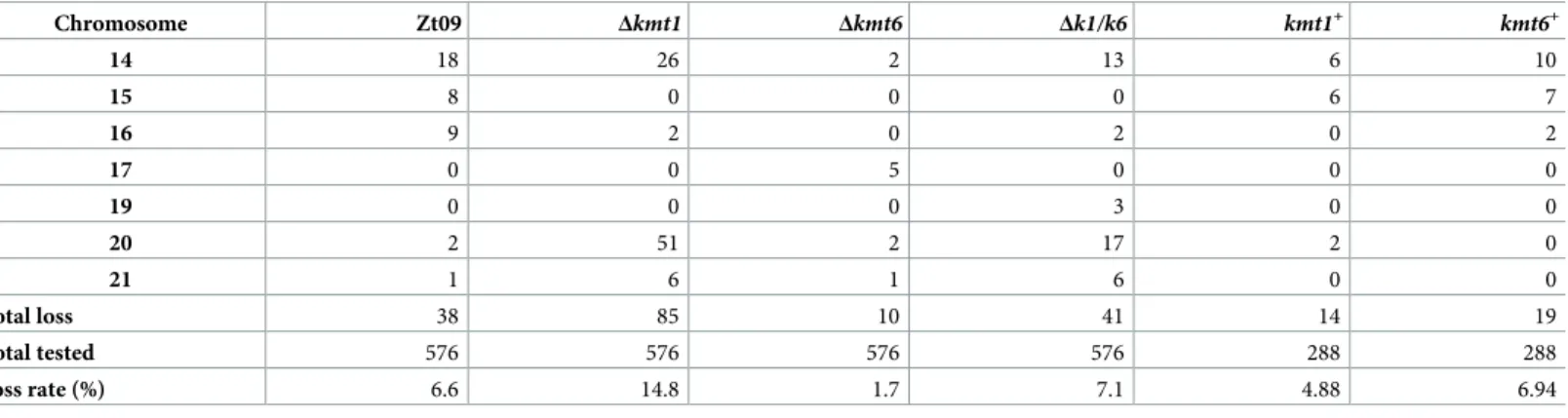
Previously, we showed that accessory chromosomes are lost at a rate of ~7% in Zt09 and we documented that accessory chromosomes 14, 15 and 16 are more frequently lost than others [46]. Here we demonstrate that, in comparison to Zt09, theΔkmt1 mutant showed a signifi-cantly increased chromosome loss rate (one sided Fisher’s exact test for count data,
p-value = 2.7 x 10⁻⁶). Interestingly, this was not due to an overall increase of accessory chromo-some loss, but rather by the dramatically increased (Fisher’s exact test,p-value = 3.7 x 10⁻⁹) fre-quency of loss for chromosome 20 (Table 2). The chromosome loss rate of the other accessory chromosomes was either comparable to Zt09 (Chr. 14, 17, 19, 21) or even significantly lower (Chr. 15 and 16, Fisher’s exact test,p-values = 1.2 x 10⁻³and 2.5 x 10⁻⁵). This suggests a special role of H3K9me3 for the maintenance of chromosome 20.
In contrast to theΔkmt1 mutants, we detected significantly fewer chromosome losses (Fish-er’s exact test,p-value = 1.2 x 10⁻⁴) in theΔkmt6 mutants. Out of 576 tested colonies, only ten had lost an accessory chromosome. This represents a four times lower chromosome loss rate compared to wild type. Therefore, absence of H3K27me3 appears to promote stability of acces-sory chromosomes. Interestingly, chromosome 17 was lost with the highest frequency in this mutant (5/10) but was not lost in any of the other mutant strains or in the wild type.
Table 2. Chromosome loss rates and frequency of individual accessory chromosome losses in the Zt091reference strain and mutants during short-term evolution
experiments. Three replicate cultures were tested per strain.
Chromosome Zt09 Δkmt1 Δkmt6 Δk1/k6 kmt1+ kmt6+ 14 18 26 2 13 6 10 15 8 0 0 0 6 7 16 9 2 0 2 0 2 17 0 0 5 0 0 0 19 0 0 0 3 0 0 20 2 51 2 17 2 0 21 1 6 1 6 0 0 total loss 38 85 10 41 14 19 total tested 576 576 576 576 288 288 loss rate (%) 6.6 14.8 1.7 7.1 4.88 6.94
1Strain Zt09 had previously spontaneously lost chromosome 18 [41].
The double deletion mutant displayed a similar chromosome loss rate as wild type but showed a chromosome loss distribution comparable to theΔkmt1 deletion strain with chro-mosome 20 being lost significantly more often (Fisher’s exact test,p-value = 1.23 10−4), and chromosomes 15 and 16 lost less frequently (Fisher’s exact test,p-values = 1.95 x 10⁻³and 9 x 10⁻³, respectively). This suggests that the increase in chromosome stability inΔk1/k6 com-pared toΔkmt1 is due to the removal of the destabilizing H3K27me3.
We considered possible reasons for the high rates of loss of chromosome 20 inΔkmt1 and Δk1/k6 mutant strains by a detailed analysis of TE content, genes and histone methylation redistribution on this chromosome. The proportion of TEs on chromosome 20 is ~23% and thereby considerably lower than the average across the eight accessory chromosomes (33.6% TE content). Consistent with patterns found on the other accessory chromosomes we find no enrichment of specific TE families [48]. Out of the 91 genes on chromosome 20, none has known or even predicted functions; it is therefore difficult to correlate potential gene functions of any of these genes to the enhanced loss rate. Chromosome 20 is one of the chromosomes that exhibits the highest extent of H3K27me3 redistribution (Fig 2) and loss of H3K27me3 in theΔk1/k6 double mutant decreases the high loss rate. However, chromosome 20 is still lost at a higher rate inΔk1/k6 compared to Zt09, indicating that other, so far unknown, factors are involved in this instability. Moreover, not all accessory chromosomes that exhibit H3K27me3 redistribution (19 and 21) are lost at higher rates, and not all accessory chromosomes are lost at the same rate in the Zt09 wild type, despite being enriched with H3K27me3, further suggest-ing that additional mechanisms are involved in the instability of chromosome 20.
Both complemented strains,kmt1+andkmt6+, showed chromosome loss rates similar to the wild type strain Zt09 and the highest loss rates for the largest accessory chromosomes (Table 2). This strongly suggests that indeed the absence of the two histone methyltransferases and the respective histone marks influence accessory chromosome dynamics.
In summary, we found that loss of H3K27me3 increases accessory chromosome stability, suggesting a mechanistic explanation for how the widespread H3K27me3 enrichment on accessory chromosomes in normal cells contributes to the previously observed extraordinary chromosome instability.
Loss of H3K9me3 promotes large-scale structural rearrangements
mediated by TE instability and redistribution of H3K27me3
In a second evolution experiment, we addressed overall genome stability over a longer period of mitotic growth. The single mutants (Δkmt1 and Δkmt6) and Zt09 were grown in triplicate cultures for ~6 months, representing ~500 asexual generations. We sequenced full genomes of progenitors and the evolved populations after 50 transfers to identify structural variations that arose during the experiment. All strains were sequenced to ~100x coverage by Illumina sequencing, and paired-end reads were mapped to the reference genome of IPO323 and nor-malized to 1x coverage for visualization [40].
We focused our analysis on large scale chromosomal rearrangements such as duplications, deletions, and translocations. Structural variation was detected computationally from sequence alignments, validated experimentally by PFGE and Southern blotting, and additional rear-rangements were identified by manual screening of mapped reads. Analysis of progenitor genomes revealed, except for the already known absence of chromosome 18 [41] and the previ-ously described variations (point mutations and short indels) in Zt09 compared to the IPO323 reference genome [46], lower sequence coverage (~0.6x) on chromosome 17 in theΔkmt6 pro-genitor strain (Fig 4A). This difference can only be explained by a lower copy number in the
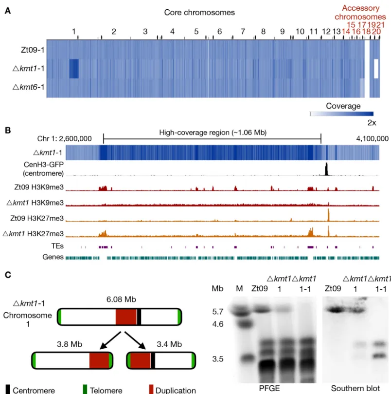
Fig 4. Genome sequencing of progenitor strains for the long-term growth experiment and analysis of structural variation in theΔkmt1 progenitor. (A) The genomes
of progenitor strains were sequenced and reads were mapped to the reference genome. Genome coverage was normalized to 1x coverage to allow identification and comparison of differences within and between strains. All strains were missing chromosome 18, as expected [41].Δkmt6 had lower coverage (0.4x) of chromosome 17. Δkmt1 lost chromosome 20 and, most notably, showed a long segment (~ 1Mb) of high-coverage (1.6x) on chromosome 1. Centromeres are indicated as black dots. (B) Examination of the high-coverage region breakpoints on chromosome 1. The first breakpoint located within a TE-rich region that is enriched with H3K9me3 in Zt09 and showed new enrichment with H3K27me3 inΔkmt1. The second breakpoint is within a gene-rich region in close proximity to relocalized H3K27me3 and very close to the centromere (~15 kb). (C) Further analysis of this high-coverage region revealedde novo telomere formation at the breakpoints indicating a chromosome breakage at both ends of the high-coverage region. To validate chromosome breakage and possible new chromosome formation, we conducted PFGE and separated the large chromosomes
sequenced pool of cells, suggesting loss of chromosome 17 in ~40% of the sequencedΔkmt6 cells, a chromosome loss that likely occurred at the very beginning of the experiment.
Unexpectedly, theΔkmt1 progenitor displayed a long high-coverage (~1.6x) region on chromosome 1 (Fig 4), suggesting that the region had been duplicated in ~60% of the sequencedΔkmt1 cells. Furthermore, this genome has a shorter chromosome 6 and does not contain chromosome 20 (Fig 4A,S9 Table). The presence of this kind of structural variation in the progenitor strain is indicative for a high degree of genome instability in absence of Kmt1. Analysis of discordant reads mapped to both ends of the ~1 Mb high-coverage region on chro-mosome 1 revealed telomeric repeats (TTAGGGn), suggesting the formation ofde novo telo-meres. Pulsed-field gel electrophoresis (PFGE) and Southern analyses confirmed the formation of two new independent chromosomes both containing the high-coverage region and either the right or left arm of chromosome 1 (Fig 4C). The breakpoint on the left side coin-cides with a large TE-rich region that is associated with H3K9me3 in the wild type. Both break-points coincide with or are in close proximity to regions that show enrichment of relocated H3K27me3 in theΔkmt1 mutant (Fig 4B), suggesting a possible link between relocated H3K27me3 and genome instability.
After six months of vegetative growth, we sequenced the pooled genomes of all nine ‘evolved’ populations. We found no evidence for large-scale genomic rearrangements in any of the evolved Zt09 orΔkmt6 populations (Fig 5A). Apart from seven small deletions or duplica-tions (S9 Table), the largest structural variation found in one of the evolvedΔkmt6 populations (Δkmt6 50–2), was a partial loss (~18 kb) at the right end of chromosome 15. However, we found variation in the read coverage of accessory chromosomes in all sequenced genomes indicating whole chromosome losses in individual cells of the sequenced population. The dis-tinct dynamics of individual accessory chromosome losses were described in the previous sec-tion as part of the short-term growth results (Table 2).
In contrast to the few variations detected in the Zt09 andΔkmt6 populations, we found numerous large-scale high-coverage regions on different core chromosomes, chromosome breakages followed byde novo telomere formation, chromosomal fusions, as well as several
smaller deletions and duplications in the evolvedΔkmt1 populations (Fig 5A,S9 Table). All three evolvedΔkmt1 populations have large duplicated regions on chromosome 1 (Fig 6), but their locations as well as the resulting structural variations differ from the one identified in the progenitor strain (Fig 6A). This can be explained by independent events, as not allΔkmt1 pro-genitor cells underwent the rearrangement of chromosome 1 (Fig 4C), or by continuous struc-tural rearrangement events as a consequence of the presence of large duplicated regions in the genome. Analyses of the affected regions and breakpoints indicate a connection between the structural variations of progenitor (compared to the reference) and evolved strains. In all evolvedΔkmt1 populations, duplicated regions fully or partially overlap with the high-coverage region of the progenitor strain (Fig 6A–6D).
Since populations reflect a mixture of distinct genotypes, we also sequenced three single Δkmt1 clones originating from the populations from transfer 50 to characterize the structural variation in more detail (Fig 5B). The single clones were selected based on different PFGE kar-yotypes (S9 Fig) and originated from populationΔkmt1-50-1 (Δkmt1-50-1-1) and Δkmt1-50-2 (50-2-1 and 50-2-2). As two of these single clones (50-1-1 and
Δkmt1-of Zt09, Δkmt1-of theΔkmt1 progenitor strain (Δkmt1-1) and of a single clone originating from the Δkmt1 progenitor strain stock (Δkmt1-1-1). Chromosome 1 (~6 Mb) is present in Zt09 andΔkmt1-1 (faint band), but not in the Δkmt1-1-1 single clone. We conducted Southern analysis on the PFGE blot using a sequence of the high-coverage region as a probe. It hybridized to the original chromosome 1 band in Zt09 andΔkmt1-1, but additionally to a ~3.4 Mb and ~3.8 Mb band in Δkmt1-1 and only to these bands inΔkmt1-1-1. This confirmed the formation of new chromosomes, both containing the high-coverage region in some cells of the progenitor strain population.
50-2-2) largely resemble the genotypes found in their respective populations, we conclude the presence of a predominant genotype in each evolved replicate population. However, Δkmt1-50-2-1 clearly differs from this genotype and therefore reveals the existence of additional, rarer genotypes in the evolved populations. Relatively small deletions and duplications (up to 30 kb) as well as chromosome breakage followed byde novo telomere formation were found on
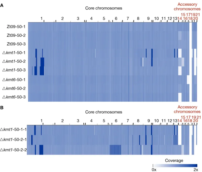
Fig 5. Genome sequencing of evolved populations and single clones originating from the long-term growth experiment. (A) Genomes of each replicate population
after 50 transfers were sequenced and mapped to the reference. Coverage is normalized to 1x. Except for coverage differences on the accessory chromosomes, there were no large structural variations detectable for the evolved Zt09 andΔkmt6 populations. In contrast, Δkmt1 populations contained multiple high-coverage regions (dark blue) on core chromosomes as well as large deletions indicated by low (light blue) or no (white) coverage. (B) To further characterize structural variation in the evolvedΔkmt1 strains, three single clones originating from populations50-1 (50-1-1) and 50-2 (50-2-1 and 50-2-2) were sequenced. Clones Δkmt1-50-1-1 andΔkmt1-50-2-2 show a very similar pattern as their respective populations, while Δkmt1-50-2-1 resembles a genotype that appears to be rare in population Δkmt1-50-2. High coverage on entire core chromosomes 13 (Δkmt1-50-2-1, 1.3x coverage) and 6 (Δkmt1-50-2-2, 1.5x coverage) indicates whole core chromosome duplications that were maintained in some nuclei. Centromeres are indicated as black dots.
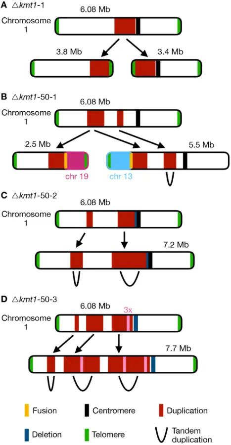
Fig 6. Different outcomes of structural variation of chromosome 1 in evolvedΔkmt1 strains. Upper chromosome
almost all chromosomes. These occurred mainly linked to annotated TEs (S10 Table) whereby loss of H3K9me3 likely promoted instability. However, major rearrangements, including chro-mosomal fusions, were always linked to large segmental duplications (S10 Fig). In two strains we detected higher coverage of entire core chromosomes indicating core chromosome dupli-cations (Fig 5B). Results from read coverage (S10 Table) and PCR analyses indicate that Δkmt1-50-2-2, as well as the majority of the Δkmt1-50-2 population, may have undergone a whole genome duplication.
To investigate whether the underlying sequence is involved in the formation of large-scale rearrangements, we analyzed the breakpoints of each duplicated region. The location of break-points does not show a clear TE-associated pattern as observed for the smaller deletions or chromosome breakages. Out of 28 analyzed breakpoints, only seven are directly located within annotated TEs, while thirteen fall into genes, seven are intergenic and one is located in the cen-tromere (S11 Table). Considering all structural rearrangements in the three sequenced single clones, we found that out of 62 events, 34 were associated (direct overlap or <5 kb distance) to regions that show enrichment for H3K27me3 (S12 Table). Based on these observations, we hypothesize that two non-exclusive pathways, namely TE-associated instability caused by loss of H3K9me3 or invasion of H3K27me3, may serve as initial events, which are followed by con-tinuous rearrangements possibly caused by increased mitotic recombination activity and defi-ciency in DNA repair resulting in a spectrum of structural variation (S11 Fig).
Discussion
H3K27me3 destabilizes accessory chromosomes
We investigated the effects of loss of two important heterochromatin-associated histone modi-fications, H3K9me3 and H3K27me3, on chromatin organization, transcription and genome stability and characterized phenotypes of the deletion mutants. Loss of H3K9me3 allows relo-calization of H3K27me3 inkmt1 deletion mutants, which has great impact on genome and
chromosome stability, resulting in numerous large-scale rearrangements. In contrast, the genomes of evolvedΔkmt6 and Zt09 strains revealed only few and relatively minor changes. Unexpectedly, the presence of H3K27me3 impacts chromosome stability by either destabiliz-ing whole chromosomes in normal cells, supported by the high loss-rate in the reference strain compared to theΔkmt6 mutants, or by mislocalization as shown by the increased sequence
higher read coverage (red) and deletion indicated by lower coverage (blue). The respective lower maps show structural rearrangements predicted by our structural variant analysis. (A) In the progenitor strain, a duplicated region was involved in the formation of two new chromosomes. At both termini of the duplication the chromosome broke and telomeric repeats were addedde novo to these breakpoints (seeFig 4). Thus, two new chromosomes were formed, both containing the duplicated sequence. This structural variation was not found in all cells in theΔkmt1 progenitor strain and the structural variation that arose in the evolved strains (B-D) can therefore be the result of rearrangements of the reference chromosome 1 or the two newly formed chromosomes. (B) In the evolved populationΔkmt1-50-1, two duplicated sequences were detected. The borders of the first region mark chromosome breakages that were fused to telomeres of other chromosomes. The first breakpoint was attached to the telomere of chromosome 13 forming a new 5.5 Mb chromosome while the second breakpoint was fused to the telomere of chromosome 19 (new 2.5 Mb chromosome). The second duplicated region represented a tandem duplication located on the new 5.5 Mb
chromosome that falls within the duplicated region of the progenitor strain. (C) PopulationΔkmt1-50-2 contained two duplicated regions, that both resembled tandem duplications. The second duplication is very similar to the one found in the progenitor strain but includes half of the centromere and had a deletion, where the breakpoint close to the centromere in the progenitor strain is located. (D) PopulationΔkmt1-50-3 displayed three duplicated sequences that all form tandem duplications resulting in the formation of a 7.7 Mb version of chromosome 1. The third duplicated region was, as in populationΔkmt1-50-2, very similar to the one in the progenitor strain. However, in this case the complete centromere-associated sequence was deleted. Furthermore, a ~50 kb region inside the third duplicated region exhibited 3x sequencing coverage and was found in between the tandem duplication of the second duplicated region (seeS9 Table).
instability in theΔkmt1 mutants. Taken together, enrichment with H3K27me3 in wild type cells is a main driver of mitotic chromosome instability.
We propose different scenarios for how chromosomes may get lost during mitosis and how H3K27me3 may be linked to these processes. For example, accessory chromosomes may not be accurately replicated whereby only one sister chromatid is transmitted. Alternatively, non-disjunction of sister chromatids during mitosis produces one cell with two copies and one cell lacking the respective chromosome. Previous cytology onZ. tritici strains expressing
GFP-tagged CENPA/CenH3 protein suggested that core and accessory chromosomes may be physi-cally separated in the nucleus [31]. Previous studies showed that H3K27me3-enriched chroma-tin localizes near the nuclear periphery, and loss of H3K27me3 enables movement of this chromatin to the nucleus core in mammals and fungi [52,53]. Proximity to the nuclear mem-brane and heterochromatic structure can furthermore result in differential, and often late, rep-lication timing [54,55]. Loss of H3K27me3 and the correlated movement to the inner nuclear matrix may alter replication dynamics of accessory chromosomes resulting in higher rates of faithfully replicated chromosomes and lower rates of mitotic loss (Fig 7).
Heterochromatic regions, especially associated with H3K27me3, tend to cluster together and form distinct foci in the nucleus ofDrosophila melanogaster visualized by cytology [56,57], and loss of H3K27me3 reduces interaction between these regions [58]. We hypothesize that enrichment of H3K27me3 on the entire accessory chromosomes maintains physical interac-tions that persist throughout mitosis. This may decrease the efficiency of separation of sister chromatids resulting in loss of the chromosome in one cell and a duplication in the other cell. So far, we have focused our screening on chromosome losses but determining the exact rates of accessory chromosome duplications is necessary to test this hypothesis. Genome sequencing ofZ. tritici chromosome loss strains revealed that duplications of accessory chromosomes can
occur [46]. Similarly, B chromosomes in rye are preferentially inherited during meiosis by non-disjunction of sister chromatids during the first pollen mitosis [59], indicating that devia-tion from normal chromosome segregadevia-tion occurs. Accessory chromosomes are commonly found in natural isolates ofZ. tritici, despite the high loss rates we demonstrated during mitotic
growth [46]. This observation implies the presence of other mechanisms that counteract the frequent losses of accessory chromosomes. Recent analyses of meiotic transmission showed that unpaired accessory chromosomes are transmitted at higher rates in a uniparental way [60,61]. We propose that H3K27me3 is involved in accessory chromosome instability and transmission both during mitosis and meiosis by influencing nuclear localization of chromo-somes and thereby altering replication or transmission (Fig 7). Future analyses with fluores-cently tagged core and accessory chromosomes and by chromosome conformation capture (Hi-C) will shed light on nuclear interactions and chromosome transmission dynamics. As not all accessory chromosomes, despite being enriched with H3K27me3, are lost at the same rate, we note that additional mechanisms likely contribute to accessory chromosome dynamics.
H3K9me3 loss allows invasion by H3K27me3 and results in genome
instability
While loss of H3K27me3 resulted in only minor differences to wild type growth and, unex-pectedly, rather promoted than decreased genome stability, we detected a high number of smaller (up to 30 kb) deletions and duplications, chromosome breakages and several gross chromosomal rearrangements linked to large duplications in theΔkmt1 mutants. Absence of H3K9me2/3 has been associated with chromosome and genome instability in other organisms [13,14,62,63]. Smaller deletions, duplications and chromosome breakages resulting in
shortened chromosomes due to loss of chromosome ends that we identified in theΔkmt1 mutants, correlate with TEs, enriched with H3K9me3 in wild type. Replication of heterochro-matin-associated DNA is challenging for the cell as repetitive sequences may form secondary structures that can stall the replication machinery [64]. Consequently, instability of repeated sequences has been linked to errors during DNA replication [65–67]. Furthermore, the struc-tural variation that arises depends on the mode of DNA repair following the DNA damage [68,69]. The structural rearrangements detected in theΔkmt1 mutants indicate that repair of double-strand breaks involves both non-homologous end joining andde novo telomere
forma-tion. We propose that the main factor for genome instability is replication-associated instabil-ity of repeated sequences subsequently promoting the formation of large-scale rearrangements (S11 Fig).
Not all breakpoints of rearrangements, especially of the large duplicated sequences, were associated with TEs, however. We found that duplicated sequences in the experimentally evolvedΔkmt1 mutants fully or partially overlap with the duplicated regions of the Δkmt1 pro-genitor strain. This suggests that structural variations are subject to continuous
Fig 7. Working model to illustrate how H3K27me3 may influence nuclear localization of whole chromosomes or landmark genomic regions. In wildtype cells (left
panel), H3K27me3 is localized in subtelomeric regions and on accessory chromosomes, directing those regions to the nuclear periphery and resulting in increased instability of these regions. Loss of H3K27me3 (middle panel) results in a relocation of former H3K27me3-enriched sequences to the inner nucleus and an increase of genome stability. Loss of the histone modification H3K9me3 enables H3K27me3 to spread, leading to mislocalization of H3K27me3, altered physical localization and chromatin interactions in the nucleus that fuel genome instability of these regions (right panel).
rearrangements, resulting in rearrangements no longer directly linked to the initial event. We note that the rearrangements and genotypes we detected are the result of selection during our long-term growth experiments and thus do not necessarily reflect the full spectrum of rear-rangements occurring inΔkmt1 mutants; many additional structural variants may have disap-peared quickly from the population or included lethal events.
Concomitant with loss of H3K9me3 in theΔkmt1 strains, we found relocalization of H3K27me3 to former H3K9me3 regions. A similar redistribution of H3K27me3 in absence of heterochromatin factors has been reported in plants and animals [70–72] and other fungi [22,27,73]. InN. crassa, redistribution of H3K27me3 in a Δkmt1 (dim-5) mutant background
results in severe growth defects and increased sensitivity to genotoxic stress that can be rescued by elimination of H3K27me3, indicating that aberrant H3K27me3 distribution severely impacts cell viability [27]. Although we did not see rescue of phenotypic defects observedin planta or in in vitro stress assays in the Δk1/k6 double mutants, the chromosome-loss rate was
reduced compared toΔkmt1 mutants, suggesting a stabilizing effect when H3K27me3 is absent. We found that some breakpoints of the rearrangements in theΔkmt1 mutants without H3K9me3 also show enrichment with the invading H3K27me3. This finding also suggests that sequences associated with H3K27me3 are more susceptible to genome instability. Regions enriched with H3K27me3 have been shown to exhibit a high degree of genetic variability in form of mutations, increased recombination, or structural variation compared to the rest of the genome [21,23,30,31,74–76]. Experimental evolution inFusarium fujikuroi showed that
increased H3K27me3 levels in subtelomeric regions coincided with increased instability [77] and we previously detected a highly increased rate of chromosomal breakage under stress con-ditions in subtelomeric H3K27me3 regions inZ. tritici [46]. These observations together with our findings strongly indicate that H3K27me3 plays a pivotal role in decreasing genome stability.
In summary, the presence of Kmt1 and H3K9me3 respectively, is essential to maintain genome integrity in this fungus. TE-mediated rearrangements may be involved in the genetic variability detected inZ. tritici isolates [78–80] and have been suggested as drivers of genome evolution in various species [81–83]. Our findings concerning the role of H3K9me3 for genome stability provide a basis for future studies focusing on the influence of heterochroma-tin on structural genome rearrangements usingZ. tritici as a model organism. We found that,
unlike for H3K9me3, presence and not absence of H3K27me3 is linked to genome instability. Surprisingly, loss of H3K27me3 does not result in dramatic changes of overt phenotypes and is also not clearly linked to transcriptional activation inZ. tritici. This allowed us to uncouple the
transcriptional and regulatory effects of H3K27me3 from the influence on chromatin stability and will in the future result in further mechanistic insights on the influence of histone modifi-cations on chromosome stability.
Materials and methods
Culturing conditions of fungal and bacterial strains
Zymoseptoria tritici strains were cultivated on solid (2% [w/v] Bacto agar) or in liquid YMS
medium (0.4% [w/v] yeast extract, 0.4% [w/v] malt extract, 0.4% [w/v] sucrose). Liquid cul-tures were inoculated from plate or directly from glycerol stocks and grown for 3–4 days at 18˚C in a shaking incubator at 200 rpm. Plates were inoculated from glycerol stocks and grown for 5–6 days at 18˚C.Escherichia coli TOP10 cells were grown overnight in dYT (1.6%
[w/v] tryptone, 1% [w/v] yeast extract, 0.5% [w/v] NaCl and 2% Bacto agar for solid medium) supplemented with antibiotics for plasmid selection (40μg/mL kanamycin) at 37˚C and at 200 rpm for liquid cultures.Agrobacterium tumefaciens strain AGL1 was grown in dYT containing
rifampicin (50μg/mL) and carbenicillin (100 μg/mL) supplemented with antibiotics for plas-mid selection (40μg/mL kanamycin) at 28˚C at 200 rpm in liquid culture for 18 h and on plate at 28˚C for two days.
Transformation of
Z. tritici
Z. tritici deletion and complementation strains were engineered using A.
tumefaciens-medi-ated transformations as described before [49,84]. Flanking regions of the respective genes were used to facilitate homologous recombination for integration at the correct genomic location. The plasmid pES61 (a derivate of the binary vector pNOV-ABCD [49]) was used for targeted gene deletion and complementation. Plasmids were assembled using a restriction enzyme-based approach or Gibson assembly [85]. Plasmids were amplified inE. coli TOP10 cells and
transformed in theA. tumefaciens strain AGL1 as described previously [86]. Gene deletions of
kmt1 (Zt09_chr_1_01919) and kmt6 (Zt09_chr_4_00551) were facilitated by replacement of
the respective ORF with a hygromycin resistance cassette (hph). The kmt1/kmt6 double
dele-tion mutant was constructed by integrating a nourseothricin resistance cassette (nat) replacing kmt1 in a kmt6 deletion mutant background. Complementation constructs containing the
respective gene and a G418 resistance cassette (neo) were integrated at the native loci in the
deletion strains. All plasmids and strains constructed in this study are listed inS1 Table. Trans-formed strains were screened by PCR for correct integrations of the construct followed by Southern blot [87] with probes generated by DIG labeling (Roche, Mannheim, Germany) fol-lowing manufacturer’s instructions.
DNA isolation for PCR screening and southern blotting
For rapid PCR screening (candidates for transformation and chromosome loss), a singleZ. tri-tici colony was resuspended in 50 μL of 25 mM NaOH, incubated at 98˚C for 10 min and
after-wards 50μL of 40 mM Tris-HCl pH 5.5 were added. Four μl of the mix was used as template for PCRs. For DNA extraction for Southern blotting, we used a standard phenol-chloroform extraction protocol [88] for DNA isolation.
Phenotypic characterization
in vitro
For thein vitro growth assays, liquid YMS cultures were inoculated with 100 cells/μL (OD600= 0.01); cells were grown in 25 mL YMS at 18˚C and 200 rpm. For each mutant and complemen-tation strain, two transformants (biological replicates), and three replicate cultures per trans-formant (technical replicates) were used. For the reference strain Zt09, two separate pre-cultures were grown as biological replicates and each pre-culture was used to inoculate three replicate cultures. OD600was measured at different time points throughout the experiment until the stationary phase was reached. The R package growthcurver [89] was used to fit the growth curve data enabling to comparein vitro growth of the different strains.
To test the tolerance of mutant and reference strains towards different stressors, we per-formed anin vitro stress assay on YMS plates. Each plate contained additives constituting
dif-ferent stress conditions. Cell suspensions containing 107/108cells/mL and a tenfold dilution series down to 100 cells/mL were prepared; 3μL of each dilution were pipetted on solid YMS containing the following additives: 0.5 M NaCl, 1 M NaCl, 1 M sorbitol, 1.5 M sorbitol, 1.5 mM H2O2, 2 mM H2O2, 300μg/mL Congo red, 0.01% MMS (methyl methane sulfonate), 0.025% MMS, 1μg/mL actinomycin D and 1.5 μg/mL actinomycin D. Furthermore, we included a H2O-agar (2% bacto agar) plate. All plates were incubated at 18˚C for six days, except for one YMS plate that was incubated at 28˚C to test for thermal stress responses.
Phenotypic characterization
in planta
Seedlings of the susceptible wheat cultivar Obelisk (Wiersum Plantbreeding BV, Winschoten, The Netherlands) were potted (three plants per pot) after four days of pre-germination and grown for seven more days. Single cell suspensions of mutant and reference strain were pre-pared (108cells / mL in H2O with 0.1% Tween 20) and brush inoculated on a marked area of the second leaf. Following inoculation, the plants were incubated in sealed plastic bags con-taining ~ 1 L of H2O for 48 h providing high humidity to promote infections. Growth condi-tions for the plants throughout the complete growth phase and infection were 16 h light (200μmol/m-2s-1) and 8 h dark at 20˚C and 90% humidity. First appearances of symptoms, necrosis or pycnidia, were assessed by manual inspection of every treated leaf. 21 or 28 days post infection, inoculated leaves were finally screened for infection symptoms. Visual inspec-tion of each leaf was performed to evaluate the percentage of leaf area covered by necrosis and pycnidia. Six different categories were differentiated based on the observed coverage (0: 0%, 1: 1–20%, 2: 21–40%, 3: 41–60%, 4: 61–80%, 5: 81–100%). Furthermore, automated symptom evaluation was performed by analysis of scanned images of infected leaf areas as described pre-viously [90].
Long-term evolution experiment
For the long-term evolution experiment (~6 months), cells were inoculated directly from the glyc-erol stocks into 20 mL liquid YMS cultures. We used Zt09,Δkmt6 (#285) and Δkmt1 (#68), each strain grown in triplicates. Every three to four days, cells were transferred to new YMS medium. Cells were grown at 18˚C and 200 rpm. For every transfer, cell density of the cultures was mea-sured by OD600and the new cultures were inoculated with a cell density of ~ 100 cells /μL (corre-lating to a transfer of 0.1% of the population). After 50 transfers, the genomes of the evolved populations and each progenitor strain were sequenced. Additionally, three genomes of single clones derived from theΔkmt1 populations after 50 transfers were sequenced to characterize genome rearrangements in more detail.
Short-term evolution experiment
For the short-term evolution experiment over a time period of four weeks, cultures were inoc-ulated from single colonies grown on solid YMS. Zt09,Δkmt6 (#285), Δkmt1 (#80), Δk1/Δk6 (#23) double mutant,kmt1+(#42) andkmt6+(#11) were grown in triplicate YMS cultures. For this experiment we used a different independentΔkmt1 mutant clone (#80), as we discovered that the strain used in the previous long-term evolution experiment (#68) was missing chro-mosome 20. Every three to four days, 900μL culture were transferred to 25 mL fresh YMS (correlating to a transfer of ~ 4% of the population). After four weeks of growth (including eight transfers to new medium) at 18˚C and 200 rpm, cultures were diluted and plated on YMS agar to obtain single colonies. These single colonies were PCR screened for presence of accessory chromosomes as described in [46].
Pulsed-field gel electrophoresis (PFGE)
Cells were grown in YMS medium for five days and harvested by centrifugation for 10 min at 3,500 rpm. We used 5 x 10⁸cells for plug preparation that were washed twice with Tris-HCl, pH 7.5, resuspended in 1 mL TE buffer (pH 8) and mixed with 1 mL of 2.2% low range ultra agarose (Bio-Rad, Munich, Germany). The mixture was pipetted into plug casting molds and cooled for 1 h at 4˚C. Plugs were placed to 50 mL screw cap Falcon tubes containing 5 mL of lysis buffer (1% SDS; 0.45 M EDTA; 1.5 mg/mL proteinase K [Roth, Karlsruhe, Germany])
and incubated for 48 h at 55˚C while the buffer was replaced once after 24 h. Chromosomal plugs were washed three times for 20 min with 1 X TE buffer before storage in 0.5 M EDTA at 4˚C. PFGE was performed with a CHEF-DR III pulsed-field electrophoresis system (BioRad, Munich, Germany). Separation of mid-size chromosomes was conducted with the settings: switching time 250 s– 1000 s, 3 V/cm, 106˚ angle, 1% pulsed-field agarose in 0.5 X TBE for 72 h. Large chromosomes were separated with the following settings: switching time 1000 s– 2000 s, 2 V/cm, 106˚ angle, 0.8% pulsed-field agarose in 1 X TAE for 96 h.Saccharomyces cerevisiae
chromosomal DNA (BioRad, Munich, Germany) was used as size marker for the for mid-size chromosomes,Schizosaccharomyces pombe chromosomal DNA (BioRad, Munich, Germany)
for the large chromosomes. Gels were stained in ethidium bromide staining solution (1μg/mL ethidium bromide in H2O) for 30 min. Detection of chromosomal bands was performed with the GelDocTM XR+ system (Bio-Rad, Munich, Germany). Southern blotting was performed as described previously [87] but using DIG-labeled probes generated with the PCR DIG label-ing Mix (Roche, Mannheim, Germany) followlabel-ing the manufacturer’s instructions.
ChIP-sequencing
Cells were grown in liquid YMS medium at 18˚C for 2 days until an OD600of ~ 1 was reached. Chromatin immunoprecipitation was performed as previously described [91] with minor modifications. We used antibodies against H3K4me2 (#07–030, Merck Millipore), H3K9me3 (#39161, Active Motif) and H3K27me3 (#39155, Active Motif). ChIP DNA was purified using SureBeads Protein G Magnetic Beads (Bio-Rad, Munich, Germany) and, replacing phenol/ chloroform extractions, we used the ChIP DNA Clean & Concentrator Kit (Zymo Research, Freiburg, Germany). We sequenced two biological and one additional technical replicate for Zt09,Δkmt1, Δkmt6, and the Δk1/k6 strains. Sequencing was performed at the OSU Center for Genome Research and Biocomputing on an Illumina HiSeq2000 or HiSeq3000 to obtain 50-nt reads and at the Max Planck Genome Center, Cologne, Germany (https://mpgc.mpipz.mpg. de/home/) on an Illumina Hiseq3000 platform obtaining 150-nt reads (S2 Table).
RNA-sequencing
For RNA extraction, cells were grown in liquid YMS at 18˚C and 200 rpm for two days until an OD600of ~ 1 was reached. Cells were harvested by centrifugation and ground in liquid nitrogen. Total RNA was extracted using TRIzol (Invitrogen, Karlsruhe, Germany) according to manufacturer’s instructions. The extracted RNA was further DNAse-treated and cleaned up using the RNA Clean & Concentrator-25 Kit (Zymo Research, Freiburg, Germany). RNA sam-ples of two biological replicates of Zt09,Δkmt1, Δkmt6, and the Δk1/k6 double mutant were sequenced. Poly(A)-captured, stranded library preparation and sequencing were performed by the Max Planck-Genome-centre Cologne, Germany (https://mpgc.mpipz.mpg.de/home/) on an Illumina Hiseq3000 platform obtaining ~ 20 million 150-nt reads per sample (S2 Table).
Genome sequencing
Genomic DNA for sequencing was prepared as described previously [92]. Library preparation and genome sequencing of the progenitor strains used for the evolution experiments were per-formed at Aros, Skejby, Denmark using an Illumina HiSeq2500 platform obtaining 100-nt paired-end reads. Library preparation (PCR-free) and sequencing of the evolved populations and the three evolved singleΔkmt1 mutants were performed by the Max Planck Genome Cen-ter, Cologne, Germany (https://mpgc.mpipz.mpg.de/home/) on an Illumina HiSeq3000 plat-form resulting in 150-nt paired-end reads (S2 Table).
Short read mapping and data analysis
A detailed list of all programs and commands used for mapping and sequencing data analyses can be found in the supplementary text S1. All sequencing data was quality filtered using the FastX toolkit ((http://hannonlab.cshl.edu/fastx_toolkit/) and Trimmomatic [93]. RNA-seq reads were mapped using hisat2 [94], mapping of ChIP and genome data was performed with Bowtie2 [95]. Conversion of sam to bam format, sorting and indexing of read alignments was done with samtools [96].
To detect enriched regions in the ChIP mappings, we used HOMER [97]. Peaks were called individually for replicates and merged with bedtools [98]. Only enriched regions found in all replicates were considered for further analyses. Genome coverage of enriched regions and overlap to genes and TEs was calculated using bedtools [98].
We used cuffdiff [99] to calculate RPKM values and to estimate expression in the different strains. Raw reads mapping on genes and TEs were counted by HTSeq [100], differential expression analysis was performed in R [101] with DESeq2 [102]. Cutoff for significantly dif-ferentially expressed genes was padj < 0.001 and |log2 fold-change| > 2. The R package topGO [103] was used to perform gene ontology enrichment analyses. Fisher’s exact test (
p-value < 0.01) was applied to detect significantly enriched terms in the category ‘biological process’.
To detect structural variation in the sequenced genomes, we used SpeedSeq [104] and LUMPY [105]. All detected variation was further verified by manual visual inspection. Visuali-zation was performed with the integrative genome browser (IGV) [106].
Supporting information
S1 Table. Plasmids, strains and primer designed for this study. Listed are all primers used to
create plasmids and probes for Southern blots. (XLSX)
S2 Table. Statistics and overview of sequencing data generated in this study.
(XLSX)
S3 Table. RPKM values of all genes of Zt09 reference and mutant strains. RPKM was
calcu-lated using cuffdiff (seeMaterial and Methods). (XLSX)
S4 Table. Genes associated to either H3K9me3 or H3K27me3 in Zt09 or mutant strains.
(XLSX)
S5 Table. Deseq2 results to identify differentially expressed genes between Zt09 reference and mutant strains. Comparisons were performed pair-wise, genes were considered to be
sig-nificantly different expressed, when log2 fold-change > 2 and padj < 0.001. (XLSX)
S6 Table. Enriched GO terms and upregulated genes in the categories DNA integration and RNA-dependent DNA replication.
(XLSX)
S7 Table. Deseq2 results to analyze expression of TEs in Zt09 reference and mutant strains.
Comparisons were performed pair-wise. (XLSX)