HAL Id: hal-01523684
https://hal.archives-ouvertes.fr/hal-01523684
Submitted on 16 May 2017
HAL is a multi-disciplinary open access
archive for the deposit and dissemination of
sci-entific research documents, whether they are
pub-lished or not. The documents may come from
teaching and research institutions in France or
abroad, or from public or private research centers.
L’archive ouverte pluridisciplinaire HAL, est
destinée au dépôt et à la diffusion de documents
scientifiques de niveau recherche, publiés ou non,
émanant des établissements d’enseignement et de
recherche français ou étrangers, des laboratoires
publics ou privés.
Smartphone-Based Collaborative System for Wounds
Tracking
Francis Faux, Rémi Bastide, Nathalie Souf, Rita Zgheib
To cite this version:
Francis Faux, Rémi Bastide, Nathalie Souf, Rita Zgheib. Smartphone-Based Collaborative System
for Wounds Tracking. 8th International Conference on e-Health, Telemedicine and Social Medicine
(eTELEMED 2016), colocated with other events part of DigitalWorld 2016, Apr 2016, Venise, Italy.
pp. 104-109. �hal-01523684�
O
pen
A
rchive
T
OULOUSE
A
rchive
O
uverte (
OATAO
)
OATAO is an open access repository that collects the work of Toulouse researchers and
makes it freely available over the web where possible.
This is an author-deposited version published in :
http://oatao.univ-toulouse.fr/
Eprints ID : 16991
The contribution was presented at eTELEMED 2016 :
https://www.iaria.org/conferences2016/eTELEMED16.html
To cite this version :
Faux, Francis and Bastide, Rémi and Souf, Nathalie and
Zgheib, Rita Smartphone-Based Collaborative System for Wounds Tracking.
(2016) In: 8th International Conference on e-Health, Telemedicine and Social
Medicine (eTELEMED 2016), colocated with other events part of DigitalWorld
2016, 24 April 2016 - 28 April 2016 (Venise, Italy).
Any correspondence concerning this service should be sent to the repository
administrator:
staff-oatao@listes-diff.inp-toulouse.fr
Smartphone-Based Collaborative System for Wounds Tracking
Francis Faux
ISIS, Universit´e de Toulouse, INU Champollion, France F81 100 Castres, France
Email: francis.faux@univ-jfc.fr
R´emi Bastide
and Nathalie Souf
and Rita Zgheib
IRIT, Universit´e de Toulouse, INU Champollion, France F31 062 Toulouse, France
Email: firstname.lastname@irit.fr
Abstract—Tracking the evolution of wounds, ulcers or bedscars is one of the important tasks of the care personnel in med-ical institutions. We present an innovative, smartphone-based software application aimed at supporting this task. By using a standardized protocol complemented by image processing techniques, the application allows for the reliable acquisition of wound photographs, the assessment of wounds evolution, as well as for the annotation of photographs with relevant metadata. We detail the user-centered, scenario-based design process of this application that has been undertaken in the Connected Health Lab (CHL), our statof-thart usability lab dedicated to e-Health applications.
Keywords–Wounds tracking; smartphone; collaborative system.
I. INTRODUCTION
Tracking the evolution of wounds is an important medical issue, and concerns health care professionals as well as patients or their family. This paper presents a mobile application attempting to go beyond the standard smartphone use, and enabling medical experts to remotely view, analyze, and track the wounds evolution. Chronic wounds are frequent. Among them, foot or leg ulcers affect million of persons suffering with type 2 diabetes. Bedsores (also called pressure sores or pressure ulcers) [1], are one of the dangerous diseases that an elder can face. They are a localized injury resulting from prolonged pressure on the skin and plague persons who have a reduced ability to move and change positions, and stay in bed or a wheelchair most of the time. In oncology, chronic wounds concern any wound even small, prone to chronicize because of the cancerous disease and its treatment. Cancerous wounds have the peculiarity to evolve according to treatments efficiency instead of following a normal healing process.
Thanks to the popularity of mobile devices in clinics and hospitals the need for a mobile collaborative wound tracking application has arisen as the image remote analysis becomes an attractive option. A smartphone application linked to a normal-ized shooting protocol allows a chronological and iconographic tracking of chronic wounds with a simple, practical, and connected tool. For medical teams, the visual examination of the healing process evolution has to be made easier and more reliable, using successive pictures taken in a standardized manner. For caregivers, this is to improve inter-professional cooperation by securing the skills transfer. In the field of tele-care, remote wound tracking improves the patients’ quality of life as patients only visit the wound clinic if necessary. Health care costs are thus reduced thanks to a limited number of both
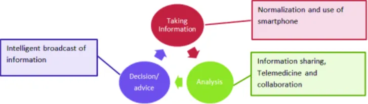
wound check appointments and travels to the clinic requiring special transportation. Many general telehealth requirements are concerned, such as quality of data transmission, privacy and security, interoperability with legacy healtchcare systems, and clear patient identification. The process of wound assessment is based on visual examination of picture by physicians, as well as on wound descriptions which provide qualitative objectives parameters (color phase peri-lesional state) and quantitative ones (wound surface and depth). However, tracking a wounds healing process across consecutive travel remains a difficult task for both clinicians and patients. Our project aims to support the following information cycle (Figure 1):
• data registering using the smartphone, • data analysis in order to share information, • decision, advice and feedback.
Figure 1. Information cycle
In this paper, wounds tracking is considered in a global and interdisciplinary approach, in relation with the patients referring doctor, in order to reduce the impact of the wound on the patients physical and psychological well-being. The presented application also has an impact on the patients environment. Thanks to an active communication with health carers, the family is taking a more active part and consequently feels reassured.
The remainder of this paper is organized as follows: Section 2 presents the related works, Section 3 outlines the key project objective. Section 4 presents the wound tracking application and Section 5 the standardized context of wound description. Future works and conclusion are presented in Section 6.
II. RELATED WORKS
Smartphones are finding their way into Healthcare systems and one of the important fields of research today is wounds assessment and tracking applications. In [2] [3], authors prove smartphones ability to ensure accurate data. This implies a
more robust report of the safety of care and a better prevention and monitoring of pressure ulcer. M-health can improve ef-ciency and patient satisfaction. Based on high resolution cam-eras integrated in smartphones, several research projects are focusing on image acquisition and analysis of chronic wounds. Topics raised in these projects are addressed in accordance with two purposes: designing a wounds processing application and defining a collaborative system fostering patient-doctor interaction. Processing wounds applications consists mainly on the analysis of wound photos in order to detect and recognize wounds size and color that are essential factors for the evaluation of wounds. Several recent papers, such as [4] aiming to automate chronic wound processing and classification, propose algorithms for size and color detection of chronic wound images taken via smartphones. Their solution is based on Mask Image components within capturing photos and Camera calibration components within the segmentation phase in order to determine the relative and absolute size of the wound. These algorithms can be useful during the wound stage recognition. Other approaches for image analysis have been proposed like size determination from camera focus and zoom [5] or detecting wound size via several different types of sensors (sensor fusion approach) for increased accuracy. In [6] the wound image analysis system for diabetic foot ulcers covers size and color topics by performing image segmentation and noise removal. The authors also present a region-detection method for the prediction of wound boundaries, and they assess the healing status based on a color evaluation method.
Several research works such as [7] [8] focus on the collab-orative facilities which may improve treatment of wounds in telemedicine. The proposed solutions aim at enhancing patient-doctor interaction offering dynamic features that may reduce the need for hospitalization and further facilitate a real-time advice for patients. In [7], the authors present an integrated ap-proach of publicly available chronic wounds databases through a telemedicine platform. It offers the possibility to take high resolution images of wounds via smartphones. They can then be stored in a database with the associated metadata in order to support their discovery, access and usability. With a unique id, the patient can get prescribed medication from doctors, which provides a smooth interaction between patient and doctors. Patient history is easily accessible remotely by several doctors, which enhances the diagnosis, treatment and routine check ups. In [8] the main focus of the authors’ is the real-time interaction via web technologies, enabling doctors to virtually collaborate with their patients and colleagues via applications working on mobile devices providing a collaborative annotation of Digital imaging and communications in medicine (DICOM) images. This research work addresses the issue of converting DICOM images to a compact version that can run on mobile devices before annotating and documenting images. Several smartphone applications such as UrgoExpert [9], INFINYS [10], Telap [11], Comedi-e [12] have been developed in order to document chronic wounds and wound care and follow-up the wound evolution. The main features of these applications are the following:
• Allowing nurses to define the wound’s class (ulcer, bedsore) and its localization in the human body (Fig-ure 2, right), as well as to visualize the wounds evolution (exudat level, healing status (wet necrosis, budding)) and the history of the treatment.
Figure 2. Comedie-e smartphone application: heel bedsore
• Photographs are captured via the smartphone camera, classified (Figure 2, bottom) and recorded in the smartphone storage card.
• Contains measurement tools. The application gen-erates wound histories in graphical and text-based formats (Figure 2, top).
• Enable accessing a medical prescription module or consulting a protocol of treatment.
• Transfer the photographs ans associated data via a secure network to the patient health record.
III. PROJECTOBJECTIVE
We propose a new software application for wound tracking. The present paper focuses on the participatory design of the application, with a practical goal of usability and efficiency. We have taken a user-centered design process, taking advan-tage of the facilities offered by the Connected Health Lab (CHL), a state-of-the-art usability lab dedicated the e-Health applications, located in the premises of the authors’ school of Engineering. In the framework of this user-centered design process, wounds pictures have been simulated using texture modeling, in order to keep them replicable. The mobile appli-cation is prototyped and assessed in the controlled environment provided by our usability lab, that faithfully replicates the working environments where the application will be deployed and used (hospital patient’s rooms, or conventional home rooms for patients under home care). Then we describe the important points on which we have to focus to allow patients and their caregivers to take a more active role in daily wound care using a collaborative annotation functionality.
IV. WOUND TRACKING
In real life conditions, wound pictures are captured each time the nurse treats the wound and changes the dressing.
a) b) c) d)
Figure 3. Telap smartphone application, top: consecutive pictures , bottom: wound contour measurement
To achieve pictures, basic recommendations have been written and proposed to the health care professionals who
are taking wound pictures. The main ideas for these recom-mendations are to improve the quality of the pictures and consequently the quality of treatment:
• Put a uniform background to better detect the wound area.
• No zoom to keep good resolution. • No move to avoid fuzzy picture.
• Pacing a ruler reference in order to be able to recal-culate the size of the wound.
• Placing reference color table in order to recalculate the colorimetry of the picture [13].
However, it is clear that difficulties remain to normalize shooting to enable a simpler and more reliable assessment of the wounds evolution. Pictures are often captured in an empirical manner without maintaining a constant angle of shooting (Figure 2, 3).
The difficulties mentioned by the healthcare professionals we are working with are:
• It is difficult to take different pictures with the same condition as the wound is changing and in on 3D surfaces (for example a leg). As the patient moves it is difficult to retrieve a similar shoot through the different patient’s visits (Figure 3). The wound evolves and the lighting conditions are varying (Figure 3 a and b). • It is very difficult to find reference points on which
to align a picture as of course wound is evolving but moreover the wound environment is not stable. For example in case of ulcer wound, bad blood irrigation induces leg swelling and even the contour lines of the leg is not stable.
a) b) c) d)
Figure 4. Overlay: a) mask (picture 5), b) c) d) mask overlaid
In order to preserve an almost unchanging angle of shoot-ing, we choose to overlaid a transparent mask image on the cur-rent capture in our smartphone application. The same method is used by T. K. Poon in [4]. The advantage of this approach is that no peripheral devices to the smartphone camera are necessary. We choose not to add temporary or permanent tattoo of dots or lines around the wound or skin marker reference points, which can serve as a pattern to ensure alignment of successive mask. The transparent mask image which is only the previous shot of the wound (Figure 4 a) is overlaid on the current capture. When using the application, first the nurse positions the smartphone in order to approximatively recover the previous shooting angle (Figure 4 b) then moves the smartphone (Figure 4 c) to align as well as possible (Figure 4 d) the mask on the current wound. The mask area page is visualized right after a new image acquisition. The outcome of the application is a set of consecutive wound pictures captured with same size and orientation.
A. Experimental simulation
Experiments have been realized in the environment of simulation of the Connected Health Lab (CHL). The use case of a heel bedsore evolution was chosen as simulation process in the context of a this Living Lab which simulate the patient route. The scenario of the study considers a seriously hurt person because of a car accident (or an accident at work). The person suffers of a polytrauma and is in a coma.
Weeks: 1, 2 3, 4 5, 6 7
Figure 5. Use case: convalescence in the Living Lab
The figure 5 presents the patient’s convalescence of a duration of 7 weeks and which includes four main phases:
1) After managing the emergency (operation, stabiliza-tion), the patient is hospitalized in intensive care for two weeks, Heel pressure ulcer appears after a week of hospitalization (week 1). Then the bedsore deteriorates because of the patient’s immobility (week 2).
2) As soon as the patient leaves the coma and wakes up, he is transferred to an orthopedic service. However the bedsore continues to deteriorate because of the lack of mobility (weeks 3 and 4).
3) The patient goes home with nursing home monitoring (week 5). His bedsore begins to heal in week 6. 4) The patient finding increasingly mobility, in particular
with the setting in the chair, the bedsore continues to heal (week 7).
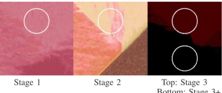
In order to realise a reproductive study, i.e., replicable model of an ulcer pressure evolution, we decided to use a serie of built picture of wounds, to serve as a benchmark for our scenario and for the evaluation of the proposed prototype. We synthesize the wound texture using Efros & Leung’s texture synthetis algorithm [14]. This algorithm is chosen because of its simplicity (Figure 6). Three reference images serve as model for different stages of bedsore evolution. A rectangular
Stage 1 Stage 2 Top: Stage 3 Bottom: Stage 3+ Figure 6. Wound texture synthesize and disk modelling the heel bedsore window (in green on Figure 7) manually selected in each image serves as texture source for the algorithm. After various tests, a 3 × 3 window is used as algorithm parameter because of the light granularity of the wound texture.
Stage 1 Stage 2 Stage 3 Figure 7. Stages of heel bedsore used for the texture synthesize
A simple disk of 16 mm of radius (area 8 mm2) models the heel pressure ulcer. For stages 1 and 2 we select the zone which looks like the real texture. The stage 3 is segmented in
TABLE I. WOUND EVALUATION
CHL ZONE INT. CARE ORT. ROOM HOME BED. ARMCH.
WEEK 1 2 3 4 5 6 7
BEDSORE STADE 1 1 2 3 3+ 3 2
two levels (called 3 and 3+) in function of level of necrosis in the wound. The bedsore evolution is summarized in the table I.
B. Analysis and results
A subjective analysis of the results (Figure 8) shows that the application is relevant in medical context. Consecutive pictures are captured with almost same scale and orientation along the seven weeks of study and for the four rooms of measurement.
The quantitative results indicate a variation of about 40% of the area and 25% of the perimeter (Figure 9). The reason of these variations comes from the difficulty to align the mask over the current wound picture especially in the intensive care room or the hospital room when the patient’s mobility is reduced. At home bedroom the shooting is easier when the patient can move its leg more easily.
Week:1 2 3 4 5 6 7
Figure 8. Result of tracking with the mask overlay
Figure 9. Area and perimeter measurements
As conclusion from these experiments in the Living Lab, the application usability is clearly insufficient in such a context. An evolution of the application is necessary. The shooting must be easier and above all automatic. As future work, we will study the possibility to introduce the correlation level between the mask overlay and the current image in order to facilitate the alignment between these two pictures. Moreover, this study will be continue for various kinds of wounds (cancerous wounds, ulcers, others bedsores) with varying areas ans complex forms.
V. STANDARDIZED CONTEXT OF WOUNDS DESCRIPTION
A. Introduction
In order to improve the informative status of the wound pictures, it is important to add descriptions to the picture itself. In this project, one of our objectives is to determine recommendations to provide efficient annotation tools able to improve efficient communication between the different actors involved in the wound care (health care professionals as well as patient or family), in the context of smartphone use. In this paper we will present some of the lines of thinking we have spotted. In order to communicate, health professionals complete the information of the medical record but also use other media as phone, mail, annotations and so on. Annotation tasks are often used in the medical area as for example post-it used for the asynchronous communication of health care professionals. As mentioned in [15] ”practitioners use
annotations to act: either to enrich the annotated document or as transitory support of knowledge used to create new knowledge (recorded or not in a document). Annotations are useful not only to keep pertinent knowledge in the health record, but also to help practitioners to collaborate when the documents show their limits”(p. 4).
Figure 10. Wounds annotations of Pange et al.
Annotation systems are proposed in many telemedicine systems using smartphones, as shown in Figure 10 for mam-mography (annotation from [16]). As friendly annotation in-terfaces are difficult to provide through smartphone devices, in order to get usable and efficient tools, we are convinced that it is extremely important to better understand how and why the annotation process should be developed.
B. Analyse
Three main issues for annotation process were detected and are presented in this paper:
Why are the annotations required. In a wound an-notation system, the objectives of the proposed anan-notations could be very different. On the one hand, some annotations are used to inform on general items such as Patient ID or Patient Name. These data are not directly involved with wound characteristics. Such annotations must be re-entered and dupli-cated on the smartphone application when the interoperability between smartphone and health care record are difficult to perform. On the other hand, some of the data bring specific awareness on an aspect of the wound : a highlighted array of interest gives the subjective point of view of a health care professional. Some annotations could then be automatically performed while improving the connection between system, adding connected objects into the wound care process (such as automatic detection of the ID of the patient) while some annotations are specifically involved on the care expertise. Being aware of these specificities will help to better understand the annotation tasks and to focus on the needed interfaces.
When and how annotations are completed. Annotations should be fed when the picture is taken or could be completed during an asynchronous task of wound image description. Items used for annotation can be from different natures: standardized description, visual annotation such as colored arrows, specific descriptions obtained after specific treatments such as wound surface calculation or colorimetry indices. By better understanding the annotation process, we hope to be able to propose efficient systems able to adapt to the different contexts of use.
What is concerned with the annotation task. Annotation tasks could bring information on three different levels of knowledge.
• The first level concerns the picture itself and the shooting environment. It could be important to remem-ber the parameters linked to the shooting so that the quality on the picture could be memorized. Quality of context description could help to qualify a further picture comparison. Metadata of picture description used in DICOM standard are some of the information to be kept, but the context of shooting referring to the recommendation can also be used.
• The second level is used in order to memorized the context of the individual who is concerned by the picture: for instance name of the patient, part of body concerned.
• Last level deals with specific wound characteristics. The goal is to establish a standardized context of wounds description in order to ensure interoperability of information systems and to improve exchanges between healthcare systems actors.
By better identifying the need of information on each level, we will be able to better determine the needed information.
The project will then address the issues related to picture description, annotation of wound pictures, and modelling of factual wounds tracking (be able to reinterpret picture in order to build meta-data useful for caregivers). We will provide a model of annotation for wound. Using the different point of view that we have mentioned on wound care annotation, we want to propose an ontological framework.
Analysis of existing annotation systems and previous work such as the one performed by the wound ontology [17], or
by the openEHR Clinical Knowledge Manager working group [18], as mentionned by Gallaguer [19] will be considered and extended by confronting them with the expertise of health professionals involved in this project. Constraints due to smart-phone use, inducing mobility, small screen, distribution of information will be also detailed.
VI. CONCLUSION AND PERSPECTIVE
We have presented a mobile, smartphone-based e-health application that supports caregivers in their important activ-ity of monitoring the evolution of wounds. The application combines innovative mobile interaction techniques with image processing and analysis in order to allow for a reliable and usable clinical use. The design of this application has fol-lowed a user-centered process, where both the standardized photograph-taking procedure and the image processing steps have been incrementally designed and validated in a controlled environment, the CHL living lab dedicated to e-Helath ap-plications. The application is currently a validated prototype. Our future steps will be to perform a clinical validation and assessment of the application, where it will be used by care professionals in a real-life setting, with real patients. We also plan to improve the image-processing components, and to take into account the interoperability of the application with legacy health information systems (e.g., electronic health records).
ACKNOWLEDGMENT
Bruno Roussel, research engineer, for his involvement in the test procedure in the Connected Health Lab and Laurent Grgoire, International coordinator, for his proofreading and editing of the English language.
REFERENCES
[1] R. Zgheib, R. Bastide, and E. Conchon, “A semantic web-of-things architecture for monitoring the risk of bedsores,” in Proceedings of the International Conference on Computational Science and Computational Intelligence (CSCI), Las Vegas, USA. IEEE, december 2015. [2] K. Wac, “Smartphone as a personal, pervasive health informatics
services platform: literature review,” Yearbook of Medical Informatics, no. 2, 2012, pp. 83–93.
[3] E. M. Quinn et al., “Clinical Unity and Community Empowerment: The Use of Smartphone Technology to Empower Community Management of Chronic Venous Ulcers through the Support of a Tertiary Unit,” PLoS one, vol. 8, no. 11, 2013, p. e78786.
[4] T. W. K. Poon and M. R. Friesen, “Algorithms for Size and Color Detection of Smartphone Images of Chronic Wounds for Healthcare Applications,” IEEE Access, vol. 3, 2015, pp. 1799–1808.
[5] P. J. F. White, W. P. Blake, and M. R. Friesen, “Algorithms for Smartphone and Tablet Image Analysis for Healthcare Applications,” IEEE Access, vol. 2, 2014, pp. 831–840.
[6] L. Wang et al., “Smartphone-Based Wound Assessment System for Patients With Diabetes,” IEEE Transactions on Biomedical Engineering, vol. 62, no. 2, 2015, pp. 477–488.
[7] C. Chakraborty, B. Gupta, and S. K. Ghosh, “Mobile metadata assisted community database of chronic wound images,” Wound Medicine, vol. 6, 2014, pp. 34–42.
[8] M. F. Pasha et al., “An android-based mobile medical image viewer and collaborative annotation: Development issues and challenges,” JDCTA: International Journal of Digital Content Technology and its Applications, vol. 6, no. 1, 2012, pp. 208–217.
[9] “UrgoExpert,” URL: http://www.urgoexpert.fr[accessed: 2016-20-03]. [10] “INFINYS,” URL: http:// www.sffpc.org[accessed: 2016-20-03]. [11] “telap,” URL: http://www.telap.org [accessed: 2016-20-03].
[12] “Comedi-e,” URL: http://www.esante-picardie.com [accessed: 2016-20-03].
[13] S. Van Poucke et al., “Automatic colorimetric calibration of human wounds,” BMC medical imaging, vol. 10, no. 1, 2010, p. 7.
[14] A. A. Efros et al., “Texture synthesis by non-parametric sampling,” in Proceedings of the Seventh IEEE International Conference on Computer Vision, vol. 2. IEEE, 1999, pp. 1033–1038.
[15] N. Bricon-Souf et al., “Informal notes to support the asynchronous collaborative activities,” International Journal of Medical Informatics, vol. 76, 2007, pp. S342–S348.
[16] A. Prange and D. Sonntag, “Smartphone sketches for instant knowledge acquisition in breast imaging,” Proceedings of the Sketch: Pen and Touch Recognition Workshop in conjunction with IUI14, 2014. [17] “Introduction to the wound ontology consortium,” URL:
http://www.slideshare.net/SvenVanPoucke/woundontology1 [accessed: 2016-20-03].
[18] “Open EHR, Clinical knowledge Manager, Inspection of open Wound,” URL: http://openehr.org/ckm/ [accessed: 2016-20-03].
[19] B. A. Gallagher, “Wound bed assessment using calibrated images and representation in openehr,” Master’s thesis, University of Dublin, Ireland, 2012.