DNA methylation in early mammalian development
by
Michelle M. Chan
B. Sc. Computer Science and Microbiology and Immunology
University of British Columbia, 2007
SUBMITTED TO THE PROGRAM OF COMPUTATIONAL AND SYSTEMS BIOLOGY IN
PARTIAL FULFILLMENT OF THE REQUIREMENTS FOR THE DEGREE OF
DOCTOR OF PHILOSOPHY IN COMPUTATIONAL AND SYSTEMS BIOLOGY
AT THE
MASSACHUSETTS INSTITUTE OF TECHNOLOGY
JUNE 2013
C 2013 Massachusetts Institute of Technology. All rights reserved.
ARCHNE
MASSACHUSETTS INSTI E OF TECHNOLOGYJUN 2
0
2013
LIBRARIES
Signature of
Author:... ... ... ... ... ... ... ... ... ... ...
...
Program in Computational and Systems Biology
May 15, 2013
Certified by:
...
Aviv Regev
Associate Professor of Biology
Thesis Supervisor
Accepted by:...
thstopher B. Burge
Professor of Biology and Biological Engineering
Chair, Computational and Systems Biology Ph.D. Graduate Committee
DNA methylation in early mammalian development
by
Michelle M. Chan
Submitted to the Program in Computational and Systems Biology
on May 15, 2013 in Partial Fulfillment of the
Requirements for the Degree of Doctor of Philosophy in
Computational and Systems Biology
ABSTRACT
All the cells in the body contain the same genome yet showcase drastically different phenotypes.
This is the result of different transcriptional programs, which are partly controlled by epigenetic
modifications, including DNA methylation. In this thesis, I analyze genome-scale DNA
methylation profiles across pre-implantation development to identify the targets and characterize
the dynamics of global demethylation that lead to totipotency and the subsequent changes to
embryonic specification. In Chapter 1, I validate and refine the decades old model for DNA
methylation in mouse embryogenesis, identify many retrotransposons with active DNA
methylation signatures at fertilization, and discover many, novel differentially methylated
regions between the gametes that exist transiently during early development. Notably, the
majority of epigenetic events unique to mammalian pre-implantation development are
characterized in mouse. In Chapter 2, 1 describe the DNA methylation dynamics in human
pre-implantation development and show that the regulatory principles that operate in mouse are
conserved, though some of their targets are species-specific and define regions of local
divergence. Finally, in Chapter 3, I compare DNA methylation dynamics of fertilization to an
artificial reprogramming process, somatic cell nuclear transfer, in mouse, and find that most
dynamics are conserved but occur at a smaller magnitude after artificial reprogramming. I
conclude this thesis with a summary of the chapters and a brief discussion of ongoing and future
work.
Thesis supervisor: Aviv Regev
Title: Associate Professor of Biology
Acknowledements
I gratefully acknowledge my thesis supervisor, Aviv Regev, my thesis advisory committee members, Chris Burge, and Mary Gehring, and co-authors for their contributions to this thesis, specifically Alexander Meissner and Zachary D. Smith. I would like to thank all past and present members of the Regev lab as well as members of the Meissner lab and the Burge lab. The computational and systems biology program has been wonderful and I would like to thank Alex, Tracy, Jonathan, Liraz, Albert, Yuanyuan, Naiyan, Emily, Jesse, Lawrence, and Danny, and also my other friends, Jackie, Ryan, Pritpaul, and Alan for making graduate school fun.
Table of Contents
Introduction ... 10
0.1 Overview ... 11
0.2 DNA m ethylation distribution and m echanism ... 12
0.3 DN A m ethylation functions... 14
0.4 DN A m ethylation in early m amm alian developm ent... 17
0.5 DNA m ethylation in som atic cell nuclear transfer ... 19
0.6 Reduced representation bisulfite sequencing... 20
0.7 References... 24
1 A unique regulatory phase of DNA methylation in the early mouse embryo ... 31
1.1 Abstract... 32
1.2 Introduction... 33
1.3 R esu lts... 3 5 1.3.1 High quality genome-scale methylation maps of murine embryogenesis ... 35
1.3.2 Global CpG methylation in the early embryo does not resemble somatic patterns.. 36
1.3.3 Two major transitions in methylation levels during early development... 37
1.3.4 The oocyte defines the early m ethylation landscape ... 38
1.3.5 Loss of methylation at fertilization is most prominent at specific repeat classes... 39
1.3.6 Sperm and the oocyte contribute distinct genomic features as heritable DMRs ... 41
1.4 Discussion... 43
1.5 M ethods... 46
1.5.1 Preparation of Sam ples ... 46
1.5.2 Preparation of Reduced Representation Bisulfite-sequencing Libraries ... 48
1.5.3 Estim ating m ethylation levels... 49
1.5.4 Genomic features ... 49
1.5.5 Identification of tiles with changing methylation levels and their enrichments ... 50
1.5.6 Identification of enriched retrotransposon families ... 50
1.5.7 N ovel DM R identification ... 50
1.5.8 Identification of SNPs... 51
1.5.9 Parent of origin m ethylation tracking ... 51
1.7 References... 68
2 DNA methylation dynamics of the human pre-implantation embryo ... 73
2.1 A bstract... 74
2.2 Introduction... 75
2.3 Results... 79
2.3.1 The human pre-implantation embryo is globally hypomethylated... 79
2.3.2 DNA methylation dynamics are largely conserved between human and mouse... 80
2.3.3 Retrotransposon dynamics are class and family specific... 82
2.3.4 Evidence of adaptive escape within LIPA lineages ... 84
2.3.5 Maternal imprint-like regions represent equivalent features but divergent targets.. 86
2.4 D iscussion... 90
2.5 Figures... 93
2.6 References... 99
3 Mouse ooplasm confers context-specific reprogramming capacity... 103
3.1 A bstract... 104
3.2 Results... 105
3.3 D iscussion... 108
3.4 M ethods... 110
3.4.1 Preparation of sam ples and genom e-scale libraries ... 110
3.4.2 Analysis of RRB S data... 110
3.4.3 Calculation of paternal m ethylation levels in zygote... 111
3.4.4 Estimation of residual host oocyte DNA in NT embryos ... 112
3.4.5 Analysis of dynam ic genomic feature sets... 113
3.4.6 Analysis of dynam ic prom oters ... 113
3.4.7 Identification of SN Ps... 114
3.4.8 Parent-of-origin m ethylation tracking... 115
3.5 Figures... 116
3.6 References... 120
C onclusion ... 122
4.1 Sum m ary... 123
4.3 References... 129
A Reduced representation bisulfite sequencing: the detection of amplification artifacts. 131 A.1 Overview ... 132
A.2 Results... 133
A.2.1 M ethylation in im print control regions ... 133
A.2.2 Single nucleotide polym orphism ratios... 133
A.2.3 Presence of recipient oocyte DNA after somatic cell nuclear transfer ... 135
A.2.4 RRBS barcoded amplicon libraries: the gold standard... 136
A.3 Figures... 138
List of Figures
1-1 Global CpG methylation dynamics across early murine embryogenesis 53
1-2 Isolation of samples and replicates for RRBS analysis 54
1-3 Comparison of RRBS performance across stages and between replicates 55
1-4 Parent of origin SNP distributions for isolated gametes and hybrid embryos 56
1-5 Reported methylation values reflect their contributions parental alleles 57
1-6 Methylation values for 100bp tiles across pre-implantation development 58
1-7 Major transitions in DNA methylation levels during early development 59
1-8 Distribution of methylation changes levels between consecutive stages 60
1-9 LINE and LTR retroelements methylation changes at fertilization 61
1-10 Methylation distributions of genomic feature annotations 62
1-11 Differentially methylated regions represent gamete specific feature classes 63
1-12 CDF of CpG densities for oocyte- and sperm-contributed DMRs 64
1-13 DMR resolution to depends on the gamete-of-origin 65
1-14 Global CpG and CpA methylation dynamics 66
1-15 A model for DNA methylation dynamics during early embryogenesis 67
2-1 DNA methylation distribution of human pre-implantation embryos 93
2-2 DNA methylation dynamics for non-repetitive sequence 94
2-3 Retrotransposon DNA methylation across human and mouse development 95
2-4 DNA methylation dynamics for LTR classes in human and mouse 96
2-5 Human LINE methylation dynamics follow class and family classifications 97
2-6 Maternally contributed methylation in human pre-implantation development 98
3-1 Genome-scale methylation profiling of nuclear transfer embryos 116
3-2 Common and distinct methylation dynamics at fertilization and nuclear transfer 117
3-3 Genomic and promoter dynamics in nuclear transfer 118
3-4 Promoter dynamics during SCNT include demethylation of gamete-specific genes 119
A-i Methylation histograms for imprinting control regions in mouse 138
A-2 SNP distributions for gametes and hybrid embryos 139
A-3 Scatterplot of untracked vs. tracked methylation values 140
A-4 SNP ratio histograms for single human blastocyst samples 141
A-6 Residual oocyte DNA read proportion for increasing read coverage levels 143
A-7 Unique proportion of reads against the number of cells in a sample 145
A-8 CDF of amplification effect for each mapped position 146
A-9 Maximum duplicate amplicon proportion histograms 147
List of Tables
Chapter 0
Introduction
Chapter 0:
Introduction
0.1 Overview
All the cells in the body contain the same genome yet showcase drastically different phenotypes. The epigenetic state of the cell determines its possible functional output by affecting cellular
gene expression programs, which in turn, are responsible for overall phenotype 3. Like gene
expression patterns, the epigenome is dynamic - it changes between developmental phases and
responds to environmental perturbations". The extent to which epigenetic marks on the genome direct vs. reflect biological processes is an active area of research. What is clear is that the epigenome has the ability to limit cellular potential through development though the exact
mechanisms remain unknown. Conversely, which modifications must be changed or relocated to reprogram a cell to a different state? Moreover, it remains unknown whether a global
reprogramming event is indiscriminate, erasing all modifications equally, or if some regions are targeted as part of ongoing regulation in the face of genome-wide erasure.
In this thesis, I analyze genome-scale DNA methylation profiles across pre-implantation development to identify the targets and characterize the dynamics of global demethylation and subsequent methylation that correspond to changes in cellular potency. I then compare the methylation changes that occur at fertilization to a process where totipotency is restored, specifically after reprogramming of the genome after somatic cell nuclear transfer. I begin this thesis with an overview of DNA methylation and its role in mammalian early development. I also introduce the reprogramming process, somatic cell nuclear transfer, and the DNA
validate and refine the decades old model for DNA methylation in mouse embryogenesis at high
resolution, identify many retrotransposons with active DNA methylation signatures at
fertilization, and discover many, novel differentially methylated regions between the gametes that exist transiently during early development. Little is known about the global DNA
methylation dynamics that occur over early human development. I show in Chapter 2 that global DNA methylation signatures are conserved between human and mouse but differences exist between the species specifically at loci, which show parentally-conferred methylation. The oocyte possesses the remarkable ability to reprogram the sperm genome upon fertilization, which is described in Chapter 1. In Chapter 3, I tum my attention to an artificial reprogramming
process, somatic cell nuclear transfer, in mouse to compare DNA methylation dynamics in this process to its natural counterpart, fertilization and find that most dynamics are conserved but occur at a smaller magnitude after artificial reprogramming. I conclude this thesis in Chapter 4 with a summary of the chapters and a brief discussion of ongoing and future work.
0.2 DNA methylation distribution and mechanism
DNA methylation is a covalent modification restricted to cytosines largely in the context of CpG
dinucleotides in mammals2 5. Non-CpG methylation, specifically in the nucleotide contexts
CHG and CHH (where H = C, T, or A), is also common in plants and has been detected at low
levels in some mammalian samples". CpGs are not distributed equally across the mammalian
genome and the CpG density of a region serves as a good predictor of methylation level6. CpGs
present in high CpG density regions, representing ~1-2% of CpGs in mouse, are usually contained within CpG islands (CGIs) and are generally unmethylated while CpGs distributed
elsewhere are largely methylated3'6. The relationship between CpG methylation and density may
thymine which, if not repaired, will introduce mutations upon replication thus depleting CpG
density where CpGs are methylated". Most CGIs are found in gene promoters with the majority
12
of the remainder existing within genic regions
DNA methyltransferases (DNMTs) are responsible for establishing and maintaining CpG
methylation. De novo methyltransferases add DNA methylation to the genome at specific
developmental transitions while the maintenance methylatransferase, DNMT1, is responsible for
mitotic inheritance of DNA methylation thereby conferring cellular memory'. The enzyme
DNMT1 is targeted to hemi-methylated DNA by the co-factor UHRF1 (ubiquitin-like containing
PHD and RING finger domains 1) during replication to ensure transmission of DNA patterns
across cell divisions
3. The de novo methyltransferases, DNMT3A and DNMT3B, are also
required for maintenance methylation at some loci in certain cell types
14. Targeting of the
DNMT3s for de novo methylation is not yet well understood but interactions with
PIWI-asscoiated RNAs (piRNAs) have been implicated in germ cells
5. DNMT3L, a non-catalytic
paralog of the DNMT3s, has been shown to interact with DNMT3A and unmethylated H3K4,
and is required for proper establishment of imprint control regions and silencing of repeat
elements'
6Numerous demethylation mechanisms have been proposed for mammals and it is clear that some
these mechanisms are not shared across different kingdoms. Active DNA demethylation in
plants is carried out by DNA glycosylases, which catylze the removal of methylated cytosines
through the base excision repair process ". Such glycosylases have not been identified in
translocation (TET) enzyme family oxidizes methylcytosine (5mC) to
5-hydroxymethylcytosine (5hmC)8'9. The role of 5hmC, a recently discovered epigenetic
modification, is not yet defined but the prevailing hypothesis is that it may represent a transient
intermediate during DNA demethylation 19,20. In support of this hypothesis, 5hmC has been
detected at low levels in mouse embryonic stem cells, and its levels have been observed to
globally increase as levels of 5mC globally decrease after fertilization in the paternal genome2 1
. To complete the demethylation process, 5hmC may be oxidized to formylcytosine and
5-carboxylcytosine, which is then cleaved by the thymine DNA glycosylase (TDG) to produce
unmethylated cytosine'8"9. Alternatively, the catalytic conversion to 5hmC may destabilize the
methylation maintenance machinery leading to passive methylation loss over replication3. This
contrasts a fully catalytic demethylation process where cytidine deaminases may deaminate 5mC or 5hmC creating single basepair mismatches in DNA which are then recognized and repaired by
the base-excision repair machinery8'1 9.
0.3 DNA methylation functions
DNA methylation serves multiple critical functions in the cell and is traditionally considered a
repressive mark. In this capacity, it is responsible for stably repressing promoters, silencing repeat regions, and establishing parental imprinting patterns. More recent studies have revealed potential, unexpected roles for DNA methylation in defining intragenic sequence features and splicing.
The correlation between DNA methylation and transcriptional repression implies the long held view that DNA methylation functions to regulate gene expression. Indeed, DNA methylation is
silencing germline promoters and promoters on the inactive X chromosomes,24. However, even
though DNA methylation is present at stably silenced promoters, it is rarely the initial directing
modification, more often reinforcing repression that is initially established through chromatin
modifications, such as H3K9 methylation which leads to heterochromatin formation-
27. DNA
hypermethylation can occur passively in cis, through the loss of activating transcription factor
binding, or can be activated in trans by the recruitment of transcriptional repressors27-29
.
H3K27
methylation is usually responsible for repressing CpG-island containing promoters, which are
generally unmethylated in vivo3 0
,
31. Transcription factor binding as well as CXXC finger
protein-i (CFP1) binding, which leads to H3K4 methylation and consequently inhibits DNMT3
recruitment, help maintain the unmethylated status of CpG island promoters
29. A further
observation showing the weakness of DNA methylation as a regulatory repressor is that it is
overcome by activating chromatin modifications at low CpG-density promoters
3.
DNA methylation silences repeat elements to maintain genomic integrity across cell divisions
and generations
32. Endogenous retrotransposons, namely long interspersed nuclear elements
(LINEs), short interspersed nuclear elements (SINEs), and long terminal repeat (LTR) elements,
make up
-40%
of mammalian genomes and are constitutively methylated in somatic cells3.Many retrotransposons undergo demethylation during two major genome reprogramming events,
germ cell and early pre-implantation development, where they show activity and must be
targeted for repression
2,34.piRNA-directed targeting has been suggested in germ cells but the
mechanism during early development is unknown
15. Studies in mouse embryonic stem cells
(ESCs), where DNMT1 is insufficient to maintain methylation at LTRs across cell divisions,
may provide some insight5. In ESCs, tripartite-motif-containing protein 28 (TRIM28) recruits
the H3K9 methyltransferase SETDB1 to LTRs through zinc finger proteins such as ZFP809,
which is specific for proviral promoters36,37. Recruitment of DNMTs follows the repression of
LTRs by H3K9 methylation though the mechanism remains unclear.
Imprinting control regions (ICRs) are portions of the genome that show monoallelic expression that is dependent on the parent-of-origin. DNA methylation is set during germ line development based on the gender of the individual, and withstands the global reprogramming that occurs in
embryonic development persisting through many cell divisions in somatic tissues2. Similar to
the mechanism for LTR silencing, TRIM28 is targeted to ICRs by a zinc finger protein, ZFP573 9
DNMT3L is required for DNA methylation of imprints in the germ line as evidenced by the lack
of imprints in female mice where both copies of DNMT3L have been disrupted40. Nevertheless,
some maternal ICRs undergo stochastic methylation after fertilization suggesting that there may
be epigenetic mechanisms that can rescue methylation defects4 1. There are approximately 20
well-characterized 'classic' ICRs covering
-100
genes in the mouse but recent work hasidentified many more loci in the brain"'9. It is unclear if these brain-specific ICRs are established
through the same mechanism as classic ICRs since they have not been observed in the early
embryo42.
As more data about the distribution of DNA methylation across the genome is described, its sole function as a repressive epigenetic mark is being challenged. It has been known that while the promoters on the inactive X chromosome are hypermethylated, the global methylation present on the inactive X chromosome is less than on the active X chromosome suggesting a distinguishable
sequencing data has revealed that DNA methylation may be a strong indicator of exons, and
specifically exon-intron boundaries, even after accounting for the asymmetrical CpG density
distributions inherent to exons and introns
44. It is also enriched at splicing regulatory motifs and
has been shown to distinguish between alternative and constitutive exons
4 5. DNA methylation in
splicing is an active field of research and insights into the possible mechanism of this novel,
regulatory role of DNA methylation will be interesting to follow.
0.4 DNA methylation in early mammalian development
The DNA methylation landscape is relatively static across somatic tissues, following the
canonical pattern of methylation that is dependent on CpG density
34. The majority of methylated
CpGs are pre-established and inherited through cell divisions
34. Generally, only a small fraction
of CpGs appears to switch their methylation levels as part of an orchestrated regulatory event34.
The two exceptions are during primordial germ cell development, which will not be reviewed
here, and pre-implantation development, where dramatic global demethylation of the genome
2,3,34
occurs
.
Studying DNA methylation during this unique phase provides an opportunity to
understand how a genome is globally reprogrammed, identifying regions where DNA
methylation is maintained, or removed and ultimately established again.
Pre-implantation development begins at fertilization, when the haploid sperm and oocyte fuse to
form the zygote. The epigenetic states of the two gametes differ greatly
8'
46'
47.The sperm
genome is hypermethylated and mostly packaged in protamines, while the oocyte genome is less
methylated and is arrested in metaphase II of
meiosis*8'49.In mouse, repeat elements retain the
most methylation in oocytes, specifically intracistemal A particle (IAP) retrotransposons
50.
paternal pronucleus and the oocyte completes meiosis, which gives rise to the second polar
body3 4. The paternal pronucleus undergoes rapid genomewide conversion of 5mC to 5hmC in
the zygote, while 5mC levels in the maternal pronucleus stay fairly constant5 1'5 2. The
maternal-effect protein Stella has been shown to protect the maternal genome, as well as some paternally
imprinted genes, from 5hmC conversion in the zygote 3. Components of the replication
elongator complex, specifically ELP3, have also been implicated in paternal genome
demethylation5 4. 5hmC is passively depleted in a replication dependent manner, and 5mC may
also be actively removed from the maternal genome, over the ensuing cleavage divisions
resulting in a minimal methylation state in the blastocyst2l'47.
The blastocyst is the first stage of specification distinguishing between the trophectoderm and
the inner cell mass (ICM)52. The ICM differentiates further to form the primitive endoderm and
the epiblast49. Cells from the ICM and epiblast are pluripotent and both have been explanted to
derive embryonic stem cell lines5556. Unlike the hypomethylated ICM, the epiblast has a
methylation profile similar to somatic tissues, its downstream lineages. Cells of the trohectoderm form extraembryonic tissues, such as the placenta, which is hypomethylated compared to somatic tissues50'". After implantation, the embryo remethylates to somatic
levels3 4.
DNA methylation studies in early mouse development have been limited to either high resolution profiling by bisulfite sequencing of single loci or low resolution global immunofluorescence staining2 2
,4 7,51,5 2,58. Consequently, the specific targets that either maintain methylation or are subject to demethylation remain unknown. Moreover, the regions affected by each phase of
paternal demethylation
-
the active demethyation upon fertilization, and a second passive
demethylation across cleavage divisions
-
are also unknown? Finally, are there other parentally
conferred imprinting control regions?
Eventhough a high resolution DNA methylation profile for mouse embryogenesis has yet to
exist, it remains the best model for mammalian preimplantation development. The dynamics of
DNA methylation across early human development is largely unexplored, presumably due to the
scarcity of samples. There is some information detailing imprinting control region methylation
in embryos, the transcriptional profile of oocytes and zygotes has been described, and the
placental methylome was recently defined, but a systematic, global characterization of DNA
methylation in normal embryos is absent'9-61. A study of DNA methylation in human
embryogenesis will not only produce a map of this epigenetic mark in an important organism but
will also provide an opportunity to compare DNA methylation dynamics across two species to
gain insight into the conservation of this process.
0.5 DNA methylation in somatic cell nuclear transfer
The ability to reprogram somatic cells to a pluripotent state has great potential in biomedical
applications and basic research. Somatic cell nuclear transfer (SCNT), also known as nuclear
cloning, demonstrates the incredible ability of the oocyte to reprogram a nucleus and remains the
most effective method to convert somatic cells to a totipotent state
62. In SCNT, a donor nucleus
is injected into an enucleated oocyte
63. The SCNT zygote is chemically activated and
chromosomes assemble into two pseudo-pronuclei using mitotic spindles present in the
ooplasm
.
SCNT embryos are cultured in vitro to the blastocyst stage before transplantation
into a surrogate female
63. The efficiency of SCNT however is low
-
-64% of SCNT embryos
develop to the blastocyst and <3% of transferred embryos develop to term7 6263. One cause of
the inefficiency is the incomplete reprogramming of epigenetic marks on the somatic
62,65
genome ',. Studies using immunofluorescence staining and bisulfite sequencing of selected
loci show a severe lack of demethylation in the SCNT zygote as well as abnormal methylation
profiles in several early developmental stages4766'6 7. Inhibition of histone deacetylases increases
the likelihood of full term development presumably through affecting chromatin modifications62.
X-inactivation also poses a problem in SCNT zygotes62,6 3. The presence of Xist decreases
X-linked expression in SCNT but when inhibited, development through implantation and
post-implantation is increased greatly62. The epigenome after SCNT remains ill defined. Similar to
mouse preimplantation development, there does not exist a genome wide high resolution DNA methylation profile for the SCNT process leaving the targets of demethylation unknown. A better understanding of the DNA methylation landscape of the somatic genome after
reprogramming by SCNT may provide insight into the causes for the low efficiency of SCNT and may also reveal potential limitations of the oocyte's demethylation machinery.
0.6 Reduced representation bisulfite sequencing
There are several methods for measuring DNA methylation levels in the cell including
immunofluorescence staining, methylation sensitive restriction digests, and bisulfite conversion of DNA. Immunofluorescence staining gives a global overview of DNA methylation levels but cannot provide locus specific information, while bisulfite conversion of DNA gives high-resolution information but typical studies only assay a small number of loci4 7,51,52,58,67
. As
genomic technology has become more common, these techniques have been modified to profile DNA methylation on a genome-wide scale. MeDIP-seq and MethylCap-seq utilize methylation specific antibodies or binding proteins to enrich for methylated regions prior to sequencing or
assaying on a microarray68. Bisulfite converted genomes are sequenced for single base pair
resolution in whole genome bisulfite sequencing (WGBS)69'70. The technology used in this
thesis is reduced representation bisulfite sequencing (RRBS) which will be the focus in this
introduction1 .
As described earlier, CpG density is unevenly distributed in mammalian genomes. CpG-rich regions are generally classified as CpG islands (CGIs) and are mostly found in promoters6. For this reason, CGIs are particularly interesting because regulation of their methylation levels may
have a direct impact on gene expression patterns in the cell. CpG-poor regions, on the other
hand, are typically static across developmental transitions and are less frequently dynamic except
as part of specific enhancer activation or repression . RRBS uses an enzymatic digestion to
enrich for CpG-rich regions, thereby focusing sequencing coverage on regions that may carry the most functional interest at a cost that facilitates the inclusion of many biological replicates that is not yet feasible using WGBS.
72
To make an RRBS library, genomic DNA is first isolated and subjected to an Msp1 digestion Msp1 is a restriction endonuclease that cuts at CCGG and thus, all end-sequenced fragments will
contain CpG methylation information72. The 3'-terminal ends of the fragments are filled in,
methylated adapter oligos are ligated, and the adapter-ligated DNA is run on an agarose gel for
size selection7 2. For libraries using mouse samples, bands corresponding to -28-300bp are
excised, as fragments within this range cover most CGIs as predicted by digestion in silico7 3.
The fragments then undergo either an extended round or two sequential rounds of bisulfite conversion to deaminate unmethylated cytosines before PCR amplification of the final
library4 2
,
7 2
. Bisulfite treatment of DNA converts unmethylated cytosines to uracil, which is
converted to thynine upon PCR amplification. Thus, bisulfite conversion creates a sequence disparity between methylated and unmethylated cytosines which can be used to estimate methylation levels. RRBS libraries are sequenced using Illumina and the resulting reads are aligned to a reference genome with software designed to handle bisulfite-converted
libraries4272, ,74.
RRBS covers -10% of the CpGs in mammalian genomes, including -85% of CGIs6 8. It
produces reproducible results with input as low as 0.5 ng of DNA and 1/10 of a HiSeq lane provides saturating coverage for one sample, though all samples used in this thesis were run in a
single lane of an Illumina Genome Analyzer 1142. RRBS can also be conducted in a 96 well
format for high throughput genome-scale DNA methylation profiling7 5'76. In contrast, traditional
WGBS requires at least 10-100ng of DNA and -3 HiSeq lanes for adequate coverage, making
the technology far more expensive than RRBS and less pragmatic for lower inputs0'7 7.
Additionally, a high throughput protocol is not yet feasible given the extensive sequencing requirement. The obvious drawback of RRBS compared to WGBS is that it does not provide genome-wide information. Additionally, many reads start from the same position in the genome as a consequence of the enzymatic digestion making amplification artifacts more difficult to detect. Different methods for investigating amplification artifacts are included in the appendix of this thesis.
Bisulfite sequencing cannot distinguish between 5-methylcytosine and 5-hydroxymethylcytosine leaving both uncoverted after bisulfite treatment. An antibody specific to 5hmC exists and can
be used in immunofluorescence staining and enrichment sequencing methods2"''47. Two
protocols that convert 5hmC but not 5mC have been developed for single basepair resolution
5hmC profiling but both rely on a paired, traditional bisulfite sequenced library to identify
regions where 5hmC
exist78'
79. These protocols have not yet been adapted to facilitate profiling
of in vivo cell populations.
0.7 References
1
Feng, S., Jacobsen, S. E. & Reik, W. Epigenetic reprogramming in plant and animal
development. Science 330, 622-627, doi: 10.1 126/science. 1190614 (2010).
2
Saitou, M., Kagiwada, S. & Kurimoto, K. Epigenetic reprogramming in mouse
pre-implantation development and primordial germ cells. Development
139,
15-3 1,
doi: 10. 1242/dev.050849 (2012).
3
Smith, Z. D. & Meissner, A. DNA methylation: roles in mammalian development. Nature
reviews. Genetics
14,
204-220, doi:10.1038/nrg3354 (2013).
4
Daxinger, L. & Whitelaw, E. Understanding transgenerational epigenetic inheritance via
the gametes in mammals. Nature reviews. Genetics 13, 153-162, doi:10.1038/nrg3188
(2012).
5
Feil, R. & Fraga, M. F. Epigenetics and the environment: emerging patterns and
implications. Nature reviews. Genetics 13, 97-109, doi: 10. 1038/nrg3142 (2011).
6
Suzuki, M. M. & Bird, A. DNA methylation landscapes: provocative insights from
epigenomics. Nature reviews. Genetics 9, 465476, doi:10.1038/nrg2341 (2008).
7
Santos, F. et al. Epigenetic marking correlates with developmental potential in cloned
bovine preimplantation embryos. Current biology: CB 13, 1116-1121 (2003).
8
Tomizawa, S. et al. Dynamic stage-specific changes in imprinted differentially
methylated regions during early mammalian development and prevalence of non-CpG
methylation in oocytes. Development 138, 811-820, doi:Doi 10.1242/Dev.061416 (2011).
9
Xie, W. et al. Base-resolution analyses of sequence and parent-of-origin dependent DNA
methylation in the mouse genome. Cell 148, 816-831, doi:10.1016/j.cell.2011.12.035
(2012).
10
Ziller, M. J. et al. Genomic distribution and inter-sample variation of non-CpG
methylation across human cell types. PLoS genetics 7, e1002389,
doi:10.1371/joumal.pgen.1002389 (2011).
11
Cohen, N. M., Kenigsberg, E. & Tanay, A. Primate CpG islands are maintained by
heterogeneous evolutionary regimes involving minimal selection. Cell 145, 773-786,
doi:10.1016/j.cell.2011.04.024 (2011).
12
Deaton, A. M. & Bird, A. CpG islands and the regulation of transcription. Genes &
13 Law, J. A. & Jacobsen, S. E. Establishing, maintaining and modifying DNA methylation
patterns in plants and animals. Nature Reviews Genetics 11, 204-220, doi:Doi
10.1038/Nrg2719 (2010).
14 Chen, T., Ueda, Y., Dodge, J. E., Wang, Z. & Li, E. Establishment and maintenance of
genomic methylation patterns in mouse embryonic stem cells by Dnmt3a and Dnmt3b. Molecular and cellular biology 23, 5594-5605 (2003).
15 Aravin, A. A. et al. A piRNA pathway primed by individual transposons is linked to de
novo DNA methylation in mice. Molecular cell 31, 785-799, doi:10.1016/j.molcel.2008.09.003 (2008).
16 Ooi, S. K. T. et al. DNMT3L connects unmethylated lysine 4 of histone H3 to de novo
methylation of DNA. Nature 448, 714-U713, doi:Doi 10.1038/NatureO5987 (2007).
17 Gehring, M., Reik, W. & Henikoff, S. DNA demethylation by DNA repair. Trends in
genetics : TIG 25, 82-90, doi: 10.101 6/j.tig.2008.12.001 (2009).
18 Bhutani, N., Burns, D. M. & Blau, H. M. DNA demethylation dynamics. Cell 146,
866-872, doi:10.1016/j.cell.2011.08.042 (2011).
19 Branco, M. R., Ficz, G. & Reik, W. Uncovering the role of 5-hydroxymethylcytosine in
the epigenome. Nature reviews. Genetics 13, 7-13, doi: 10.1038/nrg3O8O (2012).
20 Hackett, J. A. et al. Germline DNA demethylation dynamics and imprint erasure through
5-hydroxymethylcytosine. Science 339, 448-452, doi: 10.1 126/science. 1229277 (2013).
21 Inoue, A. & Zhang, Y. Replication-Dependent Loss of 5-Hydroxymethylcytosine in
Mouse Preimplantation Embryos. Science 334, 194-194, doi:Doi 10.1126/Science.1212483 (2011).
22 Iqbal, K., Jin, S. G., Pfeifer, G. P. & Szabo, P. E. Reprogramming of the paternal genome
upon fertilization involves genome-wide oxidation of 5-methylcytosine. P NatlAcad Sci USA 108, 3642-3647, doi:Doi 10.1073/Pnas.1014033108 (2011).
23 Tahiliani, M. et al. Conversion of 5-methylcytosine to 5-hydroxymethylcytosine in
mammalian DNA by MLL partner TETI. Science 324, 930-935, doi:10.1 126/science. 1170116 (2009).
24 Escamilla-Del-Arenal, M., da Rocha, S. T. & Heard, E. Evolutionary diversity and
developmental regulation of X-chromosome inactivation. Human genetics 130, 307-327, doi: 10. 1007/s00439-011-1029-2 (2011).
25 Ayyanathan, K. et al. Regulated recruitment of HP1 to a euchromatic gene induces
mitotically heritable, epigenetic gene silencing: a mammalian cell culture model of gene variegation. Genes & development 17, 1855-1869, doi:10.1101/gad.1102803 (2003).
26 Fuks, F., Hurd, P. J., Deplus, R. & Kouzarides, T. The DNA methyltransferases associate
with HP1 and the SUV39H1 histone methyltransferase. Nucleic acids research 31,
2305-2312 (2003).
27 Myant, K. et al. LSH and G9a/GLP complex are required for developmentally
programmed DNA methylation. Genome research 21, 83-94, doi:10.1101/gr.108498.110 (2011).
28 Brinkman, A. B. et al. Sequential ChIP-bisulfite sequencing enables direct genome-scale
investigation of chromatin and DNA methylation cross-talk. Genome research 22, 1128-1138, doi:10.1101/gr.133728.111 (2012).
29 Thomson, J. P. et al. CpG islands influence chromatin structure via the CpG-binding
protein Cfpl. Nature 464, 1082-Ul 162, doi:Doi 10.1038/NatureO8924 (2010).
30 Lindroth, A. M. et al. Antagonism between DNA and H3K27 methylation at the
imprinted Rasgrfl locus. PLoSgenetics 4, e1000145, doi: 10. 1371/joumal.pgen. 1000145 (2008).
31 Mikkelsen, T. S. et al. Genome-wide maps of chromatin state in pluripotent and
lineage-committed cells. Nature 448, 553-560, doi: 10. 1038/nature06008 (2007).
32 Fedoroff, N. V. Presidential Address Transposable Elements, Epigenetics, and Genome
Evolution. Science 338, 758-767 (2012).
33 McCarthy, E. M. & McDonald, J. F. Long terminal repeat retrotransposons of Mus
musculus. Genome Biol 5, doi:Artn R14 Doi 10.1 186/Gb-2004-5-3-R14 (2004).
34 Reik, W., Dean, W. & Walter, J. Epigenetic reprogramming in mammalian development.
Science 293, 1089-1093, doi:Doi 10.1126/Science.1063443 (2001).
35 Liang, G. et al. Cooperativity between DNA methyltransferases in the maintenance
methylation of repetitive elements. Molecular and cellular biology 22, 480-491 (2002).
36 Wolf, D. & Goff, S. P. TRIM28 mediates primer binding site-targeted silencing of
murine leukemia virus in embryonic cells. Cell 131, 46-57, doi:10.1016/j.cell.2007.07.026 (2007).
37 Wolf, D. & Goff, S. P. Embryonic stem cells use ZFP809 to silence retroviral DNAs. Nature 458, 1201-1204, doi: 10.1038/nature07844 (2009).
38 Leung, D. C. et al. Lysine methyltransferase G9a is required for de novo DNA
methylation and the establishment, but not the maintenance, of proviral silencing.
Proceedings of the National Academy of Sciences of the United States of America 108,
5718-5723, doi:10.1073/pnas.1014660108 (2011).
39 Li, X. J. et al. A Maternal-Zygotic Effect Gene, Zfp57, Maintains Both Maternal and
Paternal Imprints. Dev Cell 15, 547-557, doi:Doi 10.1016/J.Devcel.2008.08.014 (2008).
40 Bourc'his, D., Xu, G. L., Lin, C. S., Bollman, B. & Bestor, T. H. Dnmt3L and the
establishment of maternal genomic imprints. Science 294, 2536-2539, doi: 10.1126/science. 1065848 (2001).
41 Henckel, A. et al. Histone methylation is mechanistically linked to DNA methylation at
imprinting control regions in mammals. Human molecular genetics 18, 3375-3383, doi: 10.1093/hmg/ddp277 (2009).
42 Smith, Z. D. et al. A unique regulatory phase of DNA methylation in the early
mammalian embryo. Nature 484, 339-344, doi:10.1038/nature10960 (2012).
43 Weber, M. et al. Chromosome-wide and promoter-specific analyses identify sites of
differential DNA methylation in normal and transformed human cells. Nature genetics
37, 853-862, doi:10.1038/ng1598 (2005).
44 Gelfman, S., Cohen, N., Yearim, A. & Ast, G. DNA-methylation effect on
cotranscriptional splicing is dependent on GC architecture of the exon-intron structure. Genome research, doi:10.1 101/gr. 143503.112 (2013).
45 Anastasiadou, C., Malousi, A., Maglaveras, N. & Kouidou, S. Human epigenome data
reveal increased CpG methylation in alternatively spliced sites and putative exonic splicing enhancers. DNA and cell biology 30, 267-275, doi: 10. 1089/dna.2010.1094 (2011).
46 Smallwood, S. A. et al. Dynamic CpG island methylation landscape in oocytes and
preimplantation embryos. Nature genetics 43, 811-814, doi:10.1038/ng.864 (2011).
47 Wossidlo, M. et al. Dynamic link of DNA demethylation, DNA strand breaks and repair
48 Egli, D., Birkhoff, G. & Eggan, K. Mediators of reprogramming: transcription factors and transitions through mitosis. Nature reviews. Molecular cell biology 9, 505-516,
doi: 10.1038/nrm2439 (2008).
49 Hemberger, M., Dean, W. & Reik, W. Epigenetic dynamics of stem cells and cell lineage
commitment: digging Waddington's canal. Nat Rev Mol Cell Bio 10, 526-537, doi:Doi
10.1038/Nrm2727 (2009).
50 Popp, C. et al. Genome-wide erasure of DNA methylation in mouse primordial germ
cells is affected by AID deficiency. Nature 463, 1 101-Ul 126, doi:Doi 10.1038/NatureO8829 (2010).
51 Oswald, J. et al. Active demethylation of the paternal genome in the mouse zygote. Curr
Biol 10, 475-478, doi:Doi 10.1016/SO960-9822(00)00448-6 (2000).
52 Santos, F., Hendrich, B., Reik, W. & Dean, W. Dynamic reprogramming of DNA
methylation in the early mouse embryo. Dev Biol 241, 172-182, doi:Doi 10.1006/Dbio.2001.0501 (2002).
53 Nakamura, T. et al. PGC7/Stella protects against DNA demethylation in early
embryogenesis. Nat Cell Biol 9, 64-U81, doi:Doi 10.1038/Ncbl5l9 (2007).
54 Okada, Y., Yamagata, K., Hong, K., Wakayama, T. & Zhang, Y. A role for the elongator
complex in zygotic paternal genome demethylation. Nature 463, 554-U177, doi:Doi 10.1038/NatureO8732 (2010).
55 Evans, M. J. & Kaufian, M. H. Establishment in culture of pluripotential cells from
mouse embryos. Nature 292, 154-156 (1981).
56 Tesar, P. J. et al. New cell lines from mouse epiblast share defining features with human
embryonic stem cells. Nature 448, 196-199, doi:10.1038/nature05972 (2007).
57 Monk, M., Boubelik, M. & Lehnert, S. Temporal and regional changes in DNA
methylation in the embryonic, extraembryonic and germ cell lineages during mouse embryo development. Development 99, 371-382 (1987).
58 Mayer, W., Niveleau, A., Walter, J., Fundele, R. & Haaf, T. Embryogenesis
-Demethylation of the zygotic paternal genome. Nature 403, 501-502, doi:Doi 10.1038/35000656 (2000).
59 Huntriss, J. et al. Quantitative analysis of DNA methylation of imprinted genes in single human blastocysts by pyrosequencing. Fertility and sterility 95, 2564-2567 e2561-2568, doi:10.1016/j.fertnstert.2011.04.035 (2011).
60 Noggle, S. et al. Human oocytes reprogram somatic cells to a pluripotent state. Nature
478, 70-75, doi:10.1038/naturel 0397 (2011).
61 Schroeder, D. I. et al. The human placenta methylome. Proceedings of the National
Academy of Sciences of the United States ofAmerica 110, 6037-6042,
doi:10.1073/pnas.1215145110 (2013).
62 Narbonne, P., Miyamoto, K. & Gurdon, J. B. Reprogramming and development in
nuclear transfer embryos and in interspecific systems. Current opinion in genetics &
development 22, 450-458, doi: 10. 1016/j.gde.2012.09.002 (2012).
63 Niemann, H., Tian, X. C., King, W. A. & Lee, R. S. Epigenetic reprogramming in
embryonic and foetal development upon somatic cell nuclear transfer cloning. Reproduction 135, 151-163, doi:10.1530/REP-07-0397 (2008).
64 Egli, D. et al. Reprogramming within hours following nuclear transfer into mouse but not
human zygotes. Nature communications 2, 488, doi:10.1038/ncomms1503 (2011).
65 Rideout, W. M., Eggan, K. & Jaenisch, R. Nuclear cloning and epigenetic
reprogramming of the genome. Science 293, 1093-1098, doi:Doi 10.1 126/Science.1063206 (2001).
66 Bourc'his, D. et al. Delayed and incomplete reprogramming of chromosome methylation
patterns in bovine cloned embryos. Curr Biol 11, 1542-1546, doi:Doi
10.1016/S0960-9822(01)00480-8 (2001).
67 Dean, W. et al. Conservation of methylation reprogramming in mammalian development:
Aberrant reprogramming in cloned embryos. P NatlAcad Sci USA 98, 13734-13738, doi:Doi 10.1073/Pnas.241522698 (2001).
68 Bock, C. et al. Quantitative comparison of genome-wide DNA methylation mapping
technologies. Nature biotechnology 28, 1106-1114, doi: 10. 1038/nbt. 1681 (2010).
69 Lister, R. et al. Highly integrated single-base resolution maps of the epigenome in
Arabidopsis. Cell 133, 523-536, doi:10.1016/j.cell.2008.03.029 (2008).
70 Lister, R. et al. Human DNA methylomes at base resolution show widespread
71 Meissner, A. et al. Reduced representation bisulfite sequencing for comparative
high-resolution DNA methylation analysis. Nucleic acids research 33, 5868-5877, doi: 10.1093/nar/gki901 (2005).
72 Gu, H. et al. Preparation of reduced representation bisulfite sequencing libraries for
genome-scale DNA methylation profiling. Nature protocols 6, 468-48 1, doi:10.1038/nprot.2010.190 (2011).
73 Meissner, A. et al. Genome-scale DNA methylation maps of pluripotent and
differentiated cells. Nature 454, 766-770, doi: 10. 1038/nature07107 (2008).
74 Bock, C. Analysing and interpreting DNA methylation data. Nature reviews. Genetics 13,
705-719, doi:10.1038/nrg3273 (2012).
75 Boyle, P. et al. Gel-free multiplexed reduced representation bisulfite sequencing for
large-scale DNA methylation profiling. Genome biology 13, R92, doi:10.1 186/gb-2012-13-10-r92 (2012).
76 Smith, Z. D., Gu, H., Bock, C., Gnirke, A. & Meissner, A. High-throughput bisulfite
sequencing in mammalian genomes. Methods 48, 226-232, doi: 10.1016/j.ymeth.2009.05.003 (2009).
77 Adey, A. & Shendure, J. Ultra-low-input, tagmentation-based whole-genome bisulfite
sequencing. Genome research 22, 1139-1143, doi: 10.1 101/gr. 136242.111 (2012).
78 Booth, M. J. et al. Quantitative sequencing of methylcytosine and
5-hydroxymethylcytosine at single-base resolution. Science 336, 934-937, doi:10.1 126/science.1220671 (2012).
79 Yu, M. et al. Base-resolution analysis of 5-hydroxymethylcytosine in the mammalian
Chapter 1
A unique regulatory phase of DNA methylation in the early
mouse embryo
This work was first published as:
Zachary D. Smith*, Michelle M. Chan*, Tarjei S. Mikkelsen, Hongcang Gu, Andreas Gnirke,
Aviv Regev and Alexander Meissner. A unique regulatory phase of DNA methylation in the
early mammalian embryo. Nature 484(7394):339-44 Mar 2012. *These authors contributed
equally to this work.
ZDS and AM conceived the study and ZDS, MMC, AM facilitated its design. ZDS collected
samples and performed methylation profiling, MMC performed all analysis with assistance from
ZDS and TSM. HG and AG provided critical technical assistance and expertise. ZDS, MMC,
TSM, AR and AM interpreted the data. ZDS, MMC, and AM wrote the paper with the
assistance of the other authors.
Chapter 1:
A unique regulatory phase of DNA methylation in the early mouse embryo
1.1 Abstract
DNA methylation is highly dynamic during mammalian embryogenesis. It is broadly accepted that the paternal genome is actively depleted of global cytosine methylation at fertilization,
followed by passive depletion that reaches a minimum at the blastocyst stage. However, this model is based on limited data, and to date no base-resolution maps exist to support and refine it. Here, we generated genome-scale DNA methylation maps in mouse gametes and through post-implantation embryogenesis. We find that the oocyte already exhibits global hypomethylation, most prominently at specific families of LINE-I and LTR-containing retro-elements, which are disparate between gametes and resolve to lower methylation values in zygote. Surprisingly, the oocyte contributes a unique set of Differentially Methylated Regions (DMRs), including many
CpG Island promoter regions, that are maintained in the early embryo but are lost at the onset of embryonic specification and absent in somatic cells. In contrast, sperm contributed methylation includes retrotransposons that become completely methylated after the blastocyst stage. Our data provide a complete genome-scale, base-resolution timeline of DNA methylation in the pre-specified embryo, when this epigenetic modification is most dynamic and before returning to the canonical somatic pattern.
1.2 Introduction
Cytosine methylation in mammals is an epigenetic modification that is largely restricted to CpG dinucleotides and serves multiple critical functions including stable repression of target promoters, maintaining genomic integrity, establishing parent-specific imprinting patterns, and
silencing endogenous retrotransposon activity1,2. In somatic tissues, CpG methylation exhibits
global patterns based on relative CpG density: CpG islands at housekeeping or developmental promoters are largely unmethylated while non-regulatory CpGs distributed elsewhere in the
genome are largely methylated1'3. This landscape is relatively static across all somatic tissues that
have been examined to date where the majority of methylated CpGs are pre-established and
inherited through cell divisions4". Generally, only a small fraction of CpGs appear to switch
their methylation levels as part of an orchestrated regulatory event'.
By contrast, DNA methylation is much more dynamic during mouse germ-cell and pre-implantation development. The classical model for DNA methylation during pre-pre-implantation postulates that at fertilization, DNA methylation contributed by the paternal gamete must be
"actively" removed by a targeted, though widespread, catalytic process. This erasure is believed to occur across the genome shortly after fertilization. Recent evidence implicates a
demethylation mechanism that transitions through a hydroxymethylated (hmC) intermediate that
is catalyzed by the Tet3 member of the Tet family"'9. However, only a portion of hydroxylated
targets appears to be actively catalyzed to complete demethylation, and the identity of these
targets remains unknown0. Following this dramatic change in the zygote, there appears to be a
passive loss of global DNA methylation levels that continues until the blastocyst stage, where the
11). Again, recent evidence suggests this passive depletion may be facilitated in part by targeted
hydroxylation mediated by Tet enzymes, as the majority of accumulating hmC signal within the
paternal genome is depleted in a division-dependent manner'0. After specification of the ICM,
the embryo implants into the uterine lining in concert with gastrulation, which is accompanied by a global remethylation of the genome that is believed to contribute to lineage restriction and loss
of cellular potency' 3.
Unfortunately, little is known on a quantitative, genome-wide scale about the specific dynamics
of CpG methylation during these earliest developmental stages14. The classical model above is
drawn from observations made using either global measurements, such as
immunohistochemistry, or from limited analysis of one or several CpGs at individual loci using
sodium bisulfite treatment, PCR, and clonal sequencing1'' '~. Key questions about DNA
methylation patterns in early development remain open, including which genomic features are specifically targeted for demethylation as well as the identities of Differentially Methylated Regions (DMRs) inherited from either gamete beyond known Imprint Control Regions (ICRs).
New genomic, high-resolution methylation profiling strategies2 3'24 could contribute insight into
the underlying mechanisms and regulatory principles of CpG methylation as it functions in early mammalian development.
1.3 Results
1.3.1 High quality genome-scale methylation maps of murine embryogenesis
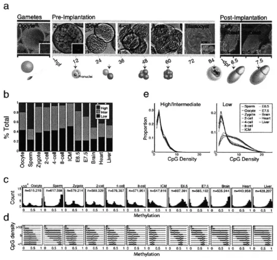
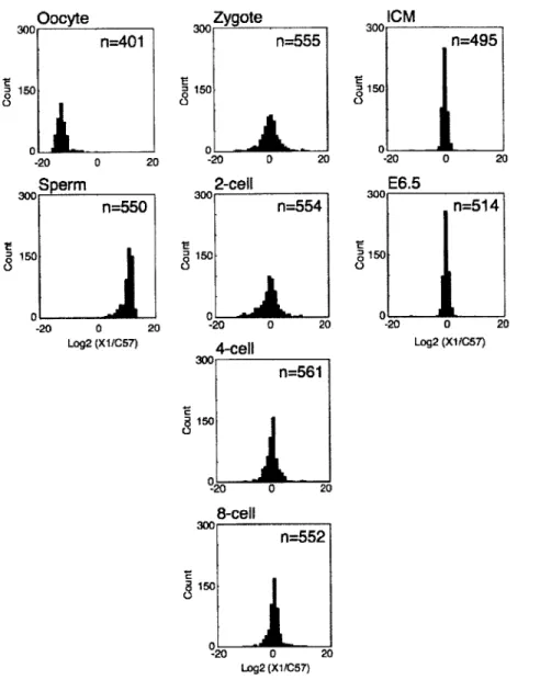
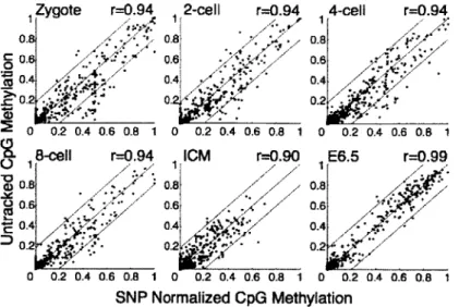
To generate a global and high-resolution view of early mammalian DNA methylation dynamics, we collected oocytes and sperm, as well as zygote, 2-, 4-, and 8- cell cleavage stage embryos, the inner cell mass (ICM) and E6.5/7.5 post-implantation embryos (Figs. 1-la, 1-2 and 1-3). All samples were extensively washed and purified to remove any somatic or meiotic contaminants. Potential maternal biasing from Meiosis I and II polar bodies (representing a Ix or 0.5x static genomic contaminant, respectively) was excluded by manually removing the polar bodies (Fig. 1-2) and assessing the paternal (129X1) to maternal (C57/B6xDBA) ratio of sequenced single nucleotide polymorphisms (SNPs) (Figs. 1-4 and 1-5). We generated reduced representation
bisulfite sequencing (RRBS)4 libraries from each stage, which were combined to provide a
comprehensive timeline of genomic DNA methylation patterns during early mouse embryogenesis.
Compared to all other genome-wide profiling strategies currently available, RRBS is optimally
suited for the low cell numbers that can be obtained from the embryonic stages in our study2 3
,2 4.
Within our range of 0.5-1Ong genomic DNA, RRBS provides high sensitivity and
reproducibility, and the expected genomic coverage (Fig. 1-3). On average, we obtained the methylation status of 953,606 CpGs for comparative analysis across our pre-implantation timeline. Different genomic features were equally well captured in libraries from the different stages (Fig. 1-3). Unfortunately, bisulfite sequencing cannot distinguish between methyl- and hydroxymethyl- cytosine (hmC), and current methods for global profiling of hmC lack the
required sensitivity to investigate the pre-implantation stages in this study9,25
cannot make any definitive statements regarding base resolution hmC distribution during the
pre-implanation stages. However, while hmC shows some unique distribution patterns in mES lines and brain tissue, it has not yet been linked to a regulatory mechanism other than to potentiate
demethylation31. Given this ambiguity, regions of high methylation, especially those retained
over multiple time points, could still be expected to function as if methylated.
1.3.2 Global CpG methylation in the early embryo does not resemble somatic patterns
The current model postulates a phase of global hypomethylation during mammalian
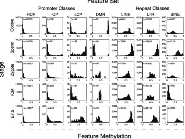
pre-implantation development that reaches a minimum at the morula/blastocyst stage. However, this model does not specify which genomic regions are affected or whether the methylation patterns in this phase still follow the general correlation with sequence context that have been established for somatic cells'. To address these questions, we investigated the global dynamics of CpG methylation using 1 00bp tiles. We calculated the methylation level of each tile by averaging over
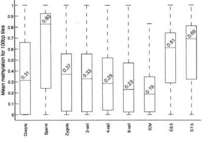
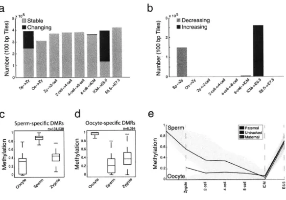
all CpGs within it that were covered with at least 5 reads. Intriguingly, we found that oocytes are already dramatically and globally hypomethylated compared to sperm (0.32 median methylation in oocyte versus 0.85 in sperm for 100bp tiles, Fig. 1-6). We next examined the relative
proportion of genomic regions at each stage falling into high (>0.8), intermediate (>0.2 and <0.8) or low (50.2) methylated categories. Notably, oocyte methylation levels more closely resembled those of early embryonic time points than the levels in sperm, post-implantation embryos, or adult tissues (Fig. 1-1b). We also observed a gradual increase in the fraction of tiles that exhibit intermediate and low methylation values from oocytes to the ICM, which is consistent with loss
We observed a dramatic difference in CpG methylation levels as a function of both CpG density
and developmental stage. Sperm and post-implantation embryos displayed the same strong
inverse relationship between CpG density and methylation levels that is present in somatic cells.
In oocyte and pre-implantation samples, this dependence was much weaker (Fig. 1-1c,d). More
specifically, we examined the relative CpG density in 'methylated tiles' (>0.2 methylation, Fig.
1-le, left) and 'hypomethylated tiles' (50.2 methylation, Fig. 1-le, right). In both pre- and
post-implantation embryos, methylated CpGs tend to be in tiles with low CpG density, thereby
recapitulating the somatic pattern (Fig. 1-le, left). In contrast, the average CpG densities in
hypomethylated tiles were lower in pre-implantation embryos and oocytes than they were in
sperm, post-implantation embryos or adult tissues (Fig. 1-le, right). This suggests that CpG
density-dependent methylation is primarily a post-implantation and adult pattern. In summary,
pre-implantation development represents a unique developmental period where methylation is
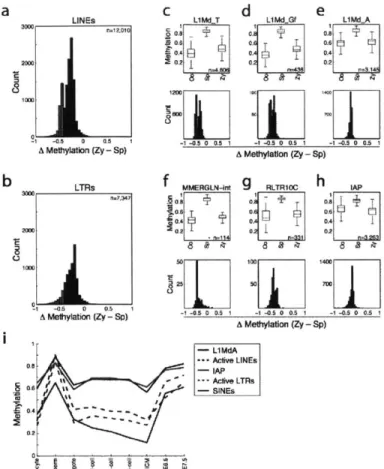
differentially positioned and regulated before being restored and maintained in a somatic fashion.
1.3.3 Two major transitions in methylation levels during early development
We next searched for substantial changes in regional DNA methylation and pinpointed the stage
at which they occurred. For each pair of consecutive stages, we compared methylation levels of
each 100bp tile and classified it as changed if the difference exceeded 0.2 and was significant
according to a FDR-corrected t-test (Fig. 1-7a).
The most dramatic changes in DNA methylation occurred during two developmental transitions:
between sperm and the zygote and between the ICM and the post-implantation embryo (Fig.
1-7a). At each of these transitions, the majority of changes were unidirectional (Fig. 1-7b): a gross
depletion of sperm hypermethylation upon fertilization (mean change = 0.48 decrease for 22% of
regions examined) and massive remethylation from the ICM to post-implantation embryos (mean
= 0.46 increase in methylation at 65% of tiles). Within our two post-implantation timepoints
(E6.5 and E7.5), the methylation levels at the majority of assayed tiles were stable or increased only slightly (Fig. 1-7b). In addition, more subtle global changes, reflecting a gradual decrease in methylation, was observed from zygote/early cleavage through the 8-cell stage and into the ICM, where methylation levels reached their lowest observed values (Fig. 1-1b,c).
1.3.4 The oocyte defines the early methylation landscape
Active demethylation is expected to occur prior to pronuclear fusion or DNA synthesis and is
generally described as complete after -6 hours post fertilization'. Indeed, when we compare
methylation patterns between sperm and zygote, the majority of regions in the genome show reduced methylation in the zygote with few additional changes from zygote to the 2-cell stage (Fig. 1-7b). Interestingly, the vast majority of tiles that are methylated at significantly different levels between gametes show higher methylation levels in sperm than in oocyte. In the zygote, their methylation is reduced to levels at or near those of the oocyte (Fig. 1-7c, d). Specifically, of
106,081 tiles that are significantly different between sperm and oocytes, 102,862 have higher methylation in sperm, but their levels more closely resemble oocyte values in the zygote stage. We confirmed this observation by tracking -65 CpGs that fell within these tiles and could be
assigned paternal or maternal specific values. Zygotes displayed a decrease in paternal
methylation in contrast to maternally contributed CpGs, which remained unmethylated (Fig. 1-7e). It is worth noting that the isolated zygotes used here are likely in earlier stages of zygotic S phase, such that either a passive, replicative based mechanism could result in the synthesis of