HAL Id: tel-01696139
https://tel.archives-ouvertes.fr/tel-01696139v2
Submitted on 9 Mar 2018
HAL is a multi-disciplinary open access
archive for the deposit and dissemination of sci-entific research documents, whether they are pub-lished or not. The documents may come from teaching and research institutions in France or abroad, or from public or private research centers.
L’archive ouverte pluridisciplinaire HAL, est destinée au dépôt et à la diffusion de documents scientifiques de niveau recherche, publiés ou non, émanant des établissements d’enseignement et de recherche français ou étrangers, des laboratoires publics ou privés.
development : insights on the ontogeny of face and
speech processing lateralization
Parvaneh Adibpour
To cite this version:
Parvaneh Adibpour. Anatomo-functional correlates of visual and auditory development : insights on the ontogeny of face and speech processing lateralization. Neurons and Cognition [q-bio.NC]. Université Pierre et Marie Curie - Paris VI, 2017. English. �NNT : 2017PA066256�. �tel-01696139v2�
1
Thèse de doctorat
de l’université Pierre et Marie Curie
Spécialité Neurosciences Cognitives
Présentée par
Parvaneh Adibpour
Anatomo-functional correlates of visual and auditory development:
insights on the ontogeny of face and speech processing lateralization
Thèse soutenue le 17 Octobre 2017
Devant le jury composé de :
Mme Christine Deruelle Rapporteur
M. Fabrice Wallois Rapporteur
M. Bruno Rossion Examinateur
Mme Nathalie George Examinateur
Mme Jessica Dubois Co-encadrante de thèse
Mme Ghislaine Dehaene-Lambertz Directrice de thèse
3
Acknowledgements
I owe many thanks to several people around the world in the path of learning over the past four years: First of all, I would like to thank my supervisors for granting me the “visa” to enter the whole new world of “infant brain”: Ghislaine Dehaene-Lambertz, for supervising this work with passion and encouraging me to move forward, for changing my vision by asking “what do we learn about the brain then?”, and for all what I learned from her in our discussions. Jessica Dubois, for her thoughtful supervision, for taking my hand and guiding me in this path with so much patience, for sitting and discussing with me closely all along, for bearing with me as a novice, and for all what she taught me, from methods to concepts!
This thesis would have not been possible without all the babies/parents who generously shared their brain/time with us. Those babies that I participated in testing them myself, brought joy to my days in the lab and discussing with the parents who valued science was a great experience of the past few years. Thus, I am more than thankful to all these babies and their parents. Part of these babies were studied in collaboration with Dr. Marie-Laure Moutard, to whom I am thankful for the opportunity to study, even if very briefly, neurodevelopmental pathologies.
I am grateful to many people that I met at Neurospin: Claire Kabdebon, definitely for her scientific share in my works and for being open to help whenever I asked for, but also for her presence as a great friend in the lab/library days, especially for the last months of writing. Jessica Lebenberg, for her help in MRI analyses, her reassuring presence from my first days in the lab and all our discussions and chats over tea pauses. François Leroy, for our discussions, and his help with MRI softwares and technical issues of the EEG room!
Vanna Santoro, not only for her precious help in carrying out the EEG experiments, but also for dealing with all the extra-administrative works with a smile and a calm attitude. UNIACT‘s clinical team for their help in carrying out the experiments, and especially Gaëlle Mediouni for being very helpful all along. Marie Palu, for her help in carrying out the ongoing experiment and for all the energy she brings to the team. Antonio Moreno and Isabelle Denghien, for their helpfulness with the technical issues and all the peaceful vibes they bring to the lab. Laurence Labruna for her helps with administration and her home-made cookies! The past and present members of the baby team: Pablo Barttfeld, for his peaceful and reassuring presence. Julia Carbajal, Ana Flo, Andrea Hisi, Irene Altarelli, Milad Ekramnia, Giulia Gennari, Karima Mersad, Karla Monzalvo, Antoinette Robert, Eric Moulton, Marie Zomeno, Kévin Da Silva, Leslie Nollace, Alexandra Hertz, and Francois-Daniel Ardellier for all the good baby-team times! Neurospin friends and colleagues across offices/open spaces/floors: Valentina Borghesani, for bringing a lot of social life to the lab. Mehdi Rahim, Darinka Trübutschek, Andres Hoyos Idrobo and Leila Azizi for their presence all along. Thien-Ly Pham, Laetitia Grabot, Martin Perez-Guevara, Elisa Castaldi, Fosca Al Roumi, Bianca Trovo, Clément Moutard, Benoit Martin, Elodie Doger de Spéville, Ana-Luisa Pinho, Baptiste Gauthier, Pedro Pinheiro Chagas,
4
Kamalakar Reddy and Aina Frau for all the time we spent together, and of course Elvis Dohmatob, for helping to save my computer!
Christophe Pallier for his help with statistics and his passion for teaching, Caroline Huron, Sébastien Marti, Stanislas Dehaene, Lucie Hertz-Panier, Florent Meyniel and Denis Riviere for all the interactions and discussions that each taught me in one way or another.
I would like to thank places too: College de France and its library, for keeping up with “science for all”! I am thankful to many people outside Neurospin as well: Anne-Caroline Fievet, for being very helpful for testing more babies in the LSCP lab, Jacqueline Fagard and Veronique Izard, for their nice feedbacks in the meetings. François Vialatte and Gerard Dreyfus, for guiding me into neuroscience at the very beginning point and before I start my PhD. Antoine Gaume, for showing me the true enthusiasm of sharing in science. Jason, for kindly helping me with corrections for English in parts of this thesis and for his moral support. Iman joon, Nahid khanoom and aghaye Momken for sharing their family love with me. Amoo Reza, for showing me his genuine way of dealing with hardships. Shervin, for being a present ear, whenever I needed. And of course, Sara, the friend for all seasons, for being there to help and to listen to me.
My family, maman, baba, Hamid and Farzaneh, for their support all along and for all what I can barely put into words. Pedarbozorg and Mamani for their care and love.
Finally, I would like to thank the jury members who accepted to take part in my jury and to read this manuscript: Dr. Christine Deruelle, Dr. Nathalie George, Dr. Bruno Rossion and Dr. Fabrice Wallois.
5
Abstract
Several cognitive functions, such as language processing, handedness, and face recognition, are lateralized in the adult human brain. The ontogeny of these functional asymmetries is still poorly understood, raising interest in studying whether they are present early on during development. To approach this issue, we attempted to map and compare the hemispheric responses in the infant brain. In particular, we aimed to evaluate the neural substrates of face and speech processing nested in the visual and auditory networks using non-invasive neuroimaging techniques and lateralized presentation of stimuli in infants.
First, we studied how the functional and structural characteristics of these two brain systems change over the first six months after birth. With event related potentials (ERP), we showed major age-related decreases in the latency of brain responses, both for early responses (visual P1 and auditory P2) and for inter-hemispheric transfer (visual P1). Using diffusion magnetic resonance imaging (MRI), we observed significant changes in myelination indices for both the visual and auditory white matter pathways. Above these relationships to infants’ age, we further demonstrated that the conduction speed of visual responses is related to the maturation of underlying tracts conducting these responses. In the auditory system, we failed to establish similar structure-function relationships, suggesting that the contribution of other pathways and cortical maturation should be further considered.
In parallel, we studied the hemispheric lateralization of face processing abilities using a discrimination paradigm of faces presented in each hemifield. Based on N290 and P400 responses in the infant ERP, we first observed that only the right hemisphere, and not the left, was able to discriminate between novel and frequently presented faces. When the frequently presented face from one side appeared on the opposite hemifield, no discrimination response was raised. We suggest that this observation is related to the transfer of face-relevant information across hemispheres.
Finally, we studied the lateralization of speech processing abilities in infants. When syllables were presented binaurally, we observed a trend for higher amplitude of P2 responses in the left relative to the right hemisphere. When syllables were presented monaurally, responses tended to be of higher amplitude and shorter latency in the contra- versus ipsi-lateral hemisphere. Ipsilateral responses were particularly delayed on the left side in typical infants but not in infants with corpus callosum agenesis, suggesting an asymmetric transfer of responses mediated by callosal fibers.
In summary, our studies highlight the potential of complementary neuroimaging approaches to study the infant brain and suggest differences in the development of visual and auditory systems and their processing biases. We also underscored the role of the corpus callosum, its protracted development, and the asymmetrical transfer of auditory information that may contribute to the reinforcement of a left-hemispheric bias for speech processing
6
Résumé
Plusieurs grandes fonctions cognitives, comme le traitement du langage, la manualité et la reconnaissance des visages, sont latéralisées dans le cerveau de l’humain adulte. Alors que l’origine de ces asymétries fonctionnelles reste encore méconnue, il apparait fondamental d’en étudier le développement. Dans ce contexte, nous avons essayé d’identifier et comparer les réponses hémisphériques dans le cerveau du bébé. Pour cela, nous avons cherché à évaluer les substrats neuronaux du traitement du visage et du langage imbriqués dans les réseaux visuels et auditifs grâce à des techniques de neuroimagerie non invasives et une présentation latéralisée des stimuli chez les nourrissons.
Premièrement, nous avons étudié comment les caractéristiques fonctionnelles et structurelles de ces deux systèmes cérébraux changent au cours des six premiers mois après la naissance. Avec les potentiels évoques, nous avons constaté des diminutions majeures de la latence des réponses cérébrales en fonction de l’âge, tant pour les réponses précoces (P1 visuel et P2 auditif) que pour le transfert interhémisphérique (P1 visuel). À l'aide de l'imagerie par résonance magnétique (l’IRM) de diffusion, nous avons observé des changements significatifs dans les indices de myélinisation pour les voies visuelles et auditives de la matière blanche. En plus de ces effets liés à l'âge des nourrissons, nous avons également démontré que la vitesse de conduction des réponses visuelles est liée à la maturation des faisceaux de fibres de substance blanche conduisant ces réponses. Dans le système auditif, nous n'avons pas pu observer de relation structure-fonction équivalente, ce qui suggère que la contribution d’autres voies ainsi que la maturation corticale devraient être étudiées en perspective.
Parallèlement, nous avons étudié la latéralisation hémisphérique du traitement des visages grâce à un paradigme de discrimination des visages présentés dans chaque hémichamp. Sur la base des réponses N290 et P400 en potentiels évoqués chez le nourrissons, nous avons d'abord observé que seul l'hémisphère droit, et non le gauche, a pu discriminer les nouveaux visages de ceux fréquemment présentés. En revanche, nous n’avons observé aucune réponse de discrimination lorsque le visage fréquemment présenté d'un côté est apparu dans l’hémichamp opposé. Nous suggérons que cette observation est due au transfert d'informations pertinentes au visage entre les hémisphères.
Enfin, nous avons étudié la latéralisation du traitement de la parole chez le nourrisson. Nous rapportons une tendance à une plus grande amplitude P2 dans l'hémisphère gauche par rapport à l'hémisphère droit en réponse à des syllabes présentées de manière binaurale. En réponse à des syllabes présentées de manière monaurale, les amplitudes évoquées ont tendance à être plus élevées, avec une latence plus courte dans l'hémisphère controlatéral versus ipsilatéral. Nous rapportons des réponses ipsilatérales particulièrement retardées dans l’hémisphère gauche chez les nourrissons typiques, mais pas chez les nourrissons avec agénésie du corps calleux, ce qui suggère un transfert asymétrique des réponses via les fibres du corps calleux.
En résumé, nos études mettent en évidence le potentiel des approches complémentaires en neuroimagerie pour étudier le cerveau du nourrisson et suggèrent des différences dans le développement des systèmes visuels et auditifs et leurs biais de traitement. Nous avons également souligné le rôle du corps calleux, son développement lent et le transfert asymétrique de l'information auditive qui peut contribuer au renforcement d'un biais hémisphérique gauche pour le traitement de la parole.
7
Contents
Abstract 5 Résume 6 1. Introduction... 171.1. The developing human brain ... 18
1.1.1. Micro- and macro- structural development of the human brain ... 19
1.1.2. Functional development ... 24
1.1.3. Early hemispheric asymmetries and functional lateralization in the developing brain .. 25
1.2. Neural bases of language ... 27
1.2.1. Language network and its lateralization in the adult brain ... 27
1.2.2. The first stages of language acquisition in infants ... 29
1.2.3. Neural bases of language development in infants ... 30
1.2.4. Is the early language network asymmetric? ... 32
1.2.5. Development of asymmetries vs. plasticity for language learning ... 36
1.3. Neural bases of face processing ... 37
1.3.1. Face processing network and its lateralization in the adult brain ... 37
1.3.2. Early face processing abilities ... 40
1.3.3. Neural bases of face processing in infants ... 41
1.3.4. Is face processing lateralized in infants? ... 44
1.3.5. Development of lateralization vs. plasticity for face processing ... 45
1.4. Lateralization versus. interhemispheric connectivity ... 46
1.4.1. Interhemispheric connectivity in the visual and auditory networks ... 49
1.4.2. Development of interhemispheric connectivity ... 51
1.4.3. Agenesis of corpus callosum: A model to study the role of interhemispheric connectivity in the development of visual and auditory networks ... 53
1.4.4. Plasticity for the establishment of interhemispheric connectivity patterns ... 54
1.5. Outline ... 55
2. Early lateralized face processing... 59
2.1. Abstract ... 59
2.2. Introduction ... 60
2.3. Materials and Methods ... 65
8
2.3.2. MRI acquisition and post-processing of diffusion MRI images ... 65
2.3.3. EEG protocol ... 66
2.3.3.1. Experimental paradigm ... 66
2.3.3.2. EEG data acquisition ... 68
2.3.4. EEG processing and ERP analyses ... 68
2.3.4.1. EEG pre-processing ... 68
2.3.4.2. Early visual perception ... 69
2.3.4.3. Discrimination of lateralized presented faces ... 70
2.4. Results ... 71
2.4.1. An efficient inter-hemispheric transfer of early visual responses in infants ... 71
2.4.2. An efficient discrimination of left-hemifield faces ... 74
2.5. Discussion ... 79
2.5.1. Fiber-specific microstructural maturation correlates with the acceleration of evoked responses ... 80
2.5.2. Lateralized presentation reveals an incompetent left hemisphere to discriminate faces ... 81
2.5.3. Efficiency of the inter-hemispheric transfer of visual information: ... 83
2.6. Conclusion: ... 84
2.7. Appendix: Follow-up experiment ... 86
3. Neural correlates of auditory development ... 93
3.1. Abstract: ... 93
3.2. Introduction: ... 94
3.3. Material and Methods: ... 96
3.3.1. Subjects ... 96
3.3.2. EEG studies: ... 97
3.3.2.1. Experimental paradigms ... 97
3.3.2.2. EEG data acquisition: ... 97
3.3.2.3. EEG pre-processing: ... 98
3.3.2.4. Auditory evoked potentials identification: ... 98
3.3.3. MRI study ... 99
3.3.3.1. MRI acquisition: ... 99
3.3.3.2. Pre-processing ... 99
3.3.3.3. Tractography ... 99
9
3.3.4. Statistical analyses ... 100
3.4. Results: ... 101
3.4.1. Development of P2 characteristics ... 102
3.4.2. Development of auditory pathways ... 103
3.4.3. Correlations between auditory pathways maturation and P2 conduction velocities .. 104
3.5. Discussion: ... 105
3.5.1. Early markers of functional lateralization and structural asymmetries ... 105
3.5.2. Functional and structural markers of maturation in the auditory system ... 107
3.5.3. Linking the functional and structural markers of maturation ... 108
3.6. Conclusion ... 109
4. Role of callosal pathways in infant auditory network ... 113
4.1. Abstract ... 113
4.2. Introduction ... 114
4.3. Materials and Methods ... 117
4.3.1. Subjects ... 117
4.3.2. EEG data acquisition ... 117
4.3.3. Stimuli ... 118
4.3.4. Experimental paradigm ... 118
4.3.5. EEG pre-processing ... 118
4.3.6. Analyses of auditory-evoked responses in typical and AgCC infants ... 119
4.4. Results ... 121
4.4.1. Different ERPs topographies in typical and AgCC infants ... 121
4.4.2. Different P2 latencies in typical and AgCC infants and across brain hemispheres ... 123
4.4.3. Different P2 latencies depending on the paradigm of auditory stimulation ... 124
4.5. Discussion ... 125
4.5.1. Altered topography of auditory responses in AgCC infants ... 126
4.5.2. A strong contribution of the ipsi-lateral pathway to auditory processing in the developing brain ... 126
4.5.3. Asymmetry of inter-hemispheric connections ... 127
4.6. Conclusions ... 128
4.7. Supplementary information ... 130
4.7.1. Do P2 amplitudes differ in typical and AgCC infants apart from differences in ERPs topographies? ... 130
10
4.7.3. Are P2 amplitudes in response to monaural stimuli stronger over the contralateral
compared to the ipsilateral hemisphere? ... 132
5. General discussion ... 134
5.1. Functional relevance of structural maturation ... 135
5.2. Neural correlates of early lateralization ... 139
5.2.1. Early lateralization of face processing ... 140
5.2.2. Early lateralization of speech processing ... 143
5.3. Early efficiency of interhemispheric communications ... 147
5.4. Perspectives for neurodevelopmental outcomes ... 150
Appendix: Neuroimaging of the developing infant brain ... 154
EEG ... 154
Neural correlates of development assessed with event-related potentials ... 155
Challenges of developmental EEG ... 155
Towards exploring EEG dimensionality ... 158
Diffusion MRI ... 159
Diffusion models ... 159
Tractography ... 159
White matter maturation ... 160
Challenges in diffusion MRI studies of developing brain ... 161
11
List of Figures
Figure 1.1: Developmental periods of occurrence and intensity of main neurogenetic events in cortical
hystogenesis ... 19
Figure 1.2: Development and maturation of gray and white matter over the course of development as revealed by post-mortem studies. ... 20
Figure 1.3. Structural imaging of cortical maturation. ... 21
Figure 1. 4. Diffusion MRI of the developing white matter ... 22
Figure 1.5. Structural imaging of cortical folding and growth ... 23
Figure 1.6. Structural imaging of asymmetrical cortical folding. ... 26
Figure 1.7. Network of brain regions implicated in linguistic processings ... 27
Figure 1.8. Hemodynamic responses to speech. ... 31
Figure 1.9. Structural asymmetries in the language network in infancy. ... 33
Figure 1.10. Functional asymmetries in the language network in infancy. ... 35
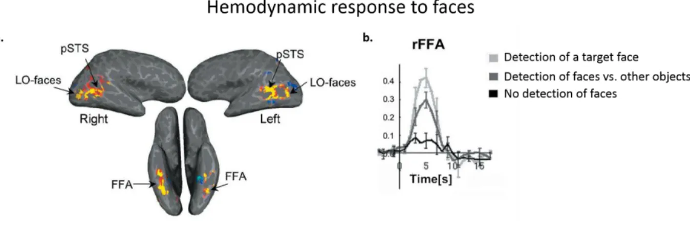
Figure 1.11. Hemodynamic response to faces ... 38
Figure 1.12. Right lateralized EEG responses to faces in infancy. ... 43
Figure 1.13. Callosal fiber connections reconstructed with tractography ... 48
Figure 2.1. EEG experimental paradigms ... 67
Figure 2.2. Examples of visual event-related responses in one infant ... 72
Figure 2.3. Structure-function relationship ... 73
Figure 2. 4. ERP according to hemifield ... 75
Figure 2.5. ERP according to the face conditions ... 76
Figure 2. 6. Comparison of N290 and P400 components across face conditions ... 77
Figure 2. 7. Examples of visual event-related responses over the group of infants ... 87
Figure 2.8. ERP according to the car conditions. ... 88
Figure 3.1. Time course of auditory evoked responses ... 101
Figure 3.2. Decrease in P2 response latency with age ... 102
Figure 3.3. Hemispheric differences for P2 response latency and amplitude ... 102
Figure 3.4. Maturation of auditory pathways quantified with DTI indices ... 104
Figure 3.5. Partial correlation between P2 speed and auditory callosal fibers maturation while controlling for age ... 105
Figure 4.1. Topographical differences between typical and AgCC infants ... 122
Figure 4.2. Comparison of P2 response latency between typical and AgCC infants ... 123
12
Sup. Figure 4.1. Auditory-evoked responses to different stimuli in typical and AgCC infants ... 130
Sup. Figure 4.2. P2 response amplitude comparison between typical and AgCC infants ... 131
Sup. Figure 4.3. Influence of the paradigm on the P2 response amplitude... 132
Figure 5. 1. Schematic model of developmental hemispheric lateralization ... 147
Figure 5.2. Microstructural maturation of auditory and visual callosal fibers. ... 149
Figure 6.1. Inter-trial phase coherence following a face stimulus. ... 155
Figure 6.2. Developmental changes in DTI parameters in the reconstructed tracts of the visual and auditory network. ... 161
13
List of Tables
Table 2. 1. Maturational synchrony across bundles: ... 73
Table 2. 2. Structure-function relationships ... 74
Table 2. 3. Comparison of N290 and P400 responses for the different face conditions ... 79
Table 2.4. Anova summary for N290 and P400 responses for the different car conditions ... 89
Table 3.1. Summary of ANCOVA analyses for P2 amplitude and latency in response to monaural syllables ... 103
Table 3.2. Partial correlations between P2 speed and maturation of acoustic radiations and auditory callosal fibers while controlling for age ... 104
Table 4.1. ANOVAs of P2 response latency in the binaural, monaural and dichotic trials ... 124
14
List of abbreviations
aCC Auditory fibers of Corpus Callosum AF Arcuate Fasciculus
AgCC Agenesis of Corpus Callosum AR Acoustic Radiations
DTI Diffusion Tensor Imaging EEG Electroencephalography EC External Capsules
EmC extreme Capsule ERP Event Related Potentials FA Fractional Anisotropy
fNIRS functional Near Infrared Spectroscopy fMRI functional Magnetic Resonance Imaging FFA Fusiform Face Area
HG Heschl's Gyrus
IFOF Inferior Fronto-Occipital Fasciculus IHTT Inter-Hemispheric Transfer Time LGN Lateral Geniculate Nucleous LO Lateral Occipital cortex MEG Magnetoencephalography MGN Medial Geniculate Nucleous MRI Magnetic Resonance Imaging
N170 face-sensitive adult’s visual ERP component N290 face-sensitive infant’s visual ERP component OR Optic Radiations
P1 First positive ERP component P2 Second positive ERP component
P400 face-sensitive infant’s visual ERP component PET Positron Emission Tomography
PT Planum Temporale
SLF Superior Longitudinal Fasciculus STS Superior Temporal Sulcus
TMS Transcranial Magnetic Stimulation UF Uncinate Fasciculus
vCC Visual fibers of Corpus Callosum <D> mean Diffusivity
𝜆┴ Transverse Diffusivity
15
Chapter 1
17
1. Introduction
Human way of inspecting the surrounding environment, acting on, and communicating with it, is unique in many aspects. Extensive amount of research has focused on understanding the neural mechanisms that support the human abilities. Accumulating evidence shows that the human brain is composed of several regions that are functionally specialized in performing a task or dealing with certain aspects of environmental inputs. These regions are similar across individuals with different cultural, linguistic, or socio-economic backgrounds, suggesting that the functional architecture of the human brain remains similar for the wide range of variability in the environmental inputs.They are not isolated with respect to the rest of the brain, but interact with other areas and send/receive information, through the connections between them, giving rise to functional circuitries and networks that have distributedregions.
The rationale behind these functionally specialized regions might be related to the speed and energy efficiency that they provide by relying on computations that are performed locally through spatially-close neuronal assemblies. Hemispheric lateralization is a special example of regional functional specialization in the brain, when certain computations remain preferentially localized in a region or a network of regions inside one hemisphere, instead of relying on neuronal assemblies across hemispheres that is limited by the timing constraints of the neural information transfer between hemispheres. It is important to note that these hemispheric lateralizations refer to hemispheric advantages rather than radical binary ability of a single hemisphere in performing a task. In the adult human brain, the existence of numerous anatomical and functional differences between the two hemispheres is supported by several post-mortem, behavioral and neuroimaging studies (Hugdahl & Westerhausen, 2010; Kimura, 1961; Toga & Thompson, 2003), and many functions such as handedness, language, face processing, visuospatial processing., etc. are biased toward one hemisphere. Yet, little is known about the ontogeny of these lateralized functions in the human brain.
The question on the ontogeny can be addressed, to some extent, by studying the infant brain, asking whether functional lateralizations and structural asymmetries are the properties of adult brain or if they are found early on as well. These questions are informative in two respects. First, infant brain provides the early machinery that facilitates the emergence of human abilities. Therefore, insights on the development of the functional lateralizations can help to understand which aspects of an early architecture, biases and constraints the architecture of the human brain to achieve its many abilities. Second, characterizing the normal development of lateralized functions is also informative for our understanding of the critical periods
18
and developmental outcomes. It becomes especially important, when we take into account the coincidence of developmental disturbances like dyslexia or autism spectrum disorder with abnormal lateralization for processing language or social stimuli such as faces.
With these questions in background, we aimed at investigating the neural correlates of face and language processing abilities, while studying the functional and structural characteristics of the visual and auditory development in the infant brain. This first chapter presents a context for our studies. Section 1.1 describes the main stages of early development and organization of the brain, with an attempt to illustrate which mechanisms can push this early architecture to develop lateralized functions. Sections 1.2 and 1.3 particularly focus on the emergence of language and face processing abilities. Starting from the description of the neural substrates of each function in the adult human brain, we will focus on their development early on. Section 1.4 attempts to review how hemispheric differences might interact with inter-hemispheric communications. Finally, section 1.5 contains the plan for the following chapters and the questions addressed in each chapter.
1.1. The developing human brain
Studying the human brain is especially difficult during the early developmental period. Post-mortem histological studies can only anatomically characterize the maturation of neural substrates and therefore lack functional significance. Behavioral studies also remain limited, not only because of the indirectness of their measures, but especially due to the limited behavioral output at this early stage. With the emergence of non-invasive neuroimaging techniques, the developing infant brain is now accessible, enabling a characterization of early neuronal circuitries and their maturation in vivo. These techniques provide information about both structural and functional development of the brain. Magnetic Resonance Imaging (MRI) provides a description of the structural development and maturation of the cerebral tissues based on their water content. A number of other techniques allow assessing the functional responsiveness of neural circuits through measuring brain electrical activity or metabolic activity. Electroencephalography (EEG) records the electrical field generated by the summation of post-synaptic potentials on the surface of the head and Magnetoencephalography (MEG) captures the magnetic field perpendicular to the generated electric field. Functional MRI (fMRI) and Near-Infrared Spectroscopy (fNIRS) measure changes in blood oxygenation related to neuronal assemblies’ oxygen consumption and its vascular consequences. In the methodological appendix of this thesis, we describe in more details, the usefulness, technical
19
considerations and limitations of EEG and diffusion-MRI techniques when employed for studying infant population, as we used these approaches to perform the studies described in this thesis.
In section 1.1.1, we briefly review the main cellular, microscopic and macroscopic phenomena involved in the development and maturation of the brain. We then discuss their relevance for the emergence of functional maturation and hemispheric lateralizations in sections 1.1.2 and 1.1.3, respectively.
1.1.1. Micro- and macro- structural development of the human brain
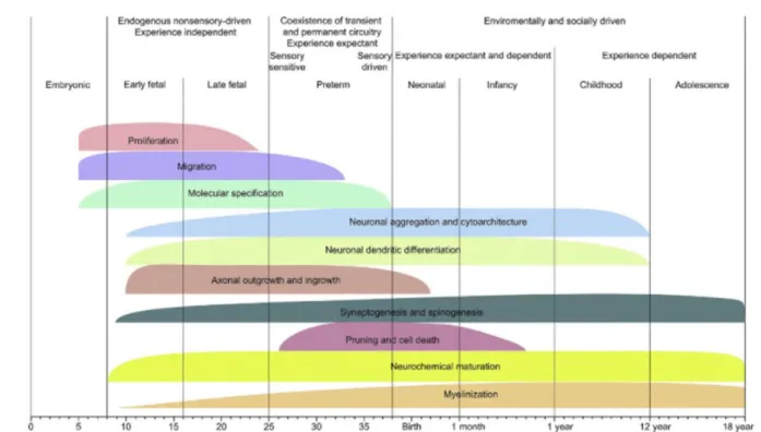
Brain development extends over a long period, starting during gestation and continuing toward the end of adolescence, and is mostly intense in the prenatal and first years of postnatal life. It relies on several maturational mechanisms that occur asynchronously, i.e. different onsets, and with different rates across brain regions and time-courses of development (Brody et al., 1987; Yakovlev. & Lecours., 1967) .Formation and maturation of the cortical layers and white matter connections rely on a succession of cellular events, driven by biological clocks or entrained by the sensory inputs. Figure 1.1 illustrates a schematic of the sequential, yet over-lapping, occurrence of these events during development (Kostović & Judaš, 2015).
Figure 1.1. Developmental periods of occurrence and intensity of main neurogenetic events in cortical hystogenesis. Note the predominance of proliferation and migration during the first trimester of gestation, growth of axons and dendrites during the second and third trimesters, and prolonged postnatal synaptogenesis, myelination and neurochemical maturation (Adapted from from Kostovic and Judas., 2015).
20
We will first describe these cellular events, and then detail their relevance in the microscopic and macroscopic developmental changes that are measured in post-mortem histological (Figure 1.2) and in vivo neuroimaging studies (Figure 1. 3 to 1.5). To illustrate the developmental processes, we combine the insights from both type of studies, without distinguishing and detailing the methods.
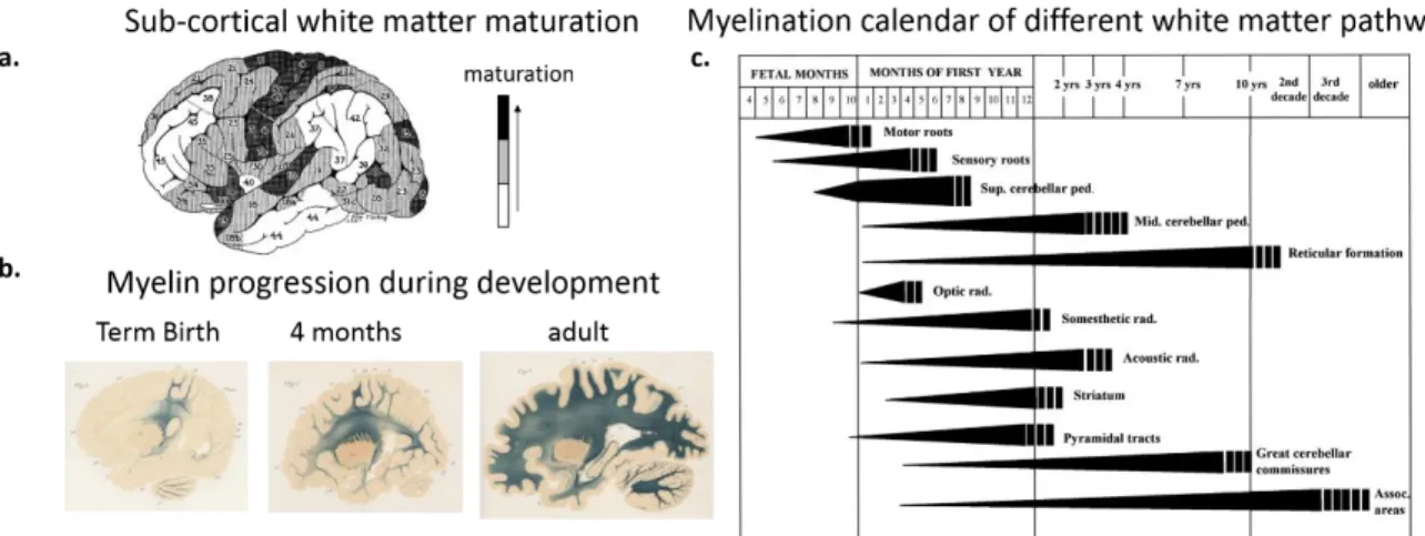
Figure 1.2. Development and maturation of gray and white matter over the course of development as revealed by post-mortem studies. Note the asynchronies across cortical regions and pathways. a) Maturation of white matter underneath cortical regions as an indicator of cortical maturation across regions (Adapted from Flechsig, 1901; 1920). The numbering of cortical regions indicates their maturation order. Darker areas are more mature than lighter areas. b) Myelin progression in developing brain. Note the progression gradient from the central to peripheral regions (Adapted from Flechsig, 1920). c) Cycles of myelination in the central nervous system during development. The width and the length of bars indicate progression in the intensity of staining corresponding to the density of myelinated fibers; the vertical stripes at the end of the graphs indicate approximate age range of termination of myelination estimated from comparison of the fetal and postnatal tissue with tissue from adults in the third and later decades of life (Adapted from Baumann & Pham-Dinh., 2001, originally adapted from Yakovlev & Lecours., 1967).
Development and maturation of cortex:
Neurons and glial cells are first produced during the embryonic period within two proliferative zones (Ventricular and Sub-Ventricular Zones). These neurons are then guided by the glial cells toward the brain periphery. They first reach the subplate zone, a transient layer underneath the future cerebral cortex that serves as a waiting compartment for the growing neurons (Pasko Rakic, 1972). The areal and laminar position of these migrating neurons are determined through signaling molecules secreted from patterning centers, i.e. commissural plate and cortical hem (O'Leary, Chou, & Sahara, 2007; Pasko Rakic, 1995), and their respective functional roles are further specified through the expression patterns of several genes (Bayatti et al., 2008; Wang et al., 2010). The cerebral cortex is progressively formed mostly in an inside-out manner, i.e. the last migrating neurons are settled in more superficial layers of cortex (Bystron, Blakemore, & Rakic, 2008; Rakic, 1995). Then the neurons are aggregated within the transient plates as well as in the cortical layers and form regions with specific cytoarchitectonic characteristics (Kostović & Judaš, 2015). Once neurons are in the cortical plate, their morphological characteristics (dendritic spines and axons),
21
start establishing (Mrzljak, Uylings, Kostovic, & van Eden, 1988; Petanjek, Judaš, Kostović, & Uylings, 2007) and cortical neural circuits are progressively formed. These circuits are first organized by overproduction of neuronal axons, dendrites, dendritic spines and also synapses and then refined and re-organized by processes like elimination of axons, later on with elimination of dendritic spines and synapses (Huttenlocher & Bonnier, 1991; Innocenti & Price, 2005; Petanjek et al., 2011). Following these events, myelination of intracortical axons that are not pruned occurs in two stages: first the proliferation and settlement of oligodendrocytes around the axons (Thomas et al., 2000) and secondly their spiral wrapping (Baumann & Pham-Dinh, 2001) and chemical maturation (Barkovich, Kjos, Jackson , & Norman, 1988; Poduslo & Jang, 1984).
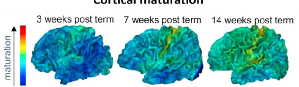
Between the fetal period and term age, the intense cortical microstructural changes can be captured in vivo with diffusion MRI. The increasing branching of dendritic spines and synaptic density within the cortex at this period, make the diffusion of water molecules more isotropic, and affects diffusion MRI parameters that are sensitive to the directionality of water molecules (Ball et al., 2013; McKinstry et al., 2002). During post-term period, cortical maturation relies on myelination of intra-cortical axons on the top of the synaptogenesis andpruningof axons, dendritic spines and synapses. These changes are also reflected in signal contrasts of MRI parameters, allowing to distinguish different maturational stages (Figure1.3)(Leroy et al., 2011), i.e. earlier maturation of primary and sensory cortices relative to the associative cortices as indicated in postmortem studies (Figure1.2.a) (Flechsig, 1920; Yakovlev. & Lecours., 1967).
Figure 1.3. Structural imaging of cortical maturation. Asynchronous maturation of cortical regions between 3 and 14 weeks of age (Adapted from Leroy et al., 2011).
Development and maturation of connections:
The axonal growth produces transient connectivity patterns during the fetal period, particularly toward the subplate zone underneath the future cortex (Kostović, Judaš, Radoš, & Hrabač, 2002). This transient connectivity starts to relocate from the subplate to the cortical plate in the early preterm period.
22
The first connections from thalamus to cortical neurons are established during this period (Dubois, Kostovic, & Judas, 2015; Kostovic & Rakic, 1990). In addition to the thalamo-cortical connections, limbic and associative cortico-cortical connections progressively develop before term (Dubois, Kostovic, & Judas, 2015; Takahashi, Folkerth, Galaburda, & Grant, 2011). This development continues after term for short fibers connecting adjacent gyri (Kostović et al., 2014). Most of the developed connections include overproduced axons that are pruned after birth in interaction with the environmental inputs, refining the connectivity in parallel with the reorganization of cortical synaptic patterns (Innocenti & Price, 2005). The pruning of axons occurs most intensely in the first postnatal year, and probably continues more slowly afterwards (Kostović et al., 2014; Petanjek et al., 2011). Parallel to the pruning of the connections, connections also become functionally more efficient through myelination of the axons that are not subject to elimination. Myelination is most intense around the term age and in the first postnatal years (Brody et al., 1987; Yakovlev & Lecours., 1967). It accelerates the speed of neural information that travel through the axons (Baumann & Pham-Dinh, 2001; van der Knaap & Valk, 2005).
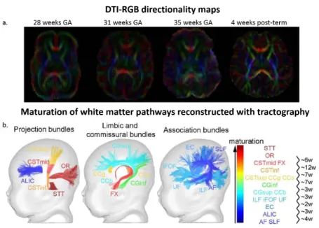
Figure 1. 4. Diffusion MRI of the developing white matter. a) DTI-RGB directionality maps of developing brain. Diffusion anisotropy increases as fibers become more compact and myelinated along development. b) The regional asynchrony in white matter maturation is highlighted in healthy infants using multi-parametric MRI. Projection, limbic, commissural and association bundles are ordered and colored according to their maturation, and maturational delays (in weeks) are computed between pairs of bundles (adapted from Dubois et al., 2015). Abbreviations: AF arcuate fasciculus; ALIC anterior limb of the internal capsule; CC corpus callosum (g/b/s genu/body/splenium); CG cingulum (inf/sup inferior/superior parts); CST cortico-spinal tract (inf/mid/sup inferior/middle/superior portions); EC external capsule; FX fornix; iFOF inferior fronto-occipital fasciculus; ILF inferior longitudinal fasciculus; OR optic radiations; SLF superior longitudinal fasciculus; STT spino-thalamic tract; UF uncinate fasciculus.
Mapping the establishment and microstructural maturation of connectivity for different pathways is possible through MRI, already from early preterm period (from ~24 gestational week onward). These
23
maps provide parameters that are sensitive to the coherence, compactness, density and myelination of fibers (Figure. 1.4.a) (Counsell et al., 2002; Deoni et al., 2012; Dubois et al., 2014; Hüppi et al., 1998) and can illustrate the asynchronous myelination of different pathways (Figure. 1.4.b) indicated by postmortem studies: It occurs earlier and faster in sensory than in motor pathways, in projection pathways (those connecting subcortical nuclei and cortical regions) than in associative ones, in proximal than distal pathways, in central regions than in peripheral ones, and in occipital lobe than in parietal, temporal and frontal lobes (Figure 1. 2.b and c)(Brody et al., 1987; Yakovlev & Lecours., 1967).
Morphological changes:
At the macroscopic level, a remarkable change is related to the increase in the brain volume. Cortical volume increases substantially before term age, from about 10 cm3 to 200 cm3 between the 21 –
40 weeks of gestational age (Kuklisova-Murgasova et al., 2011; Scott et al., 2011), and after term birth, from about 200 cm3 to 500 cm3 during the first two postnatal years (Gilmore et al., 2011; Knickmeyer et
al., 2008). White matter volume also increases with development from about 50 cm3 to 150 cm3 between
the 30– 40 weeks of gestational age (Kuklisova-Murgasova et al., 2011) and from about 150 cm3 to 200 cm3
during the first two postnatal years (Knickmeyer et al., 2008). This growth continues with a slower rate toward adolescence both for cortical areas and white matter (Groeschel, Vollmer, King, & Connelly, 2010; Shaw et al., 2008; Sowell et al., 2004).
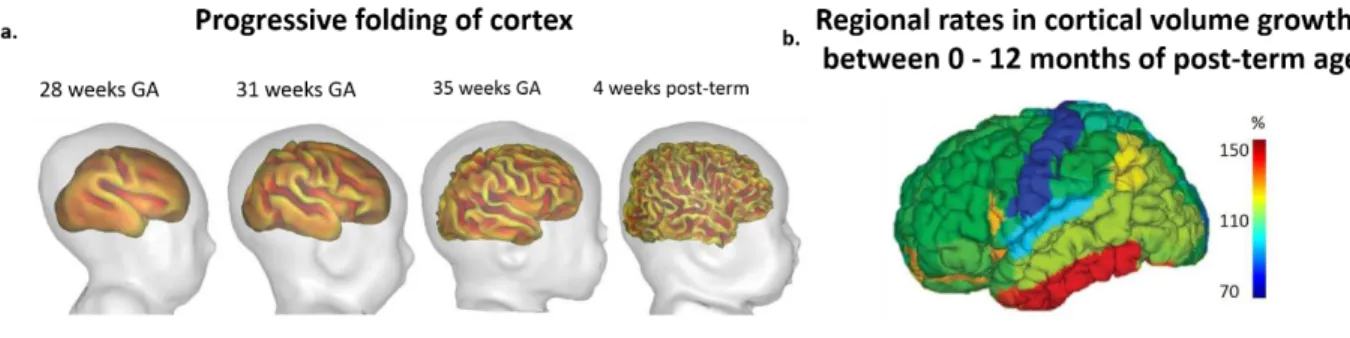
Figure 1.5. Structural imaging of cortical folding and growth. a) Inner cortical surfaces demonstrating the progressive folding and gyrification along development (Adapted from Dubois & Dehaene-Lambertz., 2015). b) Asynchronous cortical volume increase during the first post-natal year (Adapted from Gilmore et al., 2012).
Cortical volume increases asynchronously and with different rates across different regions and over different developmental periods. During the fetal period (20-28 weeks of gestation), the cortical volume increases more in the occipital and parietal regions compared to the frontal areas (Rajagopalan et al., 2011), whereas during the first two years of post-term period, it increases more rapidly in the visual ventral areas and frontal and parietal areas compared to the motor and sensory areas (Figure. 1.5.b) (Gilmore et al.,
24
2011). Parallel to the cortical and white matter volume growth, cortical folding and gyrification also occur sequentially for the primary, secondary and tertiary folds, and for the central, parietal, temporal, occipital and frontal gyri between the 20 weeks of gestational age and term age (Chi, Dooling, & Gilles, 1977; Dubois et al., 2008). At term, cortical gyrification is approximately adult-like (Hill et al., 2010) and changes much less significantly thereafter (Figure. 1.5.a).
The point of emphasis in all of the micro- and macro-structural maturational changes, is the asynchrony of occurrence onsets and rates across different regions and also developmental periods, suggesting that development of different brain networks follow very different trajectories.
1.1.2. Functional development
Parallel to the establishment and maturation of cortical circuits and structural connectivity, the activity arising from the emerging networks also changes. EEG recordings of neural activity progressively change from a discontinuous pattern at ~24 weeks of gestational age, recognized by sudden high-amplitude bursts of activity, to a more continuous pattern toward the term age (Lamblin et al., 1999; Wallois, 2010). Cortical evoked responses to sensory inputs such as sounds begin to appear in early preterm period, when thalamic afferents are relocating from the transient subplate zone to the cortical plate (Graziani et al., 1974). Myelination of fibers further decreases the latency of these sensory cortical evoked responses from the late preterm period toward infancy (Dubois et al., 2008; McCulloch, Orbach, & Skarf, 1999), referring to increasing efficiency of communication pathways.
Concurrent with these changes, fluctuations in the spontaneous neural activity of distant brain regions within a network also become more synchronized, and functional connectivity between these brain regions emerges. In infants, functional connectivity is often assessed during sleep with functional MRI. The first finger prints of emerging functional connectivity are found already during preterm period, highlighting regions and resting state networks that overlap with that of post-term period and partially with that of adults (Doria et al., 2010; Smyser et al., 2010; Thomason et al., 2014). Toward the term age, interhemispheric connectivity increases and different functional networks begin to get established (Doria et al., 2010; Fransson et al., 2009; Smyser et al., 2010; van den Heuvel et al., 2015). Yet, network measures suggest that cortical hubs, i.e. the key cortical regions interacting with many other regions and facilitating the functional integration across networks, may remain restricted to primary and sensory-motor cortices, suggesting that the functional architecture better supports basic perceptive and motor behaviors at this early stage (Fransson, Aden, Blennow, & Lagercrantz, 2010). The postnatal maturation of resting-state
25
functional networks continues asynchronously, showing a gradient from the primary sensorimotor/auditory networks, to the visual, default-mode and executive control networks (Gao et al., 2014; Gao et al., 2009).
In sum, several studies have highlighted the micro- and macro structural as well as the functional aspects of maturation in the infant brain that occur asynchronously across different regions and networks. What remains less investigated, is the link between the structural and functional aspects of maturation. We were interested in this question.
We intended to study the link between microstructural maturation of white matter pathways and functional efficiency of evoked responses for two brain networks with different developmental trajectories. We build on the rationality of a previous study in 1 to 4 months old infants, showing that the conduction speed of visual evoked responses was related more to the optic radiations (projection pathways) maturation than to the infants’ age (Dubois et al., 2008). We aimed at replicating this result for projection pathways and extending them to cortico-cortical callosal pathways in the visual network and to similar pathways of the auditory network, in chapters 2 and 3 respectively.
1.1.3. Early hemispheric asymmetries and functional lateralization in the
developing brain
In the previous sections, we described several regional asynchronies of cortical and connectivity development and maturation, e.g. earlier and faster maturation of primary and sensory areas relative to associative ones. However, we did not point that these asynchronies also exist between the left and right hemispheres, better known as asymmetries. Here, we highlight a few examples of asymmetries to draw the reader’s attention to their presence in early developmental period, but we will discuss more examples and in more details when focusing on the language and face processing networks in the rest of this chapter.
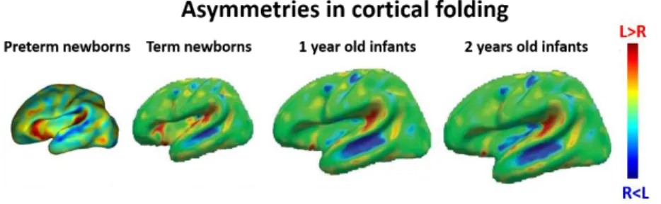
Early asymmetries are especially highlighted in the perisylvian regions. For example, cortical sulci in the temporal areas appears about 2 weeks earlier in the right than the left hemisphere in the fetal period (Figure 1.6)(Chi et al., 1977; Dubois et al., 2008), and planum temporale is larger in the left, while superior temporal sulcus is deeper in the right hemisphere during the first postnatal months (Glasel et al., 2011; Leroy et al., 2011). Asymmetries also exist at micro-structural level and in the maturational tempos of cortical regions and white matter pathways, e.g. superior temporal and inferior frontal regions and arcuate fasciculus, in the first months of infancy (Leroy et al., 2011).
26
Figure 1.6. Structural imaging of asymmetrical cortical folding. Asymmetries in cortical folding in preterm and post-term periods (Adapted from Dubois & Dehaene-Lambertz., 2015).
What gives rise to early structural asymmetries is still debated but a number of mechanisms are suggested to cause or interact with their emergence. Genetic factors, differential expression of genes, e.g. LMO4, in the two cerebral hemispheres could be one candidate (Sun et al., 2005). Random events like fluctuations in gene expressions might interfere with the pattern of brain development and make the impact of genetic factors more probabilistic (Raj & van Oudenaarden, 2008). Non-genetic factors like the position of the fetus in utero, exposing one side of the body more outward and thus channeling the environmental inputs more toward one hemisphere, may also result in differential development of the two hemispheres (Previc, 1991). The early regional asymmetries could also be the outcome of the interaction between these factors.
Asymmetries are not limited to structure and are present at the functional level too. For example, fMRI activations in response to speech in the perisylvian regions have shown to be stronger in the left than the right hemisphere in the first months of infancy (Dehaene-Lambertz, Dehaene, & Hertz-Pannier, 2002; Dehaene-Lambertz et al., 2010; Shultz, Vouloumanos, Bennett, & Pelphrey, 2014). How the structural asymmetries impact the functional architecture of the infant brain is still not clear. They might affect the dynamics of functional networks, at the time that these networks are gradually stabilizing, and result in different computational properties and responsiveness of the left and right networks of brain regions(Dehaene-Lambertz & Spelke, 2015). These differences might further give advantage to certain regions to process frequent stimuli in the environment (e.g. speech and faces) and therefore become better attuned for processing it (de Schonen,de Diaz, & Mathivet, 1986; Dehaene-Lambertz & Spelke, 2015). For example, the left hemisphere is known to be more sensitive to fast temporal changes embedded in the speech signal.
27
In sections 1.2 and 1.3, we will describe more specifically the lateralization of language and face processing networks and focus on their development.
1.2. Neural bases of language
1.2.1. Language network and its lateralization in the adult brain
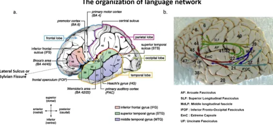
In the adult brain, linguistic processing is performed through a dedicated network of brain regions that extend around the sylvian fissure (Figure 1.7). The two key regions in this network are the inferior frontal gyrus (Broca’s area) and the posterior part of the superior temporal gyrus (Wernicke's region) that are connected by two main roads: dorsally by the arcuate fasciculus (AF) and parts of the superior longitudinal fasciculus (SLF)) and ventrally by the inferior fronto-occipital fasciculus (IFOF), extreme capsule (EmC) and external capsules (EC) and uncinate fasciculus (UF) (Axer, Klingner, & Prescher, 2013).
Figure 1.7. Network of brain regions implicated in linguistic processings. a) These regions are extended around the Sylvian fissure. Two main anatomical landmarks are Broca’s and Wernicke’s area (Adapted from Friederici., 2011. b) Connectivity of human language-related areas : including the fascicles of dorsal and ventral pathways (Adapted from Axer et al., 2013).
The perisylvian region is not symmetric in the two hemispheres. Macroscopic differences include elongated sylvian fissure and larger planum temporale (Geschwind & Levitsky, 1968), larger white matter volume underneath the Heschl’s gyrus and primary auditory cortices in the left compared to the right hemisphere (Penhune, Zatorre, MacDonald, & Evans, 1996). The arcuate fasciculus is also larger and more compact in the left than in the right hemisphere (de Schotten et al., 2011; Parker et al., 2005). Microscopically, the pyramidal neurons of the left auditory cortices are larger (Hutsler, 2003) and the white matter below the posterior superior temporal cortices has more thickly myelinated axons in the left than in the right (Anderson, Southern, & Powers, 1999). The size of the neuronal columns and the distance
28
between them is larger in the left superior temporal lobe (Seldon, 1981). Receptor density profiles of distant perisylvian regions (Broca’s area and Wernicke's region) are similar in the left but not right hemisphere (Zilles et al., 2015), in agreement with their common specialization for language processing.
Functional characteristics of the language network can be identified by localizing the brain activity when adult subjects listen to linguistic stimuli such as sentences and words. These activations appear around the perisylvian regions and are stronger in the left hemisphere, when subjects listen to their native language compared to non-native language (Mazoyer et al., 1993; Pallier et al., 2003). These observations suggest that the left perisylvian regions are responsive to features of speech specific to the native language. Yet, attributing a specific role to each of these regions is debated, as language is a complex hierarchical system that can be described at the level of phonology, syntax and semantics. Two main views exist about what aspects of language are better processed by the left hemisphere network. One view attributes this lateralization to speech and describes that spectro-temporal feature of the speech, notably the fast temporal transitions embedded in speech stimuli, are better processed by the structural properties of the auditory network in the left hemisphere (Boemio, Fromm, Braun, & Poeppel, 2005; Zatorre & Belin, 2001). This view is supported by evidence showing that brain oscillations at speech-relevant frequencies best correlate with activations in the left auditory cortices (Giraud et al., 2007). The second view relates the leftward superiority of language network to the communicative feature of language. This view is supported by evidence showing that leftward activation asymmetries are more pronounced for familiar versus unfamiliar (non-communicative) language(Pallier et al., 2003), evidence from deaf subjects showing similar leftward activations for communicative gestures of sign language (Sakai et al., 2005).
Human adults have extensive experience with language. Therefore, from adult studies alone it cannot be understood whether the described asymmetries of activations and anatomical properties are the causes for the speech (and language) to be lateralized or if they are a consequence of the exposure to linguistic stimuli with certain spectro-temporal properties. One way to approach this question is to study the ontogeny of these functional lateralizations early in life, when infants have limited knowledge of linguistic structures compared to experienced adults. This allows tackling when the functional and structural asymmetries are emerging, and how one may precede the other, to eventually shed light on their underlying mechanisms. In the following section, we describe early language acquisition and its neural bases and the emergence of their lateralizations.
29
1.2.2. The first stages of language acquisition in infants
Much before infants start to produce speech, their linguistic abilities start developing and their perceptual system is tuned to several aspects of speech stimuli. Newborns and young infants prefer speech stimuli over other auditory stimuli with similar acoustic features (Colombo & Bundy, 1981; Vouloumanos & Werker, 2007). They are able to discriminate two languages on the basis of different prosodies (Mehler et al., 1988; Nazzi, Bertoncini, & Mehler, 1998). Perceiving phonetic contrasts is yet another piece of information that is required for speech processing and infants are capable of this, very early on. Neonates and even preterm infants of 6 months gestation perceive the difference between phonetic contrasts that rely on voicing differences (e.g. /ba/ vs. /pa/) or place of articulation (e.g. /ba/ vs. /ga/) (Bertoncini et al., 1987; Eimas, Siqueland, Juscyk, & Vigorito, 1971; Mahmoudzadeh et al., 2013).
These initial preferences and discrimination abilities further develop with experience and are narrowed down to more precise processing for native language, while losing some of the sensitivity to non-native language. For example, throughout the first year of postnatal life and with more exposure to non-native language, infants improve in perceiving the phonetic contrasts of their native language, while they degrade in perceiving similar phonetic contrasts for non-native language (Kuhl et al., 2006; Werker & Tees, 1984). Many other linguistic abilities emerge during the first year of postnatal life. From 4 months on, infants are able to associate artificial words with visual items (Ozturk et al., 2013; Kabdebon., 2016). Around 6 months of age, they start building up their lexicon and know words like mommy and daddy (Tincoff & Jusczyk, 1999). They begin to understand the communicative role of speech by expecting speech to be addressed to humans rather than objects (Legerstee, Barna, & DiAdamo, 2000). Parallel to all these linguistic abilities, infants are sensitive to non-linguistic spectral cues as well. Newborns show a preference for mother’s voice, to which they had unique exposure during prenatal life (DeCasper & Spence, 1986). Even preterm infants, perceive pitch contrasts such as the difference between female and male voice (Mahmoudzadeh et al., 2013).
The presence and fast improvement of these abilities, despite the distance till the mature state, suggest that infants brain give them the ability to understand and learn many aspects of language in a biologically efficient way. In the next section, we present an overview of the neural bases that allow language learning in the developing infant brain.
30
1.2.3. Neural bases of language development in infants
Structural development of language network: Concurrent with the progressive linguistic achievements, the neural architecture of perisylvian regions develops and matures progressively. It starts already from the preterm period, with a gradient in cortical folding, primary auditory cortices preceding the associative ones (Dubois, Benders, et al., 2008). At birth, auditory cortices are still immature and their laminar organization continue to mature till childhood (Moore & Guan, 2001). Cortical microstructure continues to change in several regions of the language network along development. In early postnatal months, a gradient of maturational stages is observed across regions, with superior temporal sulcus having the least mature microstructure relative to the inferior frontal region, and further to planum temporale and Heschl’s gyrus (Leroy et al., 2011).
In the white matter, both the primary and long associative pathways are already established in the preterm period but their microstructural properties change toward childhood. Dorsal pathways lag behind the ventral pathways in terms of their microstructural maturation properties (Brauer, Anwander, Perani, & Friederici, 2013; Dubois et al., 2016), but that they have faster maturation rates between 1 to 6 months of infancy (Dubois et al., 2016). Myelination of axons also occurs at different times and with different rates for different pathways. In acoustic radiations (between thalamus and primary auditory cortices), myelination goes on from preterm period through the first postnatal year, and in cortico-cortical association fibers of language network (e.g: fronto-occipital, uncinate and arcuate fasciculus, and external capsule) it is extended from the first postnatal year toward childhood (Brody et al., 1987; Dubois et al., 2014; Yakovlev & Lecours., 1967). These examples emphasize the progressive maturation of the regions within the language network, similar to progressive emergence and improvement of language abilities in infants and children.
Functional development of language network: Understanding the development of language network also requires evaluating the responsiveness of this early neural architecture. Brain responses to linguistic stimuli can be recorded and localized in order to characterize the functional counterparts of the previously described anatomical substrates. Functional correlates of categorical phonetic discrimination abilities have been studied throughout the first postnatal year, by inspecting brain responses recorded with EEG when infants were listening to a series of repeated syllables (e.g: /ba/ /ba/ ba/ /ba/) with occasional changes in the series (e.g: /ba/ /ba/ ba/ /ga/). The so-called mismatch response, raised over the fronto-temporal brain areas, highlights the responsiveness of the developing network to a change in the synthetic speech stimuli
31
(Dehaene-Lambertz & Baillet, 1998; Dehaene-Lambertz & Dehaene, 1994; Kushnerenko et al., 2002; Pang et al., 1998). This mismatch response to phoneme contrasts have different spatial distribution over the scalp and timing than those raised to pitch contrasts (change of voice of the speaker), suggesting an early pre-specialization for phonetic processing that is different from other more general auditory abilities (Bristow et al., 2009). These EEG studies reveal the functionality of the early language network and its sensitivity for speech processing, but they do not provide information about the sources of the responses due to the low spatial resolution of EEG.
FMRI studies have succeeded to better localize these responses in neonates and young infants when listening to short periods of speech, demonstrating activations around the perisylvian regions (planum temporale, superior temporal, angular and inferior frontal gyri) (Lambertz et al., 2002; Dehaene-Lambertz et al., 2006; Perani et al., 2011; Shultz et al., 2014). These activation patterns showing several overlaps with those of adults, reveals sensitivity of these regions speech stimuli much before infants produce speech. In 3-months-olds, these responses were located around the angular gyrus when contrasted with responses to backward speech (speech stream played in reverse order, disturbing prosodic cues of natural language), suggesting that these regions are involved in processing language prosody (Dehaene-Lambertz et al., 2002). Figure 1.8 illustrates the localization of these activation patterns in infants and adults.
Figure 1.8. Hemodynamic response to speech. The activations in the peri-sylvian regions, when listening to speech stimuli in adults and 3-month old infants. Activation patterns are very similar between the two populations (adapted from Pallier et al., 2011 and Dehaene-Lambertz et al., 2002).
These activation patterns follow a temporal order in 3-months-olds, phase-lagged when moving from primary auditory cortices to superior temporal gyrus, temporal pole and inferior frontal gyrus, suggesting a hierarchical organization of the perisylvian regions at this age (Dehaene-Lambertz et al., 2006). Evidence from NIRS studies have also contributed to our understanding of the development of language network, revealing activation patterns for different levels of language processing that overlap with the
32
described characteristics of responses evidenced by EEG and fMRI. Preterm infants of 6 months of gestational age, the time when neurons are still migrating to the final location, demonstrate discrimination responses for a change in phoneme or voice that are distributed over the fronto-temporal regions (Mahmoudzadeh et al., 2013). In neonates, the detection of patterned speech structures (such as “mubaba”, with immediate repetition of syllables) among nonpatterned structures (such as “mubage”) evokes activations over similar fronto-temporal areas (Gervain et al., 2008), and in 3-months-olds processing of speech streams involves large-scale increased connectivity of fronto-temporal networks (Homae, Watanabe, Nakano, & Taga, 2011).
Overall, these studies demonstrate the functional architecture of perisylvian regions is able to perform basic linguistic processings. Next, we focus on the functional and structural lateralization of the early language network.
1.2.4. Is the early language network asymmetric?
Insights on the lateralization of the language network in infants come from indirect behavioral measures as well as direct measures of functional lateralization of the responses to linguistic stimuli and structural asymmetries. In the following, we describe the evidence that mainly support the presence of a asymmetric network in early development.
Dichotic listening evidence:
One of the early views on the emergence of lateralization was put forward by Lenneberg in 1967, proposing that lateralization is a consequence of language learning (Lenneberg, 1967). This view was soon contradicted by studies reporting lateralization of speech processing in infants before they learn a language. Some of these studies were based on observing the sucking behavior of infants for simultaneous presentation of two syllables in the two ears, for judging their reaction to similar and dissimilar pairs of syllables. Neonates and infants demonstrated better detection of dissimilar pairs of syllables (dichotic syllables), among a series of similar pairs, when the dissimilar syllable was presented to the right ear (Bertoncini et al., 1989; Entus, 1977). This observation suggested that syllabic discrimination was performed better in the left hemisphere, that receives the right-ear input through stronger contralateral pathways. However, these effects remained indirect and sometimes doubted, especially due to the inconsistencies that were found in similar studies using different behavioral assessments (Glanville, Best, & Levenson, 1977; Vargha-Khadem & Corballis, 1979). Later supports came from neuroimaging studies that were able to decipher structural and functional differences between the two hemispheres, confirming the
33
presence of an early asymmetrical language network. We describe these neural bases and their asymmetries in the next part.
Brain imaging evidence:
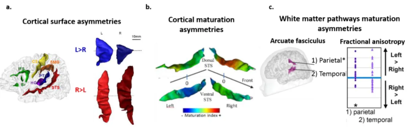
Structural asymmetries: Regarding the cortex, earliest structural asymmetries are observed during the fetal and preterm periods over the cortical gyri, and in favor of right hemisphere, with Heschl’s gyrus, superior frontal and temporal gyrus folding 1-2 weeks earlier in the right hemisphere (Dubois, Benders, et al., 2008; Kasprian et al., 2010). Asymmetries continue to emerge and become more evident in the perisylvian regions along the preterm period and early postnatal months, with thicker Heschl’s gyrus and more elongated planum temporale, and advanced growth of anterior region of the Sylvian fissure in the left relative to the right hemisphere, but deeper superior temporal sulcus in the right compared to the left hemisphere (Dubois et al., 2009; Hill et al., 2010; Glasel et al. 2011; Li et al., 2014; Smith et al., 2011; Leroy et al., 2011; Dubois & Dehaene-Lambertz., 2015). Apart from morphological differences, microstructural properties of the left and right perisylvian regions also differ. Inferior frontal sulcus (Broca’s area) is more mature on the left, whereas posterior superior temporal sulcus is more mature on the right hemisphere (Leroy et al., 2011). Figure 1.9. a. & b. illustrates these inter-hemispheric asymmetries for example regions of planum temporale and superior temporal sulcus.
Figure 1.9. Structural asymmetries in the language network in infancy. a) Cortical surface asymmetries in Planum temporale (PT) and Superior Temporal Sulcus (STS) , example of a 14 weeks old infant: PT is bigger in the left hemisphere, while STS is deeper in the right hemisphere (adapted from Glasel et al., 2011). b) Cortical maturation asymmetries in temporal areas in infants between 1-4 months of age: STS is more mature in the right hemisphere (adapted from Leroy et al., 2011. c) Maturation asymmetries in the parietal segment of Arcuate fasciculus in infants between 1-4 months of postnatal age, assessed with fractional anisotropy (Adapted from Dubois et al., 2009).
Regarding white matter, the most well-known asymmetry reported to exist at term and early postnatal months, is a leftward asymmetry of microstructure for arcuate fasciculus, similar to adults (Dubois et al., 2008; Dubois et al., 2016; Liu et al., 2010). A similar leftward asymmetry is also reported for