HAL Id: tel-02304191
https://tel.archives-ouvertes.fr/tel-02304191
Submitted on 3 Oct 2019HAL is a multi-disciplinary open access archive for the deposit and dissemination of sci-entific research documents, whether they are pub-lished or not. The documents may come from teaching and research institutions in France or abroad, or from public or private research centers.
L’archive ouverte pluridisciplinaire HAL, est destinée au dépôt et à la diffusion de documents scientifiques de niveau recherche, publiés ou non, émanant des établissements d’enseignement et de recherche français ou étrangers, des laboratoires publics ou privés.
Gene regulatory network for lateral root formation in
Arabidopsis thaliana
Duy Chi Trinh
To cite this version:
Duy Chi Trinh. Gene regulatory network for lateral root formation in Arabidopsis thaliana. Plants genetics. Université Montpellier, 2019. English. �NNT : 2019MONTG003�. �tel-02304191�
THÈSE POUR OBTENIR LE GRADE DE DOCTEUR
DE L’UNIVERSITÉ DE MONTPELLIER
En Biologie du Développement
École doctorale n°584 GAIA : Biodiversité, Agriculture, Alimentation, Environnement, Terre, Eau Unité de recherche DIADE : Diversité, Adaptation et Développement des Plantes
Présentée par TRINH Duy Chi
Le 22 mars 2019
Sous la direction de Dr. Laurent LAPLAZE
Devant le jury composé de
Propriétés du réseau de gènes contrôlant l’organisation
du primordium de racine latérale
chez Arabidopsis thaliana
Gene regulatory network for lateral root formation in
Arabidopsis thaliana
Mr Alain GOJON, Directeur de Recherche, INRA, Montpellier Mme Sandra BENSMIHEN, Chargé de Recherche, CNRS, Toulouse Mr Frédéric DOMERGUE, Chargé de Recherche, CNRS, Bordeaux
Mme Soazig GUYOMARC’H, Maitre de conférences, Université de Montpellier Mr Laurent LAPLAZE, Directeur de Recherche, IRD, Montpellier
Mme Valérie LEGUÉ, Professeur, Université Blaise Pascal de Clermont-Ferrand Mr Joop VERMEER, Professeur, Université de Zurich
Président du jury Examinateur Rapporteur Invité Directeur de thèse Rapporteur Examinateur
1
This PhD was prepared in the DIADE research unit DIversité - Adaptation - DEveloppement des plantes
Centre IRD de Montpellier 911 Avenue Agropolis 34394 Montpellier Cedex 5
2
Abstract in English
Post-embryonic lateral root organogenesis plays an essential role in defining plant root system architecture, and therefore plant growth and fitness. The aim of the thesis is to elucidate the gene regulatory network regulating lateral root development and de novo root meristem formation during root branching in the model plant Arabidopsis thaliana by combining a system-biology-based analysis of lateral root primordium transcriptome dynamics with the functional characterization of genes possibly involved in regulating lateral root organogenesis.
The first part of the thesis deals with the identification the target genes of PUCHI, an AP2/EREBP transcription factor that is involved in controlling cell proliferation and differentiation during lateral root formation. We showed that loss of PUCHI function leads to defects lateral root initiation and primordium growth and organisation. We found that several genes coding for proteins of the very long chain fatty acid (VLCFA) biosynthesis machinery are transiently induced in a PUCHI-dependent manner during lateral root development. Moreover, a mutant perturbed in VLCFA biosynthesis (kcs1-5) displays similar lateral root development defects as does puchi-1. In addition, roots of puchi-1 loss of function mutant show enhanced and continuous callus formation in auxin-rich callus induction medium, consistent with the recently reported role of VLCFAs in organizing separated callus proliferation on this inductive growing medium. Thus, our results demonstrate that PUCHI positively regulates the expression of VLCFA biosynthesis genes during lateral root development, and further support the hypothesis that lateral root and callus formation share common genetic regulatory mechanisms.
A second part of the thesis specifically addresses the issue of identifying key regulators of root meristem organization in the developing lateral root primordium. Material enabling the tracking of meristem cell identity establishment in developing primordia with live confocal microscopy was generated. A gene network inference was run to predict potential regulatory relationships between genes of interest during the time course of lateral root development. It identified potential regulators of quiescent center formation, a key step in functional organization of the lateral root primordia into a new root apical meristem. The characterization of some of these candidate genes was initiated.
Altogether, this work participated in deciphering the genetic regulation of lateral root formation in Arabidopsis thaliana.
Key words: gene regulatory network, lateral root, stem cell niche, mersitem formation, very long chain fatty acids (VLCFAs), PUCHI
3
Abstract in French
L’organogenèse post-embryonnaire des racines latérales joue un rôle essentiel dans l’établissement de l’architecture du système racinaire des plantes, et donc dans leur croissance et leur performance. L’objectif de cette thèse est de caractériser le réseau de gènes régulant le développement des racines latérales et en particulier, l’organisation fonctionnelle du primordium de racine latérale, formant un nouveau méristème racinaire, chez la plante modèle Arabidopsis
thaliana en combinant des études de biologie des systèmes appliquées à la dynamique du
transcriptome lors de la formation des racines latérales avec la caractérisation fonctionnelle de gènes candidats pour la régulation de ce phénomène d’organogenèse.
La première partie de la thèse concerne l’identification des cibles de PUCHI, un facteur de transcription de type AP2/EREBP impliqué dans le contrôle de la prolifération et de la différentiation cellulaire dans le primordium de racine latérale. Le phénotype liés à la parte de fonction de PUCHI a été caractérisé en détail et à mis en évidence un rôle de ce facteur de transcription dans l'initiation des racines latérales et le développement et l'organisation des primordia. Par l’analyse de profils spatiaux et temporels d’expression de gènes, nous avons pu mettre en évidence que l’expression de gènes codant des protéines impliquées dans la biosynthèse des acides gras à très longues chaînes (VLCFA) est transitoirement activée durant la formation de la racine latérale et que cette dynamique est dépendante de PUCHI. De plus, le mutant kcs1-5, perturbé dans la biosynthèse de VLCFAs, présente un phénotype de développement des racines latérales similaire à celui de puchi-1. Par ailleurs, la perte de fonction puchi-1 augmente fortement la formation de cals continus dans des racines cultivées sur milieu inducteur riche en auxine, ce qui est cohérent avec le rôle récemment décrit des VLCFA racinaires dans la formation et l’organisation de cals distincts lorsque la racine est cultivé sur milieu inducteur de cals. L'ensemble de nos résultats démontre que PUCHI régule positivement l’expression de gènes de biosynthèse de VLCFAs lors de la formation de racines latérales et la callogenèse. Nos résultats confortent également l’hypothèse selon laquelle la formation des racines latérales et celle de cals racinaires partagent des mécanismes de régulation communs.
La seconde partie de la thèse s’intéresse à l’identification de facteurs régulateurs clés dans l’organisation fonctionnelle du primordium de racine latérale et particulièrement, l’organisation d’un nouveau méristème racinaire. J’ai contribué à produire de nouvelles lignées de plantes permettant de suivre en temps réel par microscopie confocale la mise en place des identités cellulaires caractéristiques d’un méristème racinaire dans le primordium de racine latérale en développement. En utilisant un algorithme d’inférence de réseau de gènes, j’ai produit puis analysé les relations prédites de régulation entre gènes d’intérêt, afin d’identifier des gènes candidats potentiellement impliqués dans la formation du centre quiescent, un élément clé dans l’organisation du primordium et la mise en place du nouveau méristème racinaire. La caractérisation fonctionnelle de certains de ces gènes candidats a été initiée.
Ces travaux de thèse ont donc contribué à mieux comprendre les mécanismes de régulation de la formation des racines latérales chez Arabidopsis thaliana.
Mots clés: réseau de régulation du gène, racines latérales, niche de cellules souches, formation de mersitem, acides gras à très longue chaine (VLCFAs), PUCHI
4
ACKNOWLEDGEMENTS
First of all, I express my greatest gratitude to my PhD supervisors, Dr. Laurent Laplaze and Dr. Soazig Guyomarc’h. I feel truly privileged to be your student. You two have guided me through this journey with kindness, encouragement and great patience. Thank you very much Dr. Laurent for spending your valuable time discussing via video and answering all my requests with incredible efficiency. Thank you very much Dr. Soazig for finding the time between the university and the lab to be there for enthusiastic discussions, as well as for all the help with my everyday life. I and my little family, especially my daughter, deeply appreciate your generous support during our stay.
I thank Dr. Sandra Bensmihen, Dr Frédéric Domergue, Dr. Alain Gojon, Dr. Valérie Legué, and Dr. Joop Vermeer for participating in my PhD defense jury.
I am deeply grateful to all members of my PhD committee to my study. Thank you all very much, Dr. Thierry Joet, Dr. Patrick Lemaire, Dr. Philippe Nacry, Dr. François Parcy and Dr. Benjamin Peret, for spending many hours with me to discuss my work and to encourage me one every year.
I am grateful to the team leader Professor Pascal Gantet who introduced me to this PhD program and helped me get this study opportunity. I also thank all other members of the Cereal Root Systems team for their help during my thesis: Dr. Mikaël Lucas for network analysis using Pythonis, Dr. Antony Champion for molecular work, Daniel Moukouanga for everyday lab tasks, Dr. Alexandre Grondin, Mathieu Gonin, Jérémy Lavarenne, Nguyen Trang Hieu, Luong Ai My, Branly Wilfried Effa Effa, Marilyne DEBIEU, Carla De La Fuente Cantó and Kwanho Jeong for accompanying me through the journey with a lot of discussions and help, both in the lab and outside the lab.
I thank Prof. Malcolm Bennett (Nottingham, UK) for his valuable comments on my work. I thank Dr. Stéphane Dussert and Virginie Vaissayre (IRD, Montpellier), Frédérique Tellier and Prof. Jean-Denis Faure (Paris), and Dr. Yohann Boutté (Bordeaux) for all the hard work on fatty acids analyses. I appreciate the help of Dr. Tatsuaki Goh and Dr. Hidehiro Fukaki with research materials and helpful discussions.
I thank Myriam Collin (IRD), Carine Alcon and Alexandre Martinière (SupAgro Montpellier) for all the help with microscopy.
5
I acknowledge the essential help of Dr. Alain Ghesquiere, Bruno Barthelemy, Virginie Champion, Manuelle Rival (IRD) and Cendrine Jay-Allemand (GAIA) for processing all the paperwork necessary for my study.
My special thank goes to Dr. Luu Doan Trung (SupAgro Montpellier) for your kindness. Not only did you prepare me for the PhD, you also provided me and my family a lot of valuable help during the period.
I thank University of Science and Technology of Hanoi (USTH), Vietnam International Education Cooperation Department (VIED), French Embassy in Vietnam and Campus France for providing all financial and administrative support during the three years.
Last but not least, my special appreciation and gratitude go to my big and little family. This PhD would not be possible without your generous support and encouragement.
Table of contents 6 TABLE OF CONTENTS ABSTRACT IN ENGLISH ... 2 ABSTRACT IN FRENCH ... 3 ACKNOWLEDGEMENTS ... 4 TABLE OF CONTENTS ... 6 LIST OF FIGURES ... 9
FREQUENTLY USED ABBREVIATIONS ... 10
GENERAL INTRODUCTION ... 11
I. INTRODUCTION ... 17
II. TISSUE CONTEXT AND EARLY EVENTS OF LATERAL ROOT PRIMORDIUM ORGANOGENESIS ... 20
2.1. Competence of pericycle cells for root organogenesis ... 20
2.2. Oscillatory LR priming by endogenous cues ... 23
2.3. Lateral root founder cell specification ... 25
2.4. Lateral root primordia initiation ... 27
2.5. Control of lateral root spacing/density ... 29
III. BUILDING A NEW ORGAN: A COHERENT PROGRAM OF COORDINATED CELL DIVISIONS, WITH NEWLY SET AXIS OF GROWTH AND BOUNDARIES ... 30
3.1. Control and sequence of cell division and division planes ... 30
3.2. LRP cellular organization and anatomical patterning ... 33
3.3. Lateral root primordia boundary ... 35
3.4. LRP shape progression through surrounding tissues and LRP emergence ... 36
3.5. Connectivity between developing lateral root primordia and the primary root ... 37
IV. MERISTEM FORMATION AND ACTIVATION DURING LATERAL ROOT FORMATION ... 38
4.1. Common molecular mechanisms controlling embryonic root apical meristem and LRP meristem formation ... 38
4.2. Lateral root meristem activation ... 41
V. LRP ORGANOGENESIS AS A COMMON BASIS FOR ORGAN REGENERATION FROM CALLUS ... 43
VI. GENERAL CONCLUSIONS ... 47
CHAPTER I ... 15 Lateral root formation: building a meristem de novo
Table of contents
7
CHAPTER II ... 49
PUCHI regulates LRP initiation, positioning, patterning and emergence I. INTRODUCTION ... 50
II. RESULTS ... 51
2.1. PUCHI expression pattern ... 51
2.2. PUCHI negatively regulates LRP initiation, positioning and development ... 53
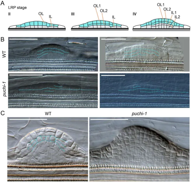
2.3. Cell division pattern is disturbed in puchi-1 LRPs ... 58
2.4. PUCHI is required for correct LRP meristem organization ... 61
2.5. puchi-1 roots generally have normal auxin response ... 62
2.6. puchi-1 pericycle is more sensitive to auxin treatment ... 64
2.7. Cytokinin signalling is altered in puchi-1 LRPs ... 65
III. DISCUSSIONS ... 66
3.1. PUCHI controls LRP initiation and spacing ... 66
3.2. Loss of PUCHI function leads to LRP development defects ... 68
3.3. PUCHI regulates cell divisions and stem cell niche establishment, possibly through hormonal signalling ... 69
IV. CONCLUSION AND PERSPECTIVES ... 72
CHAPTER III ... 73
PUCHI regulates VLCFA biosynthesis genes during LRP and callus formation I. INTRODUCTION ... 74
II. RESULTS ... 79
2.1. More on VLCFA biosynthesis genes in relevant datasets ... 79
2.2. PUCHI regulates the spatio-temporal expression patterns of VLCFA biosynthesis genes during LRP formation ... 83
2.3. VLCFA mutants and puchi-1 display similar defects in lateral root development ... 89
2.5. PUCHI is required for root but not shoot regeneration from callus ... 93
2.6. PUCHI regulates expression of KCS1 during callus formation ... 94
2.7. Mutation in PUCHI alters VLCFA composition in root calli ... 95
2.8. Probing suberin content in WT and puchi-1 roots ... 99
III. DISCUSSION AND PERSPECTIVES ... 101
3.1. VLCFA biosynthesis genes are expressed in LRPs and the endodermis ... 101
3.2. VLCFA biosynthesis genes are involved in LRP development and callus formation and are regulated by PUCHI ... 102
3.3. How do VLCFAs contribute to LRP development? ... 105
3.4. The PUCHI network regulating LRP development and callus formation ... 108
Table of contents
8
CHAPTER IV ... 115
Potential genes regulating stem cell niche formation I. INTRODUCTION ... 116
II. RESULTS ... 117
2.1. Identify potential regulators of root stem cell niche establishment in developing LRPs based on a proxy... 117
2.2. Identify potential regulators of LRP stem cell niche by whole network analysis ... 125
III. DISCUSSION AND PERSPECTIVES ... 133
SUPPLEMENTAL FIGURES ... 136
CHAPTER V ... 143
General discussions and perspectives I. PUCHI controls multiple aspects of plant development partially via regulating VLCFA biosynthesis 144 II. TDCor as a hypothesis-generating tool to identify potential genes regulating stem cell niche establishment ... 147
III. Final conclusion ... 148
CHAPTER VI ... 149
Materials and methods FRENCH SUMMARY... 158 REFERENCES ... 183 APPENDIXES ... 204 APPENDIX 1 ... 205 APPENDIX 2 ... 210 APPENDIX 3 ... 213
PUBLICATIONS, COMMUNICATIONS AND TRAINING ... 215
LIST OF PUBLICATIONS... 216
SCIENTIFIC COMMUNICATION ... 216
9
LIST OF FIGURES
Figure 1.1. Arabidopsis primary root and lateral root meristems. ... 18
Figure 1.2. Key events during lateral root primordium initiation and their regulators ... 21
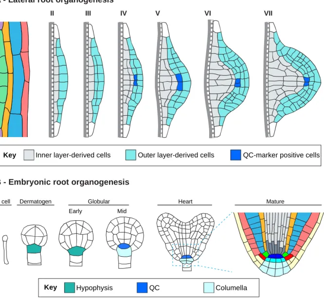
Figure 1.3. Schematic presentation of LRP organogenesis and embryonic root organogenesis. ... 31
Figure 1.4. Distribution patterns of some key regulators in different developmental contexts. ... 39
Figure 2.1. Prediction of the gene regulatory network during LRP development ... 51
Figure 2.2. Expression of GFP-PUCHI in LRPs and the primary root. ... 52
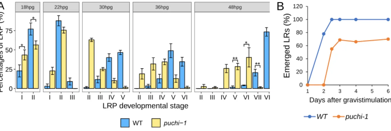
Figure 2.3. puchi-1 mutant produces more LRPs and is delayed in LRP development. ... 54
Figure 2.4. Kinetics of LRP development in WT and puchi-1. ... 56
Figure 2.5. Distribution of developmental stages of un-emerged LRPs in the marked regions in WT and puchi-1 roots. ... 57
Figure 2.6. puchi-1 LRPs are defective in cell division pattern. ... 59
Figure 2.7. Cellular organization in the presumptive LR meristem is disturbed in puchi-1 LRs. ... 60
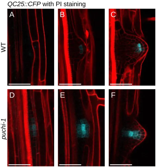
Figure 2.8. Expression of the QC-specific marker QC25::CFP is altered in puchi-1 mutant. ... 62
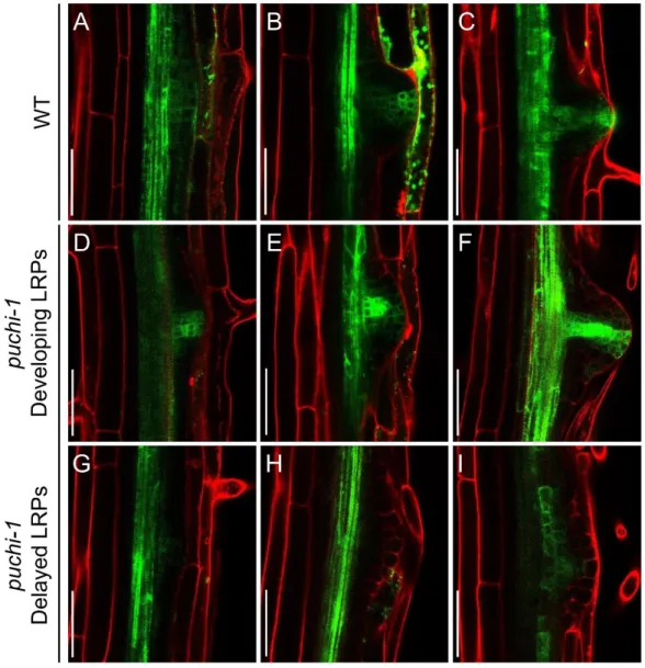
Figure 2.9. Auxin gradient as revealed by DR5::GFP reporter in WT and puchi-1 LRPs and LRs. ... 63
Figure 2.10. puchi-1 roots produce more LRPs when treated with auxin NAA. ... 65
Figure 2.11. Expression pattern of the cytokinin signaling reporter construct in WT and puchi-1 LRPs. ... 66
Figure 2.12. Summary of PUCHI roles during LRP development ... 72
Figure 3.1. Enrichment analysis using BinGO on 217 genes ... 75
Figure 3.2. Schematic representation of the VLCFA elongation cycle. ... 76
Figure 3.3. The PUCHI network inferred by TDCor ... 77
Figure 3.4. Measurement of key VLCFA biosynthetic gene expression by RT-qPCR ... 78
Figure 3.6. Expression of VLCFA biosynthesis genes in the developing LRP ... 82
Figure 3.7. VLCFA genes are expressed in LRPs and their expression patterns are dependent on PUCHI ... 85
Figure 3.8. KCS genes are expressed in LRPs and their expression patterns are dependent on PUCHI. ... 87
Figure 3.9. puchi-1 and kcs1-5 mutant produce more LRPs and are delayed in LRP development. ... 90
Figure 3.10. Callus formation was enhanced in puchi-1 and kcs1-5 roots. ... 92
Figure 3.11. puchi-1 roots have weaker root, but not shoot, regeneration capacity. ... 94
Figure 3.12. KCS1 is expressed in calli and its expression pattern is dependent on PUCHI. ... 95
Figure 3.13. An analysis of VLCFA classes ... 96
Figure 3.14. Global FA analysis revealed that mutation of PUCHI altered VLCFA composition in roots treated with CIM. ... 99
Figure 3.15. Suberin staining of roots with Fluorol yellow. ... 100
Figure 3.16. Possible PUCHI network regulating pericycle cell division and LRP formation and development.. .. 109
Figure S3.1. Expression pattern of pKCS1::GUS and pECR::GUS in the root. ... 110
Figure S3.2. Expression of pKCR1::GUS in endodermal cells ... 111
Figure S3.3. cer10-2 roots produced more lateral organs than did WT roots. ... 112
Figure S3.4. Total fatty acid profile from Arabidopsis thaliana roots. ... 112
Figure S3.5. An analysis of VLCFA classes ... 113
Figure S3.6. A subnetwork predicted by TDCor with the addition of suberin-related genes ... 114
Figure 4.1. First downstream neighbors of LBD16 ... 117
Figure 4.2. Expression profile of PISTILLATA during LRP development ... 118
Figure 4.3. Expression of PI::GFP in the WT background. ... 119
Figure 4.6. Whole network analyses reveal modules in gene network controlling LRP development. ... 126
Figure 4.7. Expression profiles extracted from the LR dataset of the genes in the three modules ... 128
Figure 4.8. Lateral root phenotype of some mutants of genes in the 3rd module. ... 132
Figure S4.1. Expression profiles in the LR dataset of some predicted regulators of PISTILLATA ... 136
Figure S4.2. Expression profiles in the LR dataset of some predicted targets of PISTILLATA ... 137
Figure S4.3. Expression profiles of genes in the third module in the LR dataset. ... 138
Figure 5.1. Illustration of the gravistimulation assay. ... 153
10
FREQUENTLY USED ABBREVIATIONS
ARF AUXIN RESPONSE FACTOR
Aux/IAA AUXIN/INDOLE-3-ACETIC ACID CIM callus-inducing medium
Dex dexamethasone DMSO dimethylsulfoxide ECR enoyl-CoA reductase FA fatty acid
FC founder cell
GFP GREEN FLUORESCENT PROTEIN
GR glucocorticoid receptor GUS β-glucuronidase
HACD hydroxyl-acyl-CoA dehydratase hpg hour post gravistimulation IAA indole-3-acetic acid
KCR β-Ketoacyl-CoA reductase KCS 3-keto-acyl-CoA synthase LR lateral root
LRIS lateral root inducible system LRP lateral root primordium MS Murashige and Skoog NAA naphthalene-1-acetic acid NPA 1-naphthylphthalamic acid PAS PASTICCINO genes PCR polymerase chain reaction
PI PISTILLATA
QC quiescent center
qRT-PCR quantitative real-time PCR RAM Root apical meristem TDCor Time Delay Correlation TF transcription factor
VLCFA very long chain fatty acid WT wild type
11
General introduction
12
Plant root system architecture (RSA) is the three-dimensional configuration of a whole root system in its living environment (Morris et al., 2017). RSA is considered a major determinant of plant viability and crop yield, and is a target for breeding to improve crop performance especially under various stresses (Smith and De Smet, 2012; Zhan et al., 2015). Root branching is of particular importance because it largely determines soil exploration of a root system and this can affect dramatically its water and nutrient acquisition (Lynch, 2013; Morris et al., 2017). Accordingly, the molecular mechanisms of root branching have been extensively studied in the model plant Arabidopsis thaliana (Arabidopsis) whose mature root system is largely derived from lateral roots (LRs) formed after germination. LRs originate from a small group of xylem-pole pericycle cells of the primary root that are primed by auxin to acquire founder cell identity (Möller et al., 2017). These founder cells undergo a succession of anticlinal and periclinal cell divisions that eventually results in the formation of a dome-shaped lateral root primordium (LRP; Malamy & Benfey, 1997; Lucas et al., 2013; Goh et al., 2016; Von Wangenheim et al., 2016). The LRP emerges through overlaying parental root tissues to become a functional LR (Swarup et al., 2008; Stoeckle et al., 2018).
Lateral root development is an excellent experimental system to study post-embryonic organogenesis. Interestingly, lateral root formation includes the de novo organization of a root apical meristem whose stem cell niche will subsequently sustain the continuous growth of the new LR (Laskowski et al., 1995; Malamy and Benfey, 1997). Moreover, recent studies have showed that lateral root formation shares common mechanisms with organ regeneration in tissue culture, especially the first step of callus formation (Perianez-Rodriguez et al., 2014; Fan et al., 2012; Sugimoto et al., 2010; Atta et al., 2009). Understanding these mechanisms is particularly relevant for many biotechnology applications in the field of plant regeneration and multiplication.
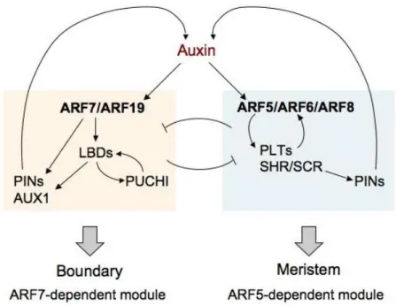
While many genes involved in lateral root development have been identified, little is known about the mechanisms that progressively organize the LRP into a root meristem (Trinh et al., 2018). LRP formation is not dependent on a stereotypical cell division pattern and therefore on cell lineage (Lucas et al., 2013; Von Wangenheim et al., 2016). LRP organization is a dynamic process dependent on complex gene regulatory networks and on cell-cell interactions including biomechanical interactions (Du and Scheres, 2017a; Stoeckle et al., 2018; Lucas et al., 2013). Interestingly, inference of the gene regulatory network involved in LR formation suggested an early patterning mechanism defining the central region and flanks of the LRP and identified genes involved in this process (Lavenus et al., 2015). The central region of a developing LRP self-organizes into a structure similar to that at the primary root apical meristem (RAM) (Laskowski et al., 1995; Malamy and Benfey, 1997). Some of the central cells express quiescent center
(QC)-General introduction
13
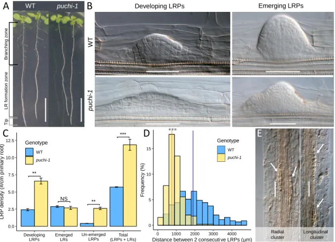
specific markers such as WOX5::GFP and QC25::CFP (Tian et al., 2014a; Goh et al., 2016; Du and Scheres, 2017b), and in the primary RAM these QC marker-positive cells are important in regulating stem cell identity and root meristem maintenance (Xu et al., 2006). Several important transcription factors (TF) controlling meristem formation during LRP development have been described, such as SHORTROOT-SCARECROW, and PLETHORAs (Goh et al., 2016; Du and Scheres, 2017b). However, there may exist many other important factors regulating meristem formation during LRP development. In LRP flanks, PUCHI encoding a AP2/EREBP-family TF was previously showed to control cell division and proliferation during LRP formation (Hirota et al., 2007). puchi-1 loss-of-function mutant produces LRPs exhibiting additional anticlinal and periclinal cell divisions from early stages and their LRs have abnormally enlarged flank cells (Hirota et al., 2007). Yet, the molecular targets regulated by PUCHI during LR development are not known.
In the frame of the research unit "Plant Diversity, Adaptation and Development" (IRD/ University of Montpellier), the research team I worked with is interested in deciphering the regulation mechanisms that control lateral root formation in various plant models, including A.
thaliana. To have a systematic view on genes possibly involved in LRP formation and
development, the team developed an algorithm called Time Delay Correlation (TDCor) (Lavenus et al., 2015) to infer genetic interaction from a time-course transcriptomic dataset profiling every stage of LRP organogenesis (Voß et al., 2015). This algorithm is based on similarity (Pearson correlation) between time-shifted expression profiles of genes in the LR dataset to suggest their possible regulator-to-target relationships (positive or negative regulation). This approach was validated experimentally using the targets of ARF7, a key player in LRP formation (Lavenus et al., 2015).
The team has exploited this inference strategy to explore the potential targets of PUCHI during LR development and looked for the molecular processes that may be influenced by PUCHI in the root. Interestingly, a number of genes coding for factors involved in the biosynthesis of very-long-chain fatty acids (VLCFAs) were found to have similar expression profiles but shifted in time to that of PUCHI. qRT-PCR further showed that expression of key VLCFA biosynthesis genes at the first stage of LRP formation was dependent on PUCHI. PUCHI was therefore hypothesized to regulate the expression of genes involved in the biosynthesis of very long chain fatty acids (VLCFA) during LR development.
My 3-year thesis initiated in that context in May 2016, with the aim to explore further and characterize experimentally the hypothesis that PUCHI acts as a master regulator of the VLCFA biosynthesis pathway during LR formation. In addition, I started a new and complementary
General introduction
14
research axis using the inference algorithm TDCor to identify upstream regulators controlling meristem establishment in the developing LRP. This PhD thesis is organized in five chapters:
Chapter I reviews recent advances on understanding LRP formation and development, with a link to plant regeneration from callus.
Chapter II describes the roles of PUCHI during LRP development through a detailed phenotyping of the loss-of-function mutant of PUCHI.
Chapter III demonstrates that PUCHI regulates the expression of VLCFA biosynthesis genes during LRP formation, and that this regulation is important for LRP formation and callus formation.
Chapter IV explores genes potentially involved in stem cell niche formation during LRP development and proposes further experiments to be done.
Chapter V provides a general discussion and perspectives resulting from this work. Chapter VI describes the materials and methods used in the work.
15
CHAPTER I
Chapter I: Lateral root formation: building a meristem de novo
16 Abstract
The complex and adaptable architecture of the plant root system in soil is of paramount importance for crop growth and performance. Root growth depends on the activity of the root apical meristem, an organized population of proliferating progenitor cells continuously replenished from a stem cell niche. Root branching, which greatly contributes to root system architecture in most dicot species, consists in de novo formation of new root meristems in existing root tissues. This phenomenon illustrates the ability of plants to repeatedly generate new tissues specialized in post-embryonic continuous growth and greatly impacts the elaboration of the root system architecture and its adaptation to environmental constraints. Here, we review the recent findings and models related to lateral root organogenesis in the dicot species Arabidopsis thaliana, with emphasis on the mechanisms controlling de novo root meristem formation. Experimental evidence suggests that critical regulatory modules are common between embryonic and post-embryonic root meristem organogenesis, and that the lateral root formation molecular pathway is in part common with organ regeneration from callus.
Keywords: lateral root, root branching, root meristem formation, Arabidopsis, auxin, organogenesis, stem cell niche
Note: this is the adapted from the review on Annual Plant Reviews online
Trinh, C.D., Laplaze, L. & Guyomarc’h, S. (2018) Lateral Root Formation: Building a Meristem de novo. In Annual Plant Reviews online. John Wiley & Sons, Ltd, Chichester, UK, pp. 1–44.
Chapter I: Lateral root formation: building a meristem de novo
17 I. INTRODUCTION
The root system fulfils multiple essential roles for the plant, including soil exploration and water and nutrient uptake, interactions with surrounding biotic and abiotic environments, plant anchorage to the substrate, and in some instances, vegetative reproduction or storage of photosynthates (Beeckman, 2009). In doing so, it greatly influences crop performance and yield (Rogers and Benfey, 2015). For example, changes in root system and water uptake explain a large part of the continuous increase in maize yield in the U.S. over the past 70 years (Hammer et al., 2009). Root system architecture (RSA), which refers to the spatial configuration of the whole root system of a plant in soil, is a potent parameter influencing root system function and crop growth (Lynch, 1995; de Dorlodot et al., 2007; Lynch, 2007; Rogers and Benfey, 2015). RSA traits have been frequently overlooked in past breeding programs due to the difficulty to access and quantify them. However, recent advances in phenotyping technologies and physiological modelling open the way for smart crop breeding programs targeting root traits and especially RSA (Smith and De Smet, 2012; Kuijken et al., 2015). These new breeding strategies offer a valuable approach to meet the demand in crop production in the current challenging context of increasing global human population and intensifying adversary soil and climatic conditions (Godfray et al., 2010; Tai et al., 2014; Smith and De Smet, 2012).
Plant RSA is modulated by 1) root growth, i.e. increase in root length, 2) root angle and 3) root branching, i.e. the formation of new roots such as lateral roots (LRs, originating from existing roots including lateral roots) and adventitious roots (emerging from shoot tissues, such as stem bases; Osmont et al., 2007; Bellini et al., 2014). Root growth relies on the activity of specialized tissues called root apical meristems that are organized populations of dividing cells including a self-maintained stem cell niche (Aichinger et al., 2012; Choe & Lee, 2017; Figure 1.1). Secondary root formation consists of de novo organogenesis of new root meristems from seemingly differentiated tissues. Contribution of post-embryonic root organogenesis to the elaboration of the plant RSA varies greatly depending on species. While the primary root and LRs emerging from it contribute to a significant extent to RSA development in dicot species, adventitious roots are predominant in RSA of most monocot species (Bellini et al., 2014). Root growth and branching are influenced by endogenous physiological cues as well as by environmental factors, such as soil texture, nutrient and water availability, and microbial interactions (Malamy, 2005; Tian et al., 2014b; Bao et al., 2014; Morris et al., 2017). This plasticity in RSA is of paramount importance for plant adaptation to environmental constraints.
Chapter I: Lateral root formation: building a meristem de novo
18
Figure 1.1. Arabidopsis primary root and lateral root meristems. Schematic organization of the apical meristem of Arabidopsis primary root (A) and emerged lateral root (B). Cell types of primary root apical meristem and presumptive cell types of lateral root apical meristems are indicated in the colour legend. In root apical meristems, the stem cells are called “initials” and the organizing centre is termed “quiescent centre” (QC).
Meristems are complex tissues gathering cells with little differentiation and retaining mitotic activity (Stahl and Simon, 2005). Division and differentiation of these cells are tightly regulated through a network of genetic factors and cell-to-cell communications providing plants with the ability to continuously generate new tissues and organs after germination. As a result, meristematic cells express repertoires of cell cycle-related genes but also specific factors influencing cell fate, such as chromatin modifiers and hormone and peptide signalling pathways (reviewed in Lee et al., 2013; Chiatante et al., 2018). Regulators of the structural properties of the tissue such as cytoskeleton organization, cell membrane dynamics, as well as primary cell wall formation are also of great importance for meristematic activity (Sassi and Traas, 2015). Importantly, emergent properties in this intricate regulation network generate long-lasting dynamic organization at the tissue scale, and especially, the maintenance of a central stem cell niche. Stem cells are undifferentiated cells able to divide with no apparent limit, renewing the stem cell pool as well as producing progenitor cells that will participate in the production of one or more
Lateral root cap
Epidermis Root cap/ Epidermis initials Cortex Endodermis Cortex/Endodermis initials Pericycle
Pericycle initials Stele initials Stele
Columella initials Columella Quiescent center
A B
Chapter I: Lateral root formation: building a meristem de novo
19
differentiated cell types (Laux, 2003; Spradling et al., 2001). In plant meristems these stem cells are prevented from differentiating by signalling from a group of other cells, the meristem organizing centre (Doerner, 1998). Specifically in root apical meristems, the stem cells are called “initials” and the organizing centre is termed “quiescent centre” (QC; Choe & Lee, 2017). As in animal stem cell niches (Ivanova, 2003; Zipori, 2004), the transcriptomic signature of plant stem cells remains elusive, although association of some specific transcription factors has been shown to be important (de Luis Balaguer et al., 2017; Galinha et al., 2007; Sarkar et al., 2007; Scheres, 2007). However, hormonal, epigenetic, and transcriptional regulators have been identified that play a critical role in stem cell niche establishment, organization, and maintenance (Choe and Lee, 2017). Not surprisingly, some factors, such as cell cycle effectors, are important for both shoot and root meristems. In addition, common schemes involving related molecules participate in both shoot and root meristem organization. For example, the ratio between auxin and cytokinin hormonal signalling greatly influences the balance between cell division and differentiation, although with seemingly different outputs in shoot and root contexts (Galinha et al., 2009; Vanstraelen and Benková, 2012). Additionally, in both shoot and root meristems transcription factors specifically expressed in the organizing centre inhibit the differentiation of neighbouring stem cells and are targets of a negative feedback mediated by non-cell autonomous peptides and membrane-located receptors (Stahl et al., 2013). Nevertheless, other aspects of meristem regulation, and especially hormonal and peptide signal transduction and its impact on cell differentiation, are specific to root or shoot development (Galinha et al., 2007).
The root apical meristem activity continuously generates new cells that participate to root growth as well as to renewal of the root cap, an important interface of the root meristem with the environment (Petricka et al., 2012; Perilli et al., 2012; Sozzani and Iyer-Pascuzzi, 2014). Still, only one or few root apical meristems are generated during plant embryogenesis. A significant proportion of the root system of a growing plant thus originates from post-embryonic root formation through a tightly regulated sequence of cell division and differentiation. This organogenesis process implies precise changes in cell cycle activities as well as modifications in cell gene expression programs (Birnbaum, 2016).
This review focuses on lateral root (LR) development, i.e. root organogenesis from existing root tissues, with emphasis on the processes generating a new functional root meristem. To that purpose, we will predominantly consider the model plant Arabidopsis thaliana, in which LR formation has been extensively studied. Arabidopsis is a dicot plant whose RSA consists of a primary root, LRs of multiple orders, e.g. tertiary roots, and few adventitious roots (Gutierrez et al., 2012; Smith and De Smet, 2012). Arabidopsis primary roots and LRs have a relatively simple
Chapter I: Lateral root formation: building a meristem de novo
20
anatomy, making them a valuable model for developmental biology studies (Lavenus et al., 2013b). The young Arabidopsis root is made of one layer of each tissue namely from the outside to the inside, the epidermis, the cortex, the endodermis and the pericycle enclosing the vascular tissues, which includes two xylem and two phloem poles (Dolan et al., 1993; Figure 1.1). In addition, Arabidopsis is amenable to LR induction protocols, live imaging techniques, and genetic reporter and mutant strategies (Jansen et al., 2013; Koornneef and Meinke, 2010). A wealth of information has been gathered on the processes regulating embryonic root meristem establishment, primary root meristem maintenance and plasticity, as well as on the cellular and molecular events underlying post-embryogenesis LR formation. Here, we synthesize recent published data highlighting observed properties of post-embryonic root meristem formation and aim to identify remaining gaps in our understanding of this biological process that greatly contributes to plant development and crop production.
II. TISSUE CONTEXT AND EARLY EVENTS OF LATERAL ROOT PRIMORDIUM ORGANOGENESIS
2.1. Competence of pericycle cells for root organogenesis
LR formation in Arabidopsis originates exclusively from pericycle cells (Figure 1.2). The pericycle is a single layer of cells representing the outermost cells of the vascular cylinder (Beeckman and De Smet, 2014). Pericycle cells are produced by inner initials of the primary root meristem. Due to this anatomical position, some pericycle cells neighbour either xylem pole or phloem pole cells. Interestingly this relative positioning is of functional importance, since lateral root primordia (LRPs) originate exclusively from xylem-pole pericycle (XPP) cells in many plants such as Arabidopsis (Dubrovsky, 2000; Parizot et al., 2007), and only from phloem pole pericycle cells in others such as maize (Jansen et al., 2012).
What makes those pericycle cells competent for LRP initiation is not clearly understood. However, a transcriptomic analysis in maize showed that compared to non-pericycle cells, pericycle cells preferentially express a subset of genes related to protein synthesis, transcription, and signal transduction, which could explain their competence for cell division (Dembinsky et al., 2007). In addition, analyses of the expression pattern of cell cycle regulators in roots suggested that stele tissues, including pericycle, retained S-phase related gene expression longer that other root tissues (Beeckman et al., 2001). Consistent with this hypothesis, a recent analysis of mitosis distribution in Arabidopsis root tips showed that, together with endodermis and vascular cells, pericycle cells retained mitotic activity longer than cortex cells or epidermis cells (Lavrekha et al., 2017). The pericycle actually consists of a heterogeneous population of cells: Arabidopsis XPP
Chapter I: Lateral root formation: building a meristem de novo 21 Figure 1 .2. K ey even ts du ri ng la ter al r oo t pr im or d iu m i ni ti at ion and the ir r egu la tor s (m odi fi ed fr o m Lave nus et al ., 2013 ). Pe ri cycl e ce ll s ar e p ri m ed par tl y th roug h auxi n rel ea se d fr o m pr o gr am m ed ce ll dea th (PC D ) in the lat er al r oo t ca p. Sub se ts of pr im ed ce ll s at oppos it e xyl em pol es t h at r et ai n st ab le st at ic aux in re spons e ar e ca ll ed p rebr anc h si tes , bu t u sua ll y on ly one w il l d eve lo p int o founde r ce ll s. Found er ce ll s then swe ll a nd th ei r nucl ei m igr at e to th e co m m o n ce ll w al l (w hi te ar row s) . A n as y m m et ri c ce ll d iv is io n ( A C D ) t h en o cc ur s m ar ki ng t he i n it ia ti o n of t he l at er al r oot or g anoge n es is p rog ram . Ex pr ess ion pa tt er n s of xyl em -p ol e pe ri cyl e ( X PP ) ce ll m ar k er s ( J1021 a nd R m 1007 ) an d dev el op m ent al zone s al o ng t he p ri m ar y r o ot a re i ndi ca ted. Pr imi ng Au xin re lea se Au xi n ma xi ma G AT A2 3 ex pr es si on Fo un de r c el l sp ec ifi ca tio n ARF 5, 6 , 7 , 8 , 1 9 IA A2 8 Au xi n Sy mp la st ic co nn ec tiv ity RA LF 34 En do de rma l vo lu me lo ss ; Pe ric yc le c el l swe llin g SHY 2/ IA A3 LB D1 6, 1 8, 3 3 ARF 7, 1 9 SL R/ IA A1 4 Au xi n Au xi n Nucl ea r mi gr at io n ACD/ In iti at io n G AT A2 3 ex pr es si on ARF 5, 6 , 7 , 8 , 1 9 IA A2 8 Au xi n G LV6 MA KR4 MA KR4 Pr eb ra nc h si te St ab le s ta tic au xi n re sp on se Ce ll cy cl e re gu la to rs P C D D ev el op m en t K e y DR5 P e ri cyc le L a te ra l ro o t ca p R m1 00 7 J01 21 X P P ma rke rs Ap ic al me ris te m Ba sa l me ris te m El on ga tio n an d dif fe re nt ia tio n zo ne
Chapter I: Lateral root formation: building a meristem de novo
22
cells in the mature root retain some meristematic cell-specific features such as fragmented vacuoles, large nuclei and dense cytoplasm, and expression of S-phase related cell cycle regulators in contrast to “differentiated” pericycle cells (Casimiro et al., 2003; Beeckman et al., 2001; Parizot et al., 2007). De Almeida Engler et al. (2009) identified 16 cell cycle genes displaying preferential expression in the Arabidopsis XPP cell file. The D-type Cyclin CYCD4;1 is expressed in XPP cells in the root meristem, and its loss of function causes a premature elongation of these cells associated with a reduced LR density (Nieuwland et al., 2009). Conversely, phloem pole pericycle cells were shown to differentiate and become quiescent significantly earlier than other pericycle cells, including XPP cells (Lavrekha et al., 2017). Longer cell cycle activity in XPP cells exiting the root meristem is dependent on the activity of ABERRANT LATERAL ROOT FORMATION 4 (ALF4) as in the alf4 mutant XPP cells only weakly express the G2-to-M transition transgene
CYCB1;1::GUS (DiDonato et al., 2004; Celenza et al., 1995). ALF4 is widely expressed in plant
tissues and encodes a plant-specific regulator modulating the activity of SCF complexes required for the signalling of the plant hormones auxin and gibberellin (Bagchi et al., 2018). Consistent with this longer cell cycle activity of pericycle cells being instrumental for LR formation, the alf4 mutant plants display a strong reduction in root branching, even in presence of the root-formation promoting hormone auxin (DiDonato et al., 2004; Bagchi et al., 2018).
Importantly, XPP cells express specific repertoires of genes as exemplified by the J0121 enhancer trap line that displays robust GFP expression in XPP cell files from the elongation zone of the meristem onwards, as well as, interestingly, in related shoot tissues competent for root formation (Casimiro et al., 2001; Laplaze et al., 2005; Sugimoto et al., 2010). Another enhancer trap line, Rm1007, showed GFP expression specifically in XPP cells and in the corresponding initials, adjacent to the root meristem quiescent centre, including in young embryos, indicating that the genomic region highlighted by the Rm1007 insertion drives very early XPP cell fate-specific expression (Parizot et al., 2007).
The close relationship between vascular pole differentiation and pericycle cell competence for LR formation suggests a robust crosstalk between these two cell populations in the primary root meristem. For example, Arabidopsis lonesome highway (lhw) mutant, that has only one xylem and one phloem pole, specifically produces LRPs along that single xylem strand (Parizot et al., 2007). Conversely, roots of the wooden leg (wol) mutant lack phloem specification and the J0121 marker is present throughout the pericycle. Interestingly, despite this characteristic, LR formation is severely affected in the wol mutant background, suggesting that both xylem and phloem specification in the primary root is required for functional LR formation (Parizot et al., 2007). Patterning of xylem and phloem vascular identities in the central domain of the root meristem is
Chapter I: Lateral root formation: building a meristem de novo
23
regulated through a complex network of transcriptional signalling and hormonal crosstalk (reviewed in Vaughan-Hirsch et al., 2018). A combination of experimental and modelling approaches suggested that mutual inhibition between auxin and cytokinin signalling patterned the alternating of xylem and phloem poles in Arabidopsis root meristem (Bishopp et al., 2011a). Signalling between vasculature identity and competence of pericycle cells for LR formation could involve auxin maxima in the xylem axis and phloem axis, in Arabidopsis and in maize, respectively (el-Showk et al., 2015; Jansen et al., 2012). Indeed, treatments by exogenous auxins or auxin transport inhibition alter the anatomical distinction between XPP and non XPP cells in Arabidopsis (Parizot et al., 2007). However, neither auxin nor cytokinin short term treatments modify the expression pattern of J0121 and Rm1007 transgenes, indicating a robust patterning mechanism of XPP cell identity in the root meristem (Parizot et al., 2007).
2.2. Oscillatory LR priming by endogenous cues
In Arabidopsis LRs originate only from XPP cells, but not every XPP cell develops into a LRP. Indeed in the basal meristem of the parental root tip, oscillating mechanisms periodically select subsets of XPP cells and prepare them to enter LR formation, a phenomenon called “priming” (Möller et al., 2017). This selection process has a major impact on RSA by defining the potential sites for root branching. The molecular processes underlying this priming event are still not fully understood, but are associated with changes in auxin signalling (De Smet et al., 2007). Monitoring expression of the synthetic auxin-responsive DR5 (DIRECT REPEAT5) promoter revealed regular pulses of auxin signalling activity in the two protoxylem strands in the basal meristem. Transcriptomic analyses showed that the expression levels of hundreds of genes were also oscillating in that segment of the root, according to the same period as auxin signal oscillations (Moreno-Risueno et al., 2010). While cells exit the basal meristem, those temporal fluctuations progressively cease and high signal stabilizes into local auxin maxima correlating with future sites of LR formation (Xuan et al., 2015; De Smet et al., 2007). The specification of these sites with high and stable DR5 signal, termed prebranch sites, is dependent on auxin signal transduction and probably on a complex network of transcription factors (Moreno-Risueno et al., 2010). The root cap plays a crucial role in creating these auxin pulses. First, a defect in auxin synthesis in the root cap impairs the amplitude of auxin signalling oscillations in the basal meristem, suggesting that auxin molecules that signal XPP priming may originate from the root cap (Xuan et al., 2015). Second, periodic programmed cell death occurs in lateral root cap cells creating rhythmic influxes of auxin into inner cells of the elongation zone of the meristem, which tightly correlate with prebranch site formation (Xuan et al., 2016). Pulsating fluxes of auxin from the root cap to the
Chapter I: Lateral root formation: building a meristem de novo
24
meristem might participate in generating peaks in auxin signalling activity in the protoxylem cell files, contributing to prebranch site specification.
Basipetal (from the root tip shootward) auxin fluxes from the columella through the lateral root cap and the root epidermis have been extensively studied in A. thaliana in particular in relation to gravitropism (Band et al., 2012; Wisniewska et al., 2006; Rashotte et al., 2000). Consistent with the model of root cap-derived auxin priming XPP cells, parental root waving, mechanical bending, or gravistimulation modulate LR formation, possibly because of gravity-modulated auxin fluxes or changes in auxin routes due to tissue bending that affects shootward auxin distribution (Lucas et al., 2008; Ditengou et al., 2008; Laskowski et al., 2008; Scheres and Laskowski, 2016). However, even agar-constrained roots, growing with only little deviation from gravity, display oscillation in auxin signalling and regularly spaced prebranched sites (Moreno-Risueno et al., 2010). This suggests that an endogenous clock mechanism produces uniformly spaced pre-branch sites to recruit pericycle cells and make them competent for de novo organogenesis.
Recently, Laskowski & ten Tusscher (2017) analysed the properties of the priming process in order to elucidate the molecular steps controlling auxin signalling oscillations and subsequent stabilization of auxin signal maximum. Both could rely on emergent properties (i.e. properties that arise from the collaborative functioning of a system, but do not belong to any one part of that system) of the global genetic system controlling auxin distribution, auxin signal transduction, and cell fate specification in the meristem (Alon, 2007). Feedback mechanisms can generate oscillating gene expression patterns that result in locally fluctuating transcriptomes (De Caluwé et al., 2016) or, when combined with cell-to-cell communication, create spatial patterning of distinct cell identities (reviewed in Green and Sharpe, 2015). Other network motifs, i.e. specific association of gene regulatory interactions, can instead buffer variations in gene expression (e.g. Vernoux et al., 2011). Particularly interesting in a developmental perspective are regulatory network properties that make the system “choose” between two potential dynamic trends, causing bifurcation in system state trajectories, distinct cell fates and potentially symmetry breaking at a tissue scale (Bishopp et al., 2011a). Such critical properties are described in various aspects of plant developmental regulation by auxin, including in root tissues. Feedback of cell parameters on auxin transporter expression and polarization can create and enhance non-uniform auxin distribution across tissues in a robust manner. As a result, asymmetric auxin distribution, locally transduced by cell specific signalling mechanisms, can create shifts in cell identity among neighbours. Van Norman et al. (2013) propose that the priming process, caused by auxin signal pulses, could enhance cell sensitivity to later auxin accumulation and its competence to translate it into the onset of a new organogenesis program. Bistable properties of auxin signalling modules could explain
Chapter I: Lateral root formation: building a meristem de novo
25
the “memorization” of that auxin signal in the prebranch sites only. Such a toggle-switch behaviour of an auxin-signalling motif has been described (Lau et al., 2011).
2.3. Lateral root founder cell specification
Within some prebranch sites, a subset of pericycle cells is later specified to become lateral root founder cells. Founder cells (FCs) refer to pre-existing cells that initiate the formation of a new organ through cell division (Laskowski et al., 1995); thus FC specification corresponds to the last apparent transition in cell identity before the onset of organogenesis. FC specification again involves a local increase in auxin signalling (De Rybel et al., 2010; Goh et al., 2012a; De Smet et al., 2007; Moreno-Risueno et al., 2010). Selective stimulation of auxin production in one or several pericycle cells is sufficient to transform them into FCs and trigger LR organogenesis (Dubrovsky et al., 2008). Conversely, in normal conditions, emergent properties of the auxin transport and signalling network may explain why, while XPP cell specification, priming and prebranch site definition may occur symmetrically on both xylem poles of the Arabidopsis vascular cylinder (De Smet et al., 2007; Goh et al., 2012a), effective initiation of LR development only occurs on one of those two sides (Goh et al., 2012a; el-Showk et al., 2015).
Auxin transcriptomic signalling is known to involve degradation of Aux/IAA proteins and activation of associated AUXIN RESPONSE FACTOR (ARF)-family transcription factors (reviewed in Weijers and Wagner, 2016). Interestingly, an auxin signalling pathway involving IAA28 and several ARFs it interacts with is required for FC specification (De Rybel et al., 2010). This auxin signalling module induces the expression of the transcription factor GATA23 in pericycle cells that will develop into a LRP before any sign of LR initiation (Figure 1.2). Artificially inducing GATA23 expression in the pericycle greatly increases LRP density, indicating that it positively regulates LR formation (De Rybel et al., 2010). GATA23 is therefore considered a marker for LR FCs, but its targets relevant for LR formation remain unknown. Interestingly,
GATA23 expression is also positively controlled by RAPID ALKALINIZATION FACTOR 34
(RALFL34), a small signalling peptide that could participate in auxin-mediated regulation of FC specification (Murphy et al., 2016).
Chromatin remodelling factors, for example Polycomb group (PcG) proteins, are known to regulate developmental transitions in both plants and animals by modulating the expression of key developmental genes (Schuettengruber et al., 2017). PcG proteins can form two multiprotein complexes: Polycomb repressive complex 1 (PRC1) and PRC2 which modify histone marks on chromatin to repress gene expression (Derkacheva and Hennig, 2014). Arabidopsis mutants impaired in subunits of the PRC2 complex CURLY LEAF (CLF) and EMBRYONIC FLOWER 2 (EMF2) displayed increased density of LR FCs and emerged LRs, suggesting that the PRC2
Chapter I: Lateral root formation: building a meristem de novo
26
complex might inhibit LR FC specification (Gu et al., 2014). CURLY LEAF is expressed in the basal meristem and in presumptive FCs, and directly represses the expression of the auxin efflux carrier-encoding gene FORMED 1 (PIN1) via histone modification (Gu et al., 2014). PIN-FORMED 1 participates to auxin signal concentration in the root tip and in LRPs (Benková et al., 2003; Blilou et al., 2005). Accordingly, an increase in the auxin-sensitive DR5 promoter activity was reported in root apical meristems and in FCs of a loss-of-function clf mutant (Gu et al., 2014). Other chromatin remodelling processes were shown to participate in the regulation of cell fate transition during LR founder cell specification, such as histone deacetylation (Singh et al., 2012), ATP-dependent chromatin remodelling (Fukaki et al., 2006; Ho et al., 2013), or histone variant deposition (Manzano et al., 2012). The precise hierarchy of these regulatory levels in combination with the auxin signalling network remains to be elucidated.
Plasma membrane-located signalling processes during FC specification are suggested by the impaired conversion of prebranch sites into LRPs in a mutant defective in the
MEMBRANE-ASSOCIATED KINASE REGULATOR4 (MAKR4) gene (Xuan et al., 2015). The MAKR4 protein
belongs to the same protein family as MAKR5 (Kang and Hardtke, 2016) and BRI1 KINASE INHIBITOR 1 (BKI1; Wang & Chory, 2006; Jiang et al., 2015), that are regulators of receptor kinases-associated signalling pathways. Expression of MAKR4, which is induced by auxin, is strongly enhanced in pericycle cells before cell division occurs and displays oscillations similar to that of DR5::LUC reporter expression. Importantly, the makr4 mutant has a normal number of prebranch sites but produces fewer LRPs, and overexpression of MAKR4 promotes LRP formation. The data indicates that MAKR4 participates in the transition of a prebranch site into a LRP (Xuan et al., 2015).
Proper cell-to-cell communication via plasmodesmata is also necessary for correct LR FC specification (Benitez-Alfonso et al., 2013). In plasmodesmal-localized b-1,3-glucanase 1 (pdbg1),
pdbg2 single and double mutants whose increased callose deposition in the stele and LRPs block
plasmodesmata, LRs are formed at higher density, sometimes even fused to each other (Benitez-Alfonso et al., 2013). Closer inspection revealed expanded domains of GATA23 expression and auxin signalling activity (as reported by DR5 promoter-based transgene expression) around FCs, indicating that the correct function of these plasmodesmata-located proteins is necessary to restrict FC specification to a subset of selected cells. How exactly symplastic communication keeps LR formation in check is currently not understood, but possibly through the movement of non-cell autonomous factors, such as transcription factors, peptides, metabolites or hormones (Benitez-Alfonso, 2014; Van Norman et al., 2011; Bishopp et al., 2011b; Yue and Beeckman, 2014; Benitez-Alfonso et al., 2013).
Chapter I: Lateral root formation: building a meristem de novo
27 2.4. Lateral root primordia initiation
In Arabidopsis, LRP initiation is first visually recognized by the swell of LR FCs, together with the shrink in volume of overlaying endodermal cells (Vermeer et al., 2014). Subsequently, nuclei of two longitudinally adjacent FCs, in each row, migrate to the common cell wall (De Rybel et al., 2010). Then these two cells undergo an anticlinal and asymmetric division yielding two shorter daughter cells next to each other (Casimiro et al., 2001; De Rybel et al., 2010). This typical cell division occurs in 4 to 6 abutting pericycle cell files concomitantly (Goh et al., 2016; Von Wangenheim et al., 2016). Because this asymmetric cell division produces two daughter cells with distinct shape and fate, it is a formative rather than a proliferative cell division (Gunning et al., 1978; Smolarkiewicz and Dhonukshe, 2013). Occurring independently of an established stem cell niche context and in a seemingly differentiated tissue, it is a landmark of the initiation of a new developmental program (De Smet and Beeckman, 2011). This first round of pericycle cell division produces the so-called stage I LRP (Malamy and Benfey, 1997). LRP initiation can also occur in a less common way from files of single pericycle cells (Dubrovsky et al., 2001; De Smet et al., 2006).
The swelling of FCs prior the first asymmetric cell division is assisted by volume loss of endodermal cells induced by auxin perception (Vermeer et al., 2014). Indeed, when endodermal cells expressed the stabilized form of SHY2, an Aux/IAA auxin signalling inhibitor, they were unable to lose their volume, hence maintained their turgidity. In those conditions underlying pericycle cells could not swell nor initiate asymmetric cell division, indicating that this space accommodation interaction between pericycle and endodermis is necessary for LR initiation (Vermeer et al., 2014). Moreover, ablation of an overlying endodermal cell triggers pericycle cell division, although in a periclinal manner, unless auxin was added (Marhavý et al., 2016).
An early characteristic feature of LR initiation is the change of cell polarity revealed by nuclear migration in pericycle cells preceding the asymmetric cell division. This is controlled by auxin-triggered expression of the LATERAL ORGAN BOUNDARIES DOMAIN (LBD) family transcription factors LBD16, LBD18 and LBD33 in pericycle cells (Goh et al., 2012a). The expression of LBD16 in XPP and FCs is dependent on the SLR/IAA14-ARF7-ARF19 auxin signalling pathway (Goh et al., 2012a). Interestingly, artificially converting LBD16 into a transcriptional repressor does not affect auxin signal in LR FCs but blocks the polar nuclear migration and subsequent anticlinal division of these cells, suggesting that the SLR/IAA14-ARF7-ARF19-LBD16 signalling cascade transduces auxin signalling into changes in pericycle cell polarity (Goh et al., 2012a). The transcription factor bZIP59 physically interacts with LBDs, including LBD16, LBD17 and LBD29 to control LRP initiation (Xu et al., 2018b).
Chapter I: Lateral root formation: building a meristem de novo
28
Transcriptomic analysis of auxin-induced LR initiation shed light on the early molecular events leading to pericycle cell division (Vanneste et al., 2005; De Smet et al., 2008; De Almeida Engler et al., 2009). Cell cycle progression is tightly regulated by an intricate and dynamic network involving transcription regulators (such as E2F and RBR) and cyclin-dependent kinases whose activity is post-transcriptionally regulated by phosphorylation, protein-protein interaction, as well as APC/C-mediated ubiquitination and degradation (Genschik et al., 2014; Breuer et al., 2014; Komaki and Sugimoto, 2012). One G1-to-S phase specific gene (CYCD3;2), one S-specific gene (CYCA2;4) and two G2-to-M related genes (CYCB2;5 and CDKB2;1) are upregulated in roots submitted to auxin-induced LR formation (Vanneste et al., 2005). Conversely, some genes encoding cell cycle inhibitors such as KRP2, KRP4, KRP7 and WEE1 are downregulated in the pericycle upon auxin treatment (De Almeida Engler et al., 2009). In normal conditions, the
CYCB1;1 gene, encoding a G2-to-M transition specific cyclin, is strongly expressed in XPP cells
undergoing the first division (Himanen, 2002; Beeckman et al., 2001).
In addition to the change in LR FC cell polarity, SLR/IAA14-dependent auxin signalling is essential for their first divisions. Either slr gain of function mutation or loss of function mutation of its two ARF partners ARF7 and ARF19 prevents LR formation because the first anticlinal and subsequent periclinal divisions of pericycle cells are blocked (Fukaki et al., 2002). The implication of SLR/IAA14 in priming and FC specification can be excluded because it is not expressed in basal meristem (Fukaki et al., 2002) and competence for LR FC specification of slr pericycle cells is still maintained (De Smet et al., 2007; De Rybel et al., 2010). The transcriptomic analysis by Vanneste et al. (2005), demonstrated that specific G1-to-S, S, and G2-to-M cell cycle regulators act downstream of IAA14-ARF7-ARF19 mediated auxin signalling. In addition, active ARF7 and ARF19 transcription factors stimulate the expression of LBD18 and LBD33 which in turn stimulate the expression of E2Fa, a potent activator of S-phase promoting gene expression (Berckmans et al., 2011). Another pathway for auxin-mediated cell cycle reactivation in XPP cells could consist in the downregulation of KRP2, an inhibitor of CYCD2;1-CDKA complex activity which could stimulate G1-to-S transition (Sanz et al., 2011).
The first asymmetric cell division in LRP formation is also regulated by a peptide belonging to the GOLVEN/root growth factor/CLE-like (GLV/RGF/CLEL) family, GLV6 (Fernandez et al., 2015). GLV6 is expressed in FCs preceding the migration of nuclei, and GLV6 silencing or overexpressing lines display decreased or increased FC division, respectively. In addition, overexpression of GLV6 or treatment of roots with GLV6 peptides disrupts the migration of FC nuclei to the common cell wall without disturbing cell cycle progression, converting the asymmetric cell division into a symmetric division (Fernandez et al., 2015). Thus cell-to-cell
Chapter I: Lateral root formation: building a meristem de novo
29
communication mediated by GLV6 and possibly other GLV/RGF/CLEL family peptides is probably involved in the correct coordination of the asymmetric LR FC division (Fernandez et al., 2015).
The double mutant impaired in AURORA kinases (aur1 aur2) shows a strong defect in division plane orientation during LR formation, as early as the first asymmetric cell division. Although LR initiation density and polar localization of FC nuclei are unaffected, the positioning of the new cell wall is uncontrolled (Van Damme et al., 2011). Interestingly, while this results in a reduced proportion of emerged LRPs, this does not totally preclude LR formation, indicating the robustness of this organogenesis program (Van Damme et al., 2011).
2.5. Control of lateral root spacing/density
Mechanisms controlling LR FC specification and LRP initiation along the primary root deeply impact the overall root branch production, i.e. LRP density (number of LRPs divided by the length of the parent root). In addition, other factors are required for efficient LRP spacing. The gene encoding the membrane localized receptor-like kinase ARABIDOPSIS CRINKLY4 (ACR4) is expressed specifically in the short daughter cells after the first asymmetric cell division. The acr4 mutant produces LRPs at higher densities and these LRPs usually stretch, sometimes even fuse together, indicating that ACR4, and possibly related kinases, control cell proliferation and spacing of newly initiated LRPs (De Smet et al., 2008). ACR4 was proposed to be the receptor of GLV6 and other related peptides (Fernandez et al., 2013), and possibly to transduce perceived signals inside the cell through a phosphorylation cascade involving the PROTEIN PHOSPHATASE 2A (PP2A; Yue et al., 2016). Cytokinin synthesized at the flanks of existing LRPs was found to act as an inhibitor of LRP initiation, thereby preventing closely formed LRPs (Chang et al., 2015). Interestingly, expression of ACR4 and GLVs were significantly reduced in mutants defective in cytokinin biosynthesis, but ACR4 and cytokinin seem to act in partially separate pathways (Chang et al., 2015). Another peptide, C-TERMINALLY ENCODED PEPTIDE 5 (CEP5), is also involved in the regulation of LRP initiation and positioning (Roberts et al., 2016). Overexpression of the CEP5 gene and exogenous treatment by the CEP5 peptide reduce LRP initiation events and induce the formation of closely positioned LRs, while CEP5 RNAi lines display an increase in LRP stage I and II density. Remarkably, CEP5 is expressed specifically in phloem pole pericycle cells and adjacent phloem cells, indicating an unknown crosstalk between different pericycle populations (Roberts et al., 2016). Similarly, the PLETHORA (PLT) transcription factors PLT3, PLT5, PLT7 act redundantly downstream of the ARF7- and ARF19-mediated auxin signalling pathway to regulate LRP spacing (Hofhuis et al., 2013).