HAL Id: hal-02620374
https://hal.inrae.fr/hal-02620374
Submitted on 25 May 2020
HAL is a multi-disciplinary open access
archive for the deposit and dissemination of
sci-entific research documents, whether they are
pub-lished or not. The documents may come from
teaching and research institutions in France or
abroad, or from public or private research centers.
L’archive ouverte pluridisciplinaire HAL, est
destinée au dépôt et à la diffusion de documents
scientifiques de niveau recherche, publiés ou non,
émanant des établissements d’enseignement et de
recherche français ou étrangers, des laboratoires
publics ou privés.
Structure of the Lipopolysaccharide from the
Bradyrhizobium sp ORS285 rfaL Mutant Strain
Flaviana Di Lorenzo, Angelo Palmigiano, Katarzyna A. Duda, Mateusz
Pallach, Nicolas Busset, Luisa Sturiale, Eric Giraud, Domenico Garozzo,
Antonio Molinaro, Alba Silipo
To cite this version:
Flaviana Di Lorenzo, Angelo Palmigiano, Katarzyna A. Duda, Mateusz Pallach, Nicolas Busset, et
al.. Structure of the Lipopolysaccharide from the Bradyrhizobium sp ORS285 rfaL Mutant Strain.
ChemistryOpen, Wiley, 2017, 6 (4), pp.541-553. �10.1002/open.201700074�. �hal-02620374�
Structure of the Lipopolysaccharide from the
Bradyrhizobium sp. ORS285 rfaL Mutant Strain
Flaviana Di Lorenzo,
[a]Angelo Palmigiano,
[b]Katarzyna A. Duda,
[c]Mateusz Pallach,
[a]Nicolas Busset,
[d]Luisa Sturiale,
[b]Eric Giraud,
[d]Domenico Garozzo,
[b]Antonio Molinaro,
[a]and Alba Silipo*
[a]1. Introduction
Bradyrhizobia are Gram-negative nitrogen-fixing bacteria capa-ble of establishing a mutualistic symbiotic relationship with plants of the Leguminosae family. The endosymbiotic interac-tion causes the development of root nodules at which bacteria can reduce atmospheric nitrogen into ammonia for the plant’s benefit.[1] Owing to their ability to develop a nitrogen-fixing
symbiosis, leguminous plants can develop in nitrogen-deficient soil and their culture does not require the addition of nitroge-nous chemical fertilizers.[2]
The establishment of this symbiosis relies on an exchange of diffusible signal molecules between the two partners in which lipochitooligosaccharides, termed nodulation factors (Nod fac-tors), synthesized by the bacteria, are essential for controlling the processes of infection and nodule organogenesis. However, Giraud et al. demonstrated that some tropical legumes of the Aeschynomene genus, such as A. indica, are nodulated by pho-tosynthetic Bradyrhizobium strains, such as BTAi1 and ORS278, that lack the canonical nodABC genes responsible for the syn-thesis of Nod factors.[3]This observation indicated, for the first
time, the existence of an alternative symbiotic pathway inde-pendent of the Nod factors. Moreover, some photosynthetic Bradyrhizobium strains, for example, ORS285, do contain the nodABC genes and display a broader host range comprising all stem-nodulating Aeschynomene species, including A. afraspera. Mutations of the nod genes in ORS285 exerted no effects on its ability to nodulate A. indica, whereas the ability to nodulate A. afraspera was completely abolished. This observation sug-gested the possibility that strain ORS285 uses distinct symbiot-ic strategies depending on the host plant, with the in-volvement of different signal molecules, which remain un-identified.[3]
Exposed on the Gram-negative bacterial surface, lipopolysac-charide (LPS) molecules have an important role in the interac-tion of bacteria with the host legume.[4,5] LPS is a tripartite
macromolecule built up of a glycolipid portion—lipid A— linked to a saccharide part that is distinguished by oligosac-charide (core OS) and polysacoligosac-charide portions, the latter being The importance of the outer membrane and of its main
con-stituent, lipopolysaccharide, in the symbiosis between rhizobia and leguminous host plants has been well studied. Here, the first complete structural characterization of the entire lipopoly-saccharide from an O-chain-deficient Bradyrhizobium ORS285 rfaL mutant is achieved by a combination of chemical analysis, NMR spectroscopy, MALDI MS and MS/MS. The lipid A structure is shown to be consistent with previously reported Bradyrhi-zobium lipid A, that is, a heterogeneous blend of penta- to hepta-acylated species carrying a nonstoichiometric hopanoid
unit and possessing very-long-chain fatty acids ranging from 26:0(25-OH) to 32:0(31-OH). The structure of the core oligosac-charide region, fully characterized for the first time here, is re-vealed to be a nonphosphorylated linear chain with methylat-ed sugar residues, with a heptose residue exclusively present in the outer core region, and with the presence of two singly substituted 3-deoxy-d-manno-oct-2-ulosonic acid (Kdo) resi-dues, one of which is located in the outer core region. The lipid A moiety is linked to the core moiety through an uncom-mon 4-substituted Kdo unit.
[a] F. Di Lorenzo, M. Pallach, A. Molinaro, Prof. A. Silipo Department of Chemical Sciences
University of Naples Federico II Via Cinthia 4, 80126, Naples (Italy) E-mail: silipo@unina.it
[b] A. Palmigiano, L. Sturiale, D. Garozzo CNR-Istituto per i Polimeri
Compositi e Biomateriali IPCB—Unit/ di Catania Via Gaifami 18, 95126, Catania (Italy)
[c] K. A. Duda
Junior Group of Allergobiochemistry
Research Center Borstel, Leibniz Center for Medicine and Biosciences Airway Research Center North (ARCN)
German Center for Lung Research, 23845 Borstel (Germany) [d] N. Busset, E. Giraud
IRD, Laboratoire des Symbioses Tropicales et M8diterran8ennes UMR IRD/SupAgro/INRA/UM2/CIRAD
Campus International de Baillarguet TA A-82/J, 34398 Montpellier Cedex 5 (France)
Supporting Information and the ORCID identification number(s) for the author(s) of this article can be found under https://doi.org/10.1002/ open.201700074.
T 2017 The Authors. Published by Wiley-VCH Verlag GmbH & Co. KGaA. This is an open access article under the terms of the Creative Commons Attribution-NonCommercial-NoDerivs License, which permits use and distribution in any medium, provided the original work is properly cited, the use is non-commercial and no modifications or adaptations are made.
termed the O-chain (or O-polysaccharide or O-antigen). De-pending on the presence or absence of this latter polysaccha-ride domain, the LPS macromolecule is defined as smooth-type (S-LPS) or rough-smooth-type LPS (R-LPS or lipooligosaccharide, LOS), respectively.[6,7] Although the general architecture of
lipid A is conserved among individuals belonging to the same species, lipid As isolated from rhizobia strains are heterogene-ous in nature in terms of sugar backbone and lipid content, that is, the nature and distribution of the acyl chains and the presence of very-long-chain fatty acids (VLCFAs).[8]
Further-more, in some bradyrhizobial strains, a triterpenoid moiety, that is, a hopanoid, was found to be covalently linked to the LPS lipid A, and was shown to have a pivotal role in the free living and symbiotic states of the bacteria.[9] The remarkable
structural diversity in the lipid A of some rhizobium species is deemed a strategy by which the bacterium can escape or at-tenuate the plant response, thus facilitating the instauration of symbiosis. The O-chain is the most variable LPS region and is directly involved in the symbiotic interaction with the host plant.[3–5,10] The structural diversity of the O-chain moiety can
contribute to modulating or suppressing the plant defense re-sponses.[11]Indeed, it has been observed that the LPS
O-poly-saccharide from the photosynthetic Bradyrhizobium BTAi1a strain, a homopolymer made up of a bicyclic monosaccharide (bradyrhizose),[12]does not trigger the innate immune response
in different plant families.[13]
Recently, we have reported two Bradyrhizobium ORS285 mu-tants in the genes gdh and rfaL, which encode a putative dTDP-glucose 4,6-dehydratase and a putative O-chain ligase, respectively;[14] these mutant strains display an LPS lacking
the O-chain moiety. However, no effect of the mutations was detected on the symbiotic properties of these strains regard-less of the Aeschynomene species considered.[13] This in
con-trast to many rhizobial species, such as B. japonicum, Rhizobi-um etli or R. phaseoli, for which the absence of the O-chain is reported to cause a dramatic decrease in symbiotic capaci-ty.[15–17]Thus, the ability of the O-antigen-lacking ORS285
mu-tants to retain nodulation-promoting properties is not yet fully understood.
In addition to the importance of determining the role of LPS in symbiotic bacteria, rhizobial LPSs have unique structural fea-tures not yet fully elucidated. Indeed, all rhizobial core OS characterized to date were found to be completely distinct from enteric LPS, including sugar composition, linkage pattern and absence of phosphate and heptose residues. With regards to the structural features, in the majority of Agrobacterium spe-cies an a-mannose (a-Man) or a-glucose (a-Glc) moiety is linked to the O-5 atom of the first Kdo unit, with the exception of Agrobacterium larrymoorei, in which an a-d-Gal unit is pres-ent, whereas in the rhizobium core there are additional galac-turonic (GalA) residues, and a third Kdo unit is present in the outer core region.[18–22]
To shed light on the peculiar rhizobial sugar arrangement, we report the structural elucidation of the LPS from the O-chain-deficient Bradyrhizobium ORS285 rfaL mutant strain (hereafter referred to as ORS285 rfaL), obtained by the use of chemical, NMR spectroscopy and mass spectrometry
tech-niques on the entire molecule and on the isolated core OS and lipid A fractions.
2. Results and Discussion
2.1. Extraction, Purification, and Compositional Analysis of ORS285 rfaL
The LPS of ORS285 rfaL was isolated from dried bacterial pellet by using hot phenol/water.[23] SDS-PAGE analysis[24] revealed
the presence in the water phase of an LOS. Intensive steps of purification with DNase, RNase and proteases followed by dial-ysis, ultracentrifugation and gel filtration chromatography were performed. Compositional analyses[25–27] revealed the
oc-currence of the following moieties: 6-substituted, 4-substituted and terminal mannose (6-Man, 4-Man, t-Man, respectively), 3-substituted 4-O-methyl-mannose (3-Man-4Me), terminal galac-turonic acid (t-GalA), 4-substituted glucose (4-Glc), 6-substitut-ed and 4,6-substitut6-substitut-ed 2,3-diamino-2,3-dideoxy-d-glucose (re-ferred to here as 4,6-DAG and 6-DAG, respectively), terminal N-methylglucosamine (t-GlcNMe), 7-substituted d,d-heptose (7-Hep), and 4- and 5-substituted 3-deoxy-d-manno-oct-2-uloson-ic acids (4-Kdo and 5-Kdo). Furthermore, a GlcNMe-(1!7)-Hep disaccharide and traces of 4-substituted d,d-heptose (4-Hep) were also detected. As further confirmation of the mutation of the rfaL gene, no bradyrhizose was detected. Fatty acid analy-sis[28] revealed the occurrence of 14:0(3-OH), 12:0(3-OH), 12:0,
12:1, and VLCFAs, among which 26:0(25-OH) and 32:0(2,31-2OH), and hopanoid, which is fully consistent with the previ-ously reported data on LPS lipid A fatty acid composition from Bradyrhizobium.[8,29]
2.2. Isolation and NMR Structural Characterization of the Core Region of ORS285 rfaL LOS
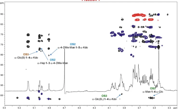
To determine the structure of the ORS285 rfaL core OS, mild acid hydrolysis was performed to cleave the acid-labile linkage between the Kdo and the lipid A disaccharide backbone. The water-soluble core OS fraction was isolated in the supernatant after a centrifugation step and further purified by means of ion exchange chromatography. Three main fractions, designated X, Y and Z, were eluted from the column and analyzed by NMR spectroscopy; the1H NMR spectra are reported in Figure 1.
Each fraction underwent a detailed NMR spectroscopic anal-ysis. Homo- and heteronuclear 2D NMR experiments (DQF-COSY, TOCSY, ROESY, NOESY, 1H–13C HSQC, HMBC and
HSQC-TOCSY) were performed to assign all the spin systems and to determine the saccharide sequence. The anomeric configura-tion of each monosaccharide unit was determined on the basis of the3J
H-1,H-2coupling constant values attained from the
DQF-COSY spectrum, whereas the values of the vicinal 3J H,H ring
coupling constants allowed the relative configuration of each sugar residue to be assigned. Finally, the combination of infor-mation attained from the inter-residue NOE connectivity and heteronuclear long-range correlations, together with methyla-tion analysis, allowed the complete characterizamethyla-tion of the core OS from ORS285 rfaL LPS.
The NMR spectra clearly revealed that fractions X and Y con-tained a mixture of OS species. Thus, fraction X was composed of three OS species, referred to hereafter as OS1, OS2 and OS3. All the sugar residues were present as pyranose rings, ac-cording to both 13C chemical shift values and the long-range
correlations between C-1/H-1 and H-5/C-5 in the1H–13C HMBC
spectrum (between C-2 and H-6 of the Kdo residue).[30] OS2
contained spin systems C, D and K2(Table 1 and Figure 1). Spin
system C (H-1 at 5.11 ppm, Table 1) and was identified as an a-manno-configured sugar unit due to the low-value coupling constants 3J
H-1,H-2 and 3JH-2,H-3 (<1 Hz), diagnostic of an H-2
equatorial orientation. Furthermore, in the TOCSY spectrum, it was possible to assign all the other ring protons resonances from H-2, allowing identification of this sugar as a heptose resi-due. Similarly, spin system D (H-1 at 5.01 ppm, Table 1) was identified as an a-mannose moiety due to the 3J
H-1,H-2 and 3J
H-2,H-3 values of 3 Hz and the intra-residual NOE contact of
H-1 only with H-2. Moreover, the downfield shift of C-4 D (76.3 ppm, Table 1 and Figure 2) was attributed to the pres-ence of a methoxy group as demonstrated by the NOE correla-tion of the signal at d=3.51 ppm (Table 1) with the signal for H-4 of a-mannose D at d=3.56 ppm (Table 1); this was further confirmed by the occurrence of a long-range correlation, ob-served in the 1H–13C HMBC spectrum, between C-4 of D and
the methoxy group. Finally, spin system K2 was attributed to
the Kdo residue on the basis of the diastereotopic methylene proton signals resonating in the high-field region of the
1H NMR spectrum at d= 1.81 (H-3
ax) and 2.05 ppm (H-3eq;
Table 1); on the basis of the H-3 proton chemical shift it was possible to assign its a-anomeric configuration.[31]
Upfield-shift-ed carbon signals were pivotal in identifying substitution at O-3 of residue D and at O-5 of the Kdo unit, whereas residue C was assigned to a terminal sugar unit, in agreement with the methylation data. The primary sequence of the OS2 structure was inferred by using the NOE connectivity measured in the ROESY spectra and by the long-range correlations in the HMBC spectrum (Figure 2). Thus, the Kdo residue K2was found to be
substituted at its O-5 position by the a-Man4Me D, which in turn was substituted at position O-3 by the a-heptose C; these data allowed us to define OS2 as the following trisaccharide (where p stands for pyranose) [Eq. (1)]:
As stated above, the NMR analysis revealed the presence, in fraction X, of a further OS species, OS1. This was identified as a tetrasaccharide (Table 1 and Figures 1 and 2) made up of spin systems K2’, D’, C’ and an extra sugar unit (E) compared
to OS2. Residue E (H-1 at 4.92 ppm; Table 1) was identified as a-GlcN: the gluco configuration was assigned from the 3J
H,H
coupling constants, whereas the C-2 signal at 54.88 ppm (Table 1) indicated it to be a nitrogen-bearing carbon atom. The analysis of the inter-residual NOE correlations and long-range contacts allowed the location of residue E to be identi-fied at position O-7 of the a-heptose C’ as a terminal sugar unit (Figures 1 and 2). Therefore, OS1 corresponded with the following tetrasaccharide [Eq. (2)]:
OS3 was composed of spin systems A, B and K1. Spin
system A was identified as an a-mannose, whereas residue B was assigned as an a-glucose moiety, as demonstrated by the large ring 3J
H,H coupling constants, and K1 was identified as
a Kdo unit. The analysis of the glycosylation shifts allowed the identification of A as a terminal mannose, of K1as a
4-substi-tuted Kdo and of B as a 4-substi4-substi-tuted glucose unit. The long-range and NOE correlations allowed the elucidation of the fol-lowing trisaccharide structure [Eq. (3)]:
Table 1.1H and13C chemical shift values [ppm] of fraction X.
1 2 3 4 5 6 7 8 OS3 A 5.27/5.26 3J H-1,H-2= 1.6/1.4 Hz 4.028 3.78 3.63 3.66/3.57 3.74/3.82 – – t-a-Man 101.46 1J C-1,H-1= 174.2 Hz 70.34 70.38 66.20 73.68 60.58 – – B 5.12 3J H-1,H-2= 3.9 Hz 3.54 3.84 3.61 3.90 3.73/3.85 – – 4-a-Glc 98.3 1J C-1,H-1= 170.5 Hz 71.62 70.70 76.06 70.82 60.60 – – B1 5.08 3J H-1,H-2= 3.37 Hz 3.52 3.88 3.60 3.90 3.83/3.75 – – 4-a-Glc 99.76 1J C-1,H-1= 170.6 Hz 71.63 70.68 76.22 70.81 60.60 – – K1/K’1 – – 2.07 1.77/(2.14) 4.02 3.98/4.10 3.68 – – 4-a-Kdop 37.41 78.80 70.19 71.90 – – OS1/OS2 C 5.11 3J H-1,H-2< 1 Hz 4.02 3.86 3.85 3.69 3.69 3.63 – t-a-Hep 102.02 1J C-1,H-1= 171.2 Hz 70.30 70.64 66.21 72.30 72.15 62.82 – C’ 5.08 3J H-1,H-2< 1 Hz 4.01 3.87 3.86 3.873 4.18 3.69/3.78 – 7-a-Hep 102.09 1J C-1,H-1= 172.4 Hz 70.36 70.60 66.21 70.62 67.49 69.17 – D/D’ 5.01/4.99 3J H-1,H-2< 1 Hz 4.08/4.10 4.02/4.04 3.56/3.54 3.85 3.74 – – 3-a-Man 101.76 1J C-1,H-1= 174.1 Hz 70.11/70.25 77.21/78.76 76.3/76.00 72.33 60.60 – – OCH3 3.51/60.23 E 4.92 3J H-1,H-2= 3.36 Hz 2.74 3.58 3.37 3.69 3.83/3.74 – – t-a-GlcN 98.97 1J C-1,H-1= 170.4 Hz 54.88 73.81 69.80 72.06 60.61 – – K2 – – 1.81/2.05 4.15 3.88 3.85 4.00 3.63/3.80 5-a-Kdop – – 37.70 70.80 79.90 72.31 68.91 62.90 K’2 – – 1.81/1.90 4.14 3.85 3.85 4.00 3.63/3.80 5-a-Kdop – – 37.62 69.53 80.58 72.31 68.90 62.90
Fraction Y (Figures 1 and 3, Table 2) was exclusively consti-tuted by the OS2 and OS3 oligosaccharides described above, and was assigned as above. Fraction Z (Figures 1 and 4) was exclusively composed of OS3’, even though the Kdo unit K1f
was present in a furanose form, as demonstrated by long-range correlation of C-2 with H-5, and further confirmed by the chemical shift values (typical of furanose rings) of H-3ax
and H-3eq resonating at 2.17/2.59 ppm, C-2 (105.0 ppm) and
C-5 (82.3 ppm) (Table 3 and Figure 4).
Thus, according to NMR and compositional analyses, the core OS isolated from Bradyrhizobium ORS285 rfaL was com-prised of two different Kdo-containing oligosaccharide skele-tons, a tri/tetrasaccharide with the structure GlcN-(1!7)-Hep-(1!3)-4-OMe-Man-(1!5)-Kdo and a trisaccharide having the structure Manp-(1!4)-Glcp-(1!4)-Kdop.
2.3. MALDI Mass Spectrometry on Intact LOS from ORS285 rfaL
An aliquot of intact LOS isolated from ORS285 rfaL was investi-gated by MALDI MS and MS/MS to determine the structure of both lipid A and core domains. The MALDI mass spectrum of intact LOS, recorded in linear mode and with positive-ion po-larity, is reported in Figure 5. These data show, in addition to an array of molecular ions, the heterogeneity of the native LOS (m/z 3600–4800). In-source decay fragments originated from cleavage (b-elimination) of the labile glycosidic bond involving the Kdo unit to yield either core OS ions (B-type ions, m/z 900–
1500) and lipid A ions (Y ions, m/z 2400–3200).[32,33]Four main
oligosaccharide species at m/z 1469.6 (OSa), 1308.5 (OSb),
1294.5 (OSc) and 925.4 (OSd) were clearly identified, together
with their corresponding ions arising from the well-known neutral loss of CO2(Dm/z 44).[32] To obtain the sequence of all
these OS structures, we performed the MALDI-TOF MS on intact LOS in the reflectron mode, followed by MALDI-TOF/TOF MS/MS analysis of the related fragments (Figure 6a) produced directly by the LOS fragmentation. In fact, they are MS3spectra
acquired with a MS/MS instrument,[34]as the precursor ions are
B-type fragments originated from the rapid in-source decay of the LOS pseudomolecular ions.
In detail, the peak at m/z 1469.6 (OSa) was consistent with
a B-ion matching with an octasaccharide composed of two Kdo, two hexose (Hex), one methyl hexose (HexMe), one hep-tose (Hep), one hexosamine (HexN) and one methylhexosa-mine (HexNMe) units (Figure 5). Its fragmentation pattern (Fig-ure 6a) showed numerous informative ions, such as those at m/z 1294.5, due to the loss of a terminal Kdo, at m/z 925.4, which matches with a fragment lacking one Kdo and two Hex, and at m/z 705.3, corresponding to an ion deprived of a further Kdo residue. Finally, the internal fragment at m/z 530.3 was in-dicative of a trisaccharide made up of one HexMe, one Hep and one Hex moiety. The OS species at m/z 1308.5 (OSb) was
assigned to a heptasaccharide sharing the same sequence— with the exception of a terminal HexN monosaccharide (Dm/z 161)—with OSa, as confirmed by MS/MS analysis.
Simi-larly, the species at m/z 1294.5 (OSc) was ascribed to a
hepta-Figure 2. Zoom-in of the overlapped1H,1H–13C HMBC (black) and1H–13C HSQC (purple and red) NMR spectra of the fraction X; the key inter-residual
Figure 3. Zoom-in of the overlapped1H,1H–13C HMBC (black) and1H–13C HSQC (purple and red) NMR spectra of the fraction Y; the key inter-residual
long-range correlations involving sugar moieties are indicated; letters are as in Table 2.
Figure 4. Zoom-in of the overlapped1H,1H–13C HMBC (black) and1H–13C HSQC (purple and red) NMR spectra of the fraction Z; the key inter-residual
Table 2.1H and13C chemical shift values [ppm] of fraction Y. 1 2 3 4 5 6 7 8 OS3 A 5.18 3J H-1,H-2=1.92 Hz 3.96 3.71 3.568 3.60 3.77/3.64 – – t-a-Man 101.35 1J C-1,H-1=174.3 Hz 70.26 70.17 66.53 73.60 60.76 – — B 5.04 3J H-1,H-2=3.9 Hz 3.47 3.78 3.56 3.83 3.71 – – 4-a-Glc 98.15 1J C-1,H-1=170.3 Hz 71.60 73.24 76.09 70.30 60.38 – – B1 5.01 3J H-1,H-2=3.85 Hz 3.44 3.75 3.53 3.83 3.68 – – 4-a-Glc 99.4 1J C-1,H-1=170.2 Hz 71.60 73.26 76.20 70.60 60.38 – – K1 – 1.71/1.99 3.94 4.06 – – – 4-a-Kdop 36.10 77.91 70.5 – – – K’1 – 1.99 3.95 3.91 – – – 4-a-Kdop 36.11 77.8 69.92 – – – OS2 C 5.03 3J H-1,H-2<1 Hz 3.94 3.79 3.79 3.75 3.67 3.62 – t-a-Hep 102.00 1J C-1,H-1=173.0 Hz 70.31 70.50 65.80 73.00 71.15 62.62 – D 4.91 3J H-1,H-2<1 Hz 4.02 3.97 3.48 3.77 3.67 – – 3-a-Man 101.7 1J C-1,H-1=171.2 Hz 70.18 77.01 76.18 72.1 60.30 – – OCH3 3.45/60.23 – K2 – – 1.74/1.98 4.06 3.81 3.78 3.93 3.63/3.73 5-a-Kdop – – 37.17 69.2 80.30 70.42 68.70 62.80 K’2 – – 1.73/1.82 3.98 3.78 3.78 3.93 3.63/3.73 5-a-Kdop – – 37.60 69.41 80.92 70.41 68.70 62.80
Table 3.1H and13C chemical shift values [ppm] of fraction Z.
1 2 3 4 5 6 7 8 A 5.17 3J H-1,H-2=1.31 Hz 3.95 3.69 3.56 3.58 3.78/3.64 – – t-a-Man 101.41 1J C-1,H-1=177.0 Hz 70.18 70.28 66.5 73.6 60.84 – – B 4.90 3J H-1,H-2=1.31 Hz 3.44 3.75 3.53 3.69 3.67/3.75 – – 4-a-Glc 96.60 1J C-1,H-1=170.9 Hz 70.98 73.39 76.04 70.18 60.50 – – K1f – – 2.17/2.59 4.52 4.41 3.63 3.43 3.71/3.56 4-a-Kdof 173.0 105.00 41.00 73.40 82.34 61.50 75.06 60.74
saccharide consisting of the same OSa sequence lacking a
ter-minal HexNMe (Dm/z 175). Finally, the B-ion at m/z 925.4 (OSd)
was identified as a pentasaccharide made up of one Kdo, one MeHex, one Hep, one HexN and one HexNMe residue, derived from the longer octasaccharide species (OSa) at m/z 1469.6, as
a result of the rupture of the labile glycosidic bond involving the intra-chain Kdo unit. The MS/MS analysis of OSd showed
two pairs of intense ions at m/z 162.1 and 764.3 and at m/z 176.1 and 750.3, corresponding to B- and Y-type ions ob-tained from the loss of a terminal HexN and a terminal HexNMe, respectively. Moreover, the internal fragment at m/z 530.2 and a further fragment at m/z 529.2 support the presence of a Hep unit substituted by two HexN residues, of which one is methylated, as outlined in Figure 6a. Indeed, the occurrence of minor OS peaks at m/z 1455.6 and 911.5 (Dm/z 14) implied that the methylation on the HexN residue was nonstoichiometric (Figure 5).
We were unable to locate, either by NMR or GC–MS, the second HexN unit, although traces of 4-Hep in the methylation analysis might lead us to conclude that the second amino sugar is located at this position. Conversely, the finding of a 4-Man residue in the methylation analysis allowed us to establish that the second Kdo unit was located at position 4 of the a-mannose. Indeed, data obtained from methylation, MS and NMR analyses led us to propose the following structure for the
main core OSb (Figures 5 and 7): a-GlcNMe-(1!7)-a-Hep-(1!
3)-a-Man4Me-(1!5)-a-Kdo-(2!4)-a-Man-(1!4)-a-Glc-(1!4)-a-Kdo.
Figure 5 shows, in the mass range between m/z 2400 and 3200, a series of peaks relative to the heterogeneous mixture of lipid A species differing in their acylation pattern, that is, in the lipid chain nature and content. The complete assignment of the lipid A fraction was achieved in detail by MS analysis in reflectron mode on intact LOS. A zoom-in view of this latter spectrum enclosing the lipid A region, is reported in Figure S1. In brief, a penta-acylated lipid A species was identified at lower masses (m/z 2128.39), composed of a pentasaccharide back-bone, constituted by a disaccharide of DAG units carrying an a-GalpA on the vicinal DAG and an a-Manp disaccharide on the distal DAG unit, acylated by two 14:0(3-OH), one 12:0(3-OH), one 12:1, and one acetylated 26:0(25-OAc) moiety. A second group of peaks was ascribed to the hexa-acylated lipid A fraction with the same acylation pattern plus a VLCFA, the major component of which ranged from C30:0(29-OH) to C32:0(2,31-2OH), in full agreement with previously published lipid A structure from B. japonicum strain BTAi1.[8]A third group
of lipid A peaks were found shifted to higher molecular mass by Dm/z 512.4, thus suggesting the presence of a hopanoid moiety, once more in accordance with previous reported data on Bradyrhizobium lipid A.[8]The MS/MS analysis on a single
Y-Figure 5. Linear MALDI-TOF mass spectrum in positive-ion mode of intact LOS from Bradyrhizobium ORS285 rfaL. Pseudomolecular ions are present in the LOS region. Further peaks are due to in-source fragmentation as sketched for the LOS species at m/z 4561.1. * Indicates contaminants.
Figure 6. a) MALDI-TOF/TOF MS/MS analysis of the main OS species (B-type ions) generated in-source over the MALDI-TOF acquisition. b) MALDI-TOF/TOF MS/MS analysis of the lipid A species (Y-type ion) at m/z 3091.29, generated in-source over the MALDI-TOF acquisition. Assignments of the fragment ions are shown on the schematic structures.
type ion allowed full characterization of the hepta-acylated lipid A species at m/z 3091.29, as reported in Figure 6b.
Finally, heterogeneous groups of ions corresponding to the native LOS, given by the combination of all the full-length core OS species (namely OSa, OSb and OSc) and the main lipid A
moieties, were detected at higher molecular masses (m/z 3600–4800), as shown in Figure 5.
3. Conclusions
Bradyrhizobia are symbiotic bacterial partners that develop ni-trogen-fixing nodules in a large diversity of leguminous spe-cies.[1] Root colonization is a key step in the interaction of
these beneficial bacteria with plants,[35] thus the molecular
mechanisms involved in such a delicate process have attracted increasing interest in several research fields. It was previously
Figure 7. Complete structure of the LPS from the ORS285 rfaL mutant strain. MALDI MS data highlighted the presence of a second amino sugar linked to the heptose residue.
reported that the bacterial outer membrane is involved in most of the host–microbe interactions, and consequently the LPS, as the main component of the outer membrane, is consid-ered one of the primary actors potentially involved in the sym-biotic process. In this context, several works suggested the im-portance of the LPS both in the very early recognition steps with the host and in the later stages of the nodulation process-es,[3,4,36] focusing the attention on the role of the LPS O-chain
moiety, presence of which is considered a pivotal prerequisite for a successful plant–microbe interaction at different stages of the nodulation event. Nevertheless, through the analysis of Bradyrhizobium ORS285 mutant strains lacking the LPS polysac-charide domain, we have recently demonstrated that the O-chain of Bradyrhizobium strains is not required for establishing symbiosis with Aeschynomene spp.[13] The reason for such
pe-culiar behavior is unresolved. However, the existence of as yet unknown symbiotic processes cannot be excluded. The possi-bility that the core OS region, as the outermost part of the Bra-dyrhizobium ORS285 mutants LPS, can be non-immunogenic and thus facilitate symbiosis, is also an important issue to be considered. This hypothesis prompted us to elucidate the com-plete structure of the LOS from the Bradyrhizobium ORS285 rfaL mutant.
The structural characterization of the core OS and lipid A was enabled by the combination of chemical, MS, MS/MS, and NMR analyses on both native LOS and its isolated constituents. The core OS region isolated from ORS285 rfaL is heterogene-ous in terms of sugar composition, as observed from NMR and MS data, and consists of a mixture of hepta- and octsacchride species containing 1) a trisacchaoctsacchride with the structure a-Man-(1!4)-a-Glc-(1!4)-a-Kdo, and 2) a tetrasaccharide having the structure a-GlcN-(1!7)-a-Hep-(1!3)-a-4OMeMan-(1!5)-a-Kdo, with the a-GlcN unit present in a nonstoichiometric amount. In addition, MS analyses showed the existence of a fur-ther GlcN unit linked to the heptose residue; furfur-thermore, nei-ther GlcN unit was stoichiometrically N-methylated. The heter-ogeneity of the native LOS was also clearly visible in the high-mass region of the MALDI MS spectrum (m/z 4100–4600, Figure 5), from which different species were identified differing in the acylation pattern of the lipid A, the length, and extent of methylation of the core region. Moreover, the lipid A com-ponent of intact LOS was fully characterized by HRMS in the reflector mode (Figure S1) and MS/MS analysis of the Y-type ions generated by in-source decay (Figure 6b). The elucidated structure was fully consistent with that previously published for Bradyrhizobium strains, that is, a mixture of penta- to hepta-acylated species carrying a nonstoichiometric hopanoid residue and both possessing two 14:0(3-OH), two 12:0(3-OH), and two VLCFAs, for example, one 26:0(25-OAc) and one VLCFA ranging from 26:0(25-OH) to 32:0(31-OH).[8]
Notably, this is the first complete structure of Bradyrhizobium LPS lipid A core region; previously, only partial structures were reported.[37] With regard to our finding, the core moiety of
ORS285 rfaL is revealed to be a new core oligosaccharide with somewhat unique structural features. In brief, ORS285 rfaL LOS displayed 1) a linear core oligosaccharide chain containing two Kdo residues, and 2) one of the two Kdo residues is placed
four residues away from the lipid A domain, that is, in the outer core region of the LOS, and represents a structural fea-ture in common with other rhizobial species, in which a Kdo unit was found in the O-chain attachment region.[18–22,38]
More-over, no phosphate groups decorating such a core OS region were detected. As a further peculiarity, the lipid A sugar back-bone is connected to the core region through a 4-substituted Kdo residue, whereas, in the majority of core structures charac-terized so far, the first Kdo is 5-monosubstituted or 4,5-disub-stituted. Finally, the occurrence of methylated sugar residues comprising the ORS285 rfaL LOS is a further characteristic in common with other Bradyrhizobium core OS moieties. In other rhizobial strains, sugar methylation has been correlated to the switch to an increase in the surface hydrophobicity from a pre-dominantly hydrophilic state.[10,39,40] It has been also
hypothe-sized that, together with the features of lipid A—the presence of VLCFA and hopanoid moieties—the presence of methylated sugar units can further contribute to increased bacterial enve-lope stability and barrier properties as well as bacterium adhe-sion ability, for example, that facilitating plant–microbe inter-actions.
Experimental Section
Bacterial Strain and LPS IsolationDried cells were washed twice with distilled water and extracted
with a hot phenol/water mixture.[17]After extensive dialysis against
distilled water, the extracted phases underwent enzymatic diges-tion and an ultracentrifugadiges-tion step in order to remove nucleic acid and protein contaminants. Both fractions were analyzed by
SDS-PAGE (13.5% acrylamide) followed by silver nitrate staining.[18]
In order to remove glucan contaminants, a phenol/chloroform/
light petroleum extraction,[41]followed by a precipitation step with
cold acetone, was performed.
Structural Analysis of LOS from ORS285 rfaL
The structure of the LOS from ORS285 rfaL was characterized using a range of methods. Monosaccharides were detected as acetylated O-methylglycoside derivatives obtained by methanolysis (1.25m HCl/MeOH, 85 8C, 24 h) followed by acetylation with acetic
anhy-dride in pyridine (858C, 30 min).[19]Linkage analysis was performed
as previously described.[20,21] In brief, the sample was methylated
with iodomethane and hydrolyzed with trifluoroacetic acid (2m, 1008C, 2 h). The carbonyl was reduced with sodium
tetradeutero-borate (NaBD4), then the sample was acetylated with acetic
anhy-dride and pyridine, and analyzed by GLC–MS.
After hydrolysis of the LOS (4m HCl, 1008C, 4 h), neutralization (5m NaOH, 1008C, 30 min) and water/chloroform extraction, fatty acids were detected in the chloroform phase by gas–liquid chromatogra-phy [HP 6890 N gas chromatograph with flame ionization detec-tion and an Agilent Technologies column (30 mV0.25 mm, film thickness 0.25 mm) of phenyl methylsiloxane HP-5MS] as methyl
esters (diazomethane, 10 min, room temperature).]28]The
tempera-ture program was 1208C for 3 min, then heating to 3208C at
Isolation of the ORS285 rfaL LOS Constituents
To study the core OS component, a portion (20 mg) of purified LOS was treated with acetate buffer (pH 4.4) for 5 h at 1008C with magnetic stirring. An appropriate mixture of chloroform and meth-anol was added to the hydrolysate to obtain a chloroform/metha-nol/hydrolysate (2:2:1.8, v/v/v) mixture. The mixture was then shaken and centrifuged. The water-soluble phase, containing the core OS fraction, was collected and freeze-dried. This fraction was then purified by high-performance anion-exchange chromatogra-phy coupled with pulsed amperometric detection (HPAEC-PAD) in alkaline conditions. Later, the samples were desalted by gel perme-ation chromatography on a G-10 column (GE Healthcare).
NMR Spectroscopy Analysis
1D and 2D NMR spectra were recorded in D2O at 278C at pD 7
with a cryoprobe-equipped Bruker 600 DRX spectrometer. Total correlation spectroscopy (TOCSY) experiments were performed with spinlock times of 100 ms using data sets (t1Vt2) of 4096V512 points. Rotating-frame Overhauser enhancement spectroscopy (ROESY) and nuclear Overhauser enhancement spectroscopy (NOESY) experiments were performed using data sets (t1Vt2) of 4096V512 points with mixing times between 100 and 400 ms. Double-quantum-filtered phase-sensitive correlation spectroscopy (DQF-COSY) experiments were performed using data sets of 4096V 912 points. The data matrix in all homonuclear experiments was zero-filled in both dimensions to give a matrix of 4 KV2 K points, and was resolution-enhanced in both dimensions using a cosine-bell function before Fourier transformation. Coupling constants were determined by 2D phase-sensitive DQF-COSY
measur-ments.[42,43] Heteronuclear single-quantum coherence (HSQC) and
heteronuclear multiple-bond correlation (HMBC) experiments were
performed in the1H-detection mode by single-quantum coherence
with proton decoupling in the 13C domain using data sets of
2048V400 points. HSQC was performed using sensitivity improve-ment and in the phase-sensitive mode using echo/antiecho
gradi-ent selection, with multiplicity editing during the selection step.[44]
HMBC was optimized on long-range coupling constants, with a low-pass J filter to suppress one-bond correlations, using gradi-ent pulses for selection. Moreover, a 60 ms delay was used for the evolution of long-range correlations. HMBC spectra were optimized for 6–15 Hz coupling constants. The data matrix in all the hetero-nuclear experiments was extended to 2048V1024 points by using a forward linear prediction extrapolation.[45]
MALDI-TOF and MALDI-TOF/TOF Mass Spectrometry
A 4800 Proteomic Analyzer (AB Sciex) was used, equipped with a Nd:YAG laser operating at a wavelength of 355 nm with a <500 ps pulse and 200 Hz firing rate. MALDI-TOF mass spectra of intact LOS were recorded either in linear or reflectron mode using positive-ion polarity. Each spectrum resulted from the sum of around 2000 laser shots. Mass spectra acquired in the reflector mode allowed detection of monoisotopic masses. External calibra-tion using a dedicated peptide calibracalibra-tion mixture (AB Sciex), pro-vided mass accuracy below 75 ppm. Data were processed using DataExplorer 4.9 software. The MS/MS experiments reported in this study were performed without use of a collision gas. Native LOS
samples were prepared as previously reported.[34]
Acknowledgements
A.S. and A.M. acknowledge the TOLLerant H2020-MSC-ETN-642157 (http://www.tollerant.eu) project.
Conflict of Interest
The authors declare no conflict of interest.
Keywords: bradyrhizobium ORS285 · lipooligosaccharides · mass spectrometry · NMR spectroscopy · symbiosis
[1] C. Masson-Boivin, E. Giraud, X. Perret, J. Batut, Trends Microbiol. 2009, 17, 458– 466.
[2] H. Ogutcu, A. Adiguzel, M. Gulluce, M. Karadayi F. Sahin, Rom. Biotech. Lett. 2009, 14, 4294 –4300.
[3] E. Giraud, L. Moulin, D. Vallenet, V. Barbe, E. Cytryn, J. C. Avarre, M. Jau-bert, D. Simon, F. Cartieaux, Y. Prin, G. Bena, L. Hannibal, J. Fardoux, M. Kojadinovic, L. Vuillet, A. Lajus, S. Cruveiller, Z. Rouy, S. Mangenot, B. Se-gurens, C. Dossat, W. L. Franck, W.-S. Chang, E. Saunders, D. Bruce, P. Ri-chardson, P. Normand, B. Dreyfus, D. Pignol, G. Stacey, D. Emerich, A. Vermeglio, C. Medigue, M. Sadowsky, Science 2007, 316, 1307 –1312. [4] N. Fraysse, F. Couderc, V. Poinsot, Eur. J. Biochem. 2003, 270, 1365 –
1380.
[5] M. A. Newman, J. M. Dow, A. Molinaro, M. Parrilli, J. Endotoxin Res. 2007, 13, 69– 84.
[6] C. R. Raetz, C. Whitfield, Annu. Rev. Biochem. 2002, 71, 635 –700. [7] F. Di Lorenzo, C. De Castro, R. Lanzetta, M. Parrilli, A. Silipo, A. Molinaro
in Carbohydrates in Drug Design and Discovery (Eds.: J. Jimenez-Barbero, F. J. Canada, S. Martin-Santamaria), RSC Publishing, London, 2015, 38– 63.
[8] A. Molinaro, O. Holst, F. Di Lorenzo, M. Callaghan, A. Nurisso, G. D’Errico, A. Zamyatina, F. Peri, R. Berisio, R. Jerala, J. Jim8nez-Barbero, A. Silipo, S. Mart&n-Santamar&a, Chem. Eur. J. 2015, 21, 500– 519.
[9] a) A. Silipo, G. Vitiello, D. Gully, L. Sturiale, C. Chaintreuil, J. Fardoux, D. Gargani, H.-I. Lee, G. Kulkarni, N. Busset, R. Marchetti, A. Palmigiano, H. Moll, R. Engel, R. Lanzetta, L. Paduano, M. Parrilli, W.-S. Chang, O. Holst, D. K. Newman, D. Garozzo, G. D’Errico, E. Giraud, A. Molinaro, Nat. Commun. 2014, 5, 5106; b) I. Komaniecka, A. Choma, A. Mazur, K. A. Duda, B. Lindner, D. Schwudke, O. Holst, J. Biol. Chem. 2014, 289, 35644 –35655.
[10] I. Lerouge, J. Vanderleyden, FEMS Microbiol. Rev. 2002, 26, 17– 47. [11] R. W. Carlson, L. S. Forsberg, E. L. Kannenberg, Subcell. Biochem. 2010,
53, 339 –386.
[12] a) A. Silipo, M. R. Leone, G. Erbs, R. Lanzetta, M. Parrilli, W.-S. Chang, M.-A. Newman, M.-A. Molinaro, Angew. Chem. Int. Ed. 2011, 50, 12610 –12612; Angew. Chem. 2011, 50, 12818–12820; b) W. Li, A. Silipo, L. B. Gersby, M.-A. Newman, A. Molinaro, B. Yu, Angew. Chem. Int. Ed. 2017, 56, 2092 –2096; Angew. Chem. 2017, 129, 2124 –2128.
[13] K. Bonaldi, D. Gargani, Y. Prin, J. Fardoux, D. Gully, N. Nouwen, S. Goor-machtig, E. Giraud, Mol. Plant-Microbe Interact. 2011, 24, 1359– 1371. [14] N. Busset, A. De Felice, C. Chaintreuil, D. Gully, J. Fardoux, S. Romdhane,
A. Molinaro, A. Silipo, E. Giraud, PLoS One 2016, 11, e0148884. [15] J.-G. Noh, H.-E. Jeon, J.-S. So, W.-S. Chang, Int. J. Mol. Sci. 2015, 16,
16778 –16791.
[16] R. W. Carlson, S. Kalembasa, D. Turowski, P. Pachori, K. D. Noel, J. Bacter-iol. 1987, 169, 4923– 4928.
[17] K. D. Noel, L. S. Forsberg, R. W. Carlson, J. Bacteriol. 2000, 182, 5317 – 5324.
[18] C. De Castro, A. Molinaro, R. Lanzetta, A. Silipo, M. Parrilli, Carbohydr. Res. 2008, 343, 1924– 1933.
[19] L. S. Forsberg, R. W. Carlson, J. Biol. Chem. 1998, 273, 2747 –2757. [20] A. Turska-Szewczuk, R. Russa, M. A. Karas´, W. Danikiewicz, G. Spjlnik,
Carbohydr. Res. 2015, 409, 1–8.
[21] B. L. Ridley, B. S. Jeyaretnam, R. W. Carlson, Glycobiology 2000, 10, 1013 –1023.
[22] A. Muszyn´ski, M. Laus, J. W. Kijne, R. W. Carlson, Glycobiology 2011, 21, 55–68.
[23] O. Westphal, K. Jann, Carbohydr. Chem. 1965, 5, 83 –91.
[24] R. Kittelberger, F. Hilbink, J. Biochem. Biophys. Methods 1993, 26, 81– 86. [25] K. Leontein, B. Lindberg, J. Lçnngren, Methods Carbohydr. Chem. 1978,
62, 359– 362.
[26] C. De Castro, M. Parrilli, O. Holst, A. Molinaro, Methods Enzymol. 2010, 480, 89–115.
[27] I. Ciucanu, F. Kerek, Carbohydr. Res. 1984, 131, 209 –217. [28] E. T. Rietschel, Eur. J. Biochem. 1976, 64, 423 –428.
[29] J. P. Young, H. L. Downer, B. D. Eardly, J. Bacteriol. 1991, 173, 2271 – 2277.
[30] A. Silipo, S. Leone, A. Molinaro, R. Lanzetta, M. Parrilli, Carbohydr. Res. 2004, 339, 2241 – 2248.
[31] O. Holst, J. E. Thomas-Oates, H. Brade, Eur. J. Biochem. 1994, 222, 183 – 194.
[32] L. Sturiale, D. Garozzo, A. Silipo, R. Lanzetta, M. Parrilli, A. Molinaro, Rapid Commun. Mass Spectrom. 2005, 19, 1829 –1834.
[33] B. W. Gibson, J. J. Engstrom, C. M. John, W. Hines, A. M. Falick, J. Am. Soc. Mass Spectrom. 1997, 8, 645 –658.
[34] L. Sturiale, A. Palmigiano, A. Silipo, Y. A. Knirel, A. P. Anisimov, R. Lanzet-ta, M. Parrilli, A. Molinaro, D. Garozzo, J. Mass. Spectrom. 2011, 46, 1135–1142.
[35] J. W. Kloepper, C. J. Beauchamp, Can. J. Microbiol. 1992, 38, 1219– 1232. [36] G. Kato, Y. Maruyama, M. Nakamura, Agric. Biol. Chem. 1980, 44, 2843 –
2855.
[37] R. W. Carlson, B. S. Krishnaiah, Carbohydr. Res. 1992, 231, 205 –219. [38] R. W. Carlson, B. Reuhs, T. B. Chen, U. R. Bhat, K. D. Noel, J. Biol. Chem.
1995, 270, 11783 –11788.
[39] E. L. Kannenberg, R. W. Carlson, Mol. Microbiol. 2001, 39, 379 –391. [40] L. Wang, Q. Wang, P. R. Reeves, Subcell Biochem. 2010, 53, 123 –152. [41] C. Galanos, O. Lederitz, O. Westphal, Eur. J. Biochem. 1969, 9, 245 –249. [42] U. Piantini, O. W. Sorensen, R. R. Ernst, J. Am. Chem. Soc. 1982, 104,
6800 –6801.
[43] M. Rance, O. W. Sorensen, G. Bodenhausen, G. Wagner, R. R. Ernst, K. Wethrich, Biochem. Biophys. Res. Commun. 1983, 117, 479– 485. [44] D. J. States, R. A. Haberkorn, D. J. Ruben, J. Magn. Reson. 1982, 48, 286 –
292.
[45] A. S. Stern, K. B. Li, J. C. Hoch, J. Am. Chem. Soc. 2002, 124, 1982– 1993.
Received: April 7, 2017
![Table 1. 1 H and 13 C chemical shift values [ppm] of fraction X.](https://thumb-eu.123doks.com/thumbv2/123doknet/13661668.429621/5.892.75.819.124.849/table-h-c-chemical-shift-values-ppm-fraction.webp)
![Table 2. 1 H and 13 C chemical shift values [ppm] of fraction Y. 1 2 3 4 5 6 7 8 OS3 A 5.18 3 J H-1,H-2 =1.92 Hz 3.96 3.71 3.568 3.60 3.77/3.64 – – t-a-Man 101.35 1 J C-1,H-1 =174.3 Hz 70.26 70.17 66.53 73.60 60.76 – — B 5.04 3 J H-1,H-2 =3.9 Hz 3.47 3.78](https://thumb-eu.123doks.com/thumbv2/123doknet/13661668.429621/8.892.75.821.125.760/table-chemical-shift-values-ppm-fraction-os-man.webp)


