HAL Id: tel-01494837
https://tel.archives-ouvertes.fr/tel-01494837
Submitted on 24 Mar 2017
HAL is a multi-disciplinary open access archive for the deposit and dissemination of sci-entific research documents, whether they are pub-lished or not. The documents may come from teaching and research institutions in France or abroad, or from public or private research centers.
L’archive ouverte pluridisciplinaire HAL, est destinée au dépôt et à la diffusion de documents scientifiques de niveau recherche, publiés ou non, émanant des établissements d’enseignement et de recherche français ou étrangers, des laboratoires publics ou privés.
and Thermodynamics of Intrinsically Disordered
Proteins
Anton Abyzov
To cite this version:
Anton Abyzov. Nuclear Magnetic Resonance Studies of the Dynamics and Thermodynamics of Intrinsically Disordered Proteins. Polymers. Université Grenoble Alpes, 2016. English. �NNT : 2016GREAY026�. �tel-01494837�
THÈSE
Pour obtenir le grade de
DOCTEUR DE LA COMMUNAUTÉ UNIVERSITÉ
GRENOBLE ALPES
Spécialité : PHYSIQUE POUR LES SCIENCES DU VIVANT
Arrêté ministériel : 7 août 2006
Présentée par
Anton ABYZOV
Thèse dirigée par Martin BLACKLEDGE et codirigée par Malene Ringkjøbing JENSEN
préparée au sein de l’équipe Flexibilité et Dynamique des Protéines par RMN de l’Institut de Biologie Structurale dans l'École Doctorale de Physique de Grenoble
Nuclear Magnetic Resonance
Studies of the Dynamics and
Thermodynamics of Intrinsically
Disordered Proteins
Thèse soutenue publiquement le 11 mars 2016, devant le jury composé de :
Prof. Franz BRUCKERT
Grenoble INP, Grenoble, Président
Dr. Carine VAN HEIJENOORT
Institut de chimie des substances naturelles, Gif sur Yvette, Rapporteur
Prof. Christian ROUMESTAND
Université de Montpellier, Montpellier, Rapporteur
Dr. Guido PINTACUDA
Institut des Sciences Analytiques, Lyon, Examinateur
Dr. Martin BLACKLEDGE
Remerciements
Je remercie d’abord mon directeur de thèse, Martin Blackledge, et mon co-‐‑directeur de thèse, Malene Ringkjøbing Jensen, pour leur investissement à mes côtés pour faire aboutir ce projet fondamental et complexe.
Je souhaite également exprimer ma reconnaissance aux membres de jury qui ont accepté d’évaluer mon travail de thèse. Je tiens à remercier Franz Bruckert pour avoir présidé mon jury. Je souhaite ensuite exprimer ma gratitude aux rapporteurs, à Christian Roumestand pour avoir analysé mon manuscrit en profondeur, et à Carine van Heijenoort pour son esprit critique. Je voudrais aussi dire un très grand merci à Guido Pintacuda, qui a fait émerger les discussions très intéressantes autour de la question de l’étude par la RMN de la dynamique des protéines lors de la soutenance de ma thèse.
Je remercie infiniment Nicola Salvi pour sa contribution à l’analyse et l’interprétation des données expérimentales, et Guillaume Bouvignies pour m’avoir aidé à différentes étapes de mon projet1. Un grand merci à Damien Maurin pour avoir assuré la production des
protéines. Je remercie aussi tous les autres membres du groupe Flexibilité et Dynamique des Protéines, présents ou anciens, pour leur contribution, les discussions intéressantes ou tout simplement pour leur présence qui a marqué mon quotidien : Elise Delaforge, Stefaniia Ivashchenko, Sigrid Milles, Loïc Salmon, Benjamin Nemoz, Koji Umezawa, Robert Schneider, Jaka Kragelj, Guillaume Communie, Valéry Ozenne, Eric Condamine, Jie-‐‑Rong Huang, Paul Guerry, Luca Mollica, Pavel Srb, Emma Weiss, Katelyn Jackson…
Je tiens à remercier également tous mes amis thésards grenoblois pour les moments inoubliables que nous avons vécu ensemble ces années : Vitaly, Boris, Elena, Pavel, Petr… Je pense aussi à mes amis œnologues du Club Œnologie de Grenoble INP : Manu, Lauriane, Auriane, Anastasia, Polina et bien sur l’incroyable magicien Ferdi, qui a presque réussi un jour à faire disparaître mon vélo… Je remercie aussi mes voisins au ‘château’, Véronique, Filiz et François-‐‑Pierre, qui sont des personnes très intelligentes et gentilles avec qui je pouvais parler de tout et qui m’ont appris diverses choses sur l’histoire et la culture française. Et merci encore à Véronique pour avoir partagé avec moi ses œuvres gastronomiques !
1 Je vous remercie aussi, Guillaume, Nicola et Loïc, pour les discussions très intéressantes sur les questions
Je souhaite aussi exprimer ma reconnaissance à la beauté de la nature autour de Grenoble, surtout dans les massifs du Vercors, de Chartreuse et de la vallée du Grésivaudan. Ils laissent des souvenirs inoubliables à ceux qui randonnent, ou à ceux qui, comme moi, montent les montagnes à vélo.
Bien sûr, je tiens à exprimer ma profonde gratitude à mes parents et à mon frère qui m’ont soutenu et conforté dans mes choix tout au long de mes études et de ma thèse. Sans vous, je n’aurais pu parcourir autant de chemin. Et enfin je ne pourrais jamais oublier l’enseignement de mon premier professeur de biologie, Ruslan Shalamov, qui a su me transmettre sa passion pour la biologie !
CONTENTS
Introduction ... 3
Chapter 1. The world of intrinsically disordered proteins ... 6
1.1 Predicting disorder ... 7
1.1.1 Amino acid composition of IDPs ... 7
1.1.2 Prediction of protein disorder by computational tools ... 8
1.2 Experimental characterization of intrinsically disordered proteins ... 9
1.2.1 Nuclear magnetic resonance (NMR) ... 10
1.2.2 Small angle X-‐‑ray scattering ... 13
1.2.3 Single-‐‑molecule approaches ... 14
1.3 Intrinsically disordered proteins in living cells ... 14
1.4 Interactions of IDPs with their partners ... 16
1.5 Role of IDPs in human diseases ... 20
Chapter 2. The context and the objectives of this work ... 22
Chapter 3. NMR relaxation ... 26
3.1 NMR basics ... 26
3.2. Origins of relaxation ... 30
3.3 Bloch-‐‑Wangsness-‐‑Redfield (BWR) theory of relaxation ... 31
3.3.1 The Master Equation ... 31
3.3.2 Spectral density function ... 39
3.4 Relaxation mechanisms and equations for relaxation rates ... 42
3.4.1 Dipolar relaxation ... 42
3.4.2 Chemical shift anisotropy (CSA) relaxation ... 43
3.4.3 Nuclear Overhauser Effect ... 45
3.4.4 Relaxation in the rotating frame ... 45
3.4.5 Interference between relaxation mechanisms ... 46
3.5 Discussion ... 49
Chapter 4. Unfolded protein dynamics and NMR studies of flexible proteins and IDPs .. 53
4.1 Description of the chain dynamics by models from polymer physics ... 53
4.2 Approaches to study dynamics in intrinsically disordered proteins ... 57
4.2.1 Theory of the model-‐‑free approach ... 58
4.2.2 Theory of reduced spectral density mapping ... 64
4.2.3 Application of classic or extended model-‐‑free approaches to study dynamics in unfolded and disordered proteins ... 68
4.2.4 Molecular dynamics studies of flexible and disordered proteins ... 74
4.2.5 Analysing the distribution of correlation times in disordered proteins ... 77
4.2.6 Recent adaptation of the model-‐‑free approach to study dynamics in disordered proteins ... 80
4.2.8 Temperature and chain length dependence of segmental motions in IDPs ... 85
4.3 Summary and discussion ... 86
Chapter 5. Materials and methods ... 88
5.1. Sample preparation ... 88
5.2. NMR measurements ... 88
5.3. Data analysis ... 93
Chapter 6. Results ... 95
6.1 Conformational sampling of NTAIL,L from chemical shifts ... 95
6.2. Initial characterisation of NTAIL at different temperatures ... 97
6.3. Article: Identification of Dynamic Modes in an Intrinsically Disordered Protein Using Temperature-‐‑Dependent NMR Relaxation ... 98
6.4. Complementary results and discussion ... 124
6.4.1 Evolution of conformational sampling of NTAIL,L with temperature ... 124
6.4.2 Temperature dependence of NTAIL,L relaxation rates in the 258K..303K range ... 124
6.4.3 Conformational sampling and dynamics in NTAIL constructs of different length ... 126
6.4.4 Sliding window model-‐‑free analysis of NTAIL constructs ... 129
6.4.5 Relaxation dispersion studies of conformational exchange in NTAIL,L ... 135
6.4.6 Discussion about the timescale of conformational exchange in MoRE of NTAIL,L ... 135
Chapter 7. Conclusion and perspectives ... 137
8. Résumé en français ... 140
8.1 Le monde des protéines intrinsèquement désordonnées ... 140
8.2 Le contexte et l’objet du travail ... 140
8.3 La relaxation RMN ... 141
8.4 La dynamique des protéines dépliées et les études RMN des protéines flexibles et intrinsèquement désordonnées ... 141
8.5 Matériels et méthodes ... 142
8.6 Résultats ... 142
Article : Identification des modes dynamiques d’une protéine intrinsèquement désordonnée à partir de la dépendance à la température de la relaxation RMN .. 142
Résultats complémentaires et discussion ... 146
8.7 Conclusions et perspectives ... 147
Bibliography ... 149
Introduction
Understanding the role of proteins in life and how they function is a quest that began a long time ago but still seems only to be in its initial stages. In the 18th century, a French
chemist Antoine de Fourcroy [1] along with other scientists, described a separate class of organic molecules that were subject to coagulation (denaturation) after being exposed to heat or acids. These molecules were given a name “proteins” only in 1838, when a Swedish chemist J. Berzelius and Dutch chemist G. Mulder, after an elemental analysis of ‘albumins’, discovered that they bore a strikingly similar composition [2]:
“Le nom protéine que je vous propose pour l'oxyde organique de la fibrine et de l'albumine, je voulais le dériver de πρωτεŧοζ, parce qu'il paraît être la substance primitive ou principale de la nutrition animale”1
— Letter from J. Berzelius to G. Mulder, 10 July 1838.
At the end of the 19th century most amino acids were discovered and at the beginning of
the 20th century a German chemist Emil Fischer experimentally proved that proteins are
composed of amino acids linked by CO-‐‑NH peptide bonds. However, at the time their role was unclear; many scientists including the Nobel laureate Richard Willstätter did not believe that enzymes were actually proteins [3]. Only in 1926 an American chemist James B. Sumner extracted and crystallized the enzyme urease, and then proved that it was a pure protein [4], thus establishing a central role for proteins in living organisms. Later he received the Nobel Prize for his work.
For a long period of time proteins were perceived as rigid molecules with a well-‐‑defined three-‐‑dimensional structure in their functional state. Intermolecular interactions were explained by the “lock and key” model developed by E. Fisher in 1894 for enzymes [5], which describes the necessity for geometrical and charge complementarity of interacting partners. In 1958, the first three-‐‑dimensional (3D) structure of a protein was obtained by X-‐‑ray crystallography [6]. The same year, some flexibility was introduced into the classical “lock and key” model by Daniel Koshland, who proposed an “induced fit” mechanism [7]: both the active site of an enzyme and the substrate can change their conformation in the course of binding. Yet, until quite recently, it was believed that the biological function of a protein is encoded in its native 3D structure, and the presence of functional domains lacking stable tertiary structure was not recognized [8]. Only at the end of the 20th and beginning of 21st century did it became apparent that a large fraction
of the human genome codes for fully disordered proteins or for proteins containing large
1 English translation: “The name protein which I propose to you for the organic oxide of fibrin and albumin,
I wanted to derive it from πρωτεŧοζ, because it seems to be the original or principal substance of animal nutrition.”
disordered domains and that these proteins play an important role in a number of human pathologies [9]. It became clear that understanding the role of the conformational flexibility of intrinsically disordered proteins (IDPs) would be necessary in order to understand their function, and eventually to develop pharmacological solutions targeting these proteins.
To study IDPs, an approach is necessary that takes into account the flexibility and dynamics of the protein chain. Nuclear Magnetic Resonance (NMR) spectroscopy has emerged as a technique particularly adapted to study motions in proteins at atomic resolution and over a wide range of time scales.
Fig. 0.1 Different timescales of motions in proteins together with the measurable NMR parameters that are sensitive to these time scales.
NMR gives us information about the behaviour of different nuclei in the protein that possess a non-‐‑zero spin. Nuclei with spin 1/2 (1H, 13C, 15N) are the easiest to study. In
theory, using appropriate NMR methods such as multidimensional experiments, as well as isotope labelling techniques to enrich the protein in 13C and 15N, it is possible to
characterize the behaviour of each 1H, 15N or 13C nucleus present in the molecule.
NMR is sensitive to motions on a broad range of timescales from picoseconds to seconds (Fig. 0.1). Of all the various NMR experiments that can be used to study protein dynamics, heteronuclear spin relaxation is particularly sensitive to protein backbone motions occurring on the pico-‐‑ to nanosecond (ps-‐‑ns) timescale.
The main objective of this thesis is to study the dynamics of IDPs in terms of time scales and motional amplitudes, on the basis of multiple types of NMR spin relaxation rates
ps ns
µs ms s
Spin relaxation
Dipolar, scalar couplings, chemical shifts
Relaxation dispersion
Real time
Enzyme catalysis Signal transduction
Ligand binding
Domain reorientations Macroscopic
diffusion Protein folding Rotational diffusion Segmental motion Internal motions Librations
measured at different magnetic field strengths and temperatures. The thesis is organized as follows:
• Chapter 1 is a general introduction to the field of intrinsically disordered proteins. Particular emphasis is placed on available approaches for the study of IDPs and on their mechanisms of interaction with partners.
• Chapter 2 presents the context and the purpose of this work. It begins with the introduction of the protein under investigation, NTAIL (the C-‐‑terminal domain of
the nucleoprotein of Sendai paramyxovirus), which is a disordered protein containing transiently populated helical structures within its molecular recognition element.
• Chapter 3 presents the main aspects of NMR relaxation theory that are relevant to this thesis, including the relationship between relaxation rates and angular correlation functions of relaxation active interactions.
• In Chapter 4, studies of polymers are highlighted and diverse approaches developed to describe their dynamics are introduced. Transition is then made to NMR studies of IDPs followed by a presentation of different models used to describe the spectral density function (a Fourier transform of correlation functions). The “model-‐‑free” approach, used in this thesis, is discussed in more detail.
• In Chapter 5, we present briefly experimental aspects of this work, in particular the pulse sequences that were used to measure relaxation rates, and some aspects of the data analysis.
• Chapter 6 is devoted to the presentation of the work carried out in this thesis. The analysis of relaxation rates using an extended-‐‑model-‐‑free-‐‑like approach, the influence of chain length and temperature and the relevance of different timescales of motions extracted from relaxation rates are presented in the form of a submitted manuscript, which represents a large part of the chapter. Complementary results are presented that were not included in the manuscript, in particular the study of slower timescale motions in NTAIL.
Chapter 1. The world of intrinsically disordered proteins
This chapter introduces the field of intrinsic protein disorder. It starts with the prediction and proof of intrinsic disorder in proteins followed by a description of in-‐‑cell studies of IDPs. In the end, the question of the role of IDPs in biology and their interaction with other proteins is raised.
In the field of molecular biology, the second part of the 20th century was marked by a
striking increase in solved and published structures of proteins. Seemingly, as the structure-‐‑function paradigm was predominant, the main purpose of structural biology was to express and purify proteins, refine crystallization conditions and solve high-‐‑ resolution structures. Yet, in the end of the previous century the question arose of the relationship between internal dynamics in proteins and their functions [10]. Rapidly, not only the role of flexibility in folded proteins, but also the existence and importance of proteins and protein regions that are fully functional in the absence of a stable fold was generally gaining credibility. These intrinsically disordered proteins (IDPs) are found to be highly abundant in nature and exhibit numerous biological functions. In recent years, an enormous interest has emerged into how these proteins function at the molecular level as witnessed by the exponentially growing number of publications and citations concerning IDPs since 1995 (Fig. 1.1).
Fig. 1.1
Number of publications, number of citations about IDPs per year and in total (inner set). Taken from [11].
The pivotal point of the transformation of the “structure-‐‑function” paradigm was linked to the onset of bioinformatics. In 1998 Romero et al., published a study [12] in which they predicted, using neural network trained on primary sequences of a relatively small number of known disordered protein regions, that not less than 15000 proteins from the SwissProt database have disordered regions of 40 consecutive amino acids or longer. This study was completed by another study, in which the authors predicted that regions
coding for intrinsically disordered segments of more than 50 residues in length are well present in genomes of organisms from the three kingdoms of life, going up to 41% for Drosophila [13]. Around 75% of signaling proteins in mammals are predicted to contain disordered regions over 30 residues, and around 25% are predicted to be fully disordered [14].
However, even these discoveries were not sufficient to convince the scientific community of the importance of intrinsic disorder in nature: the idea that a structure-‐‑less protein may be functional was considered by some as a heresy [15]. In order to overcome this widespread scepticism it was necessary to show that IDPs are really disordered, that they may stay disordered even when interacting with partners, that they retain this disorder inside living cells, that they have important biological functions, and, finally, that it is possible and necessary to study their behaviour in order to develop effective drugs. The last question is still far from being solved.
1.1 Predicting disorder
There are several methods to predict the degree of disorder from a given sequence of amino acids. Some of them depend on a database of sequences known to be disordered (mostly, those that are lacking structure in the PDB database), others rely on “ab initio” prediction.
1.1.1 Amino acid composition of IDPs
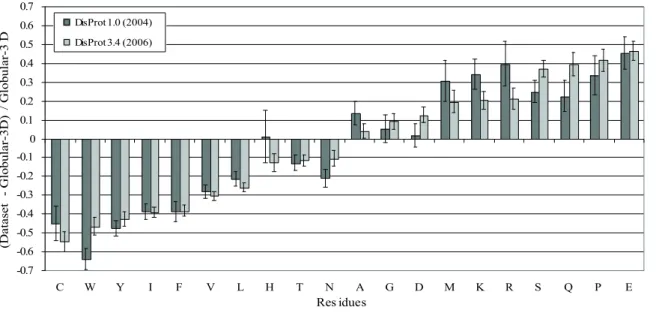
Fig. 1.2 The differences between the amino acid compositions of disordered data sets (DisProt 1.0 and Disprot 3.4) and that of an ordered data set (Globular-‐‑3D). Amino acids are arranged by the peak height as in DisProt 3.4. For details see [16].
-0.7 -0.6 -0.5 -0.4 -0.3 -0.2 -0.1 0 0.1 0.2 0.3 0.4 0.5 0.6 0.7 C W Y I F V L H T N A G D M K R S Q P E Res idues (D at as et G lo bu la r-3D ) / G lo bu la r-3 D DisProt 1.0 (2004) DisProt 3.4 (2006)
Already in 2001 it was discovered [17] that disordered and folded proteins have a different content of certain amino acids. The amino acid composition of disordered protein segments relative to globular proteins (Fig. 1.2) show that IDPs are generally enriched in charged and structure-‐‑breaking residues (Pro and Gly), and depleted in hydrophobic and aromatic residues. Proteins with this amino acid composition have low mean hydrophobicity (which is a driving force of protein compaction) and high net charge (thus contributing to intramolecular repulsion), and thus are less likely to adopt a stable fold. IDPs also have a generally lower sequence complexity [18], in particular they abound in patterns containing Pro and certain charged amino acids (such as Glu) [19].
1.1.2 Prediction of protein disorder by computational tools
It has been shown that basic physico-‐‑chemical properties of a protein can predict its degree of disorder. On a CH plot (net charge versus mean hydrophobicity) natively folded and unfolded proteins tend to form two separate clusters. FoldIndex [20], PreLink [21] and GlobPlot [22] are predictors based on this approach (see [23]).
More complicated prediction tools use databases of experimentally characterized disordered proteins: DisProt [24], IDEAL [25], MobiDB [26]. They are based on neural networks trained on databases of folded and disordered proteins, comparing sequence and/or physico-‐‑chemical properties of different segments of a given protein to those in databases. For example, the PONDR predictor [18] calculates different attributes (such as hydropathy, fractional composition of particular amino acids or sequence complexity) of a sequence over windows from 9 to 21 residues for the proteins in the databases and uses these attributes to train its neural network. The same attributes are calculated over a given sequence and used as input for the neural network. Other examples of predictors using trained neural networks are DisEMBL [27], DISOPRED2[28] and RONN [29].
The drawback of this approach is that in those databases the regions of missing electron density in PDBs are overrepresented. These regions are typically short, as extensive disorder prevents crystallization, so the above-‐‑mentioned prediction tools are necessarily trained on short disordered regions and could be less efficient in predicting long disorder.
Another way of predicting disorder is based on the idea that different amino acids have different propensities to form contacts with each other and that amino acids that avoid each other are overrepresented in IDPs because they fail to form intramolecular contacts. The FoldUnfold tool calculates the expected number of contacts per amino acid residue from the protein sequence [30]. Another predictor IUPred [31] is based on the contribution to the fold stability of each amino acid in folded proteins.
A semi-‐‑automated method called HCA (Hydrophobic Cluster Analysis) is based on a two-‐‑ dimensional graphic representation of protein sequence where hydrophobic residues forming clusters are marked (see Fig. 1.3). Disordered regions are recognizable as those depleted of hydrophobic clusters [32].
All these methods have their advantages and drawbacks. Some of them are better for short disordered regions (as DISOPRED2 and PreLink), while others (as IUPred) are more efficient in predicting long disordered segments. Finally some predictors, such as PONDR, GlobPlot, and FoldIndex, have been trained on both short and long disorder and provide a balanced performance. For this reason, to perform a reliable prediction of disordered regions, so-‐‑called “metapredictors” seek a consensus prediction based on scores obtained from different methods (PONDR-‐‑FIT is one example [33]). Another option is to apply different predictors independently and combine their output in a single analysis. The MeDor (MEtaserver of DisORder) server [34] combines the output of different disorder prediction servers and a HCA plot (see Fig. 1.3).
Fig. 1.3 MeDor output of the N-‐‑terminal domain of measles virus phosphoprotein. The upper line above the sequence shows secondary structure elements predicted by Pred2ary [35]. Below the sequence are shown the HCA plot and the output of different disorder predictors (disordered regions depicted as arrows).
At this stage of their development, disorder predictors do not in any way provide a proof of disorder in a given protein, they are rather a tool to find potentially disordered regions that merit further experimental studies. However, with the growing number of experimentally characterized IDPs, one can hope that in the future the sequence-‐‑based criteria of intrinsic disorder would be further established and experimentally validated.
1.2 Experimental characterization of intrinsically disordered proteins
One of the simplest methods to detect intrinsic disorder in proteins is circular dichroism (CD) spectroscopy. CD spectra provide estimates of secondary structure and allow for the distinction between different types of secondary structure elements (α-‐‑helices, β-‐‑ sheets, turns, etc.) [36].
IDPs can also be recognized by their peculiar behaviour in SDS polyacrylamide gel electrophoresis, gel filtration or size-‐‑exclusion chromatography, where they have unusually high apparent molecular masses (mostly due to their amino acid composition and extension of their chains) [23]. They are also much more sensitive to protease digestion [17]. Finally, IDPs are much less sensitive to high temperatures and acidic treatment than folded proteins, which may precipitate at these conditions; this property is often exploited for purification purposes [23].
1.2.1 Nuclear magnetic resonance (NMR)
Already a one-‐‑dimensional proton NMR spectrum would give us an indication about protein disorder. In an IDP, inter-‐‑residue contacts are quasi-‐‑absent, and all residues have a similar chemical environment. As the chemical shifts of protons are more sensitive to the general structural context than to the local sequence composition [37], the dispersion of the resonances of IDPs in the proton dimension is limited. But the force of NMR comes from its possibility to study proteins at the residue-‐‑specific level, using high-‐‑field spectrometers and multidimensional NMR spectroscopy.
NMR is only sensitive to nuclei with nonzero spin. Nuclei with spin more that ½ give broad resonance lines due to electric quadrupole moment and hence are more difficult to study [38]. Therefore, the most important nuclei for biomolecular NMR are those with a spin of ½, as for example 1H, 13C and 15N. In nature, the content of 13C is 1.07% and that
of 15N is 0.364%. As this is not enough for most NMR experiments, especially for
multidimensional experiments, due to limitations in accessible protein concentration, one of the most important requirements is to be able to express a protein heterologously, in a host that allows for uniform labelling of a protein. Most proteins for NMR studies are expressed in E.coli, which is the cheapest and the most simple expression system (which is still quite expensive, especially if not only 15N but also 13C labelling is required).
However, there are some recent advances in expressing labelled proteins in insect cells [39, 40].
The starting point of each protein NMR study and one of the simplest spectra to run is the HSQC (heteronuclear single quantum coherence) experiment. This two-‐‑dimensional spectrum (15N-‐‑1H HSQC) correlates the backbone nitrogen with its directly attached
amide proton and, therefore, displays a resonance at the position corresponding to the chemical shifts of the nitrogen and the amide proton. A set of three-‐‑dimensional experiments can be run in order to measure 13Cα, 13Cβ and 13CO chemical shifts and link
them to the 15N and 1HN shifts of the same and adjacent residue. As 15N and especially 13C
chemical shifts are particularly sensitive to the type of amino acid residue, it allows us to establish a direct link between peaks and the protein sequence. This process is known as backbone resonance assignment and once completed, provides access to a variety of
information about the structure and dynamical behaviour of the protein at the residue-‐‑ specific level, by running different types of NMR experiments (see also Fig. 1.1).
For unfolded and partially folded proteins, one of the most informative primary NMR observables is the chemical shift, because it is sensitive to polypeptide backbone torsion angles [23]. Secondary 13Cα, 13Cβ, 13CO and 1Hα chemical shifts are calculated as the
deviation of the experimental chemical shifts from so-‐‑called random coil values [41]. The random coil values are amino acid specific and represent the expected chemical shifts for a protein devoid of secondary and tertiary structure. These values have traditionally been obtained by measuring the chemical shift values of each of the twenty amino acids within small peptides [41]. The experimental secondary chemical shifts are sensitive to the local structure and hence provide us with information about secondary structure content in the protein [23]. It should be mentioned that this approach is sensitive to the correct frequency referencing in the NMR experiment and to overcome this problem, 13Cα and 13Cβ secondary shifts can be used simultaneously to correct for a potential reference
offset as the 13Cα and 13Cβ chemical shifts show an inverse dependence on α-‐‑helical
propensity [42].
NMR relaxation is sensitive to backbone and side chain dynamics of IDPs. The most
commonly used techniques involve the measurement of R1, R2 and heteronuclear NOEs
for backbone resonances, and allow for a detailed characterization at the residue-‐‑specific level of contributions of different motions to the dynamics of the IDP chain on the ps-‐‑ns timescale. 15N/13C R2 relaxation dispersion [43] and proton R1ρ relaxation dispersion [44]
allow the characterization of exchange in proteins on the μs – ms timescale. NMR relaxation is more extensively discussed in the subsequent chapters of this thesis.
With NMR, we are not limited to the study of local structural propensities and local dynamics. Paramagnetic relaxation enhancement (PRE) is an excellent probe of long-‐‑ range interactions in disordered proteins. This method relies on the introduction of a paramagnetic nitroxide spin-‐‑label at a specific position in the protein, usually via a conjugation with a solvent-‐‑exposed cysteine residue, which can be introduced by site-‐‑ directed mutagenesis [45, 46]. The presence of an unpaired electron causes a broadening of the nuclear spin resonances within a radius of about 25 Å [47] due to the strong dipolar interaction [48]. HSQC spectra are typically recorded with the spin-‐‑label in its paramagnetic (oxidized) and diamagnetic (reduced) state. Differences in the line width, relaxation rates, or intensity in these two spectra give an estimate of the average distance between the spin label and the given residue. The limitations of this approach are linked to the introduction of a non-‐‑native residue in the protein, and the risk of perturbation of native long-‐‑range contacts by its side chain and/or the paramagnetic label. However, it offers a possibility to elucidate a fluctuating tertiary structure in a disordered protein that would be difficult to describe using other approaches [49].
1H-‐‑1H nuclear Overhauser enhancements (NOEs) were used to calculate structure of
folded proteins since a long time [50]. In theory, NOEs can also be used to obtain more detailed information about local conformational sampling in IDPs [51]. However, quantitative interpretation of NOEs is complicated by their strong sensitivity on the range of dynamic timescales commonly encountered in unfolded proteins [49].
Another powerful method to assess local and global order in proteins is to measure
Residual Dipolar Couplings (RDCs). Dipolar couplings between different spins (nuclei)
are a very rich source of structural information because they depend only on the internuclear distance (𝑟"#), the orientation of the internuclear vector relative to the static
magnetic field, and known physical constants. In an isotropic solution, however, all orientations are sampled with the same probability, and are therefore efficiently averaged to zero. But if the molecule is partially aligned in the magnetic field, one of the orientations is preferred and so-‐‑called residual dipolar couplings appear as additional contributions to the well-‐‑known scalar couplings. To obtain this partial alignment of a protein molecule, a number of approaches are available, most of them involve dissolving the protein in some kind of anisotropic medium such as solutions of liquid crystals [52], lipid bicelles [53], filamentous bacteriophages [54] or polyacrylamide gels [55, 56].
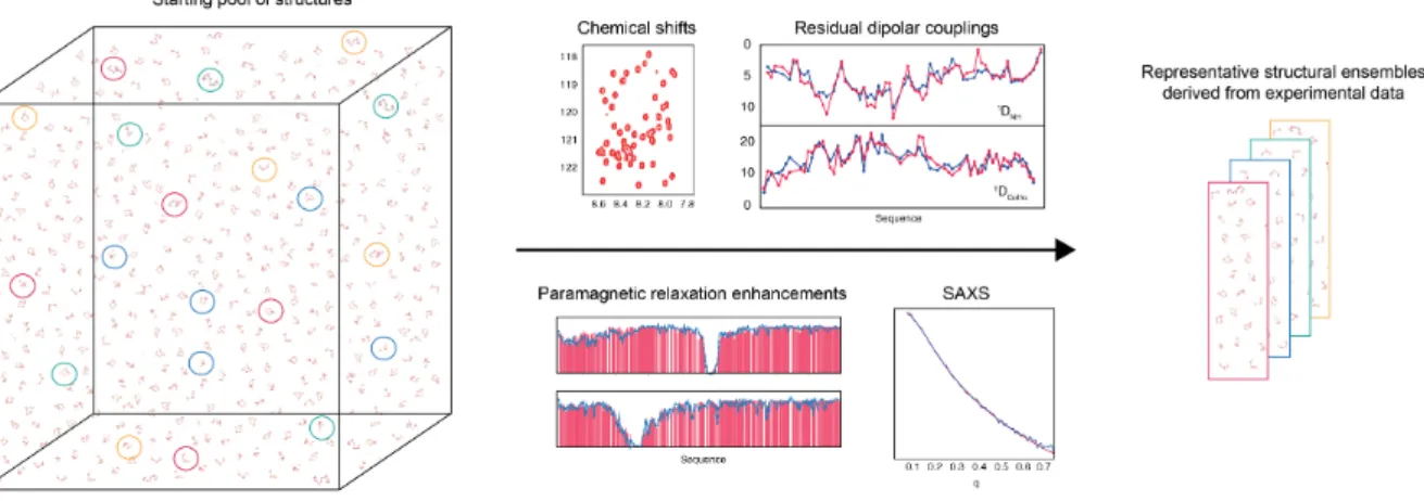
Fig. 1.4. Deriving representative ensembles of IDPs using a sample-‐‑and-‐‑select approach. Flexible-‐‑Meccano is used to generate a large pool of statistical coil conformers that is assumed to efficiently sample conformational space. Typically a pool of 20000 conformers is generated. The genetic algorithm ASTEROIDS is used to select a sub-‐‑ensemble in agreement with different complementary types of experimental data. The sizes of the sub-‐‑ensembles are adjusted depending on the protein investigated. Typical sub-‐‑ensembles contain 100-‐‑200 conformers.
Measuring RDCs has its own pitfalls; in particular, care should be taken to ensure that the protein (especially an IDP, which may have exposed hydrophobic groups) does not interact significantly with the alignment medium.
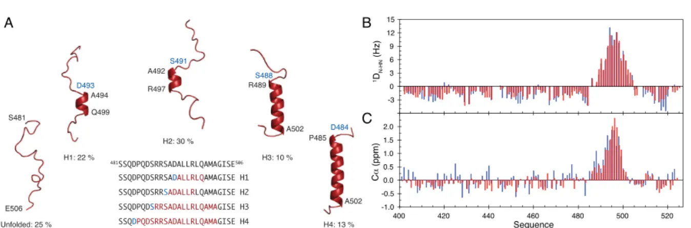
Fig. 1.5. A. Description of the conformational ensemble of the α-‐‑MoRE of the C-‐‑terminal domain, NTAIL, of
measles virus. B. Experimental (blue) and back-‐‑calculated from the ensemble (red) DN-‐‑HN RDCs for measles
virus NTAIL. C. Experimental (blue) and back-‐‑calculated Cα values.
RDCs are a valuable source of information on both structure and dynamics in disordered proteins [49, 57, 58]. An algorithm called ASTEROIDS (Fig. 1.4) was developed to use RDCs, chemical shifts and paramagnetic relaxation for the description of the conformational ensembles of IDPs. In this approach, an ensemble of random structures of an IDP is initially generated using a statistical coil generator, Flexible-‐‑Meccano [59, 60], on the basis of the primary sequence of the protein. If the experimental data are predicted correctly, then one can already conclude that a random-‐‑coil, or statistical-‐‑coil model, accurately described the conformational behaviour in solution. If there are significant deviations from coil behaviour, the genetic algorithm ASTEROIDS [61] can then be employed to select conformational ensembles that give the best fit between back-‐‑ calculated and experimentally measured RDCs and chemical shifts. As an example, using the combination of Flexible-‐‑Meccano and ASTEROIDS, the conformational ensemble representing the α-‐‑MoRE (α-‐‑helical molecular recognition element) of the disordered C-‐‑terminal domain of measles virus nucleoprotein (Measles NTAIL) was described (Fig. 1.5,
[62]).
1.2.2 Small angle X-‐ray scattering
Small-‐‑angle X-‐‑ray scattering (SAXS) is a powerful biophysical method that allows the structural characterization of biological systems up to the resolution of several nm. It is known to provide basic geometrical information about the 3D shape and size of objects, such as biomolecules, in solution (e.g. radius of gyration, shape elongation), and has been used extensively to provide ab-‐‑initio reconstructions of the overall envelope of proteins [63, 64]. SAXS is now more and more used to study IDPs [65], in general as a complementary method to NMR studies. Already standard analyses of SAXS profiles, such as the Kratky representation and Guinier analysis give us insight into the presence of transiently folded structures inside IDPs. In addition, the application of SAXS ab initio reconstruction to complexes of IDPs and folded proteins can reveal the presence of flexible chains attached to globular particles, thus allowing the study of complex
A B
formation [65]. Recently, different ensemble-‐‑selection tools were developed to describe protein flexibility by SAXS, of which the Ensemble Optimization Method (EOM, [66]) specifically addresses the question of structural ensemble descriptions of IDPs.
The major limitation of SAXS is that it requires a monodisperse sample, as the presence of oligomerization or aggregation would hamper the analysis of the shape or flexibility of macromolecules. However, if the aggregation is specific and the number of components in the system is limited (e.g. a monomer-‐‑dimer equilibrium) and the scattering profile of each oligomeric form can be predicted, it is possible to study the composition of the system [65].
1.2.3 Single-‐molecule approaches
One of the limitations of previously cited “in-‐‑bulk” approaches is that all conformations present in the sample at a given time are averaged. In single-‐‑molecule approaches, this issue is eliminated by detection of each individual molecule. Using Förster resonance
energy transfer (FRET), which probes the distance between two fluorescent dyes
attached to the protein at different positions, protein conformations as well as their time dependence down to the nanosecond time scale can be described using different experimental setups and analysis procedures [67].
The approaches based on atomic force microscopy (AFM) include high-‐‑speed AFM [68, 69], which enables the observation of dynamics in IDPs with sub-‐‑100ms to subsecond temporal resolution, as well as single-‐‑molecule force spectroscopy (SM-‐‑FS) [70]. The latter involves mechanically stretching the protein with an AFM tip and probes timescales from milliseconds to seconds and is particularly sensitive to the presence of elements of secondary structure in an IDP.
1.3 Intrinsically disordered proteins in living cells
The fact that IDPs exist and were characterized in vitro does not prove that intrinsic disorder is relevant in the context of a living cell. In the cell, a significant fraction of the total volume (20-‐‑30%) is occupied by macromolecules [71]. For example, inside an E. coli cell, the total concentration of proteins and RNA rise up to 400 mg/ml [72]. These excluded volume effects (crowding) may indeed have considerable impact on the conformational and dynamical properties of IDPs [73], and the possibility that they can force IDPs to become more extended, more compact or even fold needs to be assessed. One possibility to answer this question is to mimic the crowding by studying an IDP in the presence of crowding agents. In order to approximate the crowding conditions in a cell as closely as possible, these agents should mimic the effect of volume exclusion [72], increased viscosity of local environment [74] as well as affecting the solvation/hydration
of proteins [75, 76]. High molecular weight polymers, such as dextran or Ficoll 70, increase the viscosity and generate the excluded volume effect. Osmolytes (such as sucrose, triflouroethanol (TFE), trimethylamine N-‐‑oxide (TMAO)), interact unfavorably with the protein backbone [77] and thereby stabilize secondary structure elements in IDPs [78].
Studies carried out on various IDPs under different conditions show that dextran and Ficoll 70 elicit some compaction but not folding of the proteins [79]. Osmolytes such as TFE or TMAO promote the formation of α-‐‑helical elements in some IDPs [80]. In a recent article [73], NMR relaxation experiments on different IDPs (ProTα, TC-‐‑1 and α-‐‑synuclein) in the presence of Ficoll 70 and Dextran 70 showed a limited impact of 160mg/ml of both agents on protein dynamics. At 400 mg/ml of Dextran 70, the chain dynamics of ProTα is slowed down, however, it still retains a certain level of flexibility. Ficoll 70 was equally found to induce a minor increase of helical propensity in a relatively structured region of the TC-‐‑1 protein [73]. In a more recent study [81], SM-‐‑FRET was used to study the behavior of different IDPs in the presence of polyethylene glycol (PEG). It was found that both increasing volume fraction and size of PEG lead to IDP compaction. However, other authors discourage the use of PEG as a crowding agent due to the promotion of attractive interactions between proteins [73]. In conclusion, the crowding effect does not fold IDPs but may promote the formation of local secondary structure elements [80].
Still, mimicking the intracellular environment using artificial crowders does not provide a full answer to the question of protein disorder in cells. One should directly assess the amount of disorder under in vivo conditions. One of the possibilities is to study proteasomal degradation of IDPs “by default” (i.e. ubiquitin-‐‑independent). This approach, applied to several IDPs, including p53 [82] and p21 [83], proved that the IDPs are degraded by the 20S proteaseome and thus supported the idea that the proteins stay disordered in vivo.
Another option is to study the protein by in-‐‑cell NMR [84]. This requires the deposition of labeled proteins within the cell for example by overexpressing them under labeling conditions, by covalently linking the proteins to cell-‐‑penetrating peptides [85], by electroporation [86, 87] or by microinjection into the cell cytoplasm [88]. Thus, a labeled protein is the only NMR observable macromolecule and allows investigation of the structural and dynamic properties of the protein under conditions close to those in vivo.
Different proteins were found to stay disordered even under those conditions. Tau protein was studied in Xenopus oocytes by in-‐‑cell NMR and was found to stay partially disordered [89]. In a recent article, Binolfi et al. [90] and Waudby et al. [91] studied the structure and dynamics of α-‐‑synuclein in E. coli cells. The results show that in the bacterial cytosol, α-‐‑synuclein populates a highly dynamic state, which has the same