ORIGINAL PAPER
Identification of a CYP3A form (CYP3A126)
in fathead minnow (
Pimephales promelas)
and characterisation of putative CYP3A enzyme activity
Verena Christen&Daniel Caminada&Michael Arand&Karl Fent
Received: 3 September 2009 / Revised: 16 October 2009 / Accepted: 18 October 2009 / Published online: 8 November 2009
# Springer-Verlag 2009
Abstract Cytochrome P450-dependent monooxygenases (CYPs) are involved in the metabolic defence against xenobiotics. Human CYP3A enzymes metabolise about 50% of all pharmaceuticals in use today. Induction of CYPs and associated xenobiotic metabolism occurs also in fish and may serve as a useful tool for biomonitoring of environmental contamination. In this study we report on the cloning of a CYP3A family gene from fathead minnows (Pimephales promelas), which has been designated as CYP3A126 by the P450 nomenclature committee (GenBank no. EU332792). The cDNA was isolated, identified and characterised by extended inverse polymerase chain reaction (PCR), an alterna-tive to the commonly used method of rapid amplification of cDNA ends. In a fathead minnow cell line we identified a full-length cDNA sequence (1,863 base pairs (bp)) consisting of a 1,536 bp open reading frame encoding a 512 amino acid protein. Genomic analysis of the identified CYP3A isoenzyme revealed a DNA sequence consisting of 13 exons and 12 introns. CYP3A126 is also expressed in fathead minnow liver as demonstrated by reverse transcription PCR. Exposure of
fathead minnow (FHM) cells with the CYP3A inducer rifampicin leads to dose-dependent increase in putative CYP3A enzyme activity. In contrast, inhibitory effects of diazepam treatment were observed on putative CYP3A enzyme activity and additionally on CYP3A126 mRNA expression. This indicates that CYP3A is active in FHM cells and that CYP3A126 is at least in part responsible for this CYP3A activity. Further investigations will show whether CYP3A126 is involved in the metabolism of environmental chemicals. Keywords Fathead minnow . Fish . Cytochrome P4503A . CYP3A . CYP3A126 . Pharmaceuticals .
Interaction of chemicals with CYP3A . Diazepam . Induction and inhibition of CYP3A
Introduction
Investigations on enzymes involved in the metabolism of environmental pollutants in fish are important in providing information about their behaviour and effects in aquatic organisms. Cytochrome P450-dependent monooxygenases (CYPs) are involved in the metabolic defence against xenobiotics. In fish, induction of CYPs and associated xenobiotic metabolism is well established and may serve as a useful tool for the biomonitoring of environmental contaminations [1–4] and in the ecotoxicological assess-ment of pollutants.
In the last decade, a number of CYP3A genes have been isolated from several teleost species [5–12]. Up to date the CYP3A sub-family has much more than 100 sub-family members (Nelson, Cytochrome P450 web site:http://drnelson. utmem.edu/CytochromeP450.html). Human CYP3A enzymes metabolise about 50% of all pharmaceuticals in use today V. Christen
:
D. Caminada:
K. FentSchool of Life Sciences,
University of Applied Sciences Northwestern Switzerland, Gründenstrasse 40,
4132 Muttenz, Switzerland M. Arand
Institute of Pharmacology and Toxicology, University of Zurich, Winterthurerstrasse 190,
8057 Zurich, Switzerland K. Fent (*)
Department of Environmental Sciences, Swiss Federal Institute of Technology (ETHZ), 8092 Zurich, Switzerland
e-mail: karl.fent@bluewin.ch DOI 10.1007/s00216-009-3251-5
[13]. They are also involved in the biosynthesis and degradation of endogenous compounds such as steroids, lipids and vitamins. In both mammals and fish the CYP3A family members are considered to be important drug-metabolising enzymes. Collectively the CYP3A family comprises the largest portion of the liver and small intestinal CYPs [9,14,15]. CYP3A induction by typical mammalian CYP3A inducers rifampicin and dexamethasone was shown in zebrafish larvae [11] and grass carp in vitro and in vivo [16]. Exposure of medaka to 17 beta-estradiol suppressed CYP3A protein production [17]. Similar to mammals induction of CYP3A expression in fish is probably regulated by the pregnane X receptor (PXR) [18,19], but this still needs to be demonstrated. Exposure of Atlantic salmon to thyroxine and 1,1-dichloro-2,2-bis (p-chlorophenyl)ethylene resulted in CYP3A and PXR mRNA induction in the liver [20]. A positive correlation between CYP3A and PXR expression implies a connection, but this has to be proven. Therefore, a change in CYP3A expression could indicate an exposure of an organism to xenobiotics. Furthermore a change in CYP3A expression could provide information on the metabolism of the xenobiotics. Catalytic activities of CYP3A have been determined in different fish by use of different substrates [16,21,22]. It was found that CYP3A enzymatic activity in fish can easily be determined by application of a modified human CYP3A activity assay, in which the conversion of a substrate by CYP3A to a fluorescent product is determined [14,15,22–26].
In our present study, we report on a member of the CYP 3A family in fathead minnow (Pimephales promelas), identified and characterised in a fathead minnow cell line (FHM) and verified in adult fathead minnow liver tissue. Fathead minnow is an important fish species extensively used in aquatic toxicological research and toxicological assessment of chemicals. Therefore, identification and characterisation of its xenobiotic metabolising enzymes are important. Furthermore, we characterise the FHM cell line CYP3A enzyme activity after exposure to rifampicin (a prototypical mammalian CYP3A inducer) and diazepam (a model mammalian CYP3A substrate) using a CYP3A activity assay. In addition to the enzyme activity, we evaluate alterations in CYP3A126 mRNA levels by quantitative reverse transcription (qRT) PCR after exposure of FHM cell line to rifampicin and diazepam.
Materials and methods
Chemicals, cell culturing and exposure
Dimethyl sulfoxide (DMSO), acetonitrile (AcN), rifampicin, benzyl-trifluoromethyl coumarin (BFC), and 7-benzyloxyquinoline (BQ) were purchased from
Sigma-Aldrich, Buchs (Switzerland). Phosphate-buffered saline was purchased from Roche Diagnostics, Basel (Switzerland). Diazepam (≥99%) was kindly provided by F. Hoffmann— La Roche Ltd, Basel (Switzerland).
Stock solutions of rifampicin and diazepam were prepared in DMSO or acetonitrile (AcN) at a concentration of 100 mM if not otherwise stated. For the assay, stock solutions were diluted in cell culture medium resulting in a maximal solvent concentration of 0.1%. Further concen-trations were prepared by serial dilution.
FHM P. promelas cells (FHM) were kindly provided by T. Wahli, University Bern, Switzerland, and grown in DMEM/ F12 (LuBioScience, Lucerne, Switzerland) supplemented with 5% FBS (Sigma-Aldrich), 20 mM HEPES/pH 7.2 (Sigma-Aldrich) at room temperature (21±2 °C). Cells were split every 7 days and sub-cultured at a split ratio of about 1:3.
Huh7 The human hepatoma cell line Huh7 was kindly provided by M. Heim, University Hospital Basel, Switzerland. Cells were grown in DMEM with GlutaMAX™ (LuBio-Science) supplemented with 5% FBS in a humidified incubator with 5% CO2at 37 °C. Cells were usually split every 4 days and sub-cultured at split ratios of about 1: 6.
Exposure to rifampicin and diazepam For exposure experi-ments, 100,000 cells per well were seeded in 96-well plates and grown for 24 h in culture medium. After 24 h the medium was removed, and cells were incubated in phenol red free medium containing the pharmaceutical (10, 25 and 50μM of rifampicin or 0.1, 1, 10 and 20 μM of diazepam) or the solvent control (DMSO for rifampicin and acetoni-trile for diazepam). The cells were incubated for additional 24 h, and the CYP assay was performed as described below. For each pharmaceutical, the assay was repeated three times.
Identification and characterisation of the CYP3A126 IPCR FHM cells were directly lysed in the cell culture flask and homogenised using a QIA shredder (Qiagen, Switzerland, cat. no. 79654). Total RNA was extracted using the RNeasy mini kit (Qiagen, cat. no. 74104; Fig.1 a). Double-stranded cDNA (ds cDNA) synthesis was performed using 50 ng/µl total RNA and the cDNA Synthesis System Kit (Roche Diagnostics, cat. no. 11 117 831 001) according to manufacturer’s instructions (Fig. 1b). The ds cDNA was cleaned using the High Pure PCR product Purification Kit (Roche Diagnostics, cat. no. 1 732 668 001). Circularisation of ds cDNA was performed using 2.5 ng/µl ds DNA and 5U T4 DNA Ligase (Roche Diagnostics, cat. no. 1 004 786 001; Fig. 1c). The ligation reaction was incubated overnight at
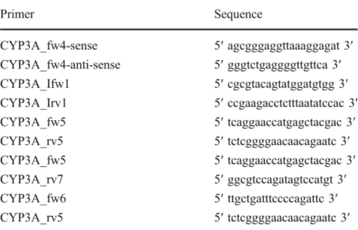
16 °C. PCR reaction was performed using 0.75 µM of each the sense (CYP3A_fw4 5′ agcgggaggttaaaggagat 3′) and anti-sense primer (CYP3A_fw4 5′ gggtctgaggggttgttca 3′) and 1 µl of the ds cDNA as template (Fig.1d). Table 1lists the primers used. The amplification conditions were 94 °C for 2 min for denaturing the template, 40 cycles of 94 °C for 30 s for denaturing, 58 °C for 30 s for annealing and 68 °C for 8 min for elongation and 68 °C for 10 min for terminal extension. The reaction was maintained at 8 °C until a second PCR was performed with an inverse primer pair (CYP3A_Ifw1 5′ cgcgtacagtatggatgtgg 3′/CYP3A_Irv1 5′ ccgaagacctctttaatatccac 3′) to amplify the ends of the unknown CYP3A cDNA (Fig.1e). PCR reaction (1 µl) was used as template. The amplification conditions were the same as mentioned above.
Sequence verification RT-PCR was performed to verify the sequence found in the FHM cell line and in FHM liver
tissue. The primers used are listed in Table 1. For that purpose the primer pair CYP3A_fw5 (5′ tcaggaaccatgagc tacgac 3′)/CYP3A_rv5 (5′ tctcggggaacaacagaatc 3′) that flanked the coding sequence (cds) of the CYP3A cDNA and the primer pairs CYP3A_fw5 (5′ tcaggaaccatgagctac gac 3′)/CYP3A_rv7 (5′ ggcgtccagatagtccatgt 3′) and CYP3A_fw6 (5′ ttgctgatttccccagattc 3′)/CYP3A_rv5 (5′ tctcggggaacaacagaatc 3′) were used. Total RNA was extracted from liver tissue using the RNeasy mini kit (Qiagen, cat. no. 79254) after lysis and homogenisation of the tissue by sonication. Total RNA from FHM cell line was extracted as mentioned above. For cDNA synthesis 50 ng/μl total RNA, 0.75 μM oligo-dT primer and Moloney murine leukaemia virus reverse transcriptase (Promega Biosciences, Inc., Wallisellen, Switzerland) were used. The reaction mixture was incubated for 5 min at 70 °C and then for 1 h at 37 °C. The reaction was stopped by heating at 95 °C for 5 min. PCR cycle conditions were 94 °C for 30 s for denaturing the template, 40 cycles of 94 °C for 30 s for denaturing, 59.1 °C for 30 s for annealing and 68 °C for 2 min for elongation and 68 °C for 7 min for terminal extension. Amplification products of the expected size were sequenced. Genomic analysis Analysis on the genomic level of the CYP3A was done in FHM cells using PCR. The PCR reaction was performed using AccuPrime Taq DNA Polymerase High Fidelity with 10× Accu Primer Buffer I (Invitrogen, Switzerland, cat. no. 12346-086) and 0.75 µM of each the sense (CYP3A_fw5) and the anti-sense primer (CYP3A_rev5). The amplification conditions consisted of initial denaturation (94 °C, 30 s), followed by 40 cycles for amplification (94 °C for 30 s for denaturing, 56 °C for 30 s for annealing and 12 min at 68 °C for elongation) and terminal extension at 68 °C for 10 min. The amplification product of about 8,300 bp was cleaned using the High Pure PCR product Purification Kit (Roche Diagnostics, cat. no. 11 732 668 001) and sequenced.
Ligation
double strand cDNA synthesis total RNA 5‘ 3‘ 3‘ 5‘ Known sequence B C D 5‘ A E AAAAA B C D 3‘ A TTTTT E B C D 3‘ A E AAAAA C D 5‘ A TTTTT E B C D A E AAAAA B C D A TTTTT E 5‘ B C D A E AAAAA C D A TTTTT 3‘ 3‘ E 5‘ 3‘ 3‘ 5‘ circulated ds cDNA E B D C A PCR PCR 5‘ (a) (b) (c) (d) (e)
Fig. 1 Schematic explanation for the obtained CYP3A amplification. a Total RNA extraction; b double strand synthesis; c circularisation of ds cDNA by ligation; d PCR with primer pair CYP3A_fw4/ CYP3A_rv4: transcripts of the circulated ds DNA template annealed together and served as a template for the polymerase; e amplification of the new CYP3A isoenzyme with the IPCR primer pair CYP3A_Ifw1/CYP3A_Irv1. CYP3A_fw4 and CYP3A_rv4 primer are marked as black arrows; CYP3A_Ifw1 and CYP3A_Irv1 inverse primer are marked as white arrows
Table 1 Primers used for inverse PCR and RT-PCR
Primer Sequence CYP3A_fw4-sense 5′ agcgggaggttaaaggagat 3′ CYP3A_fw4-anti-sense 5′ gggtctgaggggttgttca 3′ CYP3A_Ifw1 5′ cgcgtacagtatggatgtgg 3′ CYP3A_Irv1 5′ ccgaagacctctttaatatccac 3′ CYP3A_fw5 5′ tcaggaaccatgagctacgac 3′ CYP3A_rv5 5′ tctcggggaacaacagaatc 3′ CYP3A_fw5 5′ tcaggaaccatgagctacgac 3′ CYP3A_rv7 5′ ggcgtccagatagtccatgt 3′ CYP3A_fw6 5′ ttgctgatttccccagattc 3′ CYP3A_rv5 5′ tctcggggaacaacagaatc 3′
Determination of CYP3A mRNA
Determination of CYP3A mRNA: RNA isolation, reverse transcription and qPCR Total RNA was isolated from FHM and Huh7 cells using Trizol reagent according to the manufacturer's instructions. RNA was reverse-transcribed by Moloney murine leukaemia virus reverse transcriptase (Promega Biosciences, Inc.) in the presence of random hexamers (Roche) and deoxynucleoside triphosphate. The reaction mixture was incubated for 5 min at 70 °C and then for 1 h at 37 °C. The reaction was stopped by heating at 95 °C for 5 min. qPCR was performed based on SYBR green fluorescence (SYBR green PCR master mix; Roche). Table 2 lists the primers used. The primers for the FHM housekeeping gene G6PDH (glucose-6-phosphate dehydro-genase) were 5′ GTGGCATGTGTGGTTCTGAC 3′ and 5′ TTGACCTTCTCGTCCCTAACA 3′. The primers for the FHM CYP3A126 were 5′ AACGAATCGTAACGCAGCTC 3′ and 5′ TCTCGGGGAACAACAGAATC 3′. The primers for the human GAPDH (glyceraldehyde-3-phosphate dehydrogenase) were 5′ GCTCCTCCTGTTCGACAGTCA 3′ and 5′ ACCTTCCCCATGGTGTCTGA 3′. The primers for the human CYP3A4 were 5′ TCAATAACAGTCTTTC CATTCCTCAT 3′ and 5′ CTTCGAGGCGACTTTCTTTCA 3′. The amplification conditions were 95 °C for 5 min for initial denaturing, 40 cycles of 95 °C for 30 s for denaturing, 58 °C for 30 s for annealing and 72 °C for 45 s for elongation. A melting curve was run afterwards. The difference in the cycle threshold (∆CT) value was derived by subtracting the CTvalue for G6PDH, which served as an internal control, from the CTvalue for CYP3A. All reactions were run in duplicates using a BioRad real-time PCR machine (CFX 96 Real Time System). mRNA expression levels of CYP3A were expressed as a several fold increase according to the formula 2∆CT(untreated)−∆CT(treated). Determination of basic CYP3A4 catalytic activity
Assay in 24-, 48- and 98-well plates Different numbers of FHM cells were seeded in 24-, 48- and 96-well plates and
grown for 24 h in culture medium. After 24 h the medium was removed, and FHM cells were incubated in phenol red free medium containing either 50μM BFC or 60 μM BQ or AcN as solvent control, for the indicated time period. BFC and BQ are metabolised by human and fish CYP3A4 to give the highly fluorescent 7-hydroxy-4(trifluoromethyl) coumarin and the fluorescent 7-hydroxyquinoline. Both metabolites can readily be detected spectrofluorometrically [23, 27]. After this exposure time, the supernatant was transferred into a black 96-well plate mixed with the stop solution (80% AcN and 20% 0.5 M Tris base). The plate was read on a GENios Tecan reader (Tecan, Männedorf, Switzerland) using an excitation wavelength of 410 nm and an emission wavelength of 530 nm. Each experiment was repeated three times.
Determination of CYP3A catalytic activity after drug exposure Assay in 96-well plates FHM and Huh7 (control) cells (100,000 cells per well) were seeded in a 96-well plate and grown overnight. The next day, cells were incubated in phenol red free medium containing the indicated amount of model inducer rifampicin (10, 25 and 50μM and DMSO as solvent control) and inhibitor diazepam (10 and 20μM for Huh7 cells and 0.1, 1 and 10 μM for FHM cells and acetonitrile as solvent control), respectively, for 24 h. After the incubation time the CYP3A assay was performed as described above with BFC or BQ as substrate. The assay for both pharmaceuticals was repeated three times, and the number of replicates for each condition was 8.
Statistical analysis Data were graphically and statistically evaluated with GraphPad Prism 4 (GraphPad Software, Inc. San Diego, CA, USA). Data were analysed by one-way ANOVA followed by a post test (Bonferroni's multiple comparison test, significance limit p<0.05). An unpaired t test with significance limit p<0.05 was performed to analyse data of Figs.5aand6cbecause in both figures only two groups were compared with each other.
Table 2 Primers used to perform real-time PCR
Primer Sequence
FHM-Glucose-6-phosphate dehydrogenase (G6PDH)-forward 5′ GTGGCATGTGTGGTTCTGAC3′ FHM-Glucose-6-phosphate dehydrogenase (G6PDH)-reverse 5′ TTGACCTTCTCGTCCCTAACA3′
FHM CYP3A126-forward 5′ AACGAATCGTAACGCAGCTC 3′
FHM CYP3A126-reverse 5′ TCTCGGGGAACAACAGAATC3′
Human glyceraldehyde-3-phosphate dehydrogenase GAPDH-forward 5′ GCTCCTCCTGTTCGACAGTCA 3′ Human glyceraldehyde-3-phosphate dehydrogenase GAPDH-reverse 5′ ACCTTCCCCATGGTGTCTGA 3′
Human CYP3A4-forward 5′ TCAATAACAGTCTTTCCATTCCTCAT3′
Results
Full-length cDNA sequence of CYP3A126
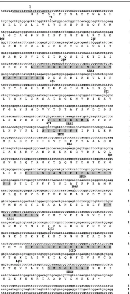
RT-PCR was performed with RNA isolated from FHM cells and universal CYP3A primers. Primers for inverse PCR (IPCR) were designed based on the sequence of the obtained RT-PCR amplification product. The full-length cDNA sequence of the CYP3A isoform was obtained in FHM cells by extended IPCR (Fig.1) [28,29]. Using this extended IPCR, an amplification product of (∼1,800 bp) was obtained and sequenced. The identified nucleotide sequence (1,863 bp) contained an open reading frame of 1,536 bp encoding a 512 amino acid protein, which comprised the putative heme binding domain—F G L G P R N C I G M R E R F A Q—and the putative substrate recognition sites (SRS) 1–6 (Fig. 2). The CYP3A cDNA contained a 5′ flanking region of 15 bp, a 3′ flanking region of 309 bp and a putative polyadenylation signal AATAAA (Fig. 2).The translated nucleotide sequence (blastx) revealed for the frame +1 a 89% identity (E value 0.0) to a CYP3A form of Ctenopharyngodon idella (GenBank access no. AAL16897; partial sequence), an 81% identity (E value 0.0) to the Danio rerio CYP3A65 amino acid sequence (GenBank accession no. AAH72702) as well as a 69% identity (E value 0.0) to both Oncorhynchus mykiss CYP3A27 (GenBank accession no. O42563) and CYP3A45 (GenBank accession no. AAK58569) and to both Fundulus heteroclitus CYP3A30 (GenBank accession no. Q9PVE8) and CYP3A56 (GenBank accession no. Q8AXY5).
RT-PCR was performed to verify the sequence found in the FHM cell line and in fathead minnow liver. Amplifi-cation products of the expected size were obtained and sequenced. The translated cds of the FHM cell line showed a 100% identity to the translated CYP3A cds received by extended IPCR. The translated cds of fathead minnow liver tissue showed a 100% similarity and 99.4% identity to the translated cds obtained by extended IPCR. In the translated sequence of fathead minnow liver the amino acid (aa) residue 42 is lysine (K), residue 217 is serine (S) and residue 321 is serine (S), whereas in the translated sequence of FHM cell line, residue 42 is aspargine (N), residue 217 is aspargine (N) and residue 321 is tyrosine (T). A sequence alignment reveals that several conserved residues in the putative SRS (SRS 1, SRS 2 and SRS 4) show high identity to the putative teleost recognition sites, which are different from other vertebrate CYP3A isoenzymes (data not shown). Analysis of expression levels of CYP3A126 was done in FHM cells using PCR. An amplification product of about 8,300 bp was received and sequenced. The sequence revealed that the CYP3A126 gene is composed of 13 exons and 12 introns (Fig.2).
Constitutive CYP3A activity in FHM cells
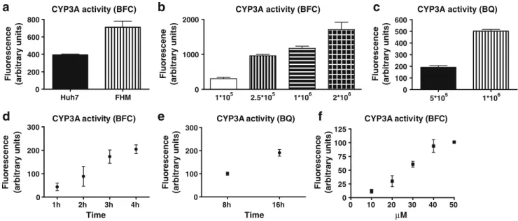
Additional to the identification of CYP3A126, general CYP3A enzyme activity of FHM cell lines was determined. For this purpose, FHM cells were seeded in 96-well plates, incubated overnight and then exposed to the CYP3A substrate BCF. Potential CYP3A activity was determined measuring the fluorescence. To control the experimental setting, potential CYP3A activity of the human liver cell line Huh7 was determined. The measured basal CYP3A activity was even higher in FHM cells than in Huh7 cells (Fig. 3a).
Dependence of CYP3A activity from cell numbers, incubation time and substrate concentration
To validate the basal CYP3A activity, enzyme activity was assessed with different numbers of FHM cells, with different incubation times with BFC and with the additional CYP3A substrate BQ and with different concentrations of BFC. First, different numbers of FHM cells were seeded in 96- and 48-well plates. After 24 h of cultivation, CYP3A activity assay was measured in culture plates. Figures 3b,
3c demonstrate that an increasing conversion of both CYP3A substrates into the fluorescent metabolites was detected with increasing numbers of cells. This clearly confirms that the detected fluorescence is based on CYP3A enzyme activity of the cells and not due to background fluorescence. Second, FHM cells grown overnight in a 96-well plate were incubated with both CYP3A substrates, and the enzyme reaction was stopped at different time points (1, 2, 3 and 4 h for BFC; 8 and 16 h for BQ). As shown in Figs. 3d, 3e, there was a correlation between incubation time and amount of measured fluorescence. This confirms that the measured fluorescence originates from cellular enzyme activity. The incubation time with BQ was much longer than with BFC due to the lower fluorescent signal of BQ [30]. Third, FHM cells, grown in 96 well plates were incubated with different concentrations of BFC (10, 20, 30, 40 and 50μM) for 4 h. As shown in Fig.3f, the measured fluorescent reached a plateau with 40 and 50μM. Based on this result, we decided to use 50 μM BFC to perform the enzyme assays.
Inhibition and induction of CYP3A activity
To confirm the potential functionality of CYP3A enzymes in FHM cells, we evaluated the induction and inhibition potential of CYP3A enzyme activity, respectively, by the mammalian model compounds rifampicin and diazepam. FHM cells and control human Huh7 cells were grown in 96-well plates and exposed to 10, 25 and 50μM rifampicin— a known human CYP3A inducer—for 24 h. After exposure
Fig. 2 Nucleotide and deduced amino acid sequence of the fathead minnow CYP3A isoenzyme. The initial ATG start codon is marked by boldface, and the stop codon has an asterisk. The putative heme binding domain, residues 440 to 455 of the amino acid sequence, and the putative SRS 1–6 are highlighted in grey. The polyadenylation signal aataaa is indicated by a double underline. PCR priming sites for the amplification of the coding region are underlined. Positions of the13 exons are indicated by their respective first nucleotide (number)
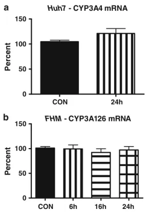
the CYP3A activity assay was performed. Treatment of Huh7 cells with rifampicin resulted in an increase in enzyme activity up to around 250% compared to solvent control cells, exposed to 0.1% DMSO (Fig.4a). The treatment of FHM cells with rifampicin also resulted in a dose-dependent induction of putative CYP3A enzyme activity up to around 150% (Fig. 4b). When performing the activity assay with BQ as a CYP3A substrate the same induction of CYP3A activity in response to rifampicin in FHM cells was found (Fig. 4c). In addition, a small but not significant increase in CYP3A4 mRNA was observed in Huh7 (Fig. 5a), but not of CYP3A126 mRNA in FHM cells (Fig. 5b). This lack of induction of CYP3A126 mRNA, when there was an increase in putative CYP3A activity, could indicate that there are more than one isoform of CYP3A in fathead minnows.
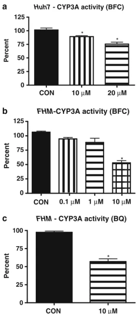
Furthermore the inhibition of the putative CYP3A activity was evaluated by treatment of FHM cells, grown in 96-well plates, with the known human CYP3A inhibitor diazepam for 24 h followed by CYP3A enzyme activity measurement. Enzyme activity was inhibited in Huh7 cells (activity assay was performed with BFC) (Fig. 6a) and in FHM cells, where activity assays were performed with BFC and BQ (Fig. 6b, c) in a dose-dependent manner. In addition to the enzyme activity, transcription of the CYP3A126 gene was inhibited as indicated by the reduction of CYP3A126 mRNA (Fig.7). The reduction of BCF activity could be the result of the observed reduction of CYP3A mRNA levels.
Discussion
The CYP3A family is a large sub-family of CYPs found in the liver and small intestine of mammals and fish [6, 31,
32]. CYP3A plays an important role in the metabolism of endogenous substances and xenobiotics including pharma-ceuticals. Furthermore, CYPs play a central role in the assessment of potential effects of pollutants present in aquatic environments [33]. Despite current knowledge on CYP3As in fish [15], there is a need for further inves-tigations because CYP3A plays a prominent role in the metabolism of pharmaceuticals and xenobiotics. It is important to obtain additional CYP3A sequences to better understand the occurrence of CYPs in fish species and their potential role in metabolism and effect of pollutants. This holds true in particular for pharmaceuticals that were identified as an emerging class of potential pollutants in the last few years [34]. Their importance is reflected by the fact that in mammals more than 50% of the pharmaceuticals are metabolised by CYP3A isoforms. Metabolism by CYP3A provides a broad defence against xenobiotics and their bioaccumulation. Therefore evaluation of the role of CYP3A in metabolism and effects of pharmaceuticals is important in light of their occurrence in aquatic systems. They may also serve as a putative biomarker for pharmaceuticals and other xenobiotics in aquatic systems. In addition to the identification of CYP3A isoforms it is also important to assess their enzyme activity, in particular with respect to possible alterations of enzyme activity induced by environmental pollutants. Huh7 FHM 0 200 400 600 800 CYP3A activity (BFC)
a
Fluorescence (arbitrary units)
1*105 2.5*105 1*106 2*106 0 1000 2000 CYP3A activity (BFC)
b
Fluorescene (arbitrary units) 5*105 1*106 0 100 200 300 400 500 600 CYP3A activity (BQ)c
Fluorescence (arbitrary units)
1h 2h 3h 4h 0 100 200 300 CYP3A activity (BFC)
d
Fluor escen c e (a rbitr a ry u n its) 8h 16h 0 100 200 300 CYP3A activity (BQ)e
Fluo rescen c e (arbit rary u n it s ) 0 10 20 30 40 50 0 25 50 75 100 125 CYP3A activity (BFC)f
µM Time Time Fluo rescen c e (a rbitrary u n its)Fig. 3 Basic CYP3A activity in FHM cells. a Basic CYP3A enzyme activity in FHM cells and Huh7 cells was measured in 96 well plates. Shown is the fluorescence in arbitrary units measured after 4-h incubation with CYP3A substrate BFC. b and c Different numbers of FHM cells were incubated with the CYP3A substrate BFC (b) for 4 h and with BQ (c) for 16 h. Shown is the measured fluorescence in arbitrary units. d
and e FHM cells were incubated with CYP3A substrate BFC (d) and with BQ (e) and enzymatic reaction stopped after different incubation periods. f FHM cells were incubated with different concentrations of the CYP3A substrate BFC for 4 h, followed by fluorescence measurement. Shown are always the results of three independently performed experiments±SD
Multiple CYP3A genes have been identified in several teleost species, e.g. CYP3A27 and CYP3A45 in rainbow trout [6, 9], CYP3A38 and CYP3A40 in medaka [7, 8], CYP3A30 and CYP3A56 in killifish [10] and CYP3A65 in D. rerio [11], but not in fathead minnow thus far. In addition to CYP3A, members of the cytochrome P450 superfamily have important metabolic functions including the conversion of C-18 androgens to C-19 estrogens. Therefore alterations in enzymatic activity or gene transcription can have pronounced consequences for the organism. Numerous studies demonstrate that it is important to assess alterations of CYP at the mRNA level and on enzyme activity after exposure to xenobiotics in fish [35–42].
In our present study, we identified a CYP3A isoform in fathead minnows, CYP3A126 (GenBank no. EU332792). First we obtained the full-length sequence of CYP3A126 in fathead minnow cells by extended IPCR. Second we confirmed this CYP3A sequence in the liver tissue of fathead minnows by RT-PCR. The analysis of the deduced amino acid sequence demonstrates a high degree of identity (up to 89%) with other teleost CYP3A isoenzymes. Furthermore the substrate recognition sites SRS1, SRS2 and SRS4 show high identity to the putative teleost recognition sites, which are different from other vertebrate CYP3A isoenzymes. This supports the hypothesis that the catalytic profile of teleost CYP3A members might differ from that of other vertebrate species [7,11].
Analysis of the complete sequences of several teleost genomes indicates that fish species contain a complement of CYP gene families similar to those found in mammals. To date, 13 teleost CYP3A genes have been identified by sequence homologies [15]. An analysis of 45 vertebrate CYP3A deduced amino acid sequences suggested that teleost, diapsid and mammalian CYP3A genes probably have undergone independent diversification and that an ancestral vertebrate genome contained a single CYP3A gene [10,31]. Whole genome duplications in teleosts have been suggested [43], and multiple CYP3A paralogs have
CON 24h
0 50 100 150
Huh7 - CYP3A4 mRNA
a
P e rcent CON 6h 16h 24h 0 50 100 150 FHM - CYP3A126 mRNAb
PercentFig. 5 Determination of CYP3A4 and CYP3A126 mRNA after rifampicin treatment. Huh7 and FHM cells were treated with rifampicin or solvent control (CON), followed by mRNA analysis. A small yet not significant increase of CYP3A4 mRNA was detected in Huh7 cells (a), but not of CYP3A126 in FHM cells (b)
CON 10 µM 25 µM 50 µM 0 100 200 300 * *
Huh7 - CYP3A activity (BFC)
a
Percent CON 10 µM 25 µM 50 µM 0 100 200 300 * * FHM - CYP3A activity (BFC)b
Percent FHM - CYP3A activity (BQ) CON 10µM 25µM 50µM 0 100 200 300 * * *c
PercentFig. 4 Dose-dependent induction of CYP3A activity by rifampicin. Treatment of Huh7 (a) and FHM (b) cells with different concen-trations of rifampicin for 24 h or with solvent control (CON), followed by incubation with BFC for 4 h. Induction of CYP3A activity was significant at 25μM (Huh7 p<0.05; FHM p<0.05) and at 50 μM (Huh7 p<0.001; FHM p<0.01) compared to solvent control samples. c Treatment of FHM cells with rifampicin for 24 h or with solvent control (CON) followed by incubation with BQ for 8 h. Induction of CYP3A activity was significant at 10μM (p<0.05), 25 μM (p<0.01) and 50μM (p<0.001) compared to solvent control samples
been identified in several species, including medaka, rainbow trout and killifish [5–9, 31, 44]. Differences in CYP catalytic activities are suggested to be determined by amino acid composition in six substrate recognition sites (SRS1 to SRS6) [45]. Key amino acids associated with CYP3A substrate specificity, binding and region-specific catalysis have been suggested by using molecular model-ling and site-directed mutagenesis. Regions in SRS1, SRS5
and SRS6 were identified that appear to be associated with a general conserved CYP3A function, whereas SRS2, SRS3 and SRS4 confer functional differences among different CYP3A enzymes [10]. Alignments of medaka CYP3A38 and CYP3A40 demonstrate that 12 of 49 amino acids differences occur in SR regions.
Additional to the identification of the CYP3A126 isoform, we used a newly established CYP3A activity assay [23] to characterise general CYP3A enzyme activity in fathead minnow cells. This in vitro assay allows to determine the potential CYP3A enzyme activity. We used BFC and BQ as substrates to investigate alterations in CYP3A activity, similar to Thibaut et al. [22]. There is still some debate whether BFC and BQ are selective enough as substrates for the CYP3A enzyme to assess CYP3A activity. Renwick et al. [46] showed that BFC metabolism appears to be primarily catalysed by CYP1A2 and CYP3A4 in humans. Stresser et al. [30] tested several substrates for their specificity to CYP3A and found that BFC and BQ dealkylation were selective to human CYP3A with little contribution of CYP1A2. In a previous work with mam-malian cells, it was already shown that BFC is a good substrate for an initial screen for CYP3A inhibition [26]. BFC was also used as a substrate to investigate CYP3A activity [27]. Therefore we are convinced that BFC and BQ are a good choice to investigate CYP3A activity, and therefore we used these substrates in the present work.
We have shown that FHM cells bear a putative constitutive CYP3A activity. The enzyme activity was dependent on cell number, and product formation was increased with incubation time. This demonstrates the potential functionality of the CYP3A enzyme(s) in FHM cells. Furthermore, we treated FHM cells with known CYP3A inducer rifampicin and inhibitor diazepam. Similar to Huh7, a dose-dependent induction of potential CYP3A enzyme activity up to 150% was noted. However, the
CON 6h 24h 0 25 50 75 100 125 * FHM - CYP3A126 mRNA Percent
Fig. 7 Determination of CYP3A126 mRNA after diazepam treat-ment. FHM cells were treated with diazepam or solvent control (CON) followed by mRNA analysis. Significant decrease of CYP3A126 mRNA level in FHM cells compared to solvent control samples after exposure to 10μM diazepam for 24 h (p<0.05)
CON 10 µM 20 µM 0 25 50 75 100 125 * *
Huh7 - CYP3A activity (BFC)
a
Percent CON 0.1 µM 1 µM 10 µM 0 25 50 75 100 125 FHM-CYP3A activity (BFC) *b
Percent CON 10 µM 0 25 50 75 100 * FHM - CYP3A activity (BQ)c
PercentFig. 6 Inhibition of CYP3A activity by diazepam. a Huh7 cells treated with 10 and 20μM diazepam for 24 h or solvent control (CON) showed a significant inhibition of CYP3A activity (10μM p< 0.05 and for 20μM p<0.01) compared to solvent control samples. b and c Treatment of FHM cells with different concentrations of diazepam for 24 h or solvent control (CON) followed by the investigation of CYP3A activity with BFC (b) and BQ (c). Diazepam (10μM) for 24 h leads to significant inhibition of CYP3A enzyme activity compared to solvent control samples by about 50% (p<0.001 for BFC and p<0.05 for BQ)
induction was weaker than in Huh7 cells. We also investigated whether the changes in enzyme activity are reflected on the mRNA level of CYP3A126. On the mRNA level little induction of CYP3A4 mRNA was observed in Huh7, but none in CYP3A126 mRNA in FHM cells. The discrepancy between CYP3A enzyme activity and CYP3A126 mRNA expression in FHM cells suggests an additional, not yet characterised mechanism in the regulation of CYP3A enzyme activity or the presence of another, not yet identified CYP3A isoform in fathead minnow. Therefore further studies are required to analyse the complexity in the regulation of the enzyme activity of the CYP3A sub-family in this species.
The exposure of FHM cells with CYP3A inhibitor diazepam inhibited the CYP3A activity in a dose-dependent manner down to 50%. Inhibition is stronger than in Huh7 cells. In addition to CYP3A enzyme activity, the transcription of the CYP3A126 gene was decreased. This suggests that diazepam interacts with CYP3A126 gene expression. Hypothetically diazepam may affect the binding and/or transcriptional activation of the PXR/RXR dimer at the response element in the CYP3A promoter. However, the goal of our present work was to show that a basal CYP3A activity, which is important for the detoxification in the liver, is present in fathead minnow and that the enzyme activity can be altered by xenobiotics. This finding is important with regard to the fact that aquatic organisms are constantly exposed to pharmaceuticals and chemicals in contaminated environ-ments. Therefore xenobiotics that decrease the enzyme activity of CYP3A can have pronounced consequences for the aquatic organisms due to disturbed detoxification in the liver. Pharmaceuticals such as anti-depressive drugs have also been detected in carp to inhibit CYP3A activity in vitro [22]. On the other hand, xenobiotics that increase the CYP3A enzyme activity may also lead to alteration of hepatic xenobiotic metabolism, as shown in carp for fenofibrate [22]. In summary, we identified a CYP3A isoform— CYP3A126—in FHM cells and in fathead minnow liver tissue. In addition, we identified a basal putative CYP3A enzyme activity in fathead minnow cells, which can be altered by pharmaceuticals. We provide indications that CYP3A126 could at least partially be responsible for the detected CYP3A activity. But we cannot exclude that there exist other CYP3A isoforms in fathead minnow that contrib-ute to the CYP3A activity. Furthermore we have shown that the FHM cell line has the potential to serve as a screening tool for the assessment of xenobiotic effects on CYP3A enzyme activity and associated metabolism. Compounds that alter CYP3A activity are interesting candidates for further mech-anistic investigations and for studies in vivo.
Acknowledgements We thank Roger Meier for his expert work and input to this paper, T. Wahli, University of Berne, for providing FHM cells, M. Heim, University of Basel for Huh7 cells, and J. Straub, F.
Hoffmann-La Roche Ltd, for providing diazepam. The designation of the CYP3A to CYP3A126 by the nomenclature committee is acknowledged. The work was supported by the Förderverein Fachhochschule Nordwestschweiz Solothurn.
References
1. Bucheli TD, Fent K (1995) Crit Rev Environ Sci Technol 25:200–268 2. Miller KA, Addison RF, Bandiera SM (2004) Mar Environ Res
57:37–54
3. Lewis NA, Williams TD, Chipman JK (2006) Toxicol Sci 92:387–393 4. Malmstrom CM, Koponen K, Lindstrom-Seppa P, Bylund G
(2004) Environ Saf 58:365–372
5. Celander M, Stegeman JJ (1997) Biochem Biophys Res Commun 236:306–312
6. Lee SJ, Wang-Buhler JL, Cok I, Yu TS, Yang YH, Miranda CL, Lech J, Buhler DR (1998) Arch Biochem Biophys 360:53–61 7. Kullman SW, Hamm JT, Hinton DE (2000) Arch Biochem
Biophys 380:29–38
8. Kullman SW, Hinton DE (2001) Mol Reprod Dev 58:149–158 9. Lee SJ, Buhler DR (2003) Arch Biochem Biophys 412:77–89 10. McArthur AG, Hegelund T, Cox RL, Stegeman JJ, Liljenberg M,
Olsson U, Sundberg P, Celander MC (2003) J Mol Evol 57:200–211 11. Tseng HP, Hseu TH, Buhler DR, Wang WD, Hu CH (2005)
Toxicol Appl Pharmacol 205:247–258
12. Vaccaro E, Salvetti A, Del Carratore R, Nencoioni S, Logo V, Gervasi PG (2007) J Biochem Molec Toxicol 21:32–40 13. Guengerich FP (1999) Annu Rev Pharmacol Toxicol 39:1–17 14. Hegelund T, Ottosson K, Radinger M, Tomberg P, Celander MC
(2004) Toxicol Chem 23:1326–1334
15. Schlenk D, Celander M, Gallagher EP, George S, James M, Kullman SW, van den Hurk P, Willett K (2008) In: Di Giulio RT, Hinton D (eds) The toxicology of fishes. CRC, Boca Raton 16. Li D, Yang XL, Lin M, Yu WJ, Hu K (2008) Comp Biochem
Physiol C 147:17–29
17. Kashiwada S, Kameshiro M, Tatsuta H, Sugaya Y, Kullman SW, Hinton DE, Goka K (2007) Comp Biochem Physiol C 145:370–378 18. Bresolin T, de Freitas Rebelo M, Celso Dias Bainy A (2005)
Comp Biochem Physiol C 140:403–407
19. Meucci V, Arukwe A (2006) Comp Biochem Physiol C 142:142–150 20. Mortensen AS, Arukwe A (2006) Environ Toxicol Chem
25:1607–1615
21. Kashiwada S, Hinton DE, Kullman SW (2005) Comp Biochem Physiol C 141:338–348
22. Thibaut R, Schnell S, Porte C (2006) Environ Sci Technol 40:5154–5160
23. Christen V, Oggier DM, Fent K (2009) Environ Toxicol Chem (in press)
24. Kullmann SW, Kashiwada S, Hinton DE (2004) Mar Environ Res 58:469–473
25. Hasselberg L, Meier S, Svardal A, Hegelund T, Celander MC (2004) Aquatic Toxicol 67:303–313
26. Stresser DM, Blanchard AP, Turner SD, Erve JC, Dandeneau AA, Miller VP, Crespi CL (2000) Drug Metab Dispos 28:1440–1448 27. Mensah-Osman EJ, Thomas DG, Tabb MM, Larios JM, Hughes
DP, Giordano TJ, Lizyness ML, Rae JM, Blumberg B, Hollenberg PF, Baker LH (2007) Cancer 109:957–965
28. Huang SH, Hu YY, Wu CH, Holcenberg J (1990) Nucleic Acids Res 18:1922
29. Arand M, Hemmer H, Durk H, Baratti J, Archelas A, Furstoss R, Oesch F (1999) Biochem J 344(Pt 1):273–280
30. Stresser DM, Turner SD, Blanchard AP, Miller VP, Crespi CL (2002) Drug Metab Dispos 30:845–852
32. Nallani SC, Goodwin B, Buckley AR, Buckley DJ, Desai PB (2004) Cancer Chemother Pharmacol 54:219–229
33. Goksoyr A, Forlin L (1992) Aquat Toxicol 22:287–311 34. Fent K, Weston AA, Caminada D (2006) Aquat Toxicol 76:122–159 35. Dorts J, Richter CA, Wright-Osment MK, Ellersieck MR, Carter
BJ, Tillitt DE (2009) Aquat Toxicol 91:44–53
36. Perkins EJ, Garcia-Reyero N, Villeneuve DL, Martinovic D, Brasfield SM, Blake LS, Brodin JD, Denslow ND, Ankley GT (2008) Mar Environ Res 66:113–115
37. Villeneuve DL, Knoebl I, Kahl MD, Jensen KM, Hammermeister DE, Greene KJ, Blake LS, Ankley GT (2006) Aquat Toxicol 76:353–368
38. Yamauchi R, Ishibashi H, Hirano M, Mori T, Kim JW, Arizono K (2008) Aquat Toxicol 90:261–268
39. Sullivan C, Mitchelmore CL, Hale RC, Van Veld PA (2007) Sci Total Environ 384:221–228
40. Salaberria I, Hansen BH, Asensio V, Olsvik PA, Andersen RA, Jenssen BM (2008) Toxicol Appl Pharmacol 234:98–106 41. Arukwe A, Nordtug T, Kortner TM, Mortensen AS, Brakstad OG
(2008) Environ Res 107:362–370
42. Liu Y, Wang J, Wie Y, Zhang H, Liu Y, Dai J (2008) Aquat Toxicol 88:183–190
43. Christoffels A, Koh EG, Chia JM, Brenner S, Aparicio S, Venkatesh B (2004) Mol Biol Evol 21:1146–1151
44. Lemaire P, Kodjabachian L (1996) Trends Genet 12:525–531 45. Gotoh O (1992) J Biol Chem 267:83
46. Renwick AB, Surry D, Price RJ, Lake BG, Evans DC (2000) Xenobiotica 30:955–969
