. . . .
. . . .
Instantaneous coronary collateral function during
supine bicycle exercise
Mario Togni
†, Steffen Gloekler
†, Pascal Meier
†, Stefano F. de Marchi, Tobias Rutz,
He´le`ne Steck, Tobias Traupe, and Christian Seiler
*
Department of Cardiology, University Hospital, CH-3010 Bern, Switzerland
Received 30 December 2009; revised 24 April 2010; accepted 20 May 2010; online publish-ahead-of-print 28 June 2010
Aims The instantaneous response of the collateral circulation to isometric physical exercise in patients with non-occlusive
coronary artery disease (CAD) is not known.
Methods and results
Thirty patients (age 59 + 9 years) undergoing percutaneous coronary intervention because of stable CAD were included in the study. Collateral function was determined before and during the last minute of a 6 min protocol of supine bicycle exercise during radial artery access coronary angiography. Collateral flow index (CFI, no unit) was determined as the ratio of mean distal coronary occlusive to mean aortic pressure both subtracted by central venous pressure. To avoid confounding due to recruitment of coronary collaterals by repetitive balloon occlusions, patients were randomly assigned to a group ‘rest first’ with CFI measurement during rest followed by CFI during exercise, and to a group ‘exercise first’ with antecedent CFI measurement during exercise before CFI at rest. Simultaneously, coronary collateral conductance (occlusive myocardial blood flow per aorto-coronary pressure drop) was determined by myocardial contrast echocardiography in the last 10 consecutive patients. Overall, CFI increased from 0.168 + 0.118 at rest to 0.262 + 0.166 during exercise (P ¼ 0.0002). The exercise-induced change in CFI did not differ statistically in the two study groups. Exercise-exercise-induced CFI reserve (CFI during exercise divided by CFI at rest) was 2.2 + 1.8. Overall, rest to peak bicycle exercise change of coronary col-lateral conductance was from 0.010 + 0.010 to 1.109 + 0.139 mL/min/100 mmHg (P , 0.0001); the respective change was similar in both groups.
Conclusion In patients with non-occlusive CAD, collateral flow instantaneously doubles during supine bicycle exercise as com-pared with the resting state.
ClinicalTrials.gov Identifier: NCT00947050.
-Keywords Coronary circulation † Collateral circulation † Physical exercise † Bicycle exercise
Introduction
Well-developed coronary collaterals have been documented to be lifesaving in patients with chronic stable coronary artery disease
(CAD).1 The possibility of maintaining resting myocardial blood
supply via collaterals has not been disputed recently, but their sus-taining an increased cardiac workload has been generally disbe-lieved. This view appears to be prevalent despite the existence of numerous experimental studies indicating the coronary collat-erals’ capability to respond with vasomotion to neurologic,
pharmacologic, and physiologic stimuli.2In humans, much less
evi-dence exists indicating that collateral vasomotor tone depends on
sympathetic stimuli such as cold pressor3,4or handgrip exercise,5
and is responding to dipyridamole6or adenosine.7However, it is
unknown so far, whether the most physiologic form of physical exercise, dynamic isometric exercise induces instantaneous coron-ary collateral function changes. The paucity of such an investigation in patients with chronic, non-occlusive CAD may relate to the fact that coronary collateral assessment requires a brief vascular occlu-sion, which has to occur during the last portion of an exercise test.
†The first three authors contributed equally to this study.
*Corresponding author. Tel:+41 31 632 36 93, Fax: +41 31 632 42 99, Email:[email protected]
In this context, the present study tested the hypothesis that cor-onary collateral function instantaneously increases in response to supine bicycle exercise.
Methods
Patients
Thirty patients (age 59 + 9 years, 28 men) with chronic stable, non-occlusive CAD eligible for percutaneous coronary intervention (PCI) of one stenotic lesion were included in the study. All underwent diag-nostic coronary angiography because of symptoms related to CAD. Patients were prospectively selected on the basis of the following cri-teria: (i) no previous transmural infarction in the myocardial area assessed for coronary collateral function, (ii) normal left ventricular (LV) ejection fraction, (iii) no congestive heart failure, (iv) no baseline electrocardiogram (ECG) ST-segment abnormalities, (v) written informed consent to participate in the study prior to the start of the invasive procedure.
Patients underwent an intra-individual comparison of collateral func-tion at rest and during supine bicycle exercise using radial artery access coronary angiography (Figure1). In order to account for the possibility of collateral recruitment following the first coronary balloon occlu-sion,8 patients were randomly assigned to a group ‘rest first’ in which the first collateral function measurement occurred at rest and the second during the last minute of the exercise test (n ¼ 15; Figure2), and vice versa to a group ‘exercise first’ (n ¼ 15).
This investigation was approved by the Ethics Committee of the Kanton of Bern, Switzerland.
Cardiac catheterization and coronary
angiography
Patients underwent left heart catheterization for diagnostic purposes from the right radial artery approach (Figure 1). This route was chosen in the context of the supine bicycle exercise protocol to prevent potentially harmful, leg-movement-induced pushing of the guiding catheter into the coronary ostium. Aortic pressure was measured using a 6F PCI guiding catheter. Central venous pressure (CVP) was obtained via the right femoral vein by a 5F pigtail catheter. Left ventricular end-diastolic pressure was determined before collat-eral function assessment. Monoplane left ventriculography was
performed followed by monoplane coronary angiography. Coronary artery stenoses were determined quantitatively as percent diameter narrowing.
Invasive coronary collateral assessment
Primary study endpoint
Coronary collateral flow relative to normal antegrade flow through the non-occluded coronary artery (collateral flow index, CFI) was deter-mined using coronary pressure measurements. A 0.014 inch pressure monitoring angioplasty guidewire (Pressure Wirew
, Radi, Uppsala, Sweden) was set at zero, calibrated, advanced through the guiding catheter, and positioned in the distal part of the vessel of interest. Col-lateral flow index was determined by simultaneous measurement of mean aortic pressure (Pao, mmHg), the distal mean coronary artery
pressure during balloon occlusion (Poccl, mmHg), and the mean CVP
(mmHg; Figure 3). Collateral flow index was calculated as (Poccl2
CVP) divided by (Pao2 CVP).9The accuracy of pressure derived CFI
measurements in comparison to ECG signs of myocardial ischaemia during occlusion and to absolute myocardial perfusion measurements has been documented previously.9–11
Secondary study endpoints
Myocardial ischaemia during the 1 min coronary occlusion was characterized by the presence or absence of angina pectoris. In the 10 last consecutive patients of the study, absolute myocardial blood flow (simultaneous to CFI) was assessed quantitatively using myocardial contrast echocardiography (MCE), whereby a previously described and validated algorithm was employed.12 Briefly, for the
calculation of absolute blood flow, the constituent factors relative myocardial blood volume rBV and its refill ratebfollowing destruc-tion of echo-contrast microbubbles were obtained during vessel occlusion. Myocardial blood flow is equal to the product of rBV and b divided by myocardial tissue density.12 Coronary collateral conductance was calculated as the ratio of myocardial blood flow during coronary occlusion (mL/min/g) per mmHg of collateral driving pressure (¼Pao2 Poccl).
Study protocol
At the start of the invasive procedure, all patients received 5000 units of heparin intravenously. No oral isosorbide dinitrate was given until after completion of the study protocol. Nitrates were withhold in
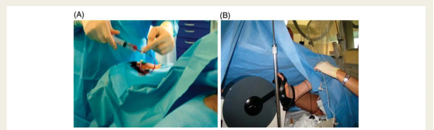
Figure 1 Invasive set-up for the implementation of the supine bicycle exercise stress protocol. (A) Right radial artery access coronary angio-graphy via a 6F sheath. The patient’s right groin is prepared in order to allow femoral vein access (central venous pressure measurement). (B) Image of the supine bicycle ergometer with the patient’s feet attached to the pedals.
order to determine nitric oxide-mediated endothelium-dependent col-lateral vasomotor function as induced by exercise and to avoid influ-ence of endothelium-independent vasomotion. Diagnostic coronary angiography was performed, and the culprit lesion responsible for the patient’s symptoms was selected (online quantitative coronary
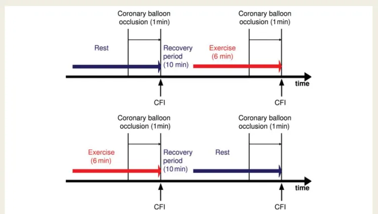
angiography). Before starting the protocol, patients were randomly assigned to the group ‘rest first’ or to the group ‘exercise first’. The randomization scheme was generated using the Web site Randomiza-tion.com (http://www.randomization.com) before study begin, whereby a closed envelope for each consecutive patient was prepared Figure 2 Chart of the study protocol. CFI, collateral flow index measurement (vertical arrows).
Figure 3 Simultaneous electrocardiogram/pressure tracings obtained in the proximal left anterior descending coronary artery during supine bicycle exercise (left side; patient of the ‘exercise first’ group) and at rest (right side). The lowest electrocardiogram lead is the intracoronary lead obtained via the pressure sensor guidewire (not attached during exercise due to motion artefacts). CVP, central venous pressure (scale 50 mmHg); Pao, aortic pressure (scale 200 mmHg); Pd, distal coronary pressure (scale 200 mmHg); Poccl, distal coronary occlusive pressure
containing the group assignment. In the ‘rest first’ group, a CFI measurement at rest in all patients with simultaneous MCE in the respective region of interest in the last 10 patients was performed during a 1 min coronary balloon occlusion at the site of the culprit lesion (low balloon inflation pressure). A 10 min recovery period fol-lowed this first collateral function assessment (Figure2). Subsequently, the 6 min supine bicycle ergometry followed in three 2 min stages at low, intermediate, and high workload. Starting with the fourth minute of the exercise test, the angioplasty balloon was again posi-tioned at the culprit lesion site, and the balloon was inflated shortly before the beginning of the sixth minute of the exercise test. CFI and—where applicable—MCE measurements were performed during 1 min until the end of the exercise test. In the ‘exercise first’ group, the protocol started with the described procedure during supine bicycle ergometry, followed by a 10 min recovery period, fol-lowed by the collateral function measurement at rest (Figure2). The recordings during both rest and exercise measurements included Pao,
Poccl, CVP, 3 to 4 ECG leads (4 peripheral and 1 intracoronary lead
via the guide wire), and—where applicable—MCE video clips. Owing to motion artefacts during maximum bicycle ergometry, the mean pressure curves necessary for CFI calculation (Figure3), and the echo-video clips were the only recordings utilizable; i.e. ECG information on myocardial ischaemia in addition to CFI and MCE was analysable only at rest and, therefore, of no use for the specific study purpose. Follow-ing the study protocol, two puffs of isosorbide dinitrate was given and PCI was performed at the site of the culprit lesion.
Statistical analysis
The sample size of the study was estimated on the basis of previous clinical coronary arteriogenesis studies.13,14 Sample size calculated prior to the study was based on the following assumptions regarding the CFI endpoint: ability to detect an increase of 50% during exercise as compared with the situation at rest (CFI mean value of 0.15) with a power of 80% (alpha error level of 5%); CFI standard deviation ¼ 0.15. Accordingly, a total sample size of 28 patients with paired measure-ments was required. All continuous data are given as mean + standard deviation. The distribution of the data for normality was tested by the Kolmogorov – Smirnov test. Baseline characteristics between the groups were analysed by Student’s t-tests for continuous data and by
x2
/Fisher’s exact tests for categorical data. Intra-individual comparison of CFI between the resting and maximum exercise condition was per-formed by a paired student’s t-test. Linear regression analysis was used to assess an association between CFI and MCE-derived coronary col-lateral conductance at rest and during exercise. Differences were con-sidered statistically significant at a two-sided P-value of ,0.05. Statistical analysis was performed using StatView.
Results
Patient characteristics and clinical data
at baseline
There were no statistically significant differences between the two groups regarding age of the patients, gender, duration of angina pectoris, history of myocardial infarction in a remote vascular area and body mass index. There were no statistical differences either in the frequency of cardiovascular risk factors, or the use
of cardiovascular medication (Table1).
Invasive and haemodynamic data
at baseline
Invasively obtained haemodynamic parameters at baseline such as heart rate, blood pressure, LV end-diastolic pressure, and LV ejection
fraction did not differ between the groups (Table2). The vessel
under-going CFI measurement at rest and during exercise and the severity of CAD as well as the severity of the stenosis being treated by PCI were similar between the groups (right-dominance in all patients). Percent diameter stenosis of the vessels not treated by PCI was 25 + 15 (no difference between the groups). Collateral function data obtained at rest were not statistically different between the ‘rest first’ and the
‘exercise first’ group (Table2).
Invasive data during supine bicycle
exercise
The maximum workload achieved was not different between the
study groups (Table3; range from 60 to 300 W). There was no
cor-relation between the maximum workload achieved and the change in CFI during exercise. Heart rate and systolic blood pressure increased significantly within both groups during exercise, and they did not
differ between the groups at maximum exercise (Table3).
Primary study endpoint
Exercise-induced increase in CFI was significant in both groups
(Table 3 and Figure 4), and the respective CFI reserve (¼CFI
during maximum exercise divided by CFI at rest) was not different
. . . .
. . . . . . . .
. . . .
. . . . Table 1 Patient characteristics and clinical data at baseline
Variable Rest first (n 5 15)
Exercise first (n 5 15) Age (years) 59 + 7 59 + 9 Male gender (%) 14 (93) 14 (93) Duration of chest pain (months) 2.4 + 2.2 5.8 + 9.2 History of prior myocardial
infarction (%)
8 (53) 6 (40)
Body mass index (kg/m2) 27 + 4 26 + 4 Cardiovascular risk factors
Dyslipidaemia (%) 13 (87) 10 (67) Diabetes mellitus (%) 1 (7) 2 (13) Systemic hypertension (%) 9 (60) 9 (60) Smoking (%) 7 (47) 4 (27) Obesity (%) 6 (40) 4 (27) Family history for coronary
artery disease (%) 7 (47) 2 (13) Cardiovascular medication Beta-blockers (%) 12 (80) 8 (53) Nitrates (%) 1 (7) 1 (7) Acetylsalicylic acid (%) 14 (93) 13 (87) Statin (%) 13 (87) 10 (67) ACE-inhibitor (%) 5 (33) 4 (27) Diuretics (%) 2 (13) 4 (27)
between the study groups (Table3). In 24 patients, CFI increased during exercise, whereas it decreased in six patients. Rate – pressure product (systolic pressure) at rest and during maximum
exercise correlated significantly with CFI (CFI ¼ 20.02+
0.00002 rate – pressure product; r2¼ 0.25, P ¼ 0.002).
Secondary study endpoints
The occurrence of angina pectoris during coronary occlusion did
not change during exercise vs. resting conditions (Table3).
Collat-eral flow index and MCE-derived coronary collatCollat-eral conductance correlated directly at rest and during exercise, and they both increased equivalently during exercise in 7 of 10 patients under-going simultaneous CFI and MCE measurements, whereas in 3 of 10 patients, CFI decreased during exercise, whereas collateral
con-ductance increased (Figure5).
Discussion
This first clinical study on coronary collateral behaviour during iso-metric physical exercise documents a two-fold increase in CFI compared with resting conditions. In a minority of every fifth patient, however, collateral function instantaneously decreases during exercise.
Instantaneous coronary collateral
function changes: non-exercise stimuli
Exercise-induced CFI increase in our entire study population was from 0.168 to 0.262 (P ¼ 0.0002). The question evolving in this context is whether such a response is solely due to supine bicycle exercise or whether it can be attributed to variables
. . . .
. . . . Table 2 Invasive data at baseline
Variable Rest first (n 5 15) Exercise first (n 5 15) P
Heart rate at rest (beats per minute) 73 + 16 71 + 10 0.69 Systolic blood pressure at rest (mmHg) 125 + 25 129 + 28 0.71 Diastolic blood pressure at rest (mmHg) 81 + 16 80 + 19 0.78 Left ventricular end-diastolic pressure (mmHg) 11 + 5 14 + 5 0.14
Left ventricular ejection fraction (%) 59 + 7 58 + 6 0.67
Vessel undergoing CFI measurement (LAD/LCX/RCA) 8/3/4 9/3/3 0.90
Number of vessels diseased 1.9 + 0.7 1.8 + 0.9 0.83
Number of stenoses 1.9 + 1.5 2.4 + 2.0 0.47
Percent diameter stenosis of vessel treated by PCI 78 + 24 76 + 32 0.82 Collateral function data at rest
Site of CFI measurement (proximal/mid/distal) 8/6/1 8/6/1 1.0 Angina pectoris during coronary occlusion (%) 10 (67) 11 (73) 0.69 ECG ST-segment elevation during coronary occlusion (%) 10 (67) 10 (67) 1.0 Collateral flow index, CFI (no unit) 0.137 + 0.117 0.200 + 0.114 0.15
LAD, left anterior descending coronary artery; LCX, left circumflex coronary artery; PCI, percutaneous coronary intervention; RCA, right coronary artery.
. . . .
. . . . Table 3 Invasive data during supine bicycle exercise
Variable Rest first (n 5 15) Exercise first (n 5 15) P
Maximum workload (watt) 114 + 14 119 + 53 0.71
Heart rate at rest (beats per minute) 73 + 16 71 + 10 0.69 Heart rate at peak exercise (beats per minute) 116 + 15* 118 + 21* 0.80 Systolic blood pressure at rest (mmHg) 125 + 25 129 + 28 0.71 Systolic blood pressure at peak exercise (mmHg) 149 + 37* 153 + 30* 0.72 Diastolic blood pressure at rest (mmHg) 81 + 16 80 + 19 0.78 Diastolic blood pressure at peak exercise (mmHg) 88 + 16 82 + 23 0.38 Collateral function data
Angina pectoris during coronary occlusion at rest (%) 10 (67) 11 (73) 0.69 Angina pectoris during coronary occlusion at peak exercise (%) 10 (67) 10 (67) 1.0 Collateral flow index at rest, CFI (no unit) 0.137 + 0.117 0.200 + 0.114 0.15 Collateral flow index at peak exercise, CFI (no unit) 0.239 + 0.140** 0.284 + 0.190** 0.47 CFI reserve (CFI during exercise/CFI at rest) 2.25 + 1.24 2.04 + 2.26 0.76
other than exercise present in the study. The key co-candidate for instantaneous collateral function enhancement is preceding ischaemia. Collateral recruitment in response to ischaemia is caused via intrinsic myocardial adenosine excretion. The design of our study accounted for this mechanism by randomly allocat-ing half the patients to a protocol with the restallocat-ing condition as the initial measurement, and the other half with the exercise condition as the first collateral assessment. In the former group without ischaemic event immediately preceding the resting con-dition, CFI was lower than in the presence of an earlier occlusion (‘exercise first’ group); this difference was not statistically significant, which could be due to the low power of the study. Thus, the influence on the study results of ischaemia-induced collateral recruitment aside from the bicycle test cannot be entirely excluded.
Instantaneous coronary collateral
function changes: exercise as stimulus
Earlier clinical studies on the acute effect of exercise on collateral function have employed thallium-201 perfusion imaging in patients with entirely collateralized, viable myocardial regions providing a qualitative, dichotomous measure of absent or present perfusion
defects.15–17Rigo et al. concluded from their study that ‘coronary
collateral vessels may help maintain relative myocardial perfusion
during exercise’.15Eng et al. studied collateral function by analysis
of exercise thallium-201 myocardial perfusion images from 31 patients who had at least one non-infarcted, entirely collateralized myocardial region. Twenty-two of 41 of the collateralized regions manifested exercise-induced perfusion defects and 19 were nor-mally perfused, whereby the latter 19 consisted of 13 with defects in other myocardial regions supplied by diseased vessels and were considered negative relative to other jeopardized
regions.17 Thus only in about one-seventh of all collateralized
regions (n ¼ 6), perfusion was adequate during maximal exercise, and in about half exercise-induced coronary steal occurred. More recent clinical work using the model of entirely collateralized anterior wall myocardium as assessed by technetium-99m per-fusion imaging has documented exercise-induced perper-fusion defects in all of the 20 patients studied, whereby 30% of them
had fixed defects (necrosis).18The study by Aboul-Enein et al.19
included 56 patients with single-vessel chronic total occlusion and viable myocardium who underwent rest-exercise myocardial perfusion imaging and coronary angiography within 6 months. The authors concluded that well-developed angiographic collat-erals could prevent resting regional wall motion abnormalities but not stress-induced perfusion defects. In comparison to these qualitative data, the present study obtained quantitative, continu-ous data to a temporarily collateralized, viable myocardial region. A parameter remotely equivalent to the extent of myocardial ischaemia in the scintigraphic studies is the rather blunt variable of angina pectoris during coronary occlusion, which remained unchanged during exercise. The more sensitive parameter of ECG ST-segment changes during occlusion indicative of ischaemia was available only at rest due to motion artefacts during exercise;
Figure 4 Individual changes (thin lines) of collateral flow index (collateral flow index; vertical axes) from the resting condition to the peak supine bicycle exercise condition in the group ‘rest first’ (left panel; triangular symbols) and vice versa in the group ‘exercise first’ (right panel; cross symbols). The thick lines indicate the mean change between the resting and exercise condition. Error bars: standard deviation.
Figure 5 Correlation between collateral flow index (collateral flow index; horizontal axis) and simultaneously obtained coron-ary collateral conductance (vertical axis) at rest (open triangles) and during peak supine bicycle exercise (cross symbols). Con-necting lines between the triangles and crosses indicate intra-individual changes between the resting and exercise con-dition. A minority of 3 of 10 patients showed an opposite change of CFI as compared with contrast echo-derived collateral conductance (dashed connecting lines).
10 of 30 patients fulfilled the ECG criteria of sufficient collaterals at rest (data not shown), and seven of them showed an increase in CFI during exercise. In 17 of 30 patients, CFI rose under exercise to a value known to represent collateral flow sufficient to prevent
signs of ischaemia during a 1 min occlusion (≥0.215).20
Instantaneous coronary collateral
function changes: steal; potential
influence of left ventricular
filling pressure
Exercise-induced collateral steal, i.e. a fall in CFI during exercise, occurred in 6 of 30 patients. Collateral steal in a study protocol such as the present one may be clinically irrelevant, but serves as quality control of collateral function measurements in two ways. First, its prevalence (20%) should be in the range of that
reported in the literature (10% in non-occlusive CAD21), the fact
of which heightens the plausibility of the data obtained. In addition, determinants of steal reported in the literature such as well-developed collaterals should correspond to those found in the present study to indicate reliability of the measurement result: patients with steal as compared with those with exercise-induced collateral flow increase had a CFI at rest of 0.227 + 0.150 and 0.154 + 0.107 (P ¼ 0.07), respectively, and a collateral conduc-tance of 0.016 + 0.017 and 0.006 + 0.005 (P ¼ 0.13), respectively. Accordingly, the prevalence of collateral steal amounts to almost
half in patients with chronic coronary occlusion.22 Second, the
occurrence of steal as obtained from pressure-derived CFI serves as indicator against the hypothesis that coronary pressure measurements during exercise reflect an elevated LV filling pressure rather than collateral perfusion pressure. In principle, it is imaginable that the elevated myocardial oxygen consumption during exercise causes more tissue ischaemia, translating into more dysfunctional, thinner myocardium with higher LV filling pressure as compared with the resting state. Considering the exist-ence of a waterfall phenomenon in the coronary circulation
oper-ative at LV filling pressures beyond 27 mmHg,23the just mentioned
hypothesis is plausible. It can be directly falsified in the six cases with steal, since in all of them collateral perfusion pressure fell during exercise. In order to account differently for this potential problem, MCE-derived collateral conductance was measured sim-ultaneously with CFI in 10 patients. The two parameters correlated with each other at rest and during exercise, and more importantly, all conductance values increased during exercise.
Study limitations
Study limitations not alluded to above are the absence of contrast myocardial perfusion data in part of the patients, and the fact that no LV pressure or pulmonary capillary wedge pressure was obtained during the exercise protocol in order to account for the potential problem discussed in the above paragraph. Both limit-ations are primarily due to the complexity of the supine bicycle protocol with radial artery access to allow a safe 1 min coronary balloon occlusion during peak exercise. It was only after the 20th patient included in the study that the operators felt secure enough to add another measurement technique simultaneous to the invasive collateral assessment. The insertion of a pigtail
catheter into the LV via a femoral artery access would have been a simple procedure, but the risk of causing ventricular arrhythmias during exercise would have been high, thus heighten-ing the risk of ventricular fibrillation and lowerheighten-ing the chance of obtaining a representative LV filling pressure. Measurement of pul-monary capillary wedge pressure via femoral-vein-access right heart catheter was not performed, because the pressure signal during peak exercise was not predicted stable enough due to the leg movements.
The principle explanation for the incomplete agreement between CFI and collateral conductance is mainly related to the difficult examination conditions for transthoracic echocardiogra-phy during exercise with the patient lying on his back on the cath lab table. In comparison to the usual left lateral supine pos-ition, respiratory artefacts impair the ultrasound image quality much more in the supine back position.
As further potential limitations, the continuing medication with vasoactive drugs until the day of the study protocol and the vari-able exercise levels achieved have to be mentioned.
Conclusion
In patients with non-occlusive CAD, collateral flow instantaneously doubles during supine bicycle exercise as compared with the resting state.
Funding
Supported by a grant from the Swiss National Science Foundation (grant # 32003B-124848/1) and the Swiss Heart Foundation. Conflict of interest: none declared.
References
1. Meier P, Gloekler S, Zbinden R, Beckh S, de Marchi S, Zbinden S, Wustmann K, Billinger M, Vogel R, Cook S, Wenaweser P, Togni M, Windecker S, Meier B, Seiler C. Beneficial effect of recruitable collaterals: a 10-year follow-up study in patients with stable coronary artery disease undergoing quantitative collateral measurements. Circulation 2007;116:975 – 983.
2. Duncker D, Bache R. Regulation of coronary blood flow during exercise. Physiol Rev 2008;88:1009 – 1086.
3. Uren N, Crake T, Tousoulis D, Seydoux C, Davies G, Maseri A. Impairment of the myocardial vasomotor response to cold pressor stress in collateral dependent myocardium. Heart 1997;78:61 – 67.
4. de Marchi S, Schwerzmann M, Billinger M, Windecker S, Meier B, Seiler C. Sym-pathetic stimulation using the cold pressor test increases coronary collateral flow. Swiss Med Wkly 2001;131:351 – 356.
5. Pohl T, Wustmann K, Zbinden S, Windecker S, Mehta H, Meier B, Seiler C. Exercise-induced human coronary collateral function: quantitative assessment during acute coronary occlusions. Cardiology 2003;100:53 – 60.
6. Vanoverschelde JLJ, Wijns W, Depre´ C, Essamri B, Heyndrckx GR, Borgers M, Bol A, Melin JA. Mechansims of chronic regional postischemic dysfunction in humans. New insights from the study of non-infarcted collateral-dependent myo-cardium. Circulation 1993;87:1513 – 1523.
7. Werner G, Surber R, Ferrari M, Fritzenwanger M, Figulla H. The functional reserve of collaterals supplying long-term chronic total coronary occlusions in patients without prior myocardial infarction. Eur Heart J 2006;27:2406 – 2412. 8. Billinger M, Fleisch M, Eberli FR, Garachemani AR, Meier B, Seiler C. Is the
devel-opment of myocardial tolerance to repeated ischemia in humans due to precon-ditioning or to collateral recruitment? J Am Coll Cardiol 1999;33:1027 – 1035. 9. Seiler C, Fleisch M, Garachemani A, Meier B. Coronary collateral quantitation in
patients with coronary artery disease using intravascular flow velocity or pressure measurements. J Am Coll Cardiol 1998;32:1272 – 1279.
10. Matsuo H, Watanabe S, Kadosaki T, Yamaki T, Tanaka S, Miyata S, Segawa T, Matsuno Y, Tomita M, Fujiwara H. Validation of collateral fractional flow reserve by myocardial perfusion imaging. Circulation 2002;105:1060 – 1065.
11. Vogel R, Zbinden R, Indermuhle A, Windecker S, Meier B, Seiler C. Collateral-flow measurements in humans by myocardial contrast echocardiography: validation of coronary pressure-derived collateral-flow assessment. Eur Heart J 2006;27:157–165. 12. Vogel R, Indermuhle A, Reinhardt J, Meier P, Siegrist PT, Namdar M, Kaufmann PA, Seiler C. The quantification of absolute myocardial perfusion in humans by contrast echocardiography: algorithm and validation. J Am Coll Cardiol 2005;45:754 – 762.
13. Seiler C, Pohl T, Wustmann K, Hutter D, Nicolet P, Windecker S, Eberli F,
Meier B. Promotion of collateral growth by granulocyte-macrophage
colony-stimulating factor in patients with coronary artery disease: a randomized, double-blind, placebo-controlled study. Circulation 2001;104:2012 – 2017. 14. Zbinden S, Zbinden R, Meier P, Windecker S, Seiler C. Safety and efficacy of
subcutaneous-only granulocyte-macrophage colony-stimulating factor for collat-eral growth promotion in patients with coronary artery disease. J Am Coll Cardiol 2005;46:1636 – 1642.
15. Rigo P, Becker L, Griffith L, Alderson P, Bailey I, Pitt B, Burow R, Wagner HJ. Influ-ence of coronary collateral vessels on the results of thallium-201 myocardial stress imaging. Am J Cardiol 1979;44:452 – 458.
16. Kolibash A, Bush C, Wepsic R, Schroeder D, Tetalman M, Lewis R. Coronary col-lateral vessels: spectrum of physiologic capabilities with respect to providing rest and stress myocardial perfusion, maintenance of left ventricular function and pro-tection against infarction. Am J Cardiol 1982;50:230 – 238.
17. Eng C, Patterson R, Horowitz S, Halgash D, Pichard A, Midwall J, Herman M, Gorlin R. Coronary collateral function during exercise. Circulation 1982;66: 309 – 316.
18. Chammas E, Hussein A, Ballane G, Helou A, Yatim A, Tarcha W, Ghanem G. Myocardial perfusion in patients with a totally occluded left anterior descending coronary artery reinjected by a normal right coronary artery: the role of collateral circulation. Angiology 2008;59:464 – 468.
19. Aboul-Enein F, Kar S, Hayes S, Sciammarella M, Abidov A, Makkar R, Friedman J, Eigler N, Berman D. Influence of angiographic collateral circulation on myocardial perfusion in patients with chronic total occlusion of a single coronary artery and no prior myocardial infarction. J Nucl Med 2004;45:950 – 955.
20. Seiler C. Collateral Circulation of the Heart, Vol. 1. 1st ed. London: Springer; 2009. 21. Seiler C, Fleisch M, Meier B. Direct intracoronary evidence of collateral steal in
humans. Circulation 1997;96:4261 – 4267.
22. Werner GS, Fritzenwanger M, Prochnau D, Schwarz G, Ferrari M, Aarnoudse W, Pijls N, Figulla H. Determinants of coronary steal in chronic total coronary occlu-sions donor artery, collateral, and microvascular resistance. J Am Coll Cardiol 2006; 48:51 – 58.
23. de Marchi S, Oswald P, Windecker S, Meier B, Seiler C. Reciprocal relationship between left ventricular filling pressure and the recruitable human coronary col-lateral circulation. Eur Heart J 2005;26:558 – 566.