2006/116
Minireview
Regulation of human cathepsin B by alternative mRNA
splicing: homeostasis, fatal errors and cell death
Antonio Baici*, Kathrin Mu¨ntener, Anna Willimann and Roman Zwicky
Department of Biochemistry, University of Zurich, Winterthurerstrasse 190, CH-8057 Zurich, Switzerland * Corresponding author
e-mail: [email protected]
Abstract
One of the control mechanisms of cathepsin B biosyn-thesis and trafficking operates through alternative splic-ing of pre-mRNA. An mRNA lacksplic-ing exon 2 is more efficiently translated than that containing all exons, and may be responsible for elevated biosynthesis and enzyme routing to the extracellular space, with critical consequences for connective tissue integrity in patholo-gies such as cancer and arthritis. mRNA missing exons 2 and 3 encodes a truncated procathepsin B form that is targeted to mitochondria. This enzyme variant is cat-alytically inactive because it cannot properly fold. How-ever, it provokes a cascade of events, which result first in morphological changes in intracellular organelles and the nucleus, finally leading to cell death.
Keywords: cysteine peptidases; mitochondria; overexpression; trafficking; unfolded protein; untranslated region.
Introduction
Cathepsin B, a cysteine peptidase of the papain family (EC 3.4.22.1, identifier C01.060 in the Merops database; Rawlings et al., 2004), has been typically classified as a lysosomal enzyme. However, it is also responsible for a variety of extracellular functions, is found at cell surfaces, and a truncated form is targeted to mitochondria. Some of these extralysosomal properties of cathepsin B are due to altered expression at the gene level, as well as to enzyme overproduction and/or atypical trafficking. Nor-mal and abnorNor-mal expression can be regulated by tran-scriptional efficiency, by different transcription starting points (Gong et al., 1993; Berquin et al., 1995), by the use of alternative promoters (Yan and Sloane, 2003), and by variable pre-mRNA splicing (Gong et al., 1993; Berardi et al., 2001; Zwicky et al., 2002, 2003; Mu¨ntener et al., 2003, 2004).
During the splicing of eukaryotic precursor mRNAs, the introns are removed and the exons are joined together. Alternative mRNA splicing is a frequent event that occurs in a cell-specific or developmental-specific manner in
approximately 60% of all human gene products (Modrek and Lee, 2002). It involves the combination of different splice junctions that give rise to functionally distinct pro-teins, allowing a gene to multiply its coding capacity and thereby to synthesize a number of structurally and func-tionally distinct protein forms far exceeding the number predicted by the genome. In this article we review our contribution to the study of alternative splicing of human cathepsin B pre-mRNA, with emphasis on the untrans-lated regions (UTR), and discuss the consequences of this event for protein synthesis, trafficking and function.
The cathepsin B gene, its transcripts and protein products
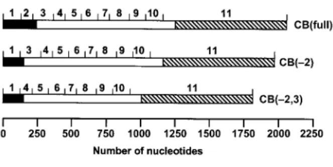
The human cathepsin B gene, located on the short arm of the 8p22 region of chromosome 8 (Fong et al., 1986; Wang et al., 1988), contains 13 exons (12 exons plus exons 2a,b) and has a coding portion of only approxi-mately 1 kb, although its total length is at least 27 kb (Figure 1). Exons 2a,b (119 bp) have been discovered in cells from a human gastric adenocarcinoma (Berquin et al., 1995). Three cathepsin B transcripts, alternatively spliced in the 59-region and relevant to this review, are shown in Figure 2. The standard 39-UTR of cathepsin B mRNA contains the complete exon 11, giving rise to a 2.2-kb RNA, while a 4-kb variant contains 141 nucleo-tides of exon 11 and the complete exon 12 (Gong et al., 1993). Another 1.7-kb transcript results from splicing of the coding region to a more downstream portion of the 39-UTR (Tam et al., 1994). The presence or absence of a 10-bp sequence in the 39-UTR is crucial for stabilization of the transcripts through a stem loop (Tam et al., 1994). As the regular start codon is located in exon 3, the full-length transcript, CB(full), and the transcript lacking exon 2, CB(-2), code for the same protein, namely preproca-thepsin B. This consists of a 17-aa-long signal peptide, followed by an inhibitory propeptide of 62 aa, the single-chain enzyme (254 aa), and a C-terminal propeptide of 6 aa. The protein is directed to the endoplasmic retic-ulum by the signal peptide, which is then removed and the protein becomes glycosylated at two sites, one in the propeptide and one in the mature protein region. The mannose-6-phosphate recognition marker, added to the propeptide in the Golgi apparatus, conveys the enzyme to the endosomes, where the mature single-chain form of 27.8 kDa is generated by removal of the propeptide through limited proteolysis. Further process-ing occurs in the endosomal-lysosomal compartment, where the C-terminal hexapeptide and two internal amino acids are trimmed off, and the N-linked oligosaccharide of the heavy chain is degraded to the level of a single
Figure 1 The human cathepsin B gene.
This scheme was generated using nucleotides 11760450– 11791965 (in reverse order) of the chromosome 8 sequence of the Homo sapiens genome (build 30) as provided on www.ncbi.nlm.nih.gov and the pDraw32 software. The location of exon 2a/b was determined using the published sequence (Berquin et al., 1995). The boundaries of all other exons (shown by numbers) were determined using the published exon-intron junction sequences (Gong et al., 1993). Exon 1, 124 bp (59-UTR); exon 2, 88 bp (59-UTR); exon 2a/b, 119 bp (59-UTR); exon 3, 151 bp (consisting of a 25-bp 59-UTR and 126-bp coding region); exon 4, 86 bp; exon 5, 115 bp; exon 6, 119 bp; exon 7, 86 bp; exon 8, 144 bp; exon 9, 117 bp; exon 10, 129 bp; exon 11, 896 bp (consisting of a 98-bp coding region and 798-bp 39-UTR); exon 12, ca. 2700 bp. The restriction sites are also shown.
Figure 2 Alternative cathepsin B messages in the 59 region and their protein products.
Exons are drawn to scale. Here only the standard 39-UTR is shown, which contains the complete exon 11. An alternative 39-UTR contains 141 nucleotides of exon 11 and the complete exon 12. Black, 59-UTR; hatched, 39-UTR; white, coding region.
N-acetylglucosamine residue. The mature lysosomal
enzyme consists of a heavy and a light chain of 22.4 and 5.2 kDa, respectively, joined by a disulfide bridge (reviewed by Mort, 2004).
Translational efficiency
The question arises as to whether there is any special reason to be interested in the 59-UTR variants CB(full) and CB(-2) if they encode the same protein. The answer is yes, especially as the 88-nt-long exon 2, which is part of an Alu element (Berquin et al., 1997), regulates the trans-lational efficiency of the transcripts. To quantitatively measure this property, and in parallel the role of the 39-UTR, we used constructs containing cathepsin B ele-ments fused to the coding sequences of the green fluorescent protein or luciferase (Zwicky et al., 2003). We analyzed the expression of the alternative transcripts as functional proteins by quantitative confocal fluorescence microscopy in living cells and by luminescence analysis using four mammalian cell lines: T/C-28a2 immortalized chondrocytes, HeLa, CHO-K1 and COS-1 cells. As a general trend, constructs missing exon 2 were biosyn-thetically more efficient than the full-length counterpart in all cell types. Cloning the luciferase reporter upstream of
the 39-UTR, downstream of the 59-UTR, or in between the 59- and 39-UTR enabled us to analyze the impact of the UTRs on cathepsin B expression. These UTRs downre-gulated luciferase biosynthesis moderately when present individually, with the 39-UTR being more efficient than the 59-UTR, but downregulated it even more when present simultaneously. Since these measurements only consid-ered the effect of parts of the 59-UTR of cathepsin B mRNA in expressing reporter proteins, and might thus be considered somewhat artificial, we addressed the question of the translational efficiency of the CB(full) and CB(-2) transcripts using antisense methods. We de-signed an antisense nucleotide consisting of 24 bases specifically designed to hybridize with CB(-2) by binding 12 bases each on exon 1 and exon 3, and considered the appropriate controls (Zwicky et al., 2002). Application of this antisense oligonucleotide to a human immortal-ized chondrocyte cell line, T/C-28a2, the mRNA of which contained only 6% of the CB(-2) splice variant, showed that this transcript was responsible for 30–50% of the total biosynthetic activity. Considering these results and the outcome for the reporter proteins described above, we conclude that the translational efficiency of CB(-2) can range between two- and 15-fold higher than that of CB(full), depending on the conditions and the particular cell.
The transcript lacking both exons 2 and 3, CB(-2,3), can be translated because of a second in-frame start codon present in exon 4 (Figure 2). However, this tran-script leads to a truncated form of procathepsin B that lacks the signal peptide (necessary for import into the endoplasmic reticulum) and 34 residues of the inhibitory propeptide. Using fusion constructs containing green flu-orescent protein and luciferase to monitor protein bio-synthesis, the translational efficiency of CB(-2,3) was shown to be maximally 4.5-fold higher than that of CB(full), depending on the cells (Zwicky et al., 2003).
Occurrence of alternative cathepsin B transcripts in cells and tissues
There are only a few reports on the occurrence of alter-natively spliced cathepsin B transcripts in human tissues (Gong et al., 1993; Berquin et al., 1995; Lemaire et al., 1997; Hizel et al., 1998; Berardi et al., 2001). The CB(full) and CB(-2) transcripts are found in variable proportions in normal and pathological tissues, with a trend to over-expression of CB(-2) in tumors (Gong et al., 1993; Hizel et al., 1998). The CB(-2,3) variant, originally presumed to be a rare tumor species (Gong et al., 1993), is also found in normal and rheumatoid synovial tissue (Lemaire et al., 1997), as well as in normal and osteoarthritic cartilage (Berardi et al., 2001). The relative proportions of CB(full), CB(-2) and CB(-2,3) in normal and osteoarthritic carti-lages vary from specimen to specimen, but there is a trend to an increase in the amount of CB(-2) and CB (-2,3) in pathological tissues (Berardi et al., 2001) com-pared to normal tissues. However, such statistical con-siderations, which refer to bulk quantitative analyses in the cartilage of a whole joint, do not make much sense. In fact, in situ investigation by RT-PCR, aimed at mapping the occurrence of the three transcripts through the depth
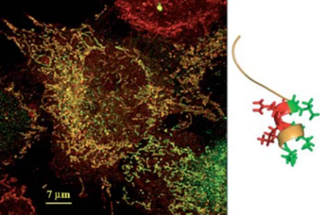
Figure 3 Human truncated procathepsin B in mitochondria. (Left) A cathepsin B-green fluorescent protein chimera contain-ing the first 65 amino acids of truncated procathepsin B startcontain-ing with position 52 (aa numbering of preprocathepsin B) was induced in HeLa cells. Confocal fluorescence microscopy shows in red metabolically active mitochondria stained with Mito-Tracker red and in green the cathepsin B chimera. The yellow color indicates co-localization of the red and green signals. (Right) The N-terminal segment of human truncated procathep-sin B showing a typical mitochondrial import motif, which con-sists of an amphipathic a-helix with a positively charged patch (red) opposite to a hydrophobic patch (green).
of articular cartilage from the surface down the subchon-dral bone, revealed remarkable differences in their distri-bution. While all transcripts were preferentially found in the vicinity of the articular surface in normal cartilage, they were distributed through the depth of the tissue in osteoarthritic cartilage, with evident accumulation at ‘more involved’ sites (Berardi et al., 2001). This finding agrees with independent localization results for cathe-psin B activity by enzyme histochemistry (Baici et al., 1995a,b).
Besides variations in the relative proportions of the 59-UTR alternatively spliced transcripts, a trend towards general enhancement of cathepsin B mRNA levels is observed in pathological tissues compared to normal counterparts (Berardi et al., 2001; Yan and Sloane, 2003). Under homeostatic conditions, the total level of cathep-sin B mRNA results in enzyme expression and trafficking, which is efficiently controlled. Considering an increase in both the total mRNA level and in the proportion of the splice variant CB(-2), a quiet housekeeping function can be ascribed to the CB(full) message, while CB(-2) appears to be responsible for uncontrolled enzyme over-production and misrouted trafficking.
Trafficking
Transient transfection experiments with cathepsin B con-structs fused to the green fluorescent protein were performed in human immortalized chondrocytes and HeLa cells. Co-localization with the trans-Golgi network and the acidic compartment was monitored using spe-cific markers. As expected, the product of CB(full) and CB(-2) appeared in the trans-Golgi network and from there it was delivered to the endosomal-lysosomal com-partment as its final location (Mu¨ntener et al., 2003). Inclusion or omission of the 6-aa-long C-terminal pro-peptide had no influence on cathepsin B trafficking. Mutation of the glycosylation site Asn 38 to Gln pre-vented the necessary glycosylation for targeting the nas-cent protein to the lumen of the endoplasmic reticulum, and the product of this construct was first detected in the rough endoplasmic reticulum before being directly delivered to the secretory pathway. However, in contrast to breast carcinoma cells (Moin et al., 2000), we could not confirm the existence of an alternative, mannose-6-phosphate receptor-independent delivery path to the acidic compartment in chondrocytes and HeLa cells. There is indeed a difference between our approach and that of Moin et al. in including (Mu¨ntener et al., 2003) or not including (Moin et al., 2000) elements of the 59-UTR in the constructs with green fluorescent protein.
After transient transfection of cells with the constructs CB(full) and CB(-2) as just described, regular targeting of procathepsin B to endosomes and lysosomes was evi-dent, and we also observed its extracellular release. Secretion was enhanced in the case of overexpression and was particularly evident with constructs lacking exon 2 from the 59-UTR. This property correlates with the known secretion of procathepsin B in cases of overex-pression, as documented, for instance, in tumors (Frosch et al., 1999) and osteoarthritis (Berardi et al., 2001). In addition to observing procathepsin B secretion from
osteoarthritic and dedifferentiated chondrocytes in cul-ture, we could extract intact glycosylated procathepsin B from human osteoarthritic cartilage (Berardi et al., 2001). We have demonstrated that procathepsin B accumulates in pathological cartilages by binding to the extracellular matrix, and that proenzyme contact with sulfated glycos-aminoglycans promotes its conversion to the active form, which can be detected using a specific enzyme histo-chemical assay (Baici et al., 1995b).
Without knowing further details, it could be supposed that truncated procathepsin B, the product of the splice variant CB(-2,3), is rapidly eliminated after biosynthesis because it cannot be conveyed to the endoplasmic retic-ulum, modified in the Golgi apparatus or sent to the lyso-somal or to the secretory pathway. Early in vitro results that reported the artificial expression of truncated pro-cathepsin B in COS cells and folding into an active enzyme (Mehtani et al., 1998) could not be confirmed by our recent studies (Mu¨ntener et al., 2005). After observing a cytoplasmic and structured localization of green fluo-rescent protein following transfection of chondrocytes and HeLa cells with a cathepsin B construct missing exons 2 and 3 fused to green fluorescent protein (Mu¨n-tener et al., 2003), a detailed study allowed us to track the final destination of truncated procathepsin B to a pre-viously unsuspected location, the mitochondrion (Mu¨n-tener et al., 2004). Co-localization of the truncated procathepsin B-green fluorescent chimeras with mito-chondria was demonstrated by specifically labeling these organelles with MitoTracker red, which fluoresces red in metabolically active mitochondria (Figure 3, left).
How truncated procathepsin B enters mitochondria and the consequences
Close examination of the N-terminal sequence of trun-cated procathepsin B, which starts with Met 52, reveals
the presence of a typical leader sequence for the import of proteins into the mitochondrial matrix (Chacinska et al., 2002). This consists of a small patch of hydrophobic amino acids, opposite to a patch of positively charged amino acids in an amphipathic a-helix, as shown in Fig-ure 3 (right). This FigFig-ure was drawn from the published structure of procathepsin B (Turk et al., 1996) by deleting amino acids up to position 51 and showing 15 residues, starting at position 52. Only the side chains of the resi-dues that make up the helix are explicitly represented. The propensity of this segment of the molecule to form an amphipathic helix in truncated procathepsin B was confirmed by computation using the program PSIPRED (McGuffin et al., 2000). Besides the co-localization exper-iments described above and illustrated in Figure 3, we directly verified the presence of truncated procathepsin B inside mitochondria after isolation of subcellular parti-cles of transfected HeLa cells by differential centrifuga-tion, followed by Western blot analysis. Further evidence that truncated procathepsin B is delivered to the mito-chondrial matrix comes from the presence, in Western blots, of two bands shortened by 2.5- and 4.4-kDa pep-tides with respect to the molecular mass of the chimera (Mu¨ntener et al., 2004). It is strongly suspected that such shortening is due to the action of mitochondrial process-ing peptidase, the enzyme responsible for cleavprocess-ing off the import presequences of proteins targeted to the mito-chondrial matrix. This enzyme preferentially catalyzes cleavage at positions having an arginine as the penulti-mate residue (-2) with an aromatic side-chain at the next position (q1) (Chacinska et al., 2002; Braun and Schmitz, 2004). There are two such potential cleavage sites in the N-terminal segment of truncated procathepsin B, namely between aa 73–74 and 89–90 (preprocathepsin B numbering).
Inside the mitochondrion, instead of behaving as an innocent bystander, truncated procathepsin B has dele-terious consequences for cell integrity. Morphological alterations are observed in the trans-Golgi network, in the acidic compartment, in mitochondria and, most promi-nently, in the nucleus, which undergoes drastic defor-mation followed by cell death (Mu¨ntener et al., 2003, 2004; Zwicky et al., 2003). Although such properties might be attributed to proteolytic events on the part of truncated procathepsin B, we demonstrated that this enzyme form is catalytically inactive (Mu¨ntener et al., 2005). The propeptide of cathepsin B exerts a double function: as a chaperone it assists correct folding of the nascent protein, and as an inhibitor it prevents substrate binding by running its chain over the active center in a direction opposite to that of a regular peptidic substrate. We tested the capability of procathepsin B to fold and to become enzymatically active by expressing protein vari-ants lacking up to 22 amino acids at the N-terminus in baculovirus-infected insect cells. The Asp11–Arg20a-helix
(proenzyme numbering) is necessary for efficient inhibi-tory activity of the propeptide. Trimming away this ele-ment or additional amino acids abolishes inhibition. Proenzyme variants, from which the N-terminal part including the Trp24–Ala26 b-sheet is missing, or which
contain an amino acid mutation directly preceding this b-sheet, are unable to fold (Mu¨ntener et al., 2005).
There-fore, procathepsin B forms lacking more than 22 amino acids at the N-terminus would potentially be enzymati-cally active, but they are destined to remain inactive because they cannot fold correctly. Truncated procathe-psin B, lacking 34 amino acids of the propeptide, has thus no chance of folding and of becoming an active enzyme.
A likely hypothesis to explain the deleterious effects of truncated procathepsin B on cell integrity is the accu-mulation of unfolded protein within the mitochondrial matrix. Figure 3 clearly reveals the structure of mito-chondria, organelles existing in two interconverting forms: as small isolated, rounded particles (the typical textbook image), and as extended filaments. These extended mitochondria represent electrically connected units, which facilitate energy delivery from the cell periph-ery to the cell core. They function as intracellular power-transmitting cables organized as a mitochondrial reticulum (Skulachev, 2001). Careful analysis of confocal images such as that shown in Figure 3 reveals changes in the structure of the mitochondrial reticulum, and the effects produced by the accumulation of unfolded trun-cated procathepsin B can be visualized after dissecting the co-localization features into the component colors (Mu¨ntener et al., 2004). The physical integrity of mito-chondria as a system of energy-delivering cables is essential for their performance. We believe that accu-mulation of unfolded truncated procathepsin B induces the cell to physically fragment its mitochondrial reticulum as a protective mechanism to prevent short circuit in the whole electrical network. However, this reaction ultimate-ly results in power failure and termination of energy deliv-ery. The subsequent morphological changes reveal features that resemble apoptosis rather than necrosis. However, we cannot precisely assign them to a given cell-death mechanism. They can possibly be placed somewhere in one of the categories of cell death described in a variety of physiological and pathological situations, such as autophagy (Bro¨ker et al., 2005), onco-sis (Otsuki et al., 2003) or paraptoonco-sis (Sperandio et al., 2000). Unfortunately, a detailed study of the mechanism of cell death induced by truncated cathepsin B is cur-rently impossible, as this represents a process that occurs slowly in an asynchronous way in only a few cells at a time. The low frequency of this process might explain the slow depletion of cells in tissues undergoing degen-eration over many years, such as osteoarthritic cartilage. In this pathological situation, characterized by overex-pression of cathepsin B at both the mRNA and protein level and with a clear presence of the CB(-2,3) mRNA splice variant (Berardi et al., 2001), cell death is a rare phenomenon, but this would be sufficient to deplete articular cartilage of its cells in one or two decades. Extending this concept, we can postulate a function of truncated procathepsin B as a physiologically relevant mechanism of cell death in tissues characterized by slow turnover and populated by long-lived cells.
Acknowledgments
This work was supported by the Swiss National Science Foun-dation, grant 32-102108/1, and by the Lydia-Hochstrasser, Hart-mann-Mu¨ller, and Olga Mayenfisch Foundations.
References
Baici, A., Ho¨rler, D., Lang, A., Merlin, C., and Kissling, R. (1995a). Cathepsin B in osteoarthritis: zonal variation of enzyme activ-ity in human femoral head cartilage. Ann. Rheum. Dis. 54, 281–288.
Baici, A., Lang, A., Ho¨rler, D., Kissling, R., and Merlin, C. (1995b). Cathepsin B in osteoarthritis: cytochemical and histochemi-cal analysis of human femoral head cartilage. Ann. Rheum. Dis. 54, 289–297.
Berardi, S., Lang, A., Kostoulas, G., Ho¨rler, D., Vilei, E.M., and Baici, A. (2001). Alternative messenger RNA splicing and enzyme forms of cathepsin B in human osteoarthritic carti-lage and cultured chondrocytes. Arthritis Rheum. 44, 1819–1831.
Berquin, I.M., Cao, L., Fong, D., and Sloane, B.F. (1995). Iden-tification of two new exons and multiple transcription start points in the 59-untranslated region of the human cathepsin-B-encoding gene. Gene 159, 143–149.
Berquin, I.M., Ahram, M., and Sloane, B.F. (1997). Exon 2 of human cathepsin B derives from an Alu element. FEBS Lett.
419, 121–123.
Braun, H.-P. and Schmitz, U.K. (2004). Mitochondrial processing peptidase. In: Handbook of Proteolytic Enzymes, A.J. Bar-rett, N.D. Rawlings, and J.F. Woessner Jr., eds. (London, UK: Elsevier), pp. 882–886.
Bro¨ker, L.E., Kruyt, F.A.E., and Giaccone, G. (2005). Cell death independent of caspases: a review. Clin. Cancer Res. 11, 3155–3162.
Chacinska, A., Pfanner, N., and Meisinger, C. (2002). How mito-chondria import hydrophilic and hydrophobic proteins. Trends Cell Biol. 12, 299–303.
Fong, D., Calhoun, D.H., Hsieh, W.T., Lee, B., and Wells, R.D. (1986). Isolation of a cDNA clone for the human lysosomal proteinase cathepsin B. Proc. Natl. Acad. Sci. USA 83, 2909–2913.
Frosch, B.A., Berquin, I., Emmert-Buck, M.R., Moin, K., and Sloane, B.F. (1999). Molecular regulation, membrane asso-ciation and secretion of tumor cathepsin B. APMIS 107, 28–37.
Gong, Q., Chan, S.J., Bajkowski, A.S., Steiner, D.F., and Frank-fater, A. (1993). Characterization of the cathepsin B gene and multiple mRNAs in human tissues: evidence for alternative splicing of cathepsin B pre-mRNA. DNA Cell Biol. 12, 299–309.
Hizel, C., Ferrara, M., Cure, H., Pezet, D., Dechelotte, P., Chippo-ni, J., Rio, P., Bignon, Y.J., and Bernard-Gallon, D. (1998). Evaluation of the 59 spliced form of human cathepsin B mRNA in colorectal mucosa and tumors. Oncol. Rep. 5, 31–34.
Lemaire, R., Flipo, R.-M., Migaud, H., Fontaine, C., Huet, G., Dacquembronne, E., and Lafyatis, R. (1997). Alternative splicing of the 59 region of cathepsin B pre-messenger RNA in rheumatoid synovial tissue. Arthritis Rheum. 40,
1540–1543.
McGuffin, L.J., Bryson, K., and Jones, D.T. (2000). The PSIPRED protein structure prediction server. Bioinformatics 16, 404–405.
Mehtani, S., Gong, Q.M., Panella, J., Subbiah, S., Peffley, D.M., and Frankfater, A. (1998). In vivo expression of an alterna-tively spliced human tumor message that encodes a
trun-cated form of cathepsin B. Subcellular distribution of the truncated enzyme in COS cells. J. Biol. Chem. 273, 13236–13244.
Modrek, B. and Lee, C. (2002). A genomic view of alternative splicing. Nat. Genet. 30, 13–19.
Moin, K., Demchik, L., Mai, J., Duessing, J., Peters, C., and Sloane, B.F. (2000). Observing proteases in living cells. Adv. Exp. Med. Biol. 477, 391–401.
Mort, J.S. (2004). Cathepsin B. In: Handbook of Proteolytic Enzymes, A.J. Barrett, N.D. Rawlings, and J.F. Woessner Jr., eds. (London, UK: Elsevier), pp. 1079–1086.
Mu¨ntener, K., Zwicky, R., Csucs, G., and Baici, A. (2003). The alternative use of exons 2 and 3 in cathepsin B mRNA con-trols enzyme trafficking and triggers nuclear fragmentation in human cells. Histochem. Cell Biol. 119, 93–101.
Mu¨ntener, K., Zwicky, R., Csucs, G., Rohrer, J., and Baici, A. (2004). Exon skipping of cathepsin B: mitochondrial targeting of a lysosomal peptidase provokes cell death. J. Biol. Chem.
279, 41012–41017.
Mu¨ntener, K., Willimann, A., Zwicky, R., Svoboda, B., Mach, L., and Baici, A. (2005). Folding competence of N-terminally truncated forms of human procathepsin B. J. Biol. Chem.
280, 11973–11980.
Otsuki, Y., Li, Z.L., and Shibata, M.A. (2003). Apoptotic detection methods – from morphology to gene. Prog. Histochem. Cytochem. 38, 275–340.
Rawlings, N.D., Tolle, D.P., and Barrett, A.J. (2004). MEROPS: the peptidase database. Nucleic Acids Res. 32 wDatabase issuex, D160–D164.
Skulachev, V.P. (2001). Mitochondrial filaments and clusters as intracellular power-transmitting cables. Trends Biochem. Sci.
26, 23–29.
Sperandio, S., de Belle, I., and Bredesen, D.E. (2000). An alter-native, nonapoptotic form of programmed cell death. Proc. Natl. Acad. Sci. USA 97, 14376–14381.
Tam, S.W., Cote-Paulino, L.R., Peak, D.A., Sheahan, K., and Murnane, M.J. (1994). Human cathepsin B-encoding cDNAs: sequence variations in the 39-untranslated region. Gene 139, 171–176.
Turk, D., Podobnik, M., Kuhelj, R., Dolinar, M., and Turk, V. (1996). Crystal structures of human procathepsin B at 3.2 and 3.3 A˚ resolution reveal an interaction motif between a papain-like cysteine protease and its propeptide. FEBS Lett.
384, 211–214.
Wang, X., Chan, S.J., Eddy, R.L., Byers, M.G., Fukushima, Y., Henry, W.M., Haley, L.L., Steiner, D.F., and Shows, T.B. (1988). Chromosome assignment of cathepsin B (CTSB) to 8p22 and cathepsin H (CTSH) to 15q24-q25. Cytogenet. Cell Genet. 46, 710–711.
Yan, S.Q. and Sloane, B.F. (2003). Molecular regulation of human cathepsin B: implication in pathologies. Biol. Chem. 384, 845–854.
Zwicky, R., Mu¨ntener, K., Goldring, M.B., and Baici, A. (2002). Cathepsin B expression and down-regulation by gene silenc-ing and antisense DNA in human chondrocytes. Biochem. J.
367, 209–217.
Zwicky, R., Mu¨ntener, K., Csucs, G., Goldring, M.B., and Baici, A. (2003). Exploring the role of 59-alternative splicing and of the 39-untranslated region of cathepsin B mRNA. Biol. Chem.