TEST YOURSELF: QUESTION
Painful fingertip swelling of the long finger
Fabio Becce
&Biljana Jovanovic
&Louis Guillou
&Nicolas Theumann
Received: 22 March 2011 / Revised: 15 May 2011 / Accepted: 20 May 2011 / Published online: 18 June 2011 # ISS 2011
Part I
A 35-year-old Caucasian woman presented with a 14-month
history of relapsing painful fingertip swelling of the left
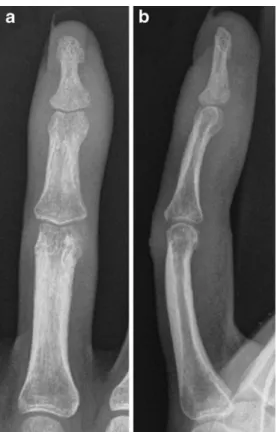
middle finger observed after a minor trauma. Conventional
radiographs (Fig.
1
) were performed at presentation. After
failure of conservative treatment, the patient underwent
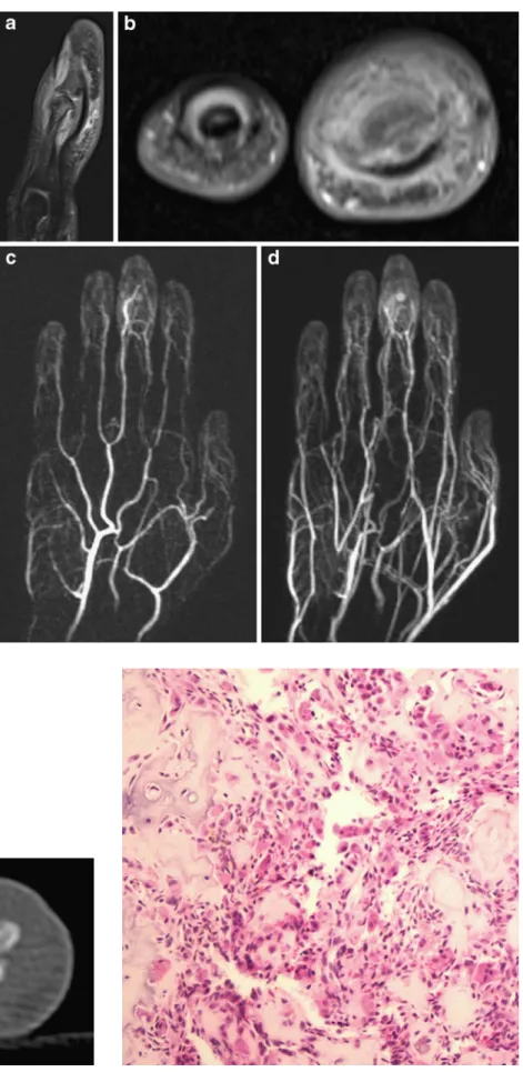
initial surgery without success. Follow-up MRI (Fig.
2
) and
CT (Fig.
3
) were obtained at our institution 6 months after
the first surgical procedure (Figs.
1a–b
,
2a–d
,
3
and
4
).
Funding None
Fig. 1 Posteroanterior (a) and lateral (b) radiographs of the left long finger
The diagnosis can be found at doi:10.1007/s00256-011-1219-y. F. Becce
:
N. Theumann (*)Department of Diagnostic and Interventional Radiology, Centre Hospitalier Universitaire Vaudois, University of Lausanne, Rue du Bugnon 46,
1011 Lausanne, Switzerland e-mail: [email protected] B. Jovanovic
Plastic and Hand Surgery Centre, 1003 Lausanne, Switzerland L. Guillou
University Institute of Pathology, Centre Hospitalier Universitaire Vaudois, 1011 Lausanne, Switzerland
Skeletal Radiol (2011) 40:1479–1480 DOI 10.1007/s00256-011-1218-z
Fig. 3 Axial unenhanced CT image of the left middle (right side of the picture) and ring fingers
Fig. 4 Histopathology image (hematoxylin and eosin stain (H&E); original magnification ×200)
Fig. 2 Sagittal (a) and axial (b) fat-suppressed gadolinium-enhanced T1-weighted turbo spin-echo MR images of the left long finger (right side of the picture in b). Four-dimensional contrast-enhanced MR angiographic images of the left hand in the arterial (c) and venous (d) phase