Images of Arterial Tissues Using Catheter Swept-Source Optical Coherence Tomography
Texte intégral
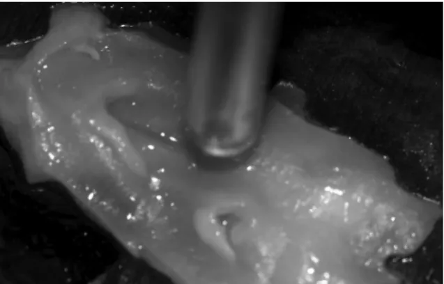
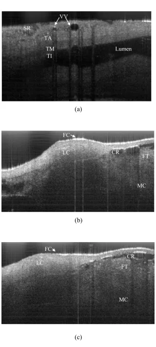
Figure


Documents relatifs
لوأ ﻦﻣ ﻠﻋ ﻞﺟو ّﺰﻋ ﷲ ﻮﻫ ﻩﺮﻜﺷ و ﻩﺪﻤﺣ ﺎﻨﻴﻠﻋ ﻲﻐﺒﻨﻳ و ةﺮﻫﺎﻇ ﻢﻌﻧ ﻦﻣ ﺎﻨﻴﻠﻋ ﻢﻌﻧأ ﺎﻣ ﻰ ﺔﻨﻃﺎﺑ ﻊﺿاﻮﺘﻤﻟا ﺚﺤﺒﻟا اﺬﻫ مﺎﻤﺗإ ﻲﻓ ﺎﻨﻟ ﻪﻧﻮﻋ ﻰﻠﻋ ﻩﺮﻜﺸﻧ.
only in large forest gaps as well as shade-tolerant understorey species, were used to test the following hypotheses: (i) CC of leaves and support tissues differ among species due
ربتعت ةسارد ةعيبط و ساسأ ضيوعتلا نع ءاهنلإا ريغ عورشملا " يفسعتلا " ةقلاعل لمعلا تاذ ةيمهأ ،ةليلق ثيحب نكمي نم اهللاخ طقف يدحت د ىدم قاطن ضيوعتلا
Through untargeted microbiome and metabolomics analyses, we show that wastewater from residential catchments is mostly composed of biomarkers derived of human activity
Alikhanyan National Science Laboratory (Yerevan Physics In- stitute) Foundation (ANSL), State Committee of Science and World Federation of Scientists (WFS), Armenia; Austrian Academy
The present study aims to know the relationship between mental health and mental hardness of the students of the Institute of Sciences and Technical of
The average and final replan prompting intervals (for only those operators that made a change from the initial interval) are also shown in Table 2. A wide range of
Materials Science Division, Argonne National Laboratory, Argonne, IL 60439, USA † Electronic supplementary information (ESI) available: Computed reduction potentials quinoxaline
