Association between arterial stiffness
and variations in estrogen-related genes
The MIT Faculty has made this article openly available.
Please share
how this access benefits you. Your story matters.
Citation
Peter, I et al. “Association Between Arterial Stiffness and Variations
in Oestrogen-related Genes.” Journal of Human Hypertension 23.10
(2009): 636–644. Web.
As Published
http://dx.doi.org/10.1038/jhh.2009.1
Publisher
Nature Publishing Group
Version
Author's final manuscript
Citable link
http://hdl.handle.net/1721.1/73662
Terms of Use
Creative Commons Attribution-Noncommercial-Share Alike 3.0
Association between Arterial Stiffness and Variations in
Estrogen-Related Genes
Inga Peter1, Alyson Kelley-Hedgepeth2, Gordon S. Huggins2, David E. Housman3, Michael E. Mendelsohn2, Joseph A. Vita4,5, Ramachandran S. Vasan4,5, Daniel Levy4,6, Emelia J. Benjamin4,5,7, and Gary F. Mitchell8
1 Department of Genetics and Genomic Sciences, Mount Sinai School of Medicine, New York, NY 2 Molecular Cardiology Research Institute, Tufts Medical Center, Boston, MA, USA
3 Center for Cancer Research, Massachusetts Institute of Technology, Cambridge, MA, USA 4 National Heart, Lung, and Blood Institute’s Framingham Heart Study, Framingham, MA, USA 5 Department of Preventive Medicine and Cardiology, Boston University School of Medicine, Boston,
MA, USA
6 Center for Population Studies, NHLBI, Bethesda, MD, USA
7 Department of Epidemiology, School of Public Health, Boston University, Boston, MA, USA 8 Cardiovascular Engineering, Inc., Norwood, MA, USA
Abstract
Increased arterial stiffness and wave reflection have been identified as cardiovascular disease risk factors. In light of significant sex differences and the moderate heritability of vascular function measures, we hypothesized that variation in the genes coding for estrogen receptors alpha (ESR1) and beta (ESR2) and aromatase (CYP19A1) is associated with aortic stiffness and pressure wave reflection as measured by noninvasive arterial tonometry. 1261 unrelated Framingham Offspring Study participants who attended the 7th examination cycle (mean age 62±10 years, 52% women) and had arterial tonometry and genotyping data were included in the study. ANCOVA was used to assess the association of polymorphisms with forward wave amplitude, augmented pressure, augmentation index, carotid-femoral pulse wave velocity, and mean arterial pressure with adjustment for potential confounders. In the sex-pooled analysis, those homozygous for the minor allele at any of four
ESR1 variants that were in strong linkage disequilibrium ((TA)n, rs2077647, rs2234693 and
rs9340799) had on average 18% higher augmented pressure and 16% greater augmentation index compared to carriers of one or two major alleles (p=0.0002–0.01). A similar magnitude of association was detected in those homozygous for the common allele at two ESR2 SNPs (p=0.007–0.02). Our results are consistent with the hypothesis that variation in ESR1 and ESR2, but not CYP19A1, is associated with increased wave reflection, which may contribute to previously demonstrated associations between these variants and adverse clinical events. Our findings will need to be replicated in additional cohorts.
Keywords
arterial stiffness; tonometry; estrogen receptor; polymorphism
NIH Public Access
Author Manuscript
J Hum Hypertens. Author manuscript; available in PMC 2010 April 1. Published in final edited form as:
J Hum Hypertens. 2009 October ; 23(10): 636–644. doi:10.1038/jhh.2009.1.
NIH-PA Author Manuscript
NIH-PA Author Manuscript
Introduction
Increased arterial stiffness and wave reflection play important roles in cardiovascular physiology. Aortic stiffness, estimated by measurement of carotid-femoral pulse wave velocity (CFPWV), has been identified as a cardiovascular disease (CVD) risk factor in 3 large, independent, community-based cohorts.1–3 Some studies show that CFPWV and pulse pressure are lower in women than men prior to 60 years of age, but increase rapidly in women thereafter, resulting in comparable CFPWV and higher pulse pressure in older women. Similarly, the incidence of CVD is low in premenopausal women as compared to their male peers, but increases sharply after menopause.4 In light of the associations between aortic stiffness and events, the disproportionate increase in aortic stiffness in older women may contribute to the higher incidence of CVD in women after the menopause. In contrast, measures of wave reflection, such as augmented pressure or augmentation index, are increased in women as compared to men at all ages, with a maximal difference just prior to menopause and a trend for values in women and men to converge after 60 years of age. Increased wave reflection in women has been attributed to shorter stature, but differences persist after adjusting for height5 and when women and men are matched for height,6 suggesting intrinsic sex differences in structure or function of the arterial system.
Estrogen is known to play an important role in endothelial function in both men and women. Premature ovarian failure has been shown to be associated with significant endothelial dysfunction.7, 8 In postmenopausal women, estrogen therapy has been shown to enhance endothelium-dependent vasodilation and reduce arterial stiffness in some studies.9, 10 Estrogen is known to have short and long-term effects on the vasculature (reviewed in 11). The rapid vasodilatory effects of estrogen are produced by estrogen-stimulated increases in endothelial cell nitric oxide (NO) synthase activity which regulates vascular tone,12 suppresses smooth muscle proliferation13 and inhibits platelet adhesion and aggregation14. Longer-term effects are mediated through estrogen-activated transcription factors, estrogen receptor (ER) α, (encoded by the ESR1 gene) and ERβ (encoded by the ESR2 gene), which are found in vascular endothelial and smooth muscle cells in both men and women.15, 16
Recently, genetic variations of the major proteins involved in steroid hormone conversion and receptor function have been shown to be associated with the increased risk of myocardial infarction17 and coronary artery disease,18–20 elevated blood pressure,21 altered lipoprotein particles concentrations,22, 23 adiposity, 24 and left ventricular mass,25–27 attracting increased attention to genes implicated in estrogen metabolism. Also, variation in the gene (CYP19A1) encoding the enzyme aromatase, which catalyzes the conversions of testosterone to estradiol and defines the estradiol/testosterone ratio, has been found to be associated with blood pressure21 and abdominal obesity.28 However, despite these and other studies, potential molecular mechanisms linking estrogen-related genes with CVD risk remain incompletely understood.
Given significant sex differences in vascular stiffness and moderate heritability of key arterial pressure waveform phenotypes,29, 30 we hypothesized that variation in ESR1, ESR2, and CYP19A1 are associated with aortic stiffness, pressure wave reflection and vasodilator function as measured by noninvasive tonometry and brachial ultrasound in the Framingham Offspring Study.
NIH-PA Author Manuscript
NIH-PA Author Manuscript
Material and Methods
Study cohort
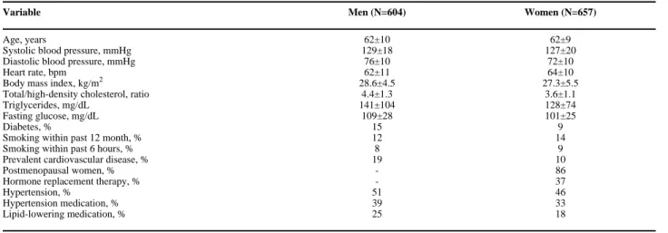
The study sample was derived from unrelated participants in the longitudinal community-based Framingham Offspring cohort of European descent and is described in detail elsewhere.31 Eligible participants consented to genetic analysis and attended the 7th examination cycle (1998–2001), during which they underwent routine medical history, physical examination, tonometry measures and brachial ultrasound. Unrelated participants (N=3539) were selected based on family structure alone without regard to any phenotypic traits and were excluded from the present analysis if they did not have tonometry measures or brachial ultrasound data (N=1508) or had missing genotype data (N=770). The final sample included 1261 individuals: 604 men and 657 women.
All participants gave written informed consent including consent for DNA analyses. The FHS protocol was approved by the Boston University Medical Center Institutional Review Board. Noninvasive Tonometry Measures
Participants were studied in the supine position after several minutes of rest as described previously.5 Arterial tonometry, with simultaneous ECG, was obtained from the brachial, radial, femoral, and carotid arteries. Transit distances were assessed by body surface
measurements from the suprasternal notch to each pulse recording site. Tonometry waveforms were analyzed as described previously.5. CFPWV was calculated from tonometry waveforms and body surface measurements. The CFPWV distance was measured with a fiberglass tape measure. The central forward wave amplitude was defined as the difference between pressure at the waveform foot and pressure at the first systolic inflection point or peak of the carotid pressure waveform. Augmented pressure was defined as the difference between central systolic pressure and pressure at the forward wave peak. Augmentation index (AI) was calculated as previously described.32 Systolic ejection period was defined as the time interval from the carotid pressure waveform foot to the dicrotic notch.
Brachial Artery Measures
Brachial artery diameter and mean flow velocity at baseline and 1-minute after reactive hyperemia, induced by 5-mintue forearm cuff occlusion, were assessed as previously described,33, 34 by using a Toshiba SSH-140A ultrasound system and commercially available software (Brachial Tools, version 3.2.3). Mean Doppler flow velocity was analyzed using a semi-automated signal averaging approach (Cardiovascular Engineering, Inc.).
Single Nucleotide Polymorphism (SNP) Selection and Genotyping
To investigate pathophysiologic connection between arterial stiffness and cardiovascular disease, we selected a priori SNPs in ESR1, ESR2, and CYP19A1 found to be associated with cardiovascular risk phenotypes in the Framingham Offspring cohort by our previous studies. 17, 21–24, 26, 27 Genomic DNA was extracted from peripheral blood leukocytes using standard methods. Genotyping for the individual SNPs in ESR1 (rs2077647, rs2234693, rs9340799, and rs1801132), ESR2 (rs944460, rs1256059, rs1256034, and rs1256031), and CYP19A1 (rs700518, rs726547), was performed as described previously.21, 35 ESR1 (TA)n, ESR1 (CA)n, ESR2 (CA)n, and CYP19A1 (TTTA)n repeat polymorphisms were genotyped using restriction fragment length analyses.36
Statistical Analysis
Observed genotype frequencies were compared with those expected under Hardy–Weinberg equilibrium (HWE) using a χ2 test. Given multiple alleles observed for the repeat
NIH-PA Author Manuscript
NIH-PA Author Manuscript
polymorphisms and their bimodal distribution, the genotype carrier status for each variant was coded using the median number of repeat sequence base pairs as a cutoff. Specifically, genotype LL was assigned if both alleles contained at least the median number of base pairs (≥176 for
ESR1 (TA)n; ≥277 for ESR1 (CA)n; ≥162 for ESR2 (CA)n, and ≥298 for CYP19A1 (TTTA)n);
SS was assigned if both alleles were ‘short’ (<176 for ESR1 (TA)n; <277 for ESR1 (CA)n; <162
for ESR2 (CA)n, and <298 for CYP19A1 (TTTA)n), and LS if one allele was ‘long’ and another one was ‘short’.
We used a general model of inheritance and sex-pooled analyses to assess relations between estrogen pathway genotypes and vascular phenotypes. Multivariable linear regression analyses were carried out to assess genetic associations with forward wave amplitude, augmented pressure, AI, and CFPWV. All analyses were adjusted for age, sex, heart rate, body mass index (or weight and height for augmented pressure and AI), mean arterial pressure (for CFPWV), total/high-density lipoprotein cholesterol ratio, triglycerides, fasting glucose, diabetes, smoking within past 6 hours, prevalent cardiovascular disease, hormone replacement therapy (in women), hypertension medication, and lipid-lowering medication. To account for sex-specific differences previously reported in relations between estrogen pathway genotypes and cardiovascular phenotypes in the Framingham sample,17, 22, 23, 27 interactions between sex and genotype were assessed by the addition of a multiplicative term to the fully adjusted model. Forearm vascular resistance was skewed and was natural log transformed to normalize the variance.
Pairwise linkage disequilibrium (LD) was evaluated by Lewontin’s D′.37 Haplotypes were inferred by using the expectation maximization algorithm.38 To account for allelic interaction, haplotypes were used as predictors in the regression models along with the above covariates. The nominal threshold for statistical significance of all analyses was set at 0.05 and was not adjusted for multiple testing. All analyses were performed using SAS/STAT and SAS/Genetics software version 9.1 (SAS Institute, Inc., Cary, North Carolina, USA).
Results
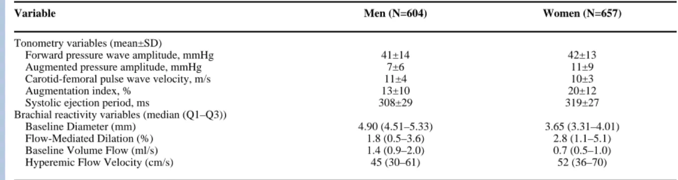
The clinical characteristics of the unrelated Framingham Offspring Study participants included in this analysis are shown in Table 1. Vascular phenotypes are summarized in Table 2 and characteristics of the genotyped polymorphisms are summarized in Table 3.
ESR1 association analysis
In the sex-pooled analysis, those homozygous for the minor alleles for ESR1 (TA)n, rs2077647, rs2234693, and rs9340799 had higher augmented pressure and AI (Table 4) as compared to carriers of one or two major alleles (recessive model, p range 0.0001–0.04). Associations were more pronounced in individuals not receiving antihypertensive treatment and in women, who had particularly higher augmented pressure and AI (data not shown). No significant sex-genotype and treatment-genotype interaction was detected. Also, we observed a modest association of several of the ESR1 variants on heart rate, which was lower in the groups with higher AI (Online Supplement Table I). In our sample, lower heart rate significantly correlated with higher augmented pressure and AI (r=−0.25 and −0.28, respectively; p<0.0001).
Strong LD detected between four of the six ESR1 markers (pairwise D′ ranges between 0.82 and 0.99, Online Supplement Figure) resulted in three common haplotypes, H5: (TA)n
[L]-rs2077647[C] - rs2234693[C] rs9340799[G] with the frequency of 32.7%, H6: (TA)n
[L]-rs2077647[C] - rs2234693[C] rs9340799[A] with the frequency of 6.9% and H12: (TA)n
[S]-rs2077647[T] - rs2234693[T] rs9340799[A] with the frequency of 45.8%. However, the data
NIH-PA Author Manuscript
NIH-PA Author Manuscript
indicate that haplotypes did not explain larger proportions of variation in augmented pressure and AI than individual SNPs.
No association was detected between ESR1 polymorphisms and forward pressure wave amplitude or CFPWV. Both of these measures showed weak to moderate correlations with augmented pressure (r=0.18, p<0.0001 and r=−0.08, p=0.009, respectively), AI (r=−0.14, p<0.0001 and r=−0.08, p=0.005, respectively) and the heart rate (r=0.04, p=0.13 and r=0.20, p<0.0001, respectively). In addition, no associations were detected between ESR1
polymorphisms and any of the brachial flow measures (p>0.05). ESR2 association analysis
Similar to the ESR1 analysis, in the sex-pooled sample, ESR2 rs944460 and rs1256034 genotypes were related to augmented pressure and AI (Table 5). Those homozygous for the major allele of ESR2 rs944460 and rs1256034, had higher augmented pressure and AI than carriers of the minor allele.
Strong LD was detected between the two pairs of ESR2 SNPs (Table 5). However, the correlation between them, or r2, was small due to low minor allele frequency for rs944460 and rs1256034 (Online Supplement Figure), which resulted in a large number of rare haplotypes. No association was detected between ESR2 polymorphisms and forward pressure wave or CFPWV. In addition, no associations were detected between ESR2 polymorphisms and any of the brachial flow measures (p>0.05).
CYP19A1 association analysis
No association was detected between the CYP19A1 SNPs (Online Supplement Figure) and studied vascular measures (Online Supplement Table II) (p>0.05).
Interaction effects
On the basis of significant interactive effects of ESR1 rs2234693 and smoking status on lipoprotein profile detected in a previous study,22 we tested the hypothesis that ESR1 rs2234693 interacts with smoking status. Regardless of genotype, in the multivariable-adjusted model smokers had higher augmented pressure and greater AI than non-smokers (10.4±5.0 vs. 8.6±4.8 mm Hg; p=0.01 and 20.0±7.1 vs. 15.8±6.4%; p<0.0001, respectively). Furthermore, homozygosity for the ESR1 rs2234693 C allele had an enhanced association with augmented pressure and AI in smokers (p=0.04 and p=0.02, respectively, for the genotype-smoking interaction term) (Figure).
Discussion
We evaluated relations between genetic variants in the ESR1, ESR2 and CYP19A1 genes and key arterial function phenotypes assessed by tonometry and brachial reactivity in unrelated individuals from the Framingham Heart Study Offspring cohort. In models that adjusted for a number of potential confounders, rare alleles of the closely related ESR1 polymorphisms and common alleles of the linked ESR2 polymorphisms were associated with higher augmented pressure and greater AI. In sex-pooled analyses, those homozygous for the minor allele at any one of the four ESR1 SNPs, which were all in strong LD, had on average 17% higher augmented pressure and 15% greater AI compared to carriers of one or two major alleles. A similar direction and magnitude of association was detected in those homozygous for the common allele at two strongly linked ESR2 SNPs. No association was found between the CYP19A1 SNPs and vascular measures. These analyses were prompted by significant associations detected between ESR1 and ESR2 gene polymorphisms and a number of cardiovascular
NIH-PA Author Manuscript
NIH-PA Author Manuscript
phenotypes in the Framingham Heart Study, including myocardial infarction,17 elevated blood pressure,21 altered lipoprotein profile,22, 23 adiposity,24 and changes in left ventricular structure.26, 27 The present findings of an association between common variants in estrogen pathway genes and measures of wave reflection suggest that abnormal pulsatile load may be involved in the pathogenesis of the foregoing adverse clinical phenotypes.
Reported associations were seen in four closely linked ESR1 SNPs, two of which may have direct relevance to vascular function. Specifically, the ESR1 TA polymorphism is located in the regulatory region of the gene ~1174 base pairs upstream of the first exon39 between promoters B and C.40 The long allele has been associated with worse cardiovascular risk profile in previous reports. Namely, compared with short allele carriers, the TA L polymorphism has been linked with more severely diseased coronary arteries,19, 41 and a higher risk of myocardial infarction.41 In the present study, ESR1 (TA)n LL homozygotes had significantly lower heart rate and higher augmented pressure amplitude and AI compared to carriers of one or two S alleles. Although the exact mechanism whereby the ESR1 TA repeat polymorphism may affect the vasculature is unclear, it is possible that this variant alters ESR1 transcript length or RNA splicing causing significant heterogeneity in ERα mRNA transcripts. It has been shown previously that alternative splicing may result in variants with deletions of exons encoding regions of the hormone-binding domain with truncated forms of ERα discovered in many tissues including vascular endothelium and showing altered ligand-activation properties (reviewed in 42).
Higher augmented pressure and AI were also detected with the C allele of another variant, ESR1 rs2234693, which is known to produce a functional myb binding site that might amplify ERα transcription or produce ERα isoforms with properties that differ from those of the full-length gene product.43
ERϐ is required for normal vasodilatation and blood pressure in both male and female mice, with loss of ERϐ causing substantial hypertension, particularly in males.44 We found associations of higher augmented pressure and AI with common variants in the two linked intronic SNPs that have no known functional effect on the receptor. These variants may regulate RNA splicing or transcription causing differences in expression levels, relative abundance of multiple ESR2 splice forms, or ligand binding affinity. However, given an unusually large distance between the ESR2 SNPs that are in LD (>30,000 base-pairs), it is possible that the observed associations may be due to the existence of as yet undetected coding variants located between and in LD with the genotyped SNPs.
We also observed a modest effect of several of the ESR1 variants on heart rate. In our sample, lower heart rate significantly correlated with higher augmented pressure and AI and could contribute to the increase in AI. Noteworthy, it has been shown that AI shares 4.6% common genetic component with heart rate.29 However, it is important to emphasize that our models included an adjustment for systolic ejection period or heart rate.
In the present study, we found no association of ER genotype carrier status with forward pressure wave amplitude or CFPWV. Both of these measures showed weak to moderate correlations with augmented pressure and heart rate. However, in an earlier study, Japanese women who were carriers of ESR1 rs2234693 CC were shown to have lower brachial-ankle pulse wave velocity, which is a measure of the spatially averaged properties of the aorta and peripheral arteries.46 Interestingly, female carriers of the common ESR1 rs2234693 C allele from the Framingham Offspring cohort had a larger average low-density lipoprotein (LDL) particle size and a lower concentration of small LDL particles, which is a protective lipid profile.23 Taken together, these data suggest that brachial-ankle pulse wave velocity may be modified through favorable estrogen-induced changes in lipoprotein properties in carriers of
NIH-PA Author Manuscript
NIH-PA Author Manuscript
the minor allele. In contrast, the same genotypes may increase arterial wave reflection because of direct effects of estrogen on vascular tone in the periphery.
In the present study we detected effect modification by smoking status. That is, among smokers, those who were homozygous for the ESR1 rs2234693 C allele had a 40% higher augmented pressure and 33% greater AI than carriers of one or two major alleles (Figure). Importantly, significant ESR1 rs2234693 - smoking status interaction in this cohort has been found with lipoprotein fraction concentrations.22 Among smokers, female carriers of ESR1 rs2234693 TT genotype had higher small LDL particle concentrations and lower LDL particle sizes than non-carriers, while no such association was detected in non-smokers. It has been shown that acute tobacco use provokes endothelial dysfunction and abolishes the protection of circulating estrogen.47 It will require further studies to determine how variation in ESR1 interacts with cigarette smoking.
There are several potential limitations of our study. The design of this study, like any other genetic association studies, does not allow drawing definitive conclusion about the exact mechanisms that may lead to increased wave reflection. Moreover, the study cohort was middle-aged to elderly and white of European descent, which may limit the generalizability of our findings to younger individuals and other ethnic backgrounds. Also, we analyzed SNPs that have been previously implicated in cardiovascular phenotypes; thus, we may have missed associations attributable to other polymorphisms not sufficiently linked to the SNPs that we evaluated. In addition, no adjustment for multiple testing was carried out. A standard correction for multiple hypothesis testing relies on the assumption that all statistical comparisons are independent. In the case of ESR1, ESR2 and CYP19A1, most of the polymorphisms were in strong LD with one another, and tonometry measures were correlated. Moreover, we tested a priori hypotheses based on reported findings with regard to these gene variants; therefore rigorous adherence to the correction for multiple testing may have increased Type II error. The consistency and biological relevance of these associations should motivate further research to verify and extend these findings. We acknowledge that our findings will need to be replicated in other cohorts.
Supplementary Material
Refer to Web version on PubMed Central for supplementary material.
Reference List
1. Mattace-Raso FU, van der Cammen TJ, Hofman A, van Popele NM, Bos ML, Schalekamp MA, et al. Arterial stiffness and risk of coronary heart disease and stroke: the Rotterdam Study. Circulation 2006;113:657–63. [PubMed: 16461838]
2. Sutton-Tyrrell K, Najjar SS, Boudreau RM, Venkitachalam L, Kupelian V, Simonsick EM, et al. Elevated aortic pulse wave velocity, a marker of arterial stiffness, predicts cardiovascular events in well-functioning older adults. Circulation 2005;111:3384–90. [PubMed: 15967850]
3. Willum-Hansen T, Staessen JA, Torp-Pedersen C, Rasmussen S, Thijs L, Ibsen H, Jeppesen J. Prognostic value of aortic pulse wave velocity as index of arterial stiffness in the general population. Circulation 2006;113:664–70. [PubMed: 16461839]
4. Colditz GA, Willett WC, Stampfer MJ, Rosner B, Speizer FE, Hennekens CH. Menopause and the risk of coronary heart disease in women. N Engl J Med 1987;316:1105–10. [PubMed: 3574358] 5. Mitchell GF, Parise H, Benjamin EJ, Larson MG, Keyes MJ, Vita JA, et al. Changes in arterial stiffness
and wave reflection with advancing age in healthy men and women: the Framingham Heart Study. Hypertension 2004;43:1239–45. [PubMed: 15123572]
6. Gatzka CD, Kingwell BA, Cameron JD, Berry KL, Liang YL, Dewar EM, et al. Gender differences in the timing of arterial wave reflection beyond differences in body height. J Hypertens 2001;19:2197– 203. [PubMed: 11725164]
NIH-PA Author Manuscript
NIH-PA Author Manuscript
7. Kalantaridou SN, Naka KK, Papanikolaou E, Kazakos N, Kravariti M, Calis KA, et al. Impaired endothelial function in young women with premature ovarian failure: normalization with hormone therapy. J Clin Endocrinol Metab 2004;89:3907–13. [PubMed: 15292326]
8. Kalantaridou SN, Naka KK, Bechlioulis A, Makrigiannakis A, Michalis L, Chrousos GP. Premature ovarian failure, endothelial dysfunction and estrogen-progestogen replacement. Trends Endocrinol Metab 2006;17:101–9. [PubMed: 16515863]
9. Sumino H, Ichikawa S, Kasama S, Takahashi T, Kumakura H, Takayama Y, et al. Different effects of oral conjugated estrogen and transdermal estradiol on arterial stiffness and vascular inflammatory markers in postmenopausal women. Atherosclerosis 2006;189:436–42. [PubMed: 16469323] 10. Gerhard M, Walsh BW, Tawakol A, Haley EA, Creager SJ, Seely EW, et al. Estradiol therapy
combined with progesterone and endothelium-dependent vasodilation in postmenopausal women. Circulation 1998;98:1158–63. [PubMed: 9743505]
11. Mendelsohn ME. Genomic and nongenomic effects of estrogen in the vasculature. Am J Cardiol 2002;90:3F–6F.
12. Moncada S, Palmer RM, Higgs EA. Nitric oxide: physiology, pathophysiology, and pharmacology. Pharmacol Rev 1991;43:109–42. [PubMed: 1852778]
13. Garg UC, Hassid A. Nitric oxide-generating vasodilators and 8-bromo-cyclic guanosine
monophosphate inhibit mitogenesis and proliferation of cultured rat vascular smooth muscle cells. J Clin Invest 1989;83:1774–7. [PubMed: 2540223]
14. Radomski MW, Palmer RM, Moncada S. Endogenous nitric oxide inhibits human platelet adhesion to vascular endothelium. Lancet 1987;2:1057–8. [PubMed: 2889967]
15. Venkov CD, Rankin AB, Vaughan DE. Identification of authentic estrogen receptor in cultured endothelial cells. A potential mechanism for steroid hormone regulation of endothelial function. Circulation 1996;94:727–33. [PubMed: 8772695]
16. Karas RH, Patterson BL, Mendelsohn ME. Human vascular smooth muscle cells contain functional estrogen receptor. Circulation 1994;89:1943–50. [PubMed: 8181116]
17. Shearman AM, Cupples LA, Demissie S, Peter I, Schmid CH, Karas RH, et al. Association between estrogen receptor alpha gene variation and cardiovascular disease. JAMA 2003;290:2263–70. [PubMed: 14600184]
18. Lu H, Higashikata T, Inazu A, Nohara A, Yu W, Shimizu M, Mabuchi H. Association of estrogen receptor-alpha gene polymorphisms with coronary artery disease in patients with familial hypercholesterolemia. Arterioscler Thromb Vasc Biol 2002;22:817–23. [PubMed: 12006396] 19. Pollak A, Rokach A, Blumenfeld A, Rosen LJ, Resnik L, Dresner PR. Association of oestrogen
receptor alpha gene polymorphism with the angiographic extent of coronary artery disease. Eur Heart J 2004;25:240–5. [PubMed: 14972425]
20. Rokach A, Pollak A, Rosen L, Friedlander Y, Blumenfeld A, Reznik L, et al. Estrogen receptor alpha gene polymorphisms are associated with the angiographic extent of coronary artery disease. J Clin Endocrinol Metab 2005;90:6556–60. [PubMed: 16159931]
21. Peter I, Shearman AM, Zucker DR, Schmid CH, Demissie S, Cupples LA, et al. Variation in estrogen-related genes and cross-sectional and longitudinal blood pressure in the Framingham Heart Study. J Hypertens 2005;23:2193–200. [PubMed: 16269961]
22. Shearman AM, Demissie S, Cupples LA, Peter I, Schmid CH, Ordovas JM, et al. Tobacco smoking, estrogen receptor alpha gene variation and small low density lipoprotein level. Hum Mol Genet 2005;14:2405–13. [PubMed: 16014638]
23. Demissie S, Cupples LA, Shearman AM, Gruenthal KM, Peter I, Schmid CH, et al. Estrogen receptor-alpha variants are associated with lipoprotein size distribution and particle levels in women: the Framingham Heart Study. Atherosclerosis 2006;185:210–8. [PubMed: 16005459]
24. Fox CS, Yang Q, Cupples LA, Guo CY, Atwood LD, Murabito JM, et al. Sex-specific association between estrogen receptor-alpha gene variation and measures of adiposity: the Framingham Heart Study. J Clin Endocrinol Metab 2005;90:6257–62. [PubMed: 16144952]
25. Leibowitz D, Dresner-Pollak R, Dvir S, Rokach A, Reznik L, Pollak A. Association of an estrogen receptor-alpha gene polymorphism with left ventricular mass. Blood Press 2006;15:45–50. [PubMed: 16492615]
NIH-PA Author Manuscript
NIH-PA Author Manuscript
26. Peter I, Huggins GS, Shearman AM, Pollak A, Schmid CH, Cupples LA, et al. Age-related changes in echocardiographic measurements: association with variation in the estrogen receptor-alpha gene. Hypertension 2007;49:1000–6. [PubMed: 17372038]
27. Peter I, Shearman AM, Vasan RS, Zucker DR, Schmid CH, Demissie S, et al. Association of estrogen receptor beta gene polymorphisms with left ventricular mass and wall thickness in women. Am J Hypertens 2005;18:1388–95. [PubMed: 16280269]
28. Baghaei F, Rosmond R, Westberg L, Hellstrand M, Eriksson E, Holm G, et al. The CYP19 gene and associations with androgens and abdominal obesity in premenopausal women. Obes Res
2003;11:578–85. [PubMed: 12690088]
29. Snieder H, Hayward CS, Perks U, Kelly RP, Kelly PJ, Spector TD. Heritability of central systolic pressure augmentation: a twin study. Hypertension 2000;35:574–9. [PubMed: 10679500]
30. Mitchell GF, DeStefano AL, Larson MG, Benjamin EJ, Chen MH, Vasan RS, et al. Heritability and a genome-wide linkage scan for arterial stiffness, wave reflection, and mean arterial pressure: the Framingham Heart Study. Circulation 2005;112:194–9. [PubMed: 15998672]
31. Kannel WB, Feinleib M, McNamara PM, Garrison RJ, Castelli WP. An investigation of coronary heart disease in families. The Framingham offspring study. Am J Epidemiol 1979;110:281–90. [PubMed: 474565]
32. Murgo JP, Westerhof N, Giolma JP, Altobelli SA. Aortic input impedance in normal man: relationship to pressure wave forms. Circulation 1980;62:105–16. [PubMed: 7379273]
33. Benjamin EJ, Larson MG, Keyes MJ, Mitchell GF, Vasan RS, Keaney JF Jr, et al. Clinical correlates and heritability of flow-mediated dilation in the community: the Framingham Heart Study. Circulation 2004;109:613–9. [PubMed: 14769683]
34. Mitchell GF, Parise H, Vita JA, Larson MG, Warner E, Keaney JF Jr, et al. Local shear stress and brachial artery flow-mediated dilation: the Framingham Heart Study. Hypertension 2004;44:134–9. [PubMed: 15249547]
35. Shearman AM, Karasik D, Gruenthal KM, Demissie S, Cupples LA, Housman DE, et al. Estrogen receptor beta polymorphisms are associated with bone mass in women and men: the Framingham Study. J Bone Miner Res 2004;19:773–81. [PubMed: 15068501]
36. Peter I, Kelley-Hedgepeth A, Fox CS, Cupples LA, Huggins GS, Housman DE, et al. Variation in Estrogen-Related Genes Associated with Cardiovascular Phenotypes and Circulating Estradiol, Testosterone, and Dehydroepiandrosterone Sulfate Levels. J Clin Endocrinol Metab 2008;93:2779– 85. [PubMed: 18445666]
37. Lewontin RC. The interaction of selection and linkage. II. Optimum models. Genetics 1964;50:757– 82. [PubMed: 14221879]
38. Elston, RC.; Olson, JM.; Palmer, L. Biostatistical genetics and genetic epidemiology. John Wiley & Sons, Ltd; 2003.
39. Sano M, Inoue S, Hosoi T, Ouchi Y, Emi M, Shiraki M, et al. Association of estrogen receptor dinucleotide repeat polymorphism with osteoporosis. Biochem Biophys Res Commun 1995;217:378–83. [PubMed: 8526937]
40. Kos M, Reid G, Denger S, Gannon F. Minireview: genomic organization of the human ERalpha gene promoter region. Mol Endocrinol 2001;15:2057–63. [PubMed: 11731608]
41. Kunnas TA, Laippala P, Penttila A, Lehtimaki T, Karhunen PJ. Association of polymorphism of human alpha oestrogen receptor gene with coronary artery disease in men: a necropsy study. BMJ 2000;321:273–4. [PubMed: 10915129]
42. Moriarty K, Kim KH, Bender JR. Minireview: estrogen receptor-mediated rapid signaling. Endocrinology 2006;147:5557–63. [PubMed: 16946015]
43. Herrington DM, Howard TD, Brosnihan KB, McDonnell DP, Li X, Hawkins GA, Reboussin DM, et al. Common estrogen receptor polymorphism augments effects of hormone replacement therapy on E-selectin but not C-reactive protein. Circulation 2002;105:1879–82. [PubMed: 11997270] 44. Zhu Y, Bian Z, Lu P, Karas RH, Bao L, Cox D, et al. Abnormal vascular function and hypertension
in mice deficient in estrogen receptor beta. Science 2002;295:505–8. [PubMed: 11799247] 45. Wilkinson IB, Qasem A, McEniery CM, Webb DJ, Avolio AP, Cockcroft JR. Nitric oxide regulates
local arterial distensibility in vivo. Circulation 2002;105:213–7. [PubMed: 11790703]
NIH-PA Author Manuscript
NIH-PA Author Manuscript
46. Hayashi K, Maeda S, Iemitsu M, Otsuki T, Sugawara J, Tanabe T, et al. Sex differences in the relationship between estrogen receptor alpha gene polymorphisms and arterial stiffness in older humans. Am J Hypertens 2007;20:650–6. [PubMed: 17531923]
47. Garcia-Fernandez R, Perez-Velasco JG, Concepcion-Millan A, Sosa S, Navaroli F, Garcia-Barreto D. Estrogen does not prevent endothelial dysfunction caused by cigarette smoking. Clin Cardiol 2004;2:71–3. [PubMed: 14979623]
NIH-PA Author Manuscript
NIH-PA Author Manuscript
Figure. Tonometry phenotypes by smoking status and the ESR1 rs2234693 genotype in the sex-pooled sample
Gene*smoking interaction adjusted
NIH-PA Author Manuscript
NIH-PA Author Manuscript
NIH-PA Author Manuscript
NIH-PA Author Manuscript
NIH-PA Author Manuscript
Table 1
Sample characteristics.
Variable Men (N=604) Women (N=657)
Age, years 62±10 62±9
Systolic blood pressure, mmHg 129±18 127±20
Diastolic blood pressure, mmHg 76±10 72±10
Heart rate, bpm 62±11 64±10
Body mass index, kg/m2 28.6±4.5 27.3±5.5
Total/high-density cholesterol, ratio 4.4±1.3 3.6±1.1
Triglycerides, mg/dL 141±104 128±74
Fasting glucose, mg/dL 109±28 101±25
Diabetes, % 15 9
Smoking within past 12 month, % 12 14
Smoking within past 6 hours, % 8 9
Prevalent cardiovascular disease, % 19 10
Postmenopausal women, % - 86
Hormone replacement therapy, % - 37
Hypertension, % 51 46
Hypertension medication, % 39 33
NIH-PA Author Manuscript
NIH-PA Author Manuscript
NIH-PA Author Manuscript
Table 2
Vascular phenotypes.
Variable Men (N=604) Women (N=657)
Tonometry variables (mean±SD)
Forward pressure wave amplitude, mmHg 41±14 42±13
Augmented pressure amplitude, mmHg 7±6 11±9
Carotid-femoral pulse wave velocity, m/s 11±4 10±3
Augmentation index, % 13±10 20±12
Systolic ejection period, ms 308±29 319±27
Brachial reactivity variables (median (Q1–Q3))
Baseline Diameter (mm) 4.90 (4.51–5.33) 3.65 (3.31–4.01)
Flow-Mediated Dilation (%) 1.8 (0.5–3.6) 2.8 (1.1–5.1)
Baseline Volume Flow (ml/s) 1.4 (0.9–2.0) 0.7 (0.5–1.0)
Hyperemic Flow Velocity (cm/s) 45 (30–61) 52 (36–70)
NIH-PA Author Manuscript
NIH-PA Author Manuscript
NIH-PA Author Manuscript
Table 3 Polymorphism characteristics. Gene dbSNP rs# SNP position Nucleotide substitution
Minor Allele Frequency
P for HWE ESR1 (TA) n Promoter (TA) n .50 * 0.95 6q25.1 rs2077647 Exon 1 (Ser10Ser) T/C .46 0.22 rs2234693 Intron 1 T/C .45 0.66 rs9340799 Intron 1 A/G .36 0.80 rs1801132 Exon 4 (Pro325Pro) C/G .23 0.15 (CA) n Intron 5 (CA) n .36 * 0.05 ESR2 rs944460 Intron 2 C/G .03 0.29 14q23.2 rs1256059 Intron 2 C/T .45 0.13 (CA) n Intron 5 (CA) n .20 * 0.28 rs1256034 Intron 6 G/A .03 0.29 rs1256031 Intron 7 T/C .49 0.59 CYP19A1 rs4646 5 ′ UTR C/A .25 0.18 15q21.1 rs700518 Exon 3 (Val80Val) A/G .48 0.01 (TTTA) n Intron 4 (TTTA) n .49 * 0.82 rs726547 Intron 4 C/T .05 0.41
NIH-PA Author Manuscript
NIH-PA Author Manuscript
NIH-PA Author Manuscript
Table 4
Tonometry phenotypes in the pooled sample by
ESR1 genotypes. SNP Genotype N Augmented pressure, mmHg Augmentation index, % LS mean±SE P LS mean±SE P (TA) n S/S L/S L/L 250 506 257 8.6±0.3 8.5±0.2 10.2±0.3 0.004 15.7±0.4 16.0±0.3 18.8±0.4 0.001 rs2077647 T/T T/C C/C 320 594 237 8.8±0.3 8.4±0.2 10.0±0.3 0.01 15.9±0.4 15.8±0.3 18.2±0.4 0.009 rs2234693 T/T T/C C/C 360 572 244 9.2±0.3 8.1±0.2 10.1±0.3 0.001 16.4±0.4 15.3±0.3 18.6±0.4 0.001 rs9340799 A/A A/G G/G 488 540 159 8.9±0.2 8.3±0.2 10.3±0.3 0.006 16.2±0.3 15.4±0.3 19.7±0.5 0.0002 rs1801132 C/C C/G G/G 683 389 67 9.1±0.2 8.4±0.2 7.7±0.6 0.80 16.6±0.3 15.9±0.3 15.6±0.8 0.98 (CA) n S/S S/L L/L 398 419 140 9.0±0.3 8.7±0.3 9.7±0.4 0.68 16.2±0.4 16.6±0.4 18.0±0.6 0.44
NIH-PA Author Manuscript
NIH-PA Author Manuscript
NIH-PA Author Manuscript
Table 5
Tonometry phenotypes in the pooled sample by
ESR2 genotypes. SNP Genotype N Augmented pressure, mmHg Augmentation index, % LS mean±SE P LS mean±SE P rs944460 C/C C/G 1108 71 8.9± 0.2 7.3± 0. 6 0.009 16.5± 0.2 13.7± 0.7 0.02 rs1256059 C/C C/T T/T 331 595 228 9.8± 0.3 8.2± 0.2 8.6± 0.3 0.12 17.6± 0.4 15.5± 0.3 16.2± 0.5 0.30 (CA) n S/S S/L L/L 757 393 43 8.9± 0.2 8.7± 0.2 8.6± 0.8 0.88 16.4± 0.2 16.4± 0.3 15.4± 1.0 0.60 rs1256034 G/G G/A 1064 70 8.9± 0.2 7.1± 0.6 0.007 16.5± 0.2 13.2± 0.8 0.01 rs1256031 T/T T/C C/C 304 601 280 10.2± 0.3 8.3± 0.2 8.7± 0.3 0.17 18.0± 0.4 15.6± 0.3 16.4± 0.4 0.48
NIH-PA Author Manuscript
NIH-PA Author Manuscript
NIH-PA Author Manuscript
Table 6
Summary What is known about this topic:
• Aortic stiffness has been identified as a cardiovascular disease risk factor in three large, independent, community-based cohorts.1–3
• Estrogen is known to play an important role in endothelial function in both men and women.
• Genetic variations of the major proteins involved in estrogen conversion and receptor function have been shown to be associated with the increased risk of myocardial infarction17 and coronary artery disease,18–20 elevated blood pressure,21 altered lipoprotein particles concentrations,22, 23 adiposity, 24 and left ventricular mass.25–27
What this Study adds:
• This 1261 unrelated Framingham Offspring Study participants shows that variation in ESR1 and ESR2, but not CYP19A1 may predispose to excess pressure wave reflection, which may contribute to morbidity from several complex diseases.
• Finding genes associated with excess wave reflection may help elucidate hemodynamic mechanisms of disease pathogenesis and enhance our ability to favorably manipulate vascular responses in genetically susceptible individuals before clinical manifestations occur.