HAL Id: hal-01595261
https://hal.archives-ouvertes.fr/hal-01595261
Submitted on 26 Sep 2017
HAL is a multi-disciplinary open access
archive for the deposit and dissemination of
sci-entific research documents, whether they are
pub-lished or not. The documents may come from
teaching and research institutions in France or
abroad, or from public or private research centers.
L’archive ouverte pluridisciplinaire HAL, est
destinée au dépôt et à la diffusion de documents
scientifiques de niveau recherche, publiés ou non,
émanant des établissements d’enseignement et de
recherche français ou étrangers, des laboratoires
publics ou privés.
Reduced adiponectin expression after high-fat diet is
associated with selective up-regulation of ALDH1A1 and
further retinoic acid receptor signaling in adipose tissue
Jean-Francois Landrier, Elnaz Kasiri, Esma Karkeni, Johanna Mihaly,
Gabriella Beke, Kathrin Weiss, Renata Lucas, Gamze Aydemir, Jérôme Salles,
Stephane Walrand, et al.
To cite this version:
Jean-Francois Landrier, Elnaz Kasiri, Esma Karkeni, Johanna Mihaly, Gabriella Beke, et al.. Reduced
adiponectin expression after high-fat diet is associated with selective up-regulation of ALDH1A1 and
further retinoic acid receptor signaling in adipose tissue. FASEB Journal, Federation of American
Society of Experimental Biology, 2017, 31 (1), pp.203-211. �10.1096/fj.201600263RR�. �hal-01595261�
THE
JOURNAL • RESEARCH • www.fasebj.org
Reduced adiponectin expression after high-fat diet is
associated with selective up-regulation of ALDH1A1
and further retinoic acid receptor signaling in
adipose tissue
Jean-Francois Landrier,*
,†,‡Elnaz Kasiri,
§,{Esma Karkeni,*
,†,‡Johanna Mih´aly,
§Gabriella B´eke,
§Kathrin Weiss,
§Renata Lucas,
§Gamze Aydemir,
§J´erome Salles,
kSt´ephane Walrand,
kAngel R. de Lera,
#and Ralph R ¨uhl
§,{,**
,1*Institut National de la Recherche Agronomique, Unit´es Mixtes de Recherche 1260, Marseille, France;†INSERM, Unit´es Mixtes de Recherche 1062, Nutrition, Ob´esit´e et Risque Thrombotique, Marseille, France;‡Aix-Marseille Universit´e, Facult´e de M´edecine, Marseille, France;
§Department of Biochemistry and Molecular Biology and{MTA-DE Public Health Research Group, Hungarian Academy of Sciences, Faculty of
Public Health, University of Debrecen, Debrecen, Hungary;kUnit´es Mixtes de Recherche, Institut National de la Recherche Agronomique (INRA) 1019 Unit´e de Nutrition Humaine, Centre de Recherches INRA de Clermont-Ferrand/Theix, Saint-Gen`es-Champanelle, France;
#
Departamento de Qu´ımica Org´anica, Universidade de Vigo, Facultad de Qu´ımica, Centro de Investigaciones Biom´edicas and Instituto de Investigaci ´on Biom´edica de Vigo, Vigo, Spain; and **Paprika Bioanalytics BT, Debrecen, Hungary
ABSTRACT:
Adiponectin is an adipocyte-derived adipokine with potent antidiabetic, inflammatory, and
anti-atherogenic activity. Long-term, high-fat diet results in gain of body weight, adiposity, further inflammatory-based
cardiovascular diseases, and reduced adiponectin secretion. Vitamin A derivatives/retinoids are involved in several
of these processes, which mainly take place in white adipose tissue (WAT). In this study, we examined adiponectin
expression as a function of dietary high-fat and high–vitamin A conditions in mice. A decrease of adiponectin
expression in addition to an up-regulation of aldehyde dehydrogenase A1 (ALDH1A1), retinoid signaling, and
retinoic acid response element signaling was selectively observed in WAT of mice fed a normal–vitamin A, high-fat
diet. Reduced adiponectin expression in WAT was also observed in mice fed a high–vitamin A diet. Adipocyte cell
culture revealed that endogenous and synthetic retinoic acid receptor (RAR)a- and RARg-selective agonists, as well
as a synthetic retinoid X receptor agonist, efficiently reduced adiponectin expression, whereas ALDH1A1 expression
only increased with RAR agonists. We conclude that reduced adiponectin expression under high-fat dietary
con-ditions is dependent on
1) increased ALDH1A1 expression in adipocytes, which does not increase all-trans-retinoic
acid levels;
2) further RAR ligand–induced, WAT-selective, increased retinoic acid response element–mediated
signaling; and
3) RAR ligand–dependent reduction of adiponectin expression.—Landrier, J.-F., Kasiri, E., Karkeni,
E., Mih´aly, J., B´eke, G., Weiss, K., Lucas, R., Aydemir, G., Salles, J., Walrand, S., de Lera, A. R., R ¨uhl, R. Reduced
adiponectin expression after high-fat diet is associated with selective up-regulation of ALDH1A1 and further
retinoic acid receptor signaling in adipose tissue. FASEB J. 31, 203–211 (2017). www.fasebj.org
KEY WORDS:
vitamin A
•nuclear hormone receptor
•obesity
•diabetes
•retinaldehyde dehydrogenase
Obesity is considered to be one of the most common
nu-tritional disorders of Western society and is characterized
by a disproportionate expansion of body fat mass
[reviewed in Gasbarrini and Piscaglia (1)]. In addition to
being an energy storage site, white adipose tissue (WAT)
also functions as a highly active metabolic regulator and
ABBREVIATIONS:9-HODE, 9-hydroxyoctadecadienoic acid; ALDH1A1, aldehyde dehydrogenase 1A1; ATRA, all-trans-retinoic acid; CTRL, control; FABP4, fatty acid binding protein 4; HF, high fat; LF, low fat; LXR, liver X receptor; NF, normal fat; PPAR, peroxisome proliferator-activated receptor; RALDH, retinaldehyde dehydrogenase; RAR, retinoic acid receptor; RARE, retinoic acid response element; RE, retinol equivalent; RETSAT, all-trans-retinol 13,14-reductase; RXR, retinoid X receptor; TG2, transglutaminase 2; VDR, vitamin D receptor; WAT, white adipose tissue
1Correspondence: Department of Preventive Medicine, Faculty of Public Health, University of Debrecen, Kassai u. 26/b, H-4028 Debrecen, Hungary.
E-mail: ralphruehl@web.de
This is an Open Access article distributed under the terms of the Creative Commons Attribution-NonCommercial 4.0 International (CC BY-NC 4.0) (http://creativecommons.org/licenses/by-nc/4.0/) which permits noncommercial use, distribution, and reproduction in any medium, provided the original work is properly cited.
doi: 10.1096/fj.201600263RR
0892-6638/17/0031-0203 © The Author(s) 203
Vol.31, No.1 , pp:203-211, February, 2017
The FASEB Journal
. 147.99.128.158 to IP
www.fasebj.org Downloaded from
major endocrine organ that secretes various adipokines
(2–4). Adiponectin is a major adipokine with strong
anti-diabetic, anti-inflammatory, and antiatherogenic activity,
and its expression is decreased in WAT under high dietary
fat conditions [reviewed in Ouchi
et al. (5)]. With the
ex-ception of the putative role of inflammation (6–8), the
precise mechanisms that mediate this down-regulation
remain to be elucidated.
Retinoids are important regulators of
adipogene-sis. Diets high in vitamin A (9), dietary excess of retinoic
acid (10, 11), and diets high in b-carotene (12–14) result
in increased adipocyte apoptosis and inhibition of
adipogenesis, while low concentrations of retinoic acid
were described to be proadipogenic (reviewed in refs.
9, 11, 15, 16). Retinoids, that is, naturally occurring and
synthetic retinol analogues (reviewed in refs. 17, 18),
are responsible for activation of specific nuclear
re-ceptors: the retinoic acid receptor (RAR) and the
ret-inoid X receptor (RXR). Bioactive retinoic acids are
formed from precursor retinaldehydes by the action
of retinaldehyde dehydrogenase enzymes (RALDHs/
ALDH1A) (19).
RALDH1/ALDH1A1–null adult mice have been
shown to be resistant to high-fat (HF) diet–induced
weight gain (20, 21), which suggested that ALDH1A1
and its metabolic products are necessary for HF
diet–induced obesity (22–24). ALDH1A1 can
synthe-size retinoic acids (25), such as all-trans-retinoic acid
(ATRA), 9-cis-retinoic acid, and, presumably, the
newly found endogenous RXR ligand,
9-cis-13,14-dihydroretinoic acid (26). The last two are ligands for
both RXRs and RARs, whereas ATRA only binds RAR.
Unfortunately, only a few studies have detected
reti-noic acids in low concentrations in adipose tissue (27,
28), but no study has addressed the presence of
reti-noic acids in WAT when comparing ALDH1A1
+/+or
ALDH1A1
2/2mice. Whether retinoic acids, and which
retinoic acids, are the major metabolites of ALDH1A1 in
WAT is yet unknown. Moreover, ALDH1A1 expression
has been shown to be regulated by liver X receptor
(LXR) (29) as well as estrogen receptor–mediated
pathways (30, 31).
Various nuclear hormone receptor pathways are
in-volved in adipokine secretion and adipocyte
differentia-tion, proliferadifferentia-tion, and lipid accumulation (reviewed in
refs. 7, 32). In particular, RXRs, the central
heterodimer-forming partners, play important roles during obesity
(33–35). RXRa as well as RXRg KO and RXR-antagonist
treatment induce resistance to weight gain after HF diet
and also promote a higher metabolic rate (36–38). RXRs
can also interact with several nuclear receptors, such as
RAR, LXR, peroxisome proliferator-activated receptor
(PPAR), vitamin D receptor (VDR), or NR4A-orphan
nuclear receptors (39, 40), and the activation of various
so-called permissive heterodimers (RXR-PPAR, -LXR,
-VDR, and -NR4A1/2) by an RXR ligand can initiate
heterodimer-mediated signaling (39–41).
The aim of our study was to find out how HF diet
reduces adiponectin expression in WAT, focusing
pri-marily on vitamin A–mediated RAR- and RXR-dependent
pathways.
MATERIALS AND METHODS
Experimental diets
Manually prepared diets were made with wheat starch
(Weizenst¨arke, Foodstar, Germany; provided by Kr¨oner-St¨arke,
Ibbenb ¨uren, Germany), saccharose (purchased from a local
supermarket in Hungary), casein (Sigma-Aldrich, Budapest,
Hungary), cellulose (Vivapur; JRS Pharma GmbH; Rosenberg,
Germany), vitamin mix (Vitamin-Vormischung C1000;
Altromin GmbH, Lage, Germany), mineral mixture
(Mineral-Spurenelemente-Vormischung C100; Altromin GmbH), and
sunflower oil (Henry Lamotte, Bremen, Germany).
Animal experiments
Animal experiments were performed in the Laboratory Animal
Core Facility of the University of Debrecen. Experiments were
performed according to Hungarian ethical guidelines.
Experiment with low-, normal-, or high-fat dietary
supplementation
After the acclimatization period, animals received a vitamin
A–deficient [0 retinol equivalent (RE)/kg diet] diet for 10 wk that
contained 5% sunflower oil as a dietary lipid, which represented
a diet with normal fat (NF) content (42). Animals were divided
into different feeding groups (n = 6 per group) and were fed for
4 wk with specific diets that contained different amounts of dietary
fat and equal amounts of vitamin A (2500 RE/kg diet, normal
vitamin A). Sunflower oil was added as dietary fat, which
con-tained either 2% [as weight %; low-fat (LF) diet], 5% (NF diet), or
25% (HF diet). The source of the fat was always sunflower
oil in different proportions added to feed. On the basis of the
analyzed feed of the NF diet, it contained 11.6% saturated fats,
20% monounsaturated fatty acids, and 68.4% polyunsaturated
fatty acids (Weiss
et al., in preparation). Furthermore, dietary
composition was 180 g/kg casein, 10 g/kg vitamin mix, 45 g/kg
mineral mix, and 20 g/kg cellulose for all applied diets (42). As a
result of the increased amount of fat in the diet, the carbohydrate
proportion was lower; the LF diet contained 29.5% sucrose and
43% starch, the NF diet 28% sucrose and 41.5% starch, and the HF
diet 17% sucrose and 32.5% starch (42).
Experiment with normal
– or high–vitamin A dietary
supplementation
For vitamin content, diets were supplemented with vitamin mix
(Vitamin-Vormischung C1000) that contained either 2500 RE/kg
as normal–vitamin A diet, or for high–vitamin A diet, an
addi-tional retinyl-palmitate (RetPal) supplement (final 326,500 RE/kg;
Sigma-Aldrich) was added to the normal–vitamin A diet (42, 43).
After euthanizing mice, blood collection was carried out by
cardiac puncture. Blood was centrifuged for 20 min and plasma
was stored at
280°C. Mice were anatomized, and WAT samples
were immediately frozen in liquid nitrogen after dissection and
later stored at
280°C until RNA extraction.
Bioimaging
Retinoic acid response element (RARE)-Luc female mice (n = 6)
were obtained from Cgene (Oslo, Norway) and received LF, NF,
or HF diets for 4 wk or the oral retinoid treatments as described
before (43, 44).
We conducted
ex vivo organ analysis by bioluminescence
imaging. All animals were treated with 120 mg/kg
D-luciferin
(Bioscience, Budapest, Hungary)
via intraperitoneal injections 15
min before euthanasia and further organ screening. Mice were
euthanized by cervical dislocation. After euthanasia, mouse liver,
WAT, intestine, and brain were collected for bioluminescence
imaging. Organs were analyzed for bioluminescence signal by
using an Andor-Ixon CCD camera (Belfast, United Kingdom),
and analysis was performed by Andor-IQ software. After
im-aging, integrated intensity/area was calculated for liver, WAT,
intestine, and brain of each treated animal.
Cell culture
3T3-L1 preadipocytes (American Type Culture Collection,
Manassas, VA, USA) were seeded in 3.5-cm-diameter dishes at a
density of 15
3 10
4cells/well. Cells were grown in DMEM that
was supplemented with 10% FBS at 37°C in a 5% CO
2humidified
atmosphere, as previously reported (45, 46). To induce
differen-tiation, 2-d postconfluent 3T3-L1 preadipocytes (day 0) were
stimulated for 48 h with 0.5 mM isobutylmethylxanthine, 0.25
mM dexamethasone, and 1 mg/ml insulin in DMEM that was
supplemented with 10% FBS. Cells were then maintained in
DMEM that was supplemented with 10% FBS and 1 mg/ml
in-sulin (47). To examine the effects on gene expression of ATRA (a
gift from BASF AG, Ludwigshafen, Germany), an RARa agonist
(BMS753), an RARg agonist (BMS189961; both were prepared in
our laboratories as described in the original patents (48, 49)], and
an RXR agonist (LG268; gift from Ligand Pharmaceuticals, San
Diego, CA, USA), 3T3-L1 adipocytes were incubated with 1 mM
of these molecules for 24 h, as previously reported (47). Data
presented are the mean of 3 independent experiments each
per-formed in triplicate.
Human adipose biopsies
Eleven lean (body mass index: 22.5
6 0.5 kg/m
2) and 14 obese
(body mass index: 31.7
6 0.9 kg/m
2) male participants were
recruited, as previously reported (50). Lean and obese volunteers
were age 44
6 7 and 44 6 5 yr, respectively. Subcutaneous
ad-ipose tissue biopsies were performed between 6:30 and 7:30
AMafter an overnight fast. Biopsies were obtained by needle
aspi-ration in the periumbilical area under local anesthesia. Adipose
tissue samples were rinsed in physiologic serum, immediately
frozen in liquid nitrogen, and stored at
280°C until RNA
ex-traction. The experimental protocol was performed in
accor-dance with the guidelines in the Declaration of Helsinki and was
approved by the Ethical Committee of the Auvergne Region
(agreement No. AU 800, March 2010). Participants gave their
written informed consent to participate in the study.
Analysis of mRNA expression
Analysis of total cellular RNA extracted from 3T3-L1 cells was
performed in France by using Trizol reagent according to
man-ufacturer instructions. Human adipose tissue sample extraction
was also performed in France, whereas WAT and liver sample
analysis from mice was done in Hungary.
For the cell culture material in the French laboratory, cDNA
was synthesized from 1 mg of total RNA in 20 ml by using random
primers and Moloney murine leukemia virus reverse
transcrip-tase. Real-time quantitative RT-PCR analyses for genes were
performed by using the Mx3005P Real-Time PCR System
(Stra-tagene, La Jolla, CA, USA) as previously described (51). For each
sample, expression was quantified in duplicate and 18S rRNA
was used as the endogenous control in the comparative cycle
threshold (C
T) method.
For WAT and liver tissue analysis of human and murine origin,
tissues were homogenized in Tri reagent solution (Thermo Fisher
Scientific, Waltham, MA, USA) and total RNA was isolated from
tissue according to manufacturer guidelines and as previously
de-scribed (52). Concentration and purity of RNA was measured
by using the NanoDrop spectrophotometer (Thermo Fisher Scientific).
For real-time quantitative PCR, total RNA was reverse
tran-scribed into cDNA by using the Super Script II First-Strand
Synthesis System (Thermo Fisher Scientific). Quantitative
real-time PCR was carried out in triplicate using predesigned MGB
assays (Thermo Fisher Scientific) on an ABI Prism 7900 (Applied
Biosystems, Villebon-sur-Yvette, France). Relative mRNA levels
were calculated by using the
C
tmethod and were normalized
to cyclophilin A mRNA. Sequence Detector Software (v. 2.1;
Applied Biosystems) was used for data analysis.
Analytical procedures
WAT samples were collected and stored in dark vials at
280°C
until analysis. Sample preparation was performed as previously
described for retinoid (53) and eicosanoid/docosanoid (54)
analysis. HPLC–tandem mass spectrometry analyses for
ret-inoids as well as eicosanoids and docosanoids, which focused
on eicosanoids with known PPAR activation potential, were
also performed as previously explained (53, 54).
ELISA assays
To examine the effect of retinoids on adiponectin secretion,
3T3-L1 adipocytes were incubated with 1 mM of the retinoids
(ATRA, RARa, RARg, or RXR ligand) for 48 h. Adiponectin
quantification was realized on the culture supernatant by using
adiponectin ELISA assay according to manufacturer protocol
(Quantikine ELISA; R&D Systems, Lille, France).
Statistics
Data are expressed as means
6
SEM. Significant differences
be-tween control and treated cells/groups were determined by
Student’s t test using Statview software (SAS Institute, Cary, NC,
USA). Values of
P , 0.05 were considered significant.
RESULTS
Effects of HF diet on body weight gain
Body weight gain was observed in animals after 4 wk
of HF diet compared with LF or NF dietary
supple-mentation (LF: 1.07
6 0.08 g; NF: 0.92 6 0.11 g; HF: 3.24 6
0.37 g; LF-HF
P = 0.03 and NF-HF P = 0.04). Food intake
slightly decreased in the HF diet group (LF: 2.93 g/d/
animal; NF: 2.70 g/d/animal; HF: 2.25 g/d/animal).
HF dietary supplementation results in
up-regulation of ALDH1A1 and down-up-regulation
of adiponectin expression
ALDH1A1 was significantly increased only in WAT (LF:
1
6 0.80; NF: 1.83 6 0.36; HF: 5.26 6 0.23) of HF diet–
fed mice compared with LF and NF diet–fed mice, and
was tissue selective for WAT (Table 1, WAT), compared
with unchanged expression in the liver (Table 1, liver).
ALDH1A2 expression remained unchanged in liver and
WAT (Table 1). ALDH1A3 was also significantly
ADIPONECTIN IS REGULATED DEPENDING ON RAR SIGNALING 205
Vol.31, No.1 , pp:203-211, February, 2017
The FASEB Journal
. 147.99.128.158 to IP
www.fasebj.org Downloaded from
increased in adipose tissue of animals fed HF and NF
diets compared with LF diet (Table 1, WAT). In addition,
expression of RAR pathway target genes, such as
CYP26A1 and CYP26B1, remained unchanged (Table 2),
whereas expression of the highly sensitive common
RAR/RXR pathway target gene transglutaminase 2 (TG2)
was strongly increased (Table 1; LF: 1
6 0.36; NF: 9.56 6
0.13; HF: 15.36
6 0.17) in HF diet–fed mice.
In addition to increased retinoid signaling, expression
of adiponectin was reduced in HF diet–fed mice (LF: 1 6
0.31; NF: 0.86
6 0.12; HF: 0.15 6 0.37).
Increased ALDH1A1 and reduced adiponectin
expression in obese volunteers
Experiments using adipose tissue biopsies from
normal-weight and obese human volunteers confirmed increased
ALDH1A1 (healthy volunteers were set as 1; 1.20
6 0.07)
and reduced adiponectin (0.85
6 0.04) expression in the
obese volunteers (Table 3).
High–vitamin A dietary supplementation
results in increased expression of ALDH1A1
and reduced adiponectin expression
Expression of ALDH1A1 increased (NF, normal vitamin A
was set as 1: 1
6 0.19; NF, high vitamin A: 2.32 6 0.47) in
the WAT of mice fed an NF diet with high vitamin A
supplementation, whereas adiponectin expression (NF,
normal vitamin A was set as 1: 1
6 0.47; NF, high vitamin
A: 0.37
6 0.21) was decreased in WAT (Table 4).
Decreased retinoic acid concentrations
present in WAT of animals fed an HF diet do
not correspond to increased RARE-mediated
signaling in RARE-Luc mice, and PPARg ligands
remain mainly unchanged
Retinol levels remained stable in the WAT of LF, NF,
and HF diet–fed animals, whereas ATRA (LF: 2.2 6
0.1 ng/g; NF: 1.7
6 0.2 ng/g; HF: 0.6 6 0.1 ng/g)
levels were lower in the WAT of HF diet–fed animals
(Table 5).
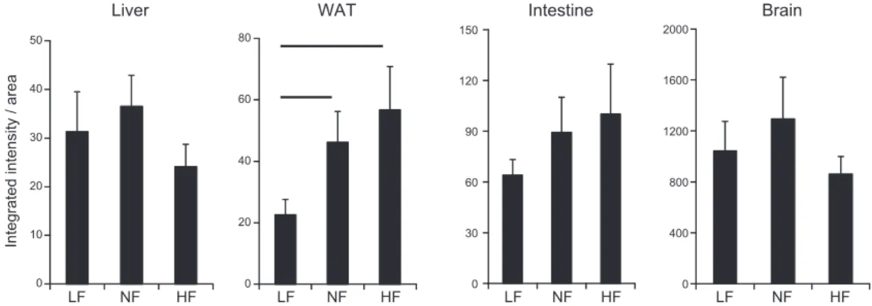
Increased retinoid signaling was confirmed in
RARE-Luc mice, with increased RARE-mediated signaling
de-tected specifically in adipose tissue of HF compared with
LF and NF diet–fed animals, whereas in liver, intestine,
and brain, no increased RARE-mediated signaling was
observed (Fig. 1).
Endogenous PPAR ligands [9-hydroxyoctadecadienoic
acid (9-HODE), 13-HODE, 13-ketooctadecadienoic acid,
12-ketoeicosatetraenoic acid, PgJ2, and d15d12PgJ2] were
mainly unchanged, except the adipose tissue–specific
PPARg ligand, hepoxilin B3, which is increased in adipose
tissue of HF diet–fed animals (Table 5).
TABLE 1. Relative adiponectin and ALDH1A1 mRNA expression
Gene
Fold activation Significance
LF NF HF LF:NF NF:HF LF:HF
WAT
ALDH1A1
1
6 0.80
1.83
6 0.36
5.26
6 0.23
0.46
0.05
0.01
ALDH1A2
1
6 0.11
1.05
6 0.14
1.09
6 0.19
0.77
0.87
0.69
ALDH1A3
1
6 0.17
1.65
6 0.08
2.59
6 0.24
0.03
0.12
0.02
Adiponectin
1
6 0.31
0.86
6 0.12
0.15
6 0.37
0.72
,0.01
0.04
Liver
ALDH1A1
1
6 0.10
1.10
6 0.11
1.39
6 0.13
0.53
0.64
0.09
ALDH1A2
1
6 0.12
0.79
6 0.09
0.74
6 0.10
0.17
0.64
0.09
Expression shown in WAT and liver of LF (set as 1), NF, and HF diet–fed mice with a normal content of vitamin A in the diet. Gene expression (all n = 6) of adiponectin and retinoic acid synthesizing enzymes (ALDH1A1, ALDH1A2, ALDH1A3). Significant values vs. LF are in italics.TABLE 2. Relative gene expression of genes involved in RAR and PPAR signaling in mouse WAT
Gene
Fold activation Significance
LF NF HF LF:NF NF:HF LF:HF
RAR pathway
CYP26A1
1
6 0.50
0.16
6 0.41
0.43
6 0.49
0.13
0.26
0.33
CYP26B1
1
6 0.63
0.58
6 0.52
0.93
6 0.66
0.60
0.65
0.94
TG2
1
6 0.36
9.56
6 0.13
15.36
6 0.17
,0.01
0.08
,0.01
PPAR pathway
PPARg
1
6 0.12
1.23
6 0.09
0.93
6 0.09
0.18
0.07
0.67
RETSAT
1
6 0.21
1.96
6 0.23
1.38
6 0.26
0.09
0.37
0.37
FABP4
1
6 0.03
1.00
6 0.10
1.08
6 0.10
0.99
0.57
0.47
FADS2
1
6 0.43
1.31
6 0.41
1.46
6 0.48
0.71
0.88
0.64
Expression in WAT of LF (set as 1), NF, and HF diet–fed mice with a normal content of vitamin A in diet (all n = 6). Significant values vs. LF are in italics.
NF and HF dietary supplementation does not
result in altered PPARg-mediated signaling
Expression of PPARg and PPARg target genes retinol
saturase (RETSAT)/fatty acid binding protein 4 (FABP4)/
FADS2 in mouse WAT remained unaffected by NF and HF
dietary supplementation compared with LF diet (Table 2).
Adiponectin expression is reduced by RAR and
RXR agonists using 3T3-L1 adipocytes
cell culture
Treatment of cultured adipocytes with synthetic
RARa-selective ligands [control (CTRL) set as 1; adiponectin:
0.22
6 0.01 and ALDH1A1 2.37 6 0.04], RARg-selective
ligands (CTRL set as 1; adiponectin: 0.22
6 0.01 and
ALDH1A1 2.64
6 0.01), and the natural RAR ligand
ATRA (CTRL set as 1; adiponectin: 0.25
6 0.03 and
ALDH1A1 3.19
6 0.07), in addition to a synthetic RXR
agonist (LG268; CTRL set as 1; adiponectin: 0.51
6 0.04
and ALDH1A1 1.04
6 0.04), resulted in increased
ALDH1A1 expression for RAR agonists, whereas
adipo-nectin expression was reduced for all RAR and RXR
li-gands. In addition, these results were confirmed at the
protein level in cell culture supernatants where
adipo-nectin secretion was reduced for all administered RAR and
RXR ligands, except for the RARg-selective ligand, which
displayed a nonsignificant decrease (Table 6).
DISCUSSION
Obesity is classically associated with a decrease of
adi-ponectin plasma level in humans and rodents, as well
as a decreased expression in adipose tissue (5). This
re-lationship between obesity and decreased adiponectin
expression is suspected to be linked to the increased
in-flammatory status of adipose tissue, as TNF-a, one of the
main inflammatory markers produced by adipose tissue
(55), is known to reduce adiponectin expression (56).
However, this mechanism is probably not exclusive, and
other pathways—RAR signaling among them—could be
involved in this regulation.
In this study, we reported that, in mice, reduced
adi-ponectin expression in WAT after HF dietary
supplemen-tation was associated with an increase of ALDH1A1
expression. Similar results were also obtained by
compar-ing lean
vs. obese WAT biopsies. Surprisingly, increased
ALDH1A1 expression in mice does not result in increased
ATRA levels in WAT.
ALDH1A1, the major enzyme for retinoic acid synthesis
using retinaldehyde as a substrate, is highly likely to play an
important role in the relationship between retinoid signaling
and obesity (20, 21, 57). Indeed, its expression is increased in
WAT during HF-induced obesity (58). In ALDH1A1
2/2-deficient adipocytes as well as in ALDH1A1
2/2mice,
adipogenesis is impaired and mice are resistant to HF
diet–inducedobesity(20),whichissuggestedtoberelatedto
altered retinoid signaling [(25) and reviewed in refs. 9, 11, 15].
Retinoic acids, the products from ALDH1A1 metabolism,
are the endogenous activators of RARs and RXRs. Reduced
retinaldehyde and retinol levels were measured in adipose
tissue of animals fed an HF diet, and ATRA levels were
speculated to be increased upon ALDH1A1 activity (20).
However, the detection and quantification of retinoic acid
levels in adipose tissue have been scarcely examined (27, 28)
and, unfortunately, the connection of retinoic acids in
response to ALDH1A1 expression in adipose tissue has
not been studied before. In the present study, we report
that increased ALDH1A1 expression in mice does not
re-sult in increased ATRA levels in WAT. On the contrary,
ATRA levels were even lower in WAT of HF
vs. LF or
NF diet–fed animals. Similar reduced levels of ATRA
were confirmed in serum and adipose tissue of obese
volunteers compared with obese volunteers after a
weight-loss diet (unpublished data), which indicates that obesity is
related to reduced local and systemic retinoid levels in
humans. These findings of reduced local retinoid levels in
adipose tissue of obese animals fit well with previous
studies on animals fed a vitamin A–deficient diet, which
were found to become obese upon reduced ATRA
syn-thesis, levels, and ATRA-mediated signaling [reviewed in
Bonet
et al. (11)]. In addition, it is well established that
retinoids, and especially ATRA, as signaling ligands, have
the ability to inhibit proliferation of adipocytes; enhance
up-regulation of genes involved in lipid oxidation,
en-ergy dissipation, and insulin response; and thereby
prevent obesity and insulin resistance [reviewed in Bonet
et
al. (11)], probably by targeting adipocyte oxidative
phos-phorylation and mitochondriobiogenesis (59).
As a result of this unclear evidence and inconclusive
determination of retinoic acids levels in WAT, we opted,
like others [(21, 57) plus follow-up reviews in refs. 15, 22],
for an indirect method of detection of retinoid signaling by
using a RARE-reporter mouse model (44) and we
con-firmed increased WAT-selective, RARE-mediated
signal-ing in the WAT of HF
vs. LF diet–fed mice (57). Previous
experimental studies claimed, without any analytical proof,
TABLE 4. Relative adiponectin and ALDH1A1 mRNA expression
levels in WAT depending on vitamin A
Gene Fold activation Significance Normal vitamin A High vitamin A
ALDH1A1
1
6 0.19
2.32
6 0.47
0.05
Adiponectin
1
6 0.47
0.37
6 0.21
0.02
Expression in WAT of mice fed an NF diet with normal or high vitamin A supplementation (set as 1; n = 6). Significant values vs. NF, normal vitamin A are in italics.TABLE 3. Relative expression from adiponectin and ALDH1A1 in
human WAT
Gene Fold activation Significance NV, n = 20 OB, n = 26ALDH1A1
1.00
6 0.08
1.20
6 0.07
0.03
Adiponectin
1.00
6 0.04
0.85
6 0.04
0.01
Expression in WAT of obese (OB) and normal volunteers (NV). Significant values vs. NV are in italics. NV was calculated to be set as 1.
ADIPONECTIN IS REGULATED DEPENDING ON RAR SIGNALING 207
Vol.31, No.1 , pp:203-211, February, 2017
The FASEB Journal
. 147.99.128.158 to IP
www.fasebj.org Downloaded from
the involvement of ALDH1A1-synthesized ATRA in
adi-pose tissue and, only on the basis of increased RARE
sig-naling (21, 57), that ATRA is the metabolite of ALDH1A1 in
adipose tissue and that the described effects of
ALDH1A1, by consequence, are mediated by ATRA-RAR
signaling. Furthermore, they claimed that the ALDH1A1
product ATRA must be involved in the
ALDH1A1-mediated increase of adipose tissue expansion and
diet-induced obesity. Our solid data, generated by using
HPLC–tandem mass spectrometry quantification of ATRA
in adipose tissue, contradicts these claims and warns about
the common obtainment of false-positive data from
RARE-Luc activation models (60).
We concluded that either a still-uncharacterized
endog-enous RAR ligand must be synthesized in the WAT of HF
diet–fed mice to induce WAT-selective, RARE-mediated
signaling or alternative mechanisms that possibly involve
transporter protein–mediated signaling (reviewed in ref. 61
and speculated upon in ref. 62) or post-translational
modi-fications in adipocytes [(21) and reviewed in ref. 63] must
be taken into consideration. With regard to ligands other
than ATRA, it is still unknown which RAR- and/or
RXR-activating ligands could be synthesized upon
ALDH1A1 expression in WAT, and we could not
conclusively suggest a possible structure using our current
analytical expertise (26, 53). However, several known and
unknown candidates, including 9-cis- and
all-trans-13,14-dihydroretinoic acid, retinal, apo-lycopenoic acids,
apo-139-carotenone, apo-109-carotenoic acid, and apo-149-carotenoic
acid (20, 26, 44, 47, 64–72), were recently identified and could
constitute potential endogenous retinoids.
To further exclude the involvement of PPARg, the
key regulator of adipogenesis (7, 73), as a major nuclear
receptor responsible for adiponectin reduction after
an HF-supplemented diet, endogenous PPAR ligands
were determined. Levels in adipose tissue were mainly
unaltered after LF, NF, or HF dietary supplementation
(74–77). Only the levels of the endogenous and adipose
tissue–specific PPARg ligand, hepoxilin B3, (78, 79) were
significantly increased in HF
vs. LF diet–fed animals. In
addition, our data show no increased expression of
PPARg and known PPARg target genes, RETSAT, FABP4,
and FADS2, in the WAT of HF diet–fed mice, which, in
part, contrasts with previous studies. In general, increased
PPARg expression in adipose tissue after HF diet is mainly
related to omental and not subcutaneous fat in humans, as
reviewed in (80). In mice, increased PPARg expression is
observable just after diets with extreme HF conditions,
Figure 1. Integrated intensity areas of bioluminescence imaging of various organs of RARE-LUC mice (n = 6) that were fed LF,
NF, and HF diets, with normal vitamin A content in the diet. The line over the bars indicates statistical significance.
TABLE 5. HPLC–tandem mass spectrometry analysis of retinoids and eicosanoids in WAT
Compound Level (ng/g) Significance LF NF HF LF:NF NF:HF LF:HF
Retinoid
ATRA
2.2
6 0.1
1.7
6 0.2
0.6
6 0.1
0.19
0.01
,0.01
ROL
1461
6 97
1591
6 94
1520
6 50
0.35
0.38
0.41
Eicosanoid
13-HODE
557
6 46
605
6 79
803
6 83
0.40
0.21
0.11
9-HODE
186
6 17
157
6 18
211
6 27
0.29
0.22
0.35
13-KODE
228
6 18
674
6 266
433
6 95
0.22
0.34
0.16
12-KETE
10.3
6 2.4
7.6
6 1.1
19.6
6 4.6
0.31
0.12
0.20
PgJ2
0.2
6 0.0
0.4
6 0.0
0.2
6 0.2
0.08
0.09
0.44
d15d12PgJ2
UQL
UQL
UQL
HXB3
0.5
6 0.2
2.2
6 0.5
5.3
6 1.1
0.08
0.13
0.03
Analysis of retinoids, ATRA and retinol, as well as the endogenous relevant PPAR ligands, 13-HODE, 9-HODE, 13-ketooctadecadienoic acid (KODE), 12-ketoeicosatetraenoic acid (KETE), PgJ2, d15d12PgJ2, and hepoxilin B3 (HXB3); all in ng/g6SEMof WAT samples from LF, NF, and HF diet–fed mice with a normal content of vitamin A in diet (all n = 4). Significant values vs. LF are in italics. ROL, retinol; UQL, under the quantification limit.
after strong weight gain, and after a long time of HF
di-etary supplementation (,8 wk) (81–84). Finally, it is well
established that PPARg signaling activation increases
secretion of adiponectin rather than decreases it (85). In
summary, all these data strongly imply that
PPARg-mediated signaling in adipose tissue of animals fed an
HF-supplemented diet is unlikely to be of major
impor-tance for further reduced adiponectin expression.
To evaluate RAR- and RXR-mediated signaling
path-ways in adipocytes directly, 3T3-L1 adipocyte cell culture
models were used, and we determined that ALDH1A1
was increased after administration of ATRA and synthetic
RARa- and RARg-selective RAR ligands, and not by a
synthetic RXR ligand, whereas adiponectin expression
and secretion in the cell supernatant were decreased after
administration of RAR or RXR agonists. We conclude,
therefore, that this direct down-regulation of adiponectin
is an RAR- or RXR-mediated pathway and that ALDH1A1
expression is just regulated by an RAR ligand.
In summary (Fig. 2), we found that reduced adiponectin
expression in the WAT of mice is under the control of
retinoid-mediated signaling, mainly
via RAR-mediated
signaling pathways. We suggest that altered retinoid
sig-naling in adipose tissue is an important mechanism of HF
diet–induced obesity. In particular, ALDH1A1 seems to be
the key enzyme that is responsible for the synthesis of
al-ternative endogenous RAR ligands selectively in WAT. This
increased ALDH1A1 and reduced adiponectin expression
was also confirmed to occur in adipose tissue from obese
human volunteers. Endogenous as well as synthetic RAR
ligands were shown to further directly inhibit adiponectin
expression in cultured adipocytes. The nature of the
en-dogenous RAR/RXR agonists or antagonists synthesized
by ALDH1A1 in WAT remains elusive and is the topic of
future studies. Characterization of these novel endogenous
retinoids with mainly RAR, as well as potential RXR, ligand
activation potential and their metabolic pathways can help
clarify the controversy of the altered retinoid signaling in
adipose tissue. On the basis of these data, novel strategies
can be developed to selectively inhibit distinct retinoid
sig-naling, especially that which involves ALDH1A1 products
under HF diet, focused on adipose tissue to enable sufficient
beneficial adiponectin expression.
AUTHOR CONTRIBUTIONS
R. R¨
uhl and J.-F. Landrier designed the experiments;
E. Kasiri, E. Karkeni, J. Mih´aly, G. B´eke, K. Weiss, R. Lucas,
G. Aydemir, J. Salles, and S. Walrand performed the
experiments; E. Karkeni, J. Mih´aly, G. B´eke, and G. Aydemir
analyzed the data; and A. R. de Lera provided reagents.
REFERENCES
1. Gasbarrini, A., and Piscaglia, A. C. (2005) A natural diet versus modern Western diets? A new approach to prevent“well-being syn-dromes.” Dig. Dis. Sci. 50, 1–6
2. Ahima, R. S. (2006) Adipose tissue as an endocrine organ. Obesity (Silver Spring) 14, 242S–249S
3. Sakurai, T., Ogasawara, J., Kizaki, T., Ishibashi, Y., Sumitani, Y., Takahashi, K., Ishida, H., Miyazaki, H., Saitoh, D., Haga, S., Izawa, T., and Ohno, H. (2012) Preventive and improvement effects of exercise training and supplement intake in white adipose tissues on obesity and lifestyle-related diseases. Environ. Health Prev. Med. 17, 348–356 4. Maury, E., and Brichard, S. M. (2010) Adipokine dysregulation,
adipose tissue inflammation and metabolic syndrome. Mol. Cell. Endocrinol. 314, 1–16
Figure 2. Simplified scheme showing how HF diet induces
ALDH1A1 expression, increased RAR ligand (RAR-LIG), and
reduced adiponectin expression selectively in WAT.
TABLE 6. Relative adiponectin concentrations and relative adiponectin and ALDH1A1 mRNA expression in 3T3-adipocytes
AdiponectinALDH1A1 Significance
Retinoid ELISA REX REX ELISA Adiponectin REX ALDH1A1 REX
ATRA
0.57
6 0.14
0.25
6 0.03
3.19
6 0.07
0.02
,0.01
,0.01
RARa-LIG
0.67
6 0.04
0.22
6 0.01
2.37
6 0.04
0.05
,0.01
0.01
RARg-LIG
0.81
6 0.04
0.22
6 0.01
2.64
6 0.01
0.20
,0.01
,0.01
RXR-LIG
0.64
6 0.04
0.51
6 0.04
1.04
6 0.04
0.05
0.03
0.14
Expression after 24 h in cultured 3T3-L1 adipocytes with ATRA (1 mM), an RARa-specific agonist BMS753/RARa-LIG (1 mM), an RARg-specific agonist BMS189961/RARg-LIG (1 mM), and an RXR ligand RXR-LIG/LG268 (1 mM) calculated with CTRL treatments set as 1. Significance andSEMare based on n = 6 parallel treatments. Significant values vs. CTRL are in italics. LIG, ligand. REX, relative expression.
ADIPONECTIN IS REGULATED DEPENDING ON RAR SIGNALING 209
Vol.31, No.1 , pp:203-211, February, 2017
The FASEB Journal
. 147.99.128.158 to IP
www.fasebj.org Downloaded from
5. Ouchi, N., Parker, J. L., Lugus, J. J., and Walsh, K. (2011) Adipokines in inflammation and metabolic disease. Nat. Rev. Immunol. 11, 85–97
6. Karki, S., Chakrabarti, P., Huang, G., Wang, H., Farmer, S. R., and Kandror, K. V. (2011) The multi-level action of fatty acids on adipo-nectin production by fat cells. PLoS One 6, e28146
7. Iwaki, M., Matsuda, M., Maeda, N., Funahashi, T., Matsuzawa, Y., Makishima, M., and Shimomura, I. (2003) Induction of adiponectin, a fat-derived antidiabetic and antiatherogenic factor, by nuclear re-ceptors. Diabetes 52, 1655–1663
8. Lagishetty, V., Nandiwada, V. B., Kalashikam, R. R., and Manchala, R. (2007) Effect of maternal vitamin and mineral restrictions on the body fat content and adipocytokine levels of WNIN rat offspring. Nutr. Metab. (Lond.) 4, 21
9. Bonet, M. L., Ribot, J., Felipe, F., and Palou, A. (2003) Vitamin A and the regulation of fat reserves. Cell. Mol. Life Sci. 60, 1311–1321 10. Bonet, M. L., Puigserver, P., Serra, F., Ribot, J., V´azquez, F., Pico, C.,
and Palou, A. (1997) Retinoic acid modulates retinoid X receptor alpha and retinoic acid receptor alpha levels of cultured brown adipocytes. FEBS Lett. 406, 196–200
11. Bonet, M. L., Ribot, J., and Palou, A. (2012) Lipid metabolism in mammalian tissues and its control by retinoic acid. Biochim. Biophys. Acta 1821, 177–189
12. Lobo, G. P., Amengual, J., Li, H. N., Golczak, M., Bonet, M. L., Palczewski, K., and von Lintig, J. (2010) Beta,beta-carotene decreases peroxisome proliferator receptor gamma activity and reduces lipid storage capacity of adipocytes in a beta,beta-carotene oxygenase 1-dependent manner. J. Biol. Chem. 285, 27891–27899
13. Canas, J. A., Damaso, L., Altomare, A., Killen, K., Hossain, J., and Balagopal, P. B. (2012) Insulin resistance and adiposity in relation to serum b-carotene levels. J. Pediatr. 161, 58–64.e1–2
14. Amengual, J., Gouranton, E., van Helden, Y. G., Hessel, S., Ribot, J., Kramer, E., Kiec-Wilk, B., Razny, U., Lietz, G., Wyss, A., Dembinska-Kiec, A., Palou, A., Keijer, J., Landrier, J. F., Bonet, M. L., and von Lintig, J. (2011) Beta-carotene reduces body adiposity of mice via BCMO1. PLoS One 6, e20644
15. Yasmeen, R., Jeyakumar, S. M., Reichert, B., Yang, F., and Ziouzenkova, O. (2012) The contribution of vitamin A to autocrine regulation of fat depots. Biochim. Biophys. Acta 1821, 190–197 16. Marcotorchino, J., Tourniaire, F., and Landrier, J. F. (2013) Vitamin
D, adipose tissue, and obesity. Horm. Mol. Biol. Clin. Investig. 15, 123–128
17. R¨uhl, R. (2007) Effects of dietary retinoids and carotenoids on immune development. Proc. Nutr. Soc. 66, 458–469
18. Blomhoff, R., and Blomhoff, H. K. (2006) Overview of retinoid metabolism and function. J. Neurobiol. 66, 606–630
19. Napoli, J. L. (1999) Interactions of retinoid binding proteins and enzymes in retinoid metabolism. Biochim. Biophys. Acta 1440, 139–162 20. Ziouzenkova, O., Orasanu, G., Sharlach, M., Akiyama, T. E., Berger, J. P., Viereck, J., Hamilton, J. A., Tang, G., Dolnikowski, G. G., Vogel, S., Duester, G., and Plutzky, J. (2007) Retinaldehyde represses adipogenesis and diet-induced obesity. Nat. Med. 13, 695–702 21. Reichert, B., Yasmeen, R., Jeyakumar, S. M., Yang, F., Thomou, T.,
Alder, H., Duester, G., Maiseyeu, A., Mihai, G., Harrison, E. H., Rajagopalan, S., Kirkland, J. L., and Ziouzenkova, O. (2011) Concerted action of aldehyde dehydrogenases influences depot-specific fat formation. Mol. Endocrinol. 25, 799–809
22. Petrosino, J. M., Disilvestro, D., and Ziouzenkova, O. (2014) Aldehyde dehydrogenase 1A1: friend or foe to female metabolism? Nutrients 6, 950–973
23. Mcilroy, G. D., Delibegovic, M., Owen, C., Stoney, P. N., Shearer, K. D., McCaffery, P. J., and Mody, N. (2013) Fenretinide treatment prevents diet-induced obesity in association with major alterations in retinoid homeostatic gene expression in adipose, liver, and hypothalamus. Diabetes 62, 825–836
24. Zhang, M., Liu, C., Hu, M. Y., Zhang, J., Xu, P., Li, F., Zhong, Z. Y., Liu, L., and Liu, X. D. (2015) High-fat diet enhanced retinal de-hydrogenase activity, but suppressed retinol dede-hydrogenase activity in liver of rats. J. Pharmacol. Sci. 127, 430–438
25. Gagnon, I., Duester, G., and Bhat, P. V. (2003) Enzymatic characterization of recombinant mouse retinal dehydrogenase type 1. Biochem. Pharmacol. 65, 1685–1690
26. R ¨uhl, R., Krzy˙zosiak, A., Niewiadomska-Cimicka, A., Rochel, N., Szeles, L., Vaz, B., Wietrzych-Schindler, M., ´Alvarez, S., Szklenar, M., Nagy, L., de Lera, A. R., and Kre˛ ˙zel, W. (2015) 9-cis-13,14-dihydroretinoic acid is an endogenous retinoid acting as RXR ligand in mice. PLoS Genet. 11, e1005213
27. Obrochta, K. M., Kane, M. A., and Napoli, J. L. (2014) Effects of diet and strain on mouse serum and tissue retinoid concentrations. PLoS One 9, e99435
28. Kane, M. A., Folias, A. E., Wang, C., and Napoli, J. L. (2008) Quantitative profiling of endogenous retinoic acid in vivo and in vitro by tandem mass spectrometry. Anal. Chem. 80, 1702–1708
29. Huq, M. D., Tsai, N. P., Gupta, P., and Wei, L. N. (2006) Regulation of retinal dehydrogenases and retinoic acid synthesis by cholesterol metabolites. EMBO J. 25, 3203–3213
30. R¨uhl, R., Fritzsche, B., Vermot, J., Niederreither, K., Neumann, U., Schmidt, A., Schweigert, F. J., and Doll´e, P. (2006) Regulation of expression of the retinoic acid-synthesising enzymes retinaldehyde dehydrogenases in the uteri of ovariectomised mice after treatment with oestrogen, gestagen and their combination. Reprod. Fertil. Dev. 18, 339–345
31. Vermot, J., Fraulob, V., Doll´e, P., and Niederreither, K. (2000) Expression of enzymes synthesizing (aldehyde dehydrogenase 1 and reinaldehyde dehydrogenase 2) and metabolizaing (Cyp26) retinoic acid in the mouse female reproductive system. Endocrinology 141, 3638–3645
32. Masoodi, M., Kuda, O., Rossmeisl, M., Flachs, P., and Kopecky, J. (2015) Lipid signaling in adipose tissue: connecting inflammation & metabolism. Biochim. Biophys. Acta 1851, 503–518
33. Desvergne, B. (2007) RXR: from partnership to leadership in metabolic regulations. Vitam. Horm. 75, 1–32
34. Szanto, A., Narkar, V., Shen, Q., Uray, I. P., Davies, P. J., and Nagy, L. (2004) Retinoid X receptors: X-ploring their (patho)physiological functions. Cell Death Differ. 11, S126–S143
35. Shulman, A. I., and Mangelsdorf, D. J. (2005) Retinoid X receptor heterodimers in the metabolic syndrome. N. Engl. J. Med. 353, 604–615 36. Yamauchi, T., Waki, H., Kamon, J., Murakami, K., Motojima, K., Komeda, K., Miki, H., Kubota, N., Terauchi, Y., Tsuchida, A., Tsuboyama-Kasaoka, N., Yamauchi, N., Ide, T., Hori, W., Kato, S., Fukayama, M., Akanuma, Y., Ezaki, O., Itai, A., Nagai, R., Kimura, S., Tobe, K., Kagechika, H., Shudo, K., and Kadowaki, T. (2001) Inhibition of RXR and PPARgamma ameliorates diet-induced obe-sity and type 2 diabetes. J. Clin. Invest. 108, 1001–1013
37. Imai, T., Jiang, M., Chambon, P., and Metzger, D. (2001) Impaired adipogenesis and lipolysis in the mouse upon selective ablation of the retinoid X receptor alpha mediated by a tamoxifen-inducible chi-meric Cre recombinase (Cre-ERT2) in adipocytes. Proc. Natl. Acad. Sci. USA 98, 224–228
38. Metzger, D., Imai, T., Jiang, M., Takukawa, R., Desvergne, B., Wahli, W., and Chambon, P. (2005) Functional role of RXRs and PPARgamma in mature adipocytes. Prostaglandins Leukot. Essent. Fatty Acids 73, 51–58 39. Mangelsdorf, D. J., Thummel, C., Beato, M., Herrlich, P., Sch¨utz, G.,
Umesono, K., Blumberg, B., Kastner, P., Mark, M., Chambon, P., and Evans, R. M. (1995) The nuclear receptor superfamily: the second decade. Cell 83, 835–839
40. Mangelsdorf, D. J., and Evans, R. M. (1995) The RXR heterodimers and orphan receptors. Cell 83, 841–850
41. Perlmann, T., and Jansson, L. (1995) A novel pathway for vitamin A signaling mediated by RXR heterodimerization with NGFI-B and NURR1. Genes Dev. 9, 769–782
42. Weiss, K., Mih´aly, J., Liebisch, G., Marosv¨olgyi, T., Garcia, A. L., Schmitz, G., Decsi, T., and R¨uhl, R. (2014) Effect of high versus low doses of fat and vitamin A dietary supplementation on fatty acid composition of phospholipids in mice. Genes Nutr. 9, 368
43. Mih´aly, J., Gericke, J., Aydemir, G., Weiss, K., Carlsen, H., Blomhoff, R., Garcia, J., and R¨uhl, R. (2012) Reduced retinoid signaling in the skin after systemic retinoid-X receptor ligand treatment in mice with potential relevance for skin disorders. Dermatology (Basel) 225, 304–311 44. Aydemir, G., Carlsen, H., Blomhoff, R., and R¨uhl, R. (2012) Lycopene induces retinoic acid receptor transcriptional activation in mice. Mol. Nutr. Food Res. 56, 702–712
45. Landrier, J. F., Gouranton, E., El Yazidi, C., Malezet, C., Balaguer, P., Borel, P., and Amiot, M. J. (2009) Adiponectin expression is induced by vitamin E via a peroxisome proliferator-activated receptor gamma-dependent mechanism. Endocrinology 150, 5318–5325
46. Marcotorchino, J., Gouranton, E., Romier, B., Tourniaire, F., Astier, J., Malezet, C., Amiot, M. J., and Landrier, J. F. (2012) Vitamin D reduces the inflammatory response and restores glucose uptake in adipocytes. Mol. Nutr. Food Res. 56, 1771–1782
47. Gouranton, E., Aydemir, G., Reynaud, E., Marcotorchino, J., Malezet, C., Caris-Veyrat, C., Blomhoff, R., Landrier, J. F., and R ¨uhl, R. (2011) Apo-109-lycopenoic acid impacts adipose tissue biology via the retinoic acid receptors. Biochim. Biophys. Acta 1811, 1105–1114
48. Swann, R. T., Smith, D. E., Tramposch, K. M., and Zusi, F. C. (1996) Preparation and RARg-specific retinoic receptor transacivation of retinobenzoic acid derivatives. U.S. Patent: 5624957 A. Washington, DC, April 29, 1997
49. Zusi, F. C., Reczek, P. R., and Ostrowski, J. (1998) Preparation of 5-substituted-1,1,3,3-ttramethyl-2-ketoindanes as retinoid-like compounds. European Patent 98912117.3. Munich, Germany, January 19, 2000 50. Tourniaire, F., Romier-Crouzet, B., Lee, J. H., Marcotorchino, J., Gouranton, E., Salles, J., Malezet, C., Astier, J., Darmon, P., Blouin, E., Walrand, S., Ye, J., and Landrier, J. F. (2013) Chemokine expression in inflamed adiposetissue ismainly mediated by NF-kB. PLoS One 8,e66515 51. Landrier, J. F., Malezet-Desmoulins, C., Reboul, E., Marie Lorec, A., Josephe Amiot, M., and Borel, P. (2008) Comparison of different vehicles to study the effect of tocopherols on gene expression in intestinal cells. Free Radic. Res. 42, 523–530
52. Karkeni, E., Marcotorchino, J., Tourniaire, F., Astier, J., Peiretti, F., Darmon, P., and Landrier, J. F. (2015) Vitamin D limits chemokine expression in adipocytes and macrophage migration in vitro and in male mice. Endocrinology 156, 1782–1793
53. R¨uhl, R. (2006) Method to determine 4-oxo-retinoic acids, retinoic acids and retinol in serum and cell extracts by liquid chromatography/ diode-array detection atmospheric pressure chemical ionisation tandem mass spectrometry. Rapid Commun. Mass Spectrom. 20, 2497–2504
54. Szklenar, M., Kalkowski, J., Stangl, V., Lorenz, M., and R¨uhl, R. (2013) Eicosanoids and docosanoids in plasma and aorta of healthy and atherosclerotic rabbits. J. Vasc. Res. 50, 372–382
55. Gregor, M. F., and Hotamisligil, G. S. (2011) Inflammatory mechanisms in obesity. Annu. Rev. Immunol. 29, 415–445
56. Fasshauer, M., Klein, J., Neumann, S., Eszlinger, M., and Paschke, R. (2002) Hormonal regulation of adiponectin gene expression in 3T3-L1 adipocytes. Biochem. Biophys. Res. Commun. 290, 1084–1089 57. Yasmeen, R., Reichert, B., Deiuliis, J., Yang, F., Lynch, A., Meyers, J.,
Sharlach, M., Shin, S., Volz, K. S., Green, K. B., Lee, K., Alder, H., Duester, G., Zechner, R., Rajagopalan, S., and Ziouzenkova, O. (2013) Autocrine function of aldehyde dehydrogenase 1 as a determinant of diet- and sex-specific differences in visceral adiposity. Diabetes 62, 124–136
58. Kiefer, F. W., Vernochet, C., O’Brien, P., Spoerl, S., Brown, J. D., Nallamshetty, S., Zeyda, M., Stulnig, T. M., Cohen, D. E., Kahn, C. R., and Plutzky, J. (2012) Retinaldehyde dehydrogenase 1 regulates a thermogenic program in white adipose tissue. Nat. Med. 18, 918–925 59. Tourniaire, F., Musinovic, H., Gouranton, E., Astier, J., Marcotorchino, J., Arreguin, A., Bernot, D., Palou, A., Bonet, M. L., Ribot, J., and Landrier, J. F. (2015) All-trans retinoic acid induces oxidative phosphorylation and mitochondria biogenesis in adipocytes. J. Lipid Res. 56, 1100–1109
60. Napoli, J. L. (2012) Physiological insights into all-trans-retinoic acid biosynthesis. Biochim. Biophys. Acta 1821, 152–167
61. Frey, S. K., and Vogel, S. (2011) Vitamin A metabolism and adipose tissue biology. Nutrients 3, 27–39
62. Noy, N. (2013) The one-two punch: Retinoic acid suppresses obesity both by promoting energy expenditure and by inhibiting adipo-genesis. Adipocyte 2, 184–187
63. Ahmadian, M., Suh, J. M., Hah, N., Liddle, C., Atkins, A. R., Downes, M., and Evans, R. M. (2013) PPARg signaling and metabolism: the good, the bad and the future. Nat. Med. 19, 557–566
64. Aydemir, G., Kasiri, Y., Bart´ok, E. M., Birta, E., Fr¨ohlich, K., B¨ohm, V., Mihaly, J., and R¨uhl, R. (2016) Lycopene supplementation restores vitamin A deficiency in mice and possesses thereby partial pro-vitamin A activity transmitted via RAR signaling. [E-pub ahead of print] Mol. Nutr. Food Res. doi: 10.1002/mnfr.201600031
65. Moise, A. R., Kuksa, V., Blaner, W. S., Baehr, W., and Palczewski, K. (2005) Metabolism and transactivation activity of 13,14-dihydroretinoic acid. J. Biol. Chem. 280, 27815–27825
66. Sun, J., Narayanasamy, S., Curley, R. W., Jr., and Harrison, E. H. (2014) b-Apo-13-carotenone regulates retinoid X receptor transcriptional activity through tetramerization of the receptor. J. Biol. Chem. 289, 33118–33124
67. Wang, C. X., Jiang, H., Yuen, J. J., Lee, S. A., Narayanasamy, S., Curley, R. W., Jr., Harrison, E. H., and Blaner, W. S. (2015) Actions of b-apo-carotenoids in differentiating cells: differential effects in P19 cells and 3T3-L1 adipocytes. Arch. Biochem. Biophys. 572, 2–10
68. Eroglu, A., Hruszkewycz, D. P., dela Sena, C., Narayanasamy, S., Riedl, K. M., Kopec, R. E., Schwartz, S. J., Curley, R. W., Jr., and Harrison, E. H. (2012) Naturally occurring eccentric cleavage products of provitamin A b-carotene function as antagonists of retinoic acid receptors. J. Biol. Chem. 287, 15886–15895
69. Bonet, M. L., Canas, J. A., Ribot, J., and Palou, A. (2015) Carotenoids and their conversion products in the control of adipocyte function, adiposity and obesity. Arch. Biochem. Biophys. 572, 112–125
70. Sima, A., Manolescu, D. C., and Bhat, P. (2011) Retinoids and retinoid-metabolic gene expression in mouse adipose tissues. Biochem. Cell Biol. 89, 578–584
71. Aydemir, G., Kasiri, Y., Birta, E., B´eke, G., Garcia, A. L., Bart´ok, E. M., and R¨uhl, R. (2013) Lycopene-derived bioactive retinoic acid receptors/retinoid-X receptors-activating metabolites may be relevant for lycopene’s anti-cancer potential. Mol. Nutr. Food Res. 57, 739–747 72. De Lera, A. R., Krezel, W., and R¨uhl, R. (2016) An endogenous
mammalian retinoid X receptor ligand, at last! ChemMedChem 11, 1027–1037
73. Tishinsky, J. M., Ma, D. W., and Robinson, L. E. (2011) Eicosapentaenoic acid and rosiglitazone increase adiponectin in an additive and PPARg-dependent manner in human adipocytes. Obesity (Silver Spring) 19, 262–268
74. Dozsa, A., Mihaly, J., Dezso, B., Csizmadia, E., Keresztessy, T., Marko, L., R ¨uhl, R., Remenyik, E., and Nagy, L. (2016) Decreased peroxisome proliferator-activated receptor g level and signalling in sebaceous glands of patients with acne vulgaris. Clin. Exp. Dermatol. 41, 547–551
75. Dozsa, A., Dezso, B., Toth, B. I., Bacsi, A., Poliska, S., Camera, E., Picardo, M., Zouboulis, C. C., B´ır´o, T., Schmitz, G., Liebisch, G., R¨uhl, R., Remenyik, E., and Nagy, L. (2014) PPARg-mediated and arachidonic acid-dependent signaling is involved in differentiation and lipid production of human sebocytes. J. Invest. Dermatol. 134, 910–920
76. Nagy, L., Tontonoz, P., Alvarez, J. G., Chen, H., and Evans, R. M. (1998) Oxidized LDL regulates macrophage gene expression through ligand activation of PPARgamma. Cell 93, 229–240 77. Flachs, P., Rossmeisl, M., Bryhn, M., and Kopecky, J. (2009) Cellular
and molecular effects of n-3 polyunsaturated fatty acids on adipose tissue biology and metabolism. Clin. Sci. 116, 1–16
78. Hallenborg, P., Jørgensen, C., Petersen, R. K., Feddersen, S., Araujo, P., Markt, P., Langer, T., Furstenberger, G., Krieg, P., Koppen, A., Kalkhoven, E., Madsen, L., and Kristiansen, K. (2010) Epidermis-type lipoxygenase 3 regulates adipocyte differentiation and peroxisome proliferator-activated receptor gamma activity. Mol. Cell. Biol. 30, 4077–4091
79. Hallenborg, P., Petersen, R. K., Kouskoumvekaki, I., Newman, J. W., Madsen, L., and Kristiansen, K. (2016) The elusive endogenous adipogenic PPARg agonists: lining up the suspects. Prog. Lipid Res. 61, 149–162
80. Larsen, T. M., Toubro, S., and Astrup, A. (2003) PPARgamma agonists in the treatment of type II diabetes: is increased fatness commensurate with long-term efficacy? Int. J. Obes. Relat. Metab. Dis-ord. 27, 147–161
81. Jones, J. R., Barrick, C., Kim, K. A., Lindner, J., Blondeau, B., Fujimoto, Y., Shiota, M., Kesterson, R. A., Kahn, B. B., and Magnuson, M. A. (2005) Deletion of PPARgamma in adipose tissues of mice protects against high fat diet-induced obesity and insulin resistance. Proc. Natl. Acad. Sci. USA 102, 6207–6212
82. Inoue, M., Ohtake, T., Motomura, W., Takahashi, N., Hosoki, Y., Miyoshi, S., Suzuki, Y., Saito, H., Kohgo, Y., and Okumura, T. (2005) Increased expression of PPARgamma in high fat diet-induced liver steatosis in mice. Biochem. Biophys. Res. Commun. 336, 215–222
83. Gao, M., Ma, Y., and Liu, D. (2015) High-fat diet-induced adiposity, adipose inflammation, hepatic steatosis and hyperinsulinemia in outbred CD-1 mice. PLoS One 10, e0119784
84. Kubota, N., Terauchi, Y., Miki, H., Tamemoto, H., Yamauchi, T., Komeda, K., Satoh, S., Nakano, R., Ishii, C., Sugiyama, T., Eto, K., Tsubamoto, Y., Okuno, A., Murakami, K., Sekihara, H., Hasegawa, G., Naito, M., Toyoshima, Y., Tanaka, S., Shiota, K., Kitamura, T., Fujita, T., Ezaki, O., Aizawa, S., and Kadowaki, T., et al. (1999) PPAR gamma mediates high-fat diet-induced adipocyte hypertrophy and insulin resistance. Mol. Cell 4, 597–609
85. Maeda, N., Takahashi, M., Funahashi, T., Kihara, S., Nishizawa, H., Kishida, K., Nagaretani, H., Matsuda, M., Komuro, R., Ouchi, N., Kuriyama, H., Hotta, K., Nakamura, T., Shimomura, I., and Matsuzawa, Y. (2001) PPARgamma ligands increase expression and plasma concentrations of adiponectin, an adipose-derived protein. Diabetes 50, 2094–2099
Received for publication February 12, 2016. Accepted for publication September 22, 2016.
ADIPONECTIN IS REGULATED DEPENDING ON RAR SIGNALING 211
Vol.31, No.1 , pp:203-211, February, 2017
The FASEB Journal
. 147.99.128.158 to IP
www.fasebj.org Downloaded from