Double arterial perfusion strategy for extensive thoracic aortic surgery
to avoid lower body hypothermic circulatory arrest
Martin Czerny
a,*, Markus Mach
a, Florian Schönhoff
a, Reto Basciani
b, Hansjörg Jenni
a, Thierry P. Carrel
aand
Jürg Schmidli
aa Department of Cardiovascular Surgery, University Hospital Berne, Berne, Switzerland
b Department of Anesthesiology, University Hospital Berne, Berne, Switzerland
* Corresponding author. Department of Cardiovascular Surgery, University Hospital Berne, Freiburgstrasse 18, Berne CH-3010, Switzerland. Tel: +41-31-6322376; fax: +41-31-6322919; e-mail: martin.czerny@insel.ch (M. Czerny).
Received 28 February 2013; received in revised form 24 June 2013; accepted 2 July 2013
Abstract
OBJECTIVE: To analyse our results of using a double arterial perfusion strategy to avoid lower body hypothermic circulatory arrest after ex-tensive thoracic aortic surgery.
METHODS: We analysed the intra- and perioperative courses of 10 patients (median age 58 years, median logistic EuroSCORE 14.6) who underwent extensive thoracic aortic surgery with a double arterial perfusion strategy. The main goal of double arterial perfusion is to separ-ate myocardial and supra-aortic from systemic perfusion. Aortic repair starts at the most distal level of the descending aorta, followed by re-insertion of the supra-aortic vessels, and ends with completion of the proximal anastomosis or by any kind of root repair as needed. RESULTS: Seven of 10 patients had prior surgery of the thoracic aorta. Indications for surgery were post-dissection aneurysm in 4 patients, true aneurysm in 3, anastomotic aneurysms in 2 and Type B aortic dissection with pseudo-coarctation in 1. Surgical access was performed through median sternotomy with left hemi-clamshell extension in all cases. There was no in-hospital mortality, but perioperative neuro-logical symptoms occurred in 2 patients. These 2 patients developed delayed stroke (after awaking) after an initial uneventful clinical course, and in 1 of them, neurological symptoms resolved completely during follow-up. The median follow-up was 7 (±13) months. There was no death and no need for additional redo surgery during this observational period.
CONCLUSIONS: Extensive surgery of the thoracic aorta using a double arterial perfusion technique in order to avoid lower body hypother-mic circulatory arrest is an attractive option. Further refinements of this technique may enable the safe and effective simultaneous multiseg-mental treatment of thoracic aortic pathology in patients who would otherwise have to undergo a two-step surgical approach.
Keywords:Double arterial perfusion• Extensive thoracic aortic surgery • Lower body perfusion
INTRODUCTION
Endovascular therapy is becoming increasingly accepted as a standard therapy in patients presenting with increasingly complex thoracic aortic disease. However, there is still a substantial number of patients who are not ideal candidates for such procedures. For these patients, a surgical approach remains the only curative option, particularly in complex situations when patients had already undergone previous surgery on the aortic root or the ascending aorta with or without proximal aortic arch repair. The most frequent pathology probably remains chronic Type B aortic dissection after previous Type A repair [1,2]. These operations are challenging and may require a two-step approach to cure the entire aortic arch and descending aortic disease [3, 4]. As arch repair is part of the procedure, hypothermic circulatory arrest remains necessary in the majority of patients, as part of the con-ceptual approach [5,6]. We have developed a double arterial per-fusion strategy to avoid lower body hypothermic circulatory arrest
in patients requiring extensive repair of the aortic arch and the descending aorta.
The aim of this report was to analyse the intra- and periopera-tive results in patients with single-stage extensive repair of the aortic arch and the descending aorta.
PATIENTS AND METHODS
Patients
This brief series consists of 10 patients (median age 58 years, median logistic EuroSCORE 14.6) operated on between 2005 and 2012. All patients were operated on through median sternotomy, with left clamshell extension in 9 patients and right hemi-clamshell extension in 1. Early and mid-term outcomes were eval-uated.
© The Author 2013. Published by Oxford University Press on behalf of the European Association for Cardio-Thoracic Surgery. All rights reserved.
Definition of clinical parameters
Preoperative parameters were defined according to EuroSCORE I guidelines [7]. Mortality was defined as in-hospital death.
Neurological injury was defined as any new sensomotoric deficit (including those with subclinical manifestation) persisting at the time of discharge in combination with a morphological correlate in computed tomography (CT) scan or magnetic resonance (MR) imaging.
Data collection and follow-up protocol
Data were collected prospectively using the Dendrite clinical data-base system. Following surgery, all patients were seen in our aortic outpatient clinic on a regular basis. Those who did not come for the outpatient consultation were contacted via general practici-oners or directly via phone calls. Follow-up was complete in all patients.
Technique of double arterial perfusion, surgical
exposure, perfusion management and
neuroprotection
The main goal of double arterial perfusion is to separate myocar-dial and supra-aortic from systemic (visceral and peripheral) per-fusion. As such, the cannulation site for supra-aortic perfusion is via the ascending aorta, a previous ascending aortic graft or via the right subclavian artery, whereas the cannulation site for systemic perfusion is either via the distal descending aorta or via the left ex-ternal iliac artery.
The reason for not choosing the femoral artery is that, by using the external iliac artery, we erased the problem of any kind of groin infection or lymphaticfistulation.
We routinely perform a hemi-clamshell approach in the fourth intercostal space, accomplishing sternotomy in all cases. Thefifth rib is routinely transsected posteriorly in order to prevent fracture potentially being caused by the additional retractor. We do not think that a separate thoracotomy would provide similar exposure, as a crucial step is the ability to elevate the free edge of the costal arch to a left lateral position. This manoeuvre seems crucial for us to get full access to the distal arch and the descending aorta.
After establishment of double arterial perfusion, the principal steps are as follows. Patients are cooled to a core temperature between 26 and 32°C bladder temperature, depending on the extent of surgery. Anα-stat pH strategy is used during cooling and rewarming. Vasodilators such as sodium nitroprosside and phen-tolamine are used to achieve homogenous cooling by reducing the peripheral vascular resistance. Distal aortic clamping, prefer-ably with both clamps distal to the left subclavian in order to min-imize myocardial ischaemic time, is performed. From then, supra-aortic and lower body perfusion are separated, but perfused by the same pump. After accomplishment of the distal descending anastomosis, proximal repair is done stepwise by selectively reim-planting supra-aortic vessels in a retrograde fashion. From this step onwards, selective antegrade supra-aortic perfusion (SACP) is initiated. In our setting, SACP is routinely done by a small roller pump usually used for cardioplegia administration. Therefore, we have the option of choosing different blood temperatures for supra-aortic and systemic perfusion. The temperature of the
cerebral perfusate is usually 20°C. Bispectral index and near infra-red spectroscopy (NIRS) are used to monitor cerebral function [8]. In our setting, we do not apply a strictflow or pressure strategy, but use NIRS oriented flow rates aiming to maintain pre-SACP NIRS values.
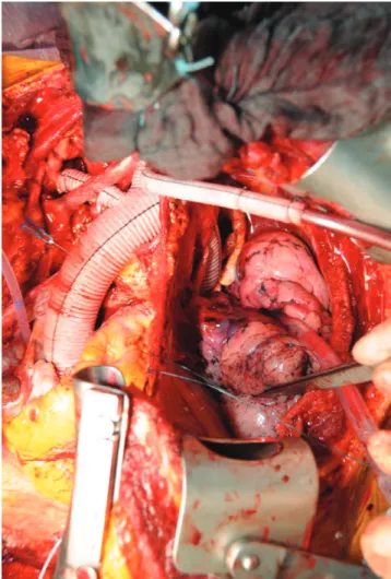
According to the individual situation, cardiac arrest is initiated. Additional cardiac procedures are then performed. Figure1depicts an overview of the completed repair. Figure2depicts the tech-nique of selective reimplantation of the supra-aortic branches used mainly in patients with connective tissue disease or very friable aortic arch tissue. Rewarming is performed until a target core temperature of 35° is reached. We do not routinely use cere-brospinal fluid drainage in these cases as the remaining risk of symptomatic spinal cord injury is low. We routinely perform MR angiography to identify important suppliers to the spinal cord. However, in this series, no selective reimplantation was warranted as leading suppliers were identified in downstream segments not involved in the repair. In post-dissection cases, a tounge-shaped excision of the distal dissection membrane is performed.
After weaning from cardiopulmonary bypass, reversal of heparin with protamine ratio 1 : 1 (1 mg protamine per 100 IU heparin) is performed. Intraoperative autologous transfusion using a cell-saver device is used in all patients. Intraoperative and post-operative transfusion thresholds are guided by in-hospital stan-dards supplemented by rotational thrombelastometry (ROTEM,
Figure 1:Overview of surgical access and completed total thoracic aortic repair.
A O RT IC S U RGE R Y
Pentapharm GmbH, Munich, Germany) and analysis of selected parameters of coagulation.
RESULTS
Chronic health conditions, risk factors and previous
surgical approaches
Patient demographics are summarized in Table 1. Seven of 10 patients had already undergone proximal thoracic aortic surgery (Table1).
Indications
The most common indication was post-dissection aneurysm for-mation in a remaining chronic Type B aortic dissection following repair of a previous Type A aortic dissection. Three patients had true aneurysms involving several thoracic aortic segments. Two patients had anastomotic aneurysms—one of them with contained rupture into the left upper lobe after previous coarctation repair. The last patient had received prior aortic valve replacement and suffered from Type B aortic dissection resulting in pseudo-coarctation with medically refractory arterial hypertension in the upper extremities (Table2). Figure3A shows the morphology of a
typical patient undergoing this approach. Figure3B and C shows a sagittal CT scan before surgery and a sagittal CT scan after surgery, respectively.
Table 1: Descriptive characteristics of the cohort (N = 10) Demographics
Age, median (±SD) 58 (±16)
Female,n 2
Chronic health conditions and risk factors
Hypertension,n 10
Chronic obstructive pulmonary disease,n 0
Diabetes mellitus,n 0
Serum creatinine >200 mmol/l,n 0
Coronary artery disease,n 3
Extracardiac arteriopathy,n 2
Logistic EuroSCORE, median (±SD) 14.6 (±12)
Redo surgery,n 7
Previous surgical approach (thoracic aorta)
Root/asc/hemiarch,n 3 including CABG 1 including TEVAR 1 Ascending/hemiarch,n 1 Coarctational repair,n 2 AVR 1
Classification of chronic health conditions and risk factors according to EuroSCORE criteria.
root/asc/hemiarch: aortic root, ascending and hemiarch replacement; CABG: coronary artery bypass grafting; TEVAR: thoracic endovascular aortic repair; Ascending/hemiarch: ascending aortic and hemiarch replacement; AVR: aortic valve replacement.
Table 2: Surgical characteristics of the cohort (N = 10) Surgical indications
Post-dissection aneurysm,n 4
Non-dissection aneurysm,n 3
Anastomotic aneurysm,n 2
Type B dissection with coarctation,n 1
Double arterial cannulation sites and extent of aortic replacement
Ascending (prosthetic) and descending,n 5
Ascending (prosthetic) and inguinal,n 3
Subclavian and descending,n 1
Subclavian and inguinal,n 1
Asc/arch/ 1/3 descending,n 4
Asc/arch/ 2/3 descending,n 4
Asc/arch/half descending,n 2
Additional cardiac and vascular procedures
CABG,n 3
Aortic valve replacement,n 2
Composite graft,n 1
LSA aneurysm repair/pulmonary embolectomy,n 1
Subclavian/carotid transposition,n 1
ECC times
ECC in minutes, median (±SD) 220 (±52)
Myocardial ischaemia in minutes, median (±SD) 107 (±38)
SACP, min (±SD) 30 (±23)
Asc: ascending aorta; arch: aortic arch; 1/3 descending: proximal third of the descending aorta; 2/3 descending: proximal two-thirds of the descending aorta; half descending: proximal half of the descending aorta; CABG: coronary artery bypass grafting; LSA: left subclavian artery; ECC: extracorporeal circulation; SACP: selective antegrade cerebral perfusion.
Double arterial cannulation sites
A combination of direct cannulation of the ascending aorta (or a previously inserted ascending aortic graft) and the distal descend-ing aorta was performed in the majority of patients, followed by the combination of cannulation of the ascending aorta (or a previ-ously inserted ascending aortic graft) and the left external iliac artery. The combination for double proximal and distal arterial perfusion used in the case in Figure3A–C is shown in Fig.3D.
Extent of repair
Four of 10 patients underwent replacement of the ascending aorta, the aortic arch and the proximal third of the descending aorta, whereas another 4 had the procedure extended to the
proximal two-thirds of the descending aorta. Two patients under-went a distal repair extending to the proximal half of the descend-ing aorta (Table2).
Additional cardiac and vascular procedures
Concomittant coronary artery bypass grafting was performed in 3 patients. Other additional cardiac procedures performed are pre-sented in Table3. Concomittant right-sided intrathoracic subclavian-to-carotid artery transposition was performed in 1 patient who had a Lusorian artery with its offspring from a distal aortic arch an-eurysm and 1 patient underwent left subclavian artery anan-eurysm repair as well as left-sided pulmonary embolectomy (because of subacute pulmonary embolism).
Figure 3:(A) Aneurysm on the basis of a remaining Type B aortic dissection after Type A repair and failed TEVAR. (B) Preoperative CT scan of the same patient as in
Fig.3A. (C) Postoperative CT scan of the same patient as in Figures 3A,B. (D) Scheme, depicting upper and lower body arterial access sites in the same patient using the double arterial perfusion concept.
A O RT IC S U RGE R Y
Outcome and follow-up
There was no in-hospital mortality, and perioperative neurological injury occurred in 2 patients who developed a delayed stroke occur-ing after an initial uneventful clinical course. One of them resolved completely during follow-up. One patient sustained a left recurring laryngeal nerve palsy. The median follow-up was 7 (±13) months. There was no death and no need for redo surgery in this period.
COMMENT
Extensive thoracic aortic surgery using a double arterial perfusion technique to avoid lower body hypothermic circulatory arrest is an attractive option for patients presenting with aortic arch and descending aortic aneurysms. Further experience and refinement of this technique may enable safe and effective simultaneous mul-tisegmental treatment of thoracic aortic pathology in patients who would otherwise have to undergo a two-step surgical approach.
The majority of patients in this small series had previous prox-imal thoracic aortic surgery [9]. Due to improved follow-up pro-grammes in many institutions, late pathologies in different segments are being increasingly observed. Two main groups can be differen-tiated: thefirst consists of patients who have undergone surgery for Type A repair and later develop aneurysmal formation in the remaining chronic Type B dissection. The second group is con-stituted of patients after previous coarctation repair. This is an interesting pathology since a large number of patients received coarctation repair in the last decades and some of them may develop late aneurysmal formation (especially after patch plasty). It is not sure that these patients are followed adequately.
The correct application of this technique requires a different view of the potential indications. We are convinced that this ap-proach is most suitable for any multisegmental thoracic aortic pathology extending clearly beyond the offspring of the left sub-clavian artery, endingfinally at the level of the thoracoabdominal transition. The most frequent indication for surgery was aneurysm in a chronic Type B dissection after Type A repair. A substantial number of these patients may exhibit the maximal diameter of the aneurysm in the distal aortic arch, while aortic dimensions become regular before the thoracoabdominal transition. For these patients, median sternotomy combined with left hemi-clamshell incision represents an ideal approach to access the entire thoracic aorta from the aortic root to the thoracoabdominal transition [10]. The alternative to this approach is a two-step surgical approach consisiting of proximal thoracic repair including the aortic arch
combined with a conventional or frozen elephant trunk (FET) im-plantation with secondary surgical (or in suitable cases thoracic endovascular aortic repair, TEVAR) repair via a lateral thoracotomy approach [11–13]. In the present series, the majority of the patients would not have been suitable for any combined vascular or endo-vascular procedure [14].
Using this double arterial cannulation technique, true lower body hypothermic circulatory arrest is definitely not necessary. In simple situations in which the duration of arch repair is limited because only a hemiarch or a proximal descending anastomosis has to be performed, this strategy may be critized as overtreat-ment. However, in complex morphologies, this concept may prove to be very advantageous, because there is continuous blood supply to all affected organ systems. Certain similarities on the one hand, but clear differences on the other, compared with the arch first technique should be mentioned. Regarding access, the archfirst technique uses a bilateral hemisclamshell approach with a trans-verse sternotomy, whereas we use a median sternotomy with a single-sided hemi-clamshell incision. We perform the descending anastomosis and not the arch anastomosisfirst. Thereby our myo-cardial ischaemic time is one-third shorter compared with the arch first approach. Finally, with our approach, lower body ischaemia is completely avoided [10,11]. As such, it might be argued that our technique primarily aims at avoiding lower body circulatory arrest.
Furthermore, FET implantation might be considered in some of these cases. However, it remains questionable if a suitable circumfer-ential layer of native tissue would have been available tofix the FET. We are convinced that especially in aneurysms on the basis of a post-dissection process—either remaining Type B after Type A repair of chronic Type B should form the exception and not the rule for FET approaches as it will be a very rare event that complete exclu-sion of the false lumen will be achieved due to the lack of a defined distal landing zone. Open surgery effectively treats the entire thor-acic aortic disease, whereas an FET will rather be a trade-off.
The hemi-clamshell extension allows perfect exposure of the entire thoracic aorta [10]. Nevertheless, some issues are important regarding nerval anatomy. It is of utmost importance to preserve the phrenic as well as the vagus and recurring laryngeal nerve. Injury to these structures may substantially prolong the post-operative course with regard to respiratory issues. Interestingly, we have not observed pulmonary complications such as pneumonia requiring prolonged intubation or any other major pulmonary complication in this series. A reason for that may be that manipu-lation on the left lung is minimal and is limited to the distal anasto-mosis on the descending aorta.
The percentage of additional cardiac and vascular procedures was fairly high and reflects the issue that aortic lesions are frequent-ly accompanied by valvular degeneration as well as by anatomical abnormalities andfinally, aneurysmal formation of branch vessels.
The results presented in this small series are very satisfying with regard to survival and other major clinical endpoints. They illus-trate that the most vulnerable organ in aortic arch surgery remains the brain, and that complications such as prolonged periods of hypotension are poorly tolerated. Future strategies have to em-phasize a better understanding and consequenttly an improve-ment of neuroprotective strategies.
Limitations and strengths
The number of patients is low and indications may have been heterogenous. However, this brief report is the proof of concept Table 3: Outcome characteristics of the cohort (N = 10)
Early in-hospital complications
In-hospital mortality,n 0
Acute renal failure,n 1
Acute myocardial infarction,n 1
Neurological injury,n 2
Late outcome
Follow-up in months, median (±SD) 7 (±13)
Late death,n 0
Redo surgery,n 0
Classification of complications according to society of thoracic surgeons criteria.
that a double arterial cannulation strategy is a very effective perfu-sional approach for patients suffering from complex multisegmen-tal thoracic aortic pathologies. Furthermore, this approach is against the mainstream of what is currently followed by a large number of surgeons. We feel that it remains important to report the advances in conventional surgical approaches that take longer, definitely are more quiet but are finally highly likely to be more durable, particularly in highly complex patients such as those pre-sented here.
Summarizing, extensive proximal thoracic aortic surgery using a double arterial perfusion technique in order to avoid lower body hypothermic circulatory arrest is an attractive conceptual option. Further refinements of this technique will enable a safe and effect-ive simultaneous multisegmental treatment of thoracic aortic pathology in patients who would otherwise have to undergo a two-step surgical approach.
Conflict of interest: none declared.
REFERENCES
[1] Krähenbühl E, Maksimovic S, Sodeck G, Reineke D, Schoenhoff F, Schmidli
Jet al. What makes the difference between the natural course of a
remain-ing type B dissection after type A repair and a primary type B aortic dissec-tion? Eur J Cardiothorac Surg 2012;41:e110–5.
[2] Czerny M, Barchichat I, Meszaros K, Sodeck GH, Reineke D, Englberger L et al. Long-term results after proximal thoracic aortic redo surgery. PLoS One 2013;8:e57713.
[3] Ius F, Hagl C, Haverich A, Pichlmaier M. Elephant trunk procedure 27 years after Borst: what remains and what is new? Eur J Cardiothorac Surg 2011; 40:1–11.
[4] Estrera AL, Miller CC III, Porat EE, Huynh TT, Winnerkvist A, Safi HJ. Staged
repair of extensive aortic aneurysms. Ann Thorac Surg 2002;74:S1803–5.
[5] Czerny M, Krähenbühl E, Reineke D, Sodeck G, Englberger L, Weber A et al. Mortality and neurologic injury after surgical repair with hypothermic circulatory arrest in acute and chronic proximal thoracic aortic pathology: effect of age on outcome. Circulation 2011;124:1407–13.
[6] Pacini D, Di Marco L, Leone A, Di Bartolomeo R, Sodeck G, Englberger L et al. Antegrade selective cerebral perfusion and moderate hypothermia in aortic arch surgery: clinical outcomes in elderly patients. Eur J Cardiothorac Surg 2012;42:249–53.
[7] Roques F, Nashef SA, Michel P, Gauducheau E, de Vincentiis C, Baudet E et al. Risk factors and outcome in European cardiac surgery: analysis of the EuroSCORE multinational database of 19030 patients. Eur J Cardiothorac Surg 1999;15:816–22.
[8] Harrer M, Waldenberger FR, Weiss G, Folkmann S, Gorlitzer M, Moidl R et al. Aortic arch surgery using bilateral antegrade selective cerebral perfu-sion in combination with near-infrared spectroscopy. Eur J Cardiothorac
Surg 2010;38:561–7.
[9] Czerny M, Bachet J, Bavaria J, Bonser RS, Borger MA, De Paulis Ret al. The
future of aortic surgery in Europe. Eur J Cardiothorac Surg 2013;43:226–30.
[10] Kouchoukos NT, Masetti P, Mauney MC, Murphy MC, Castner CF.
One-stage repair of extensive chronic aortic dissection using the arch-first
technique and bilateral anterior thoracotomy. Ann Thorac Surg 2008;86:
1502–9.
[11] Kouchoukos NT. One-stage repair of extensive thoracic aortic disease. J Thorac Cardiovasc Surg 2010;140:S150–3.
[12] Pacini D, Tsagakis K, Jakob H, Mestres CA, Armaro A, Weiss Get al.
The frozen elephant trunk for the treatment of chronic dissection of the thoracic aorta: a multicenter experience. Ann Thorac Surg 2011;92: 1663–70.
[13] Shrestha M, Pichlmaier M, Martens A, Hagl C, Khaladj N, Haverich A. Total aortic arch replacement with a novel four-branched frozen elephant trunk
graft:first-in-man results. Eur J Cardiothorac Surg 2013;43:406–10.
[14] Czerny M, Roedler S, Fakhimi S, Sodeck G, Funovics M, Dumfarth Jet al.
Midterm results of thoracic endovascular aortic repair in patients with aneurysms involving the descending aorta originating from chronic type B dissections. Ann Thorac Surg 2010;90:90–4.
European Journal of Cardio-Thoracic Surgery 45 (2014) 465–466
EDITORIAL COMMENT
doi:10.1093/ejcts/ezt451 Advance Access publication 23 September 2013
Re: Double arterial perfusion strategy for extensive thoracic aortic
surgery to avoid lower body hypothermic circulatory arrest
Nicholas T. Kouchoukos*
Missouri Baptist Medical Center, St. Louis, MO, USA* Corresponding author. 3009 N. Ballas Rd, Suite 360 C, St. Louis, MO 63131, USA. Tel: +1-314-9965287; fax: +1-314-4326068; e-mail: ntkouch@aol.com (N.T. Kouchoukos)
Keywords:Double arterial perfusion• Extensive thoracic aortic surgery • Lower body perfusion
Czerny and his co-workers report a technique of double arterial perfusion to avoid lower body hypothermic circulatory arrest in 10 patients undergoing extensive thoracic aortic repairs [1]. A median sternotomy combined with a left fourth intercostal space incision was utilized to provide exposure of the entire thoracic aorta. There was no hospital mortality, 2 patients (20%) sustained a stroke, and one each developed acute renal failure and myocardial infarction.
Their technique represents another modification of the one-stage approach to treat extensive thoracic aortic aneurysmal disease, some of which develops after previous procedures on the thoracic aorta such as repair of acute type A dissection. The authors employed a double arterial perfusion strategy and double-clamping of the descending thoracic aorta to avoid lower body hypothermic circulatory arrest, and selectively perfused the brachiocephalic vessels with a separate roller pump at a
A O RT IC S U RGE R Y