Review
Regulating cellular oxygen sensing by hydroxylation
Joachim Fandrey
a,⁎
, Thomas A. Gorr
b, Max Gassmann
b aInstitut für Physiologie, Universität Duisburg-Essen, Hufelandstrasse 55, D-45122 Essen, Germany b
Institute of Veterinary Physiology, Vetsuisse Faculty and Zurich Center for Integrative Human Physiology (ZIHP), University of Zurich, Winterthurerstrasse 260, 8057 Zurich, Switzerland
Received 17 February 2006; received in revised form 19 April 2006; accepted 3 May 2006 Available online 10 May 2006
Time for primary review 28 days
Abstract
Oxygen homeostasis under conditions of limited O2supply requires hypoxia-dependent gene regulation. The transcription factor complex
hypoxia-inducible factor 1 (HIF-1) has been recognized as the master regulator that mediates the adaptational genetic response to ensure restoration of energy supply. This review will focus on the recent advances in understanding the hypoxia-induced cellular response with particular respect to cellular O2sensing for adequate control of HIF-1 activation.
© 2006 European Society of Cardiology. Published by Elsevier B.V. All rights reserved. Keywords: HIF-1; Hypoxia; Hypoxia-induced gene expression
1. O2sensing via HIFα factors
Crucial to linking reduced oxygen supply to changes in gene expression are hypoxia-inducible transcription factors, or HIFs [1]. Across the animal kingdom, HIF is a heterodimer ofα- and β subunits. Both subunits belong to the family of basic-helix-loop-helix (bHLH)/PAS1 transcrip-tion factors. Under physiological oxygen partial pressures (pO2), HIF is quickly destabilized. In contrast, α and β
subunits heterodimerize in the nucleus in response to oxygen deprivation. Here, the dimer specifically binds to target gene motifs called hypoxia response elements (HREs) to either induce or suppress gene expression. A decade after the seminal identification of HIF-1 as the responsible factor for the hypoxia-induced transcription of the red cell producing hormone erythropoietin[2], this transcription factor has now been implicated to possibly control even up to several hundreds (i.e. ∼2–5% of genome) of hypoxia-responsive
genes in humans [3]. In mammals, the 70 HIF targets validated to date[4]typically fall into two main categories whose functions aim to restore energy and O2homeostasis
by either cell-autonomous or systemic means:
(a) increasing anaerobic energy production via stimulated glycolytic substrate flux;
(b) improving tissue oxygenation via stimulated angio-genesis, vasodilation and erythropoiesis.
HIF signaling – although not the only pathway linking declining pO2with changes in DNA activity (see Cummins
and Taylor [5] for a recent review on HIF-independent transcription responses to hypoxia) – has thus been appreciated as a major component in the homeostatic adaptations and survival of cells under both physiological and tumorigenic-pathological oxygen limitations [6]. Not-withstanding this key importance for hypoxic survival, the mechanisms and players behind this switch from active (hypoxia) to inactive (normoxia, reoxygenation) states of HIF signaling were only poorly understood until recently. Fortunately, there has been a remarkable progress made during the last 5 years, advancing our comprehension of
Cardiovascular Research 71 (2006) 642–651
www.elsevier.com/locate/cardiores
⁎ Corresponding author. Tel./fax: +49 201 723 4600/4648. E-mail address:[email protected](J. Fandrey).
1PAS: acronym for PER, ARNT, SIM, the first proteins discovered to contain this domain.
0008-6363/$ - see front matter © 2006 European Society of Cardiology. Published by Elsevier B.V. All rights reserved. doi:10.1016/j.cardiores.2006.05.005
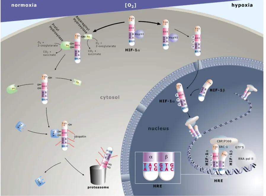
Fig. 1. HIF-α mediated pathway of O2sensing. Molecules are not drawn to scale. Prolyl hydroxylase in the figure represents PHD1–3. For further details see text. 643 ey et al. / Car diovascular Resear ch 71 (2006) 642 – 651
cellular oxygen sensing onto an entirely new level. To record this recent progress, with a particular focus on the hydroxylase-mediated regulation of the HIF signaling pathway, is the goal of this current review.
From nematodes to crustaceans to flies to vertebrates, HIF activity is controlled at the protein level through oxidative modifications of the α subunits [7]. Multiple isoforms of these regulatory HIF proteins exist in fish, amphibians, birds and mammals[8]. Three HIF-α subunits (HIF-1α, -2α and -3α) have been reported in human and rodent cells, with five and six splice variant transcripts known to occur for the human 1α and 3α isoforms, respectively[9,10]. All threeα subunit factors share the ability to heterodimerize with the ubiquitously expressed HIF-1β subunit, originally known as the aryl hydrocarbon receptor nuclear translocator or ARNT, thus producing HIF-1, -2 and -3. While both stability and activity of the 1α, 2α and some 3α (i.e. 3α1–3α3) subunits
are quickly impaired by normoxia or reoxygenation, the steady state levels of the ARNT protein are unaffected by changes in oxygen tension[11]. As outlined in detail below, and summarized in Fig. 1, the HIF oxygen sensors, that control the abundance of HIF-α proteins in the cell are a family of novel prolyl hydroxylases named PHD1-3, for the characteristic prolyl hydroxylase domains contained within the reading frame[12–14]. In the presence of oxygen, these PHDs catalyze the Fe(II)-dependent hydroxylation of specific prolyl residues (Pro402 and Pro564 in human HIF-1α) contained within the oxygen-dependent degradation domain (ODD) of HIF-1α[15], -2α or -3α1–3subunits (see
Fig. 1: ODD hydroxy-prolines as“OH”). Once hydroxylat-ed, the HIF-α subunits rapidly bind to the von Hippel-Lindau (pVHL) tumor suppressor protein that is the recognition component of an E3 ubiquitin ligase complex, thereby tying prolyl hydroxylation to ubiquitination and proteasomal degradation of these regulatory constituents under high or rising pO2(e.g.[16,17]; seeFig. 1). In addition, acetylation
of ODD-contained lysines (e.g. K532in human HIF-1α) via acetyltransferases such as ARD1 might be another, though currently far more controversially discussed, enzymatic trigger for enhanced pVHL recognition and destabilization of alpha subunits [18–20]. Unlike PHDs, the activity of acetyltransferases is not known to be influenced by changes in oxygen tension, thus making it hard to see how normoxia should lead to differentially high rates of protein substrate acetylations (for review see [21,22]). Notwithstanding our incomplete mechanistic understanding, the efficacy of these hydroxylation- (and potentially acetylation-) based post-translational controls can best be appreciated by the < 5 min half-life of HIF-1α upon reoxygenation[11]and, conversely, the instantaneous accumulation of the transcription factor in vitro[23]and in vivo[24]during declining oxygen tensions. A second O2-requiring hydroxylation, that of a single
asparagine (Asn803in human HIF-1α) within the C-terminal transactivation domain of HIF-1α and -2α (seeFig. 1: TAD-C, hydroxyl-asparagine as “OH”) by an asparaginyl hydroxylase dubbed FIH-1 (factor inhibiting HIF-1), leads
to steric hindrance of the interaction betweenα subunits and the coactivator proteins p300/CBP (note: lack of TAD-C domains in 3α isoforms). This hindrance prohibits the transactivation of target genes under high pO2[25,26]. The
absolute requirement of PHD- and FIH-1 activities for oxygen and Fe2+ [27–29] has made these hydroxylation reactions susceptible to inhibition not only by declining pO2
but also by hypoxia-mimicking agents such as transition metals (e.g., Co2+) and iron chelators (e.g., desferrioxamine, DFO; ciclopiroxolamine (CPX) [12,30]). Long before the discovery of PHD-mediated O2 sensing, Co2+, DFO and
CPX were empirically known to be able to induce erythropoietin, i.e. HIF target, gene responses under normoxia [31,32]. As a strength of concept, insights into the catalytic requirements of PHDs can now incorporate these earlier data into a plausible model of HIF control. Upon exposure to hypoxia, cobalt or iron chelators, hydroxylation of prolyl and asparaginyl residues is reduced (see Fig. 1). This inhibition enables theα subunit to escape proteolytic degradation and, assisted by various interacting factors (e.g. heat shock protein 90, HSP 90)[4,33] and/or stimulatory kinase signals (reviewed in[6]), to efficiently translocate into the nucleus where the phosphorylated form (P) dimerizes with ARNT (reviewed in [22]) through intermolecular interactions between the HLH and PAS domains (see Fig. 1). Next, theα:β heterodimer associates with the transcrip-tional coactivators p300/CBP and Src-1 to regulate target gene transcription via HRE binding sites. Specific recogni-tion of HRE motifs is accomplished through the N-terminal basic (b) region in both factors (see Fig. 1: 5′TACGTG3′ given as HRE example; note:α subunit occupancy of 5′ TAC nucleotides, andβ subunit binding to 3′ GTG nucleotides). The HIF/coactivator complex is now endowed to modulate the activity of the general transcription factor (GTF)/RNA polymerase II machinery in a hypoxia-responsive fashion. 2. Regulation of HIFα by hydroxylation
As mentioned above, both the regulation of O2-dependent
lability and thus protein abundance as well as transcriptional activity of HIF-α subunits depend on hydroxylation. So far, PHD1, PHD2, PHD3 and FIH-1 have been identified to perform this post-translational modification. They now appear as today's best candidates for cellular HIF oxygen sensors. A fourth prolyl hydroxylase termed PH-4 has been isolated from a database approach [34]. Although this protein showed great similarity to the known PHDs and was able to suppress HIF-accumulation and HIF-dependent reporter gene activation, it failed to show activity in an HIF-1α-pVHL in vitro interaction assay. Therefore the role as an HIF prolyl hydroxylase, particularly with O2dependent
activity, is currently not clear.
Initially, the search for HIF-associated O2sensors focused
on heme proteins that would– in response to altered states in oxygenation– change their heme conformation and valency and thereby generate a signal to control O2-dependent gene 644 J. Fandrey et al. / Cardiovascular Research 71 (2006) 642–651
expression [1,31]. However, the post-translational hydrox-ylation of HIF-α implied O2-dependent enzymatic activity.
Subsequently, it was demonstrated that modification of proline residues in the ODD of HIF-α controls abundance while asparagine hydroxylation is the important switch for regulating the binding of coactivator p300/CBP [17,35]. Conservation of the O2-sensing system down to C. elegans
helped to define a new family of dioxygenases that regulate HIF-α by prolyl hydroxylation [12,13,36]. Although the human homologues were variously named HPH-1/EGLN3/ PHD3, HPH-2/EGLN1/PHD2, and HPH-3/EGLN2/PHD1 [37]we will use the initial term PHD1, -2, and -3. Shortly thereafter the enzyme hydroxylating the TAD-C previously termed FIH-1 was identified as an asparaginyl hydroxylase that fell into the same family of dioxygenases[26].
By definition, dioxygenases metabolize molecular oxy-gen and incorporate both atoms of oxyoxy-gen into their products. PHDs and FIH-1 are Fe2+, 2-oxoglutarate (2OG) and ascorbate dependent non-heme binding oxygenases of which the enzymatic activity is regulated by O2availability
(see Fig. 1). With respect to cellular localization and O2
regulated enzymatic activity they differ from the well-known collagen prolyl hydroxylase (C-P4H1) whoseα(I) subunit is a HIF-1 target gene[38]. Like PHDs and FIH-1, the C-P4H1 releases CO2 and succinate upon completion of the
enzymatic cycle and its activity depends on substrate and ascorbate concentration as it is well known from scurvy. However, the most significant difference related to their function was found when the affinity for O2was determined
for the PHDs and FIH-1 and compared to C-4PH1. While the Km value for O2of the C-P4H1 was around 40μM, values
initially reported for the PHDs were between 230 and 250μM which exceeds even the concentration of O2in air
[39]. For FIH-1, the Km was found to be considerably lower, 90μM[40], but still more than twice as high as that of C-P4H1 (40 μM; [41]). These Km values were determined using the 14CO2 release from labeled 2OG catalyzed by
recombinant enzymes and short 19 amino acid (aa) HIF-α peptides as substrates. A caveat with respect to these Km measurements has been expressed since interpretation of the O2affinity would need to consider the reaction kinetics of
the enzymes[42]. In general, information on the kinetics of prolyl hydroxylases is mainly based on data from C4-PH1, but not from PHDs. In C4-PH1, O2appears to be bound first
to the enzymatic complex. Subsequently, the enzyme–O2
complex is stabilized by binding pro-collagen polypeptide substrate [43]. More recently, this order of binding was questioned. Instead, it was proposed that a preformed enzyme–substrate complex binds O2[44], which would be
in line with crystallography data on FIH-1, where such a preformed enzyme–Fe2+–2OG–substrate complex was de-scribed to bind O2 [45]. This order of binding implies,
however, that substrate bound to the enzyme could affect the Km for O2. Indeed, when the length of HIF-α peptides used
for in vitro assays was extended to 200 aa the Km value for O2of the PHDs dropped to 100 μM (Johanna Myllyharju,
personal communication, 2006). Whether full length HIF-α proteins would lower the Km even further is still a matter of debate. Full length HIF-α proteins are almost insoluble and have not been used in the in vitro enzymatic measurements. Even a Km of 100μM corresponds with the maximal O2
concentrations in the body, the alveolar pO2 in the lung.
Oxygen tensions in the tissue, particularly in the kidney where HIF signaling is required for erythropoietin expres-sion[46], or in the placenta where HIF is required for regular development [47], are certainly lower than any of the Km values determined so far. Measurement of tissue pO2under
physiological conditions has been notoriously difficult. Most of the technical pO2 sensors, e.g. of Clark-type, consume
oxygen and thus blur measurements of pO2. In addition,
tissue pO2 is determined by the ratio of the rate of
consumption to the rate of supply which may vary even within tissue areas very close to each other[48]. However, a range of oxygen concentration from 40 down to 4μM may reasonably be assumed. As such, the high Km for oxygen would place the steep slope range of the activity curve determined for PHDs and FIH-1 right into the range of physiological pO2values. Any incremental reduction in the
pO2would thus trigger a marked lowering of hydroxylation
capacity [42]. This was demonstrated already in the first description of PHDs when a decrease in HIF hydroxylation between pO2 values of 150 mm Hg and 75 mm Hg was
found[12]. Interestingly, the Km of FIH-1 is lower than for PHDs and indeed, experiments indicate that FIH-1 control dominates HIF activation during exposures to a lower pO2
range [49]. The emerging picture of graded hydroxylase activities that operate with maximal sensitivity in the range of physiological tissue pO2 thus fits initial in vitro
observations, where half-maximal activation of HIF signal-ing was reported at oxygen concentrations of 15 to 20μM [50]. Detection of nuclear HIF-1α in cells, under conditions where the pericellular pO2 was measured and oxygen
consumption was taken into account [51], indicates that HIF accumulation may be achieved at higher pO2values but
that, in addition, cell-specific differences may exist [52]. Collectively, however, there is no doubt that the absolute requirement for O2causes lack of enzymatic activity under
anoxia. Upon a graded decrease of the pO2 within the
physiological range overall hydroxylation capacity of PHDs and FIH-1 will become limiting and thus the overall process of hydroxylation will be rate-limiting in the system that regulates HIF abundance and activity. As such, the enzymes fulfill the criteria of oxygen sensors. In addition, the pO2
range in which the oxygen sensors work may be affected by abundance of the enzymes themselves [42]. This would, in fact, explain different oxygen sensitivity for cells in culture under identical physical conditions[52].
3. Hydroxylase abundance and interacting proteins Prolyl- and asparaginyl-hydroxylases are non-equilibrium enzymes because they only catalyze the hydroxylation of
their substrate but not the reverse reaction. Thus the abundance of HIF-hydroxylases will determine HIF-α accumulation and activity at distinct pO2's. Expression of
the PHD iso-enzymes varies between tissues but no specific role for this difference has been determined in vivo [53]. PHD1 is highly expressed in the testis whereas PHD3 mRNA is abundant in the heart. A recent study using newly developed monoclonal antibodies determined protein distri-bution throughout several organs and revealed tissue specific distribution and also differences in staining intensity between normal and malignant tissue[54]. Knock-out mice for PHD1 to 3 have been generated and are currently characterized with respect to their phenotype. Homozygous PHD2−/− mice are not viable, but PHD1−/− and PHD3−/− are. An interesting phenotype was reported for PHD1−/− mice which showed a significant protection from hypoxia induced muscle damage in a hind limb ischemia model (P. Carmeliet, Keystone Symposium January 2006, Brecken-ridge, CO).
A strong stimulus that regulates PHD2 and PHD3 abundance is hypoxia itself. Initially it was observed that the half-life of HIF-1α was significantly shortened after prolonged or severe hypoxic periods[55]. It was found that the pVHL/HIF pathway regulated hypoxic inducibility of PHD2 and 3[56]and recently functional HREs were isolated and characterized in the promoter of the PHD2 gene[57]as well as of the PHD3 gene [58]. In contrast, PHD1 is not induced by hypoxia, potentially even reduced in a particular subset of cells[59]. Several negative regulatory elements in the PHD1 promoter, and a possible role for ARNT in this hypoxic down-regulation, have been reported [60]. Like-wise, levels of FIH-1 are not affected by hypoxia [61]. Hypoxic induction of PHD2 and 3 therefore forms a negative feedback loop whose function is not yet entirely clear. It has recently been suggested that higher levels of hydroxylase expression may extend the regulatory range of the oxygen sensors to more severe hypoxia [42]. On the other hand, induction, notably of PHD3, appears to be of particular importance for the reoxygenation phase when higher PHD3 levels may increase the dynamics of the hypoxic response by shortening the half-life of HIF-1α protein. Very recently, two alternatively processed PHD3 transcripts, designated PHD3Δ1 and PHD3Δ4 were identified[62]. The expression of both PHD3 and PHD3Δ1 was observed in all tissues and cell lines tested, although the expression of the novel PHD3Δ4 appeared to be restricted to primary cancer tissues. The function of PHD3Δ4 was assessed in transfection experiments showing a preserved prolyl hydroxylase activity although the impact on O2 sensing remains to be fully
elucidated.
Another open question is whether the localization of PHDs is affected after hypoxic induction. PHD enzymes show a distinct intracellular distribution pattern with PHD1 mostly, if not exclusively, localized in the nucleus whereas PHD2 predominantly resides in the cytoplasm and PHD3 is found in both cellular compartments [61]. A recent study
using novel antibodies more or less confirmed the localiza-tion of PHD2 and 3 while PHD1 was also found in the cytoplasm[54]. This may indicate that cell-specific differ-ences exist or that particular circumstances affect the translocation of the oxygen sensors. It has for example been described that PHD1 is estrogen-dependent although no effects on subcellular localization were studied so far[63]. Very recent work, however, provides evidence that cellular localization is a dynamic process, particularly that PHD2 may also be found in the nucleus upon induction [64]. Irrespective of the cellular localization and the tissue distribution, the current consensus is that PHD2 is the main enzyme that regulates steady state levels of HIF-1α. This is indicated by the fact that knock-down of PHD2 by siRNA is sufficient to increase HIF-1α levels under normoxic condition [65]. In addition, HIF target genes were up-regulated indicating that inhibition of PHD2 alone is sufficient for HIF-1 target gene activation although FIH-1 activity was not reduced. This notion has further been tested in a study where the relative importance of PHD2 at different levels of hypoxia was found to be most important for normoxic and moderately hypoxic situations whereas FIH-1 appears to regulate HIF activity at more severe hypoxia[49]. This fits in fact the lower Km value for oxygen of FIH-1 as mentioned above.
Most probably, however, regulation of PHDs comprises several more levels of complexity. PHD activity is inhibited when intracellular calcium is chelated although no obvious calcium binding site has been identified [66]. This could indicate that other calcium dependent cofactors are required for full PHD activity. The ring finger E3-ubiquitin ligases Siah1a/2 were recognized to co-immunoprecipitate with PHD1 and 3 [67]. Siah1a/2 appear to particularly affect PHD3 levels by increased proteolysis. Siah1a/2-deficient cells showed higher levels of PHD3 and a loss of hypoxic HIF-1α induction which could be reversed by siRNA against PHD3. Interestingly, Siah proteins themselves are hypoxia-inducible suggesting that, under hypoxic conditions, PHD3 proteolysis will increase. On the other hand, PHD3 is HIF-1α target gene and PHD3 expression is induced in hypoxia [58,61]. Thus hypoxia increases expression and degradation of PHD3 which in turn controls HIF-1α levels. How this intricate regulation and counter-regulation affects physio-logical oxygen sensing is unclear at present although Siah2−/− mice subjected to hypoxia displayed an impaired erythropoietic response which might indicate physiological relevance of this regulation[67].
When the initial biochemical purification of an HIF-1 prolyl hydroxylase revealed a 320–440 kDa complex that appeared much larger in mass than individual PHDs[68], it became obvious that other proteins associate with PHDs. In an attempt to characterize such proteins, the chaperonin complex TriC (aka CCT) was found to co-purify with PHD3. Whether PHD3 represents a folding substrate for TriC is currently not clear[69]. Interaction of PHD2 with OS-9, a widely expressed protein of yet unassigned function, appears
to increase PHD2 activity and thus to promote degradation of HIF-1α [70]. Knock-down of OS-9 by siRNA increased HIF-1α protein levels, HIF-1-mediated transcription, and expression of the HIF-1 target gene VEGF under non-hypoxic conditions. Interaction of the tumor suppressor protein ING4 with PHD2 surprisingly did not affect either PHD2 activity or HIF stability[71]. Instead, ING4 binding to PHD2 appears to interfere with transcriptional activation of HIF-1α either by interacting with the TAD-N domain within the ODD or through hindrance of the correct folding of the active HIF complex between the C-terminus and the DNA binding domains [71]. This field is now under active investigation, particularly with respect to other PHD targets [72]. In this regard, the groups of R. Wenger and D. Katschinski recently reported several yeast-two-hybrid based identifications of novel PHD1 (i.e. onconeural cerebellar degeneration-related protein 2, Cdr2), PHD2 (i.e. peptidyl-prolyl cis/trans isomerase FK506-binding protein 38, FKBP38) and PHD3 (i.e. activating transcription factor-4, ATF-4) interacting factors that might either represent PHD hydroxylation targets (Cdr2, ATF-4) or PHD activity modulators (FKBP38) (see Acta Physiol. 2006, vol. 186, suppl. 1, PM07P-4, PM08A-5, PM08A-7). Moreover, the novel WD-repeat protein Morg1 was identified to act as a molecular scaffold for PHD3 [73]. Morg1 was shown to support the specific activity of PHD3 and may even be important for maximal activity of the enzyme under certain conditions.
4. Effect of divalent metal ions on PHD activity
Classically it had been reported that cobalt chloride can mimic the anemic or hypoxic induction of erythropoietin production and in fact cobalt chloride was used as an erythropoietic agent[74]. Later, in vivo and in vitro studies revealed that this effect is due to cobalt chloride dependent induction of HIF-1[46]. Since nickel (Ni2+) and manganese (Mn2+) acted in a similar way and induced erythropoietin production it was initially proposed that those divalent transition metal ions would replace iron in the potential heme protein sensor, thereby locking this protein in its active deoxy conformation[31]. This concept has been perpetuated when the Fe2+-dependent PHD enzymes were identified as O2 sensors and is currently used to explain why divalent
metal ions increase HIF-1α protein, even though PHDs turned out to be non-heme binding dioxygenases (i.e. iron is directly coordinated with the protein). It has indeed been found that hypoxia mimetics like Co2+ and iron chelators inhibit PHD activity[12]. However, a competing hypothesis has been proposed, whereby cobalt and nickel deplete intracellular ascorbate, which is also required for full enzymatic activity (see below; [75]). Nevertheless, the absolute requirement of Fe2+ for HIF hydroxylase activity is beyond doubt. When recombinant PHD enzymes were recently purified and tested for their Km values it was found that iron was so tightly bound to these enzymes that Km
values for iron could only be estimated with lower than 0.1 μM [76]. For FIH-1, a Km for iron of 0.5 μM was measured, which still is considerably lower than the Km of 2μM for C-P4H1. Although the values for PHDs were only estimated, high affinity binding of the metal by PHDs is supported by the finding that, in vitro, the iron chelator desferrioxamine (DFO) and the divalent metal ions (Co2+, Ni2+, Mn2+) were more effective in inhibiting FIH-1 than PHD preparations (see Fig. 1). However, the obvious and important question whether cellular iron availability affects physiological hydroxylase function has not yet been fully addressed. One recent report found changes in iron distribution in response to NO treatment and may thus indicate that cellular iron distribution is in fact able to affect oxygen sensing[77]. Although the direct addition of iron to cultures reduced HIF-1α levels, it may be doubted whether this iron is in fact transferred to the PHD without prior oxidation and may thus affect other yet unknown ways of HIF-α accumulation[78].
5. Substrate specificity, ascorbate requirement and ROS Both in HIF-1α and HIF-2α the proline residues to be hydroxylated are in the context of an LXXLAP consensus sequence. This sequence is not recognized by C-P4H1, but by PHDs. Within this consensus are the two critical proline residues 402 and 564 in human HIF-1α which are both recognized by PHD1 and 2 [79], but not by PHD3. PHD3 will only hydroxylate proline 564. In addition, Leu574 in human HIF-1α was found to be essential for pVHL binding [80]and PHD2 recruitment to the Pro564 hydroxylation site [81]. High substrate specificity of PHDs to recognize these particular proline residues, therefore, results from the conserved presence of the LXXLAP consensus together with adjacent‘recruiter residues’. Consequently, the Km for 19 aa model peptides including the consensus sequence was as low as 7 to 8 μM [39]. Despite this high substrate specificity, in vitro mutagenesis studies unexpectedly revealed that prolyl hydroxylation is rather tolerant towards a range of substitutions in the vicinity of the target proline [79]. It has been reasoned that these effects may be due to high concentration of short peptides used for in vitro studies. Therefore X-ray structure studies of PHDs and HIF-α protein substrate are mostly needed. This was so far only achieved for FIH-1 bound to the TAD-C of HIF[45]. Here the higher stringency and particularly the requirement for valine 802 for hydroxylation activity became obvious when an extensive set of hydrogen bonds of the TAD-C was found to be formed with FIH-1 to bring the asparagine 803 close to the catalytic iron of the active site of the enzyme [45]. Moreover, structural changes have been observed between a state where the TAD-C is bound to FIH-1, which would cause hydroxylation and inhibition of activation, and an active state where p300 is bound to the TAD-C [82,83]. Collectively, recognition of the substrate, both by PHDs and FIH-1, appears to depend on multiple residues within the
surrounding protein which ensure highly specific substrate recognition.
Ascorbate has been proposed to protect prolyl hydroxylases from uncoupled reactions where no substrate is hydroxylated and reactive oxygen species (ROS) may be generated that could damage the enzymes[41]. In fact, ascorbate deficiency has been found to be limiting for PHD function in human tumor cells causing high HIF-1α levels[78]. Usually C-4PH1 enzymes catalyze the uncoupled decarboxylation of 2OG at a rate of less than 1% of the complete reaction [41]. When proline is hydroxylated, a number of cycles can be run without ascorbate, but then Fe3+ needs to be reduced for continued activity of the enzyme. In the case of uncoupled reactions, e.g. if no available substrate is recognized, ascorbate may act as an alternative O2acceptor to limit the
generation of reactive oxygen species. Under these condi-tions, ascorbate is consumed stoichiometrically. For other 2OG-dependent oxygenases the generation of ROS has been described and can inactivate the related oxygenases by self-oxidation which might cause fragmentation of the enzymes [84]. So far, uncoupling has not been observed for PHDs, but about 1% of the maximum activity of FIH-1 did not account for hydroxylation in the presence of an HIF substrate[40]. While a great percentage of uncoupled reactions may be harmful to the enzymes and potentially other targets in the cell as pointed out above, the precise mechanism is not fully elucidated. One particular open question is whether the HIFα substrate binds prior to or after oxygen has been bound to the enzyme. On the one hand, only substrate-bound protein would bind oxygen and thus substrate recognition would indeed reduce the potential for ROS generation. On the other side, oxygen affinity may well be modulated by substrate binding as pointed out above [37]. Another under-investi-gated issue concerns our limited understanding when comparing the regulation of HIF-hydroxylase activities between organisms capable of de novo ascorbate synthesis (e.g. rodents) and others for which vitamin C is an essential nutrient (e.g. humans, primates, guinea pigs).
With respect to ROS it is noteworthy that the model of cellular oxygen sensing by oxygen-dependent enzymatic activity of PHDs is in competition with the hypothesis that mitochondria and/or other cellular oxidases generate ROS to control oxygen dependent gene expression [85]. Reduced levels of ROS, particularly H2O2, have been observed under
hypoxia and were correlated with increased HIF-dependent erythropoietin expression [86]. This was later associated with a perinuclear Fenton type reaction that reduced HIF-1α levels and would thus indicate that locally increased ROS diminish HIF activation[87]. On the other hand, ROS have been found to inhibit PHD activity which would explain how ROS can increase HIF-1α levels [88]. Increased ROS production by mitochondria, however, has often been achieved by the use of inhibitors of mitochondrial respiration which have recently been shown to completely alter cellular oxygen supply [89]. Due to diffusion limited oxygen availability to cells in culture[51], any inhibition of oxygen
consumption would significantly alter pericellular oxygen tensions. As such, it has recently been proposed that inhibition of the mitochondrial electron transport chain would not necessarily produce reactive species that act as signaling molecules, but could instead increase oxygen availability for PHD enzymes[89]. So far, it has not been possible to clearly define intracellular O2 levels and,
particularly, O2 gradients that occur with lowest values
around mitochondria where oxygen is consumed. However, an interesting scenario has been proposed for the action of nitric oxide (NO) with respect to inhibition of HIF accumulation under hypoxic conditions. NO is known to inhibit mitochondrial O2consumption. Under the influence
of NO, oxygen, which is no longer consumed by the mitochondria, may be redistributed to PHD enzymes causing increased activity and thus HIF-1α degradation[90]. Similar mechanisms may account for inhibition of HIF-α accumu-lation under the influence of inhibitors of mitochondrial respiration or depletion of mitochondrial DNA (ρ0 cells). This would not require the action of ROS for oxygen sensing and will explain results that were recently reported to substantiate this hypothesis[91,92]. Although a direct effect of ROS on Fe2+ in PHD enzymes was claimed [88], this finding contradicts the observation that upon reoxygenation where, without doubt, major amounts of ROS are produced, rapid degradation of HIF-α due to PHD activity is not impaired.
6. Linking oxygen sensing with tricarboxylic acid cycle activity
Recently some exciting reports on the effects of tricarboxylic acid cycle (TCA) intermediates affecting PHD activity have been published [93–95]. Since PHDs are 2OG-dependent enzymes and since 2OG is an interme-diate in the TCA, an obvious follow-up question is: Does cross-talk between these two important cellular systems affect oxygen sensing capacity? While the Km values for 2OG of PHDs are in the range of 55–60 μM, and for FIH–1 around 25μM[39,40], the exact 2OG concentration within the cell or even in a subcellular compartment is difficult to calculate. It was therefore important to dissect that hereditary cancer syndromes associated with defects in succinate dehydrogenase (subunits B, C or D) displayed competitive inhibition of PHD activity through increased levels of succinate[93]. While this would biochemically be referred to as product inhibition, another hereditary cancer syndrome where fumarate hydratase is deficient, revealed that increased fumarate levels are likewise able to significantly inhibit 2OG-dependent PHD activity and thus increase HIF-1α levels[94]. In addition, in an earlier study where the role of co-substrates for prolyl hydroxylases was investigated, it was found that the endogenous 2-oxoacids pyruvate and oxaloacetate can both act as competitive inhibitors of 2OG-dependent oxygenases [96]. More recently, pyruvate and oxaloacetate have also been found to induce HIF-1[97]. In
summary, the effect of intermediates of the citric acid cycle (TCA intermediates, seeFig. 1) on oxygen sensing provides an explanation for the pathology observed with defects in these hereditary cancer syndromes. Whether this cross-talk is also found under physiological circumstances will be an interesting field of investigation.
Acknowledgments
The authors wish to acknowledge Ms. Marianne Mathys (Audiovisual Services, University of Zurich) for her expert assistance in creatingFig. 1. We thank Johanna Myllyharju for sharing unpublished data on the Km for O2of PHDs.
This work was supported by the Deutsche Forschungsge-meinschaft (FA 225/18, 225/19 and 225/20) to J. F., the Swiss National Science Foundation and the EU's Sixth Framework Programme project EUROXY (LSCH-CT-2003-502932) to M.G. and project PULMOTENSION (LSHM-CT-2005-018725) to J.F and M.G.
References
[1] Bunn HF, Poyton RO. Oxygen sensing and molecular adaptation to hypoxia. Physiol Rev 1996;76:839–85.
[2] Wang GL, Semenza GL. Purification and characterization of hypoxia-inducible factor 1. J Biol Chem 1995;270:1230–7.
[3] Manalo DJ, Rowan A, Lavoie T, Natarajan L, Kelly BD, Ye SQ, et al. Transcriptional regulation of vascular endothelial cell responses to hypoxia by HIF-1. Blood 2005;105:659–69.
[4] Wenger RH, Stiehl DP, Camenisch G. Integration of oxygen signaling at the consensus HRE. Sci STKE 2005;306:re12.
[5] Cummins E, Taylor C. Hypoxia-responsive transcription factors. Pflügers Arch 2005;450:363–71.
[6] Höpfl G, Ogunshola O, Gassmann M. HIFs and tumors—causes and consequences. Am J Physiol Regul Integr Comp Physiol 2004;286: R608–23.
[7] Gorr TA, Gassmann M, Wappner P. Sensing and responding to hypoxia via HIF in model invertebrates. J Insect Physiol 2006;52:349–64. [8] Nikinmaa M, Rees BB. Oxygen-dependent gene expression in fishes.
Am J Physiol Regul Integr Comp Physiol 2005;288:R1079–90. [9] Maynard MA, Qi H, Chung J, Lee EHL, Kondo Y, Hara S, et al.
Multiple splice variants of the human HIF-3α locus are targets of the von Hippel-Lindau E3 ubiquitin ligase complex. J Biol Chem 2003;278:11032–40.
[10] Lee KH, Park JW, Chun YS. Non-hypoxic transcriptional activation of the aryl hydrocarbon receptor nuclear translocator in concert with a novel hypoxia-inducible factor-1alpha isoform. Nucleic Acids Res 2004;32:5499–511.
[11] Huang LE, Arany Z, Livingston DM, Bunn HF. Activation of hypoxia-inducible transcription factor depends primarily upon redox-sensitive stabilization of itsα-subunit. J Biol Chem 1996;271:32253–9. [12] Epstein AC, Gleadle JM, McNeill LA, Hewitson KS, O'Rourke J,
Mole DR, et al. C. elegans EGL-9 and mammalian homologs define a family of dioxygenases that regulate HIF by prolyl hydroxylation. Cell 2001;107:43–54.
[13] Ivan M, Kondo K, Yang H, Kim W, Valiando J, Ohh M, et al. HIFalpha targeted for VHL-mediated destruction by proline hydroxylation: implications for O2sensing. Science 2001;292:464–8.
[14] Jaakkola P, Mole DR, Tian YM, Wilson MI, Gielbert J, Gaskell SJ, et al. Targeting of HIF-alpha to the von Hippel-Lindau ubiquitylation complex by O2-regulated prolyl hydroxylation. Science 2001;292: 468–72.
[15] Huang LE, Gu J, Schau M, Bunn HF. Regulation of hypoxia-inducible factor 1alpha is mediated by an O2-dependent degradation domain via the ubiquitin–proteasome pathway. Proc Natl Acad Sci U S A 1998; 95:7987–92.
[16] Salceda S, Caro J. Hypoxia-inducible factor 1alpha (HIF-1alpha) protein is rapidly degraded by the ubiquitin–proteasome system under normoxic conditions. Its stabilization by hypoxia depends on redox-induced changes. J Biol Chem 1997;272:22642–7.
[17] Maxwell PH, Wiesener MS, Chang GW, Clifford SC, Vaux EC, Cockman ME, et al. The tumour suppressor protein VHL targets hypoxia-inducible factors for oxygen-dependent proteolysis. Nature 1999;399:271–5.
[18] Jeong JW, Bae MK, Ahn MY, Kim SH, Sohn TK, Bae MH, et al. Regulation and destabilization of HIF-1α by ARD1-mediated acetylation. Cell 2002;111:709–20.
[19] Arnesen T, Kong X, Evjenth R, Gromyko D, Varhaug JE, Lin Z, et al. Interaction between HIF-1 alpha (ODD) and hARD1 does not induce acetylation and destabilization of HIF-1 alpha. FEBS Lett 2005;579:6428–32.
[20] Bilton R, Mazure N, Trottier E, Hattab M, Dery MA, Richard DE, et al. Arrest-defective-1 protein, an acetyltransferase, does not alter stability of hypoxia-inducible factor (HIF)-1α and is not induced by hypoxia or HIF. J Biol Chem 2005;280:31132–40.
[21] Lee JW, Bae SH, Jeong JW, Kim SH, Kim KW. Hypoxia-inducible factor (HIF-1)α: its protein stability and biological functions. Exp Mol Med 2004;36:1–12.
[22] Brahimi-Horn C, Mazure N, Pouyssegur J. Signalling via the hypoxia-inducible factor-1α requires multiple posttranslational modifications. Cell Signal 2005;17:1–9.
[23] Jewell UR, Kvietikova I, Scheid A, Bauer C, Wenger RH, Gassmann M. Induction of HIF-1alpha in response to hypoxia is instantaneous. FASEB J 2001;15:1312–4.
[24] Stroka DM, Burkhardt T, Desbaillets I, Wenger RH, Neil DA, Bauer C, et al. HIF-1 is expressed in normoxic tissue and displays an organ-specific regulation under systemic hypoxia. FASEB J 2001;15: 2445–53.
[25] Lando D, Peet DJ, Whelan DA, Gorman JJ, Whitelaw ML. Asparagine hydroxylation of the HIF transactivation domain a hypoxic switch. Science 2002;295:858–61.
[26] Lando D, Peet DJ, Gorman JJ, Whelan DA, Whitelaw ML, Bruick RK. FIH-1 is an asparaginyl hydroxylase enzyme that regulates the transcriptional activity of hypoxia-inducible factor. Genes Dev 2002;16:1466–71.
[27] Lando D, Gorman JJ, Whitelaw ML, Peet DJ. Oxygen-dependent regulation of hypoxia-inducible factors by prolyl and asparaginyl hydroxylation. Eur J Biochem 2003;270:781–90.
[28] Acker T, Acker H. Cellular oxygen sensing need in CNS function: physiological and pathological implications. J Exp Biol 2004;207:3171–88.
[29] Schofield CJ, Ratcliffe PJ. Oxygen Sensing by HIF hydroxylases. Nat Rev Mol Cell Biol 2004;5:343–54.
[30] Wanner RM, Spielmann P, Stroka DM, Camenisch G, Camenisch I, Scheid A, et al. Epolones induce erythropoietin expression via hypoxia-inducible factor-1 alpha activation. Blood 2000;96: 1558–65.
[31] Goldberg MA, Dunning SP, Bunn HF. Regulation of the erythropoietin gene: evidence that the oxygen sensor is a heme protein. Science 1988;242:1412–5.
[32] Wang GL, Semenza GL. Desferrioxamine induces erythropoietin gene expression and hypoxia-inducible factor 1 DNA-binding activity: implications for models of hypoxia signal transduction. Blood 1993;82:3610–5.
[33] Ibrahim NO, Hahn T, Franke C, Stiehl DP, Wirthner R, Wenger RH, et al. Induction of the hypoxia-inducible factor system by low levels of heat shock protein 90 inhibitors. Cancer Res 2005;65:11094–100. [34] Oehme F, Ellinghaus P, Kolkhof P, Smith TJ, Ramakrishnan S, Hutter
4-hydroxylase, modulates activity of hypoxia-inducible transcription factors. Biochem Biophys Res Commun 2002;296:343–9.
[35] Mahon PC, Hirota K, Semenza GL. FIH-1: a novel protein that interacts with HIF-1alpha and VHL to mediate repression of HIF-1 transcriptional activity. Genes Dev 2001;15:2675–86.
[36] Bruick RK, McKnight SL. A conserved family of prolyl-4-hydro-xylases that modify HIF. Science 2001;294:1337–40.
[37] Dann III CE, Bruick RK. Dioxygenases as O2-dependent regulators of the hypoxic response pathway. Biochem Biophys Res Commun 2005;338:639–47.
[38] Takahashi Y, Takahashi S, Shiga Y, Yoshimi T, Miura T. Hypoxic induction of prolyl 4-hydroxylase alpha (I) in cultured cells. J Biol Chem 2000;275:14139–46.
[39] Hirsilä M, Koivunen P, Gunzler V, Kivirikko KI, Myllyharju J. Characterization of the human prolyl 4-hydroxylases that modify the hypoxia-inducible factor. J Biol Chem 2003;278:30772–80. [40] Koivunen P, Hirsilä M, Günzler V, Kivirikko KI, Myllyharju J.
Catalytic properties of the asparaginyl hydroxylase (FIH) in the oxygen sensing pathway are distinct from those of its prolyl 4-hydroxylases. J Biol Chem 2004;279:9899–904.
[41] Myllyharju J, Kivirikko KI. Characterization of the iron- and 2-oxoglutarate-binding sites of human prolyl 4-hydroxylase. EMBO J 1997;16:1173–80.
[42] Schofield CJ, Ratcliffe PJ. Signalling hypoxia by HIF hydroxylases. Biochem Biophys Res Commun 2005;338:617–26.
[43] Kivirikko KI, Myllyharju J. Prolyl 4-hydroxylases and their protein disulfide isomerase subunit. Matrix Biol 1998;16:357–68.
[44] Hausinger RP. FeII/alpha-ketoglutarate-dependent hydroxylases and related enzymes. Crit Rev Biochem Mol Biol 2004;39:21–68. [45] Elkins JM, Hewitson KS, McNeill LA, Seibel JF, Schlemminger I,
Pugh CW, et al. Structure of factor-inhibiting hypoxia-inducible factor (HIF) reveals mechanism of oxidative modification of HIF-1α. J Biol Chem 2003;278:1802–6.
[46] Fandrey J. Oxygen-dependent and tissue-specific regulation of erythropoietin gene expression. Am J Physiol Regul Integr Comp Physiol 2004;286:R977–88.
[47] Schäffer L, Vogel J, Breymann C, Gassmann M, Marti HH. Preserved placental oxygenation and development during severe systemic hypoxia. Am J Physiol Regul Integr Comp Physiol 2005:00237. [48] Baumgartl H, Zimelka W, Lubbers DW. Evaluation of PO(2) profiles
to describe the oxygen pressure field within the tissue. Comp Biochem Physiol A Mol Integr Physiol 2002;132:75–85.
[49] Stolze IP, Tian YM, Appelhoff RJ, Turley H, Wykoff CC, Gleadle JM, et al. Genetic analysis of the role of the asparaginyl hydroxylase factor inhibiting hypoxia-inducible factor (HIF) in regulating HIF transcrip-tional target genes. J Biol Chem 2004;279:42719–25.
[50] Jiang BH, Semenza GL, Bauer C, Marti HH. Hypoxia-inducible factor 1 levels vary exponentially over a physiologically relevant range of O2 tension. Am J Physiol 1996;271:C1172–80.
[51] Metzen E, Wolff M, Fandrey J, Jelkmann W. Pericellular PO2and O2 consumption in monolayer cell cultures. Respir Physiol 1995;100: 101–6.
[52] Stolze I, Berchner-Pfannschmidt U, Freitag P, Wotzlaw C, Rossler J, Frede S, et al. Hypoxia-inducible erythropoietin gene expression in human neuroblastoma cells. Blood 2002;100:2623–8.
[53] Lieb ME, Menzies K, Moschella MC, Ni R, Taubman MB. Mammalian EGLN genes have distinct patterns of mRNA expression and regulation. Biochem Cell Biol 2002;80:421–6.
[54] Soilleux EJ, Turley H, Tian YM, Pugh CW, Gatter KC, Harris AL. Use of novel monoclonal antibodies to determine the expression and distribution of the hypoxia regulatory factors PHD-1, PHD-2, PHD-3 and FIH in normal and neoplastic human tissues. Histopathology 2005;47:602–10. [55] Berra E, Richard DE, Gothie E, Pouyssegur J. HIF-1-dependent transcriptional activity is required for oxygen-mediated HIF-1alpha degradation. FEBS Lett 2001;491:85–90.
[56] Del Peso L, Castellanos MC, Temes E, Martin-Puig S, Cuevas Y, Olmos G, et al. The von Hippel Lindau/hypoxia-inducible factor (HIF)
pathway regulates the transcription of the HIF-proline hydroxylase genes in response to low oxygen. J Biol Chem 2003;278:48690–5. [57] Metzen E, Stiehl DP, Doege K, Marxsen JH, Hellwig-Bürgel T,
Jelkmann W. Regulation of the prolyl hydroxylase domain protein 2 (phd2/egln-1) gene: identification of a functional hypoxia-responsive element. Biochem J 2005;387:711–7.
[58] Pescador N, Cuevas Y, Naranjo S, Alcaide M, Villar D, Landazuri MO, et al. Identification of a functional hypoxia-responsive element that regulates the expression of the egl nine homologue 3 (egln3/phd3) gene. Biochem J 2005;390:189–97.
[59] Appelhoff RJ, Tian YM, Raval RR, Turley H, Harris AL, Pugh CW, et al. Differential function of the prolyl hydroxylases PHD1, PHD2, and PHD3 in the regulation of hypoxia-inducible factor. J Biol Chem 2004;279:38458–65.
[60] Erez N, Stambolsky P, Shats I, Milyavsky M, Kachko T, Rotter V. Hypoxia-dependent regulation of PHD1: cloning and characterization of the human PHD1/EGLN2 gene promoter. FEBS Lett 2004;567:311–5. [61] Metzen E, Berchner-Pfannschmidt U, Stengel P, Marxsen JH, Stolze I,
Klinger M, et al. Intracellular localisation of human HIF-1 alpha hydroxylases: implications for oxygen sensing. J Cell Sci 2003;116: 1319–26.
[62] Cervera AM, Apostolova N, Luna-Crespo F, Sanjuan-Pla A, Garcia-Bou R, McCreath KJ. An alternatively spliced transcript of the PHD3 gene retains prolyl hydroxylase activity. Cancer Lett 2006;233:131–8. [63] Seth P, Krop I, Porter D, Polyak K. Novel estrogen and tamoxifen induced genes identified by SAGE (Serial Analysis of Gene Expression). Oncogene 2002;21:836–43.
[64] Jokilehto T, Rantanen K, Luukkaa M, Heikkinen P, Grenman R, Minn H, et al. Overexpression and nuclear translocation of hypoxia-inducible factor prolyl hydroxylase PHD2 in head and neck squamous cell carcinoma is associated with tumor aggressiveness. Clin Cancer Res 2006;12:1080–7.
[65] Berra E, Benizri E, Ginouves A, Volmat V, Roux D, Pouyssegur J. HIF prolyl-hydroxylase 2 is the key oxygen sensor setting low steady-state levels of HIF-1alpha in normoxia. EMBO J 2003;22:4082–90. [66] Berchner-Pfannschmidt U, Petrat F, Doege K, Trinidad B, Freitag P,
Metzen E, et al. Chelation of cellular calcium modulates hypoxia-inducible gene expression through activation of hypoxia-hypoxia-inducible factor-1alpha. J Biol Chem 2004;279:44976–86.
[67] Nakayama K, Frew IJ, Hagensen M, Skals M, Habelhah H, Bhoumik A, et al. Siah2 regulates stability of prolyl-hydroxylases, controls HIF1α abundance, and modulates physiological responses to hypoxia. Cell 2004;117:941–52.
[68] Ivan M, Haberberger T, Gervasi DC, Michelson KS, Gunzler V, Kondo K, et al. Biochemical purification and pharmacological inhibition of a mammalian prolyl hydroxylase acting on hypoxia-inducible factor. Proc Natl Acad Sci U S A 2002;99:13459–64.
[69] Masson N, Appelhoff RJ, Tuckerman JR, Tian YM, Demol H, Puype M, et al. The HIF prolyl hydroxylase PHD3 is a potential substrate of the TRiC chaperonin. FEBS Lett 2004;570:166–70.
[70] Baek JH, Mahon PC, Oh J, Kelly B, Krishnamachary B, Pearson M, et al. OS-9 interacts with hypoxia-inducible factor 1α and prolyl hydroxylases to promote oxygen-dependent degradation of HIF-1α. Mol Cell 2005;17:503–12.
[71] Ozer A, Wu LC, Bruick RK. The candidate tumor suppressor ING4 represses activation of the hypoxia inducible factor (HIF). Proc Natl Acad Sci U S A 2005;102:7481–6.
[72] Kuznetsova AV, Meller J, Schnell PO, Nash JA, Ignacak ML, Sanchez Y, et al. von Hippel-Lindau protein binds hyperphosphorylated large subunit of RNA polymerase II through a proline hydroxylation motif and targets it for ubiquitination. Proc Natl Acad Sci U S A 2003;100:2706–11.
[73] Hopfer U, Hopfer H, Jablonski K, Stahl RAK, Wolf G. The novel WD-repeat protein MORG1 acts as a molecular scaffold for HIF prolyl-hydroxylase 3 (PHD3). J Biol Chem 2006;281:8645–55.
[74] Jelkmann W. Erythropoietin: structure, control of production, and function. Physiol Rev 1992;72:449–89.
[75] Salnikow K, Donald SP, Bruick RK, Zhitkovich A, Phang JM, Kasprzak KS. Depletion of intracellular ascorbate by the carcinogenic metals nickel and cobalt results in the induction of hypoxic stress. J Biol Chem 2004;279:40337–44.
[76] Hirsilä M, Koivunen P, Xu L, Seeley T, Kivirikko KI, Myllyharju J. Effect of desferrioxamine and metals on the hydroxylases in the oxygen sensing pathway. FASEB J 2005;19:1308–10.
[77] Callapina M, Zhou J, Schnitzer S, Metzen E, Lohr C, Deitmer JW, et al. Nitric oxide reverses desferrioxamine- and hypoxia-evoked HIF-1α accumulation—implications for prolyl hydroxylase activity and iron. Exp Cell Res 2005;306:274–84.
[78] Knowles HJ, Raval RR, Harris AL, Ratcliffe PJ. Effect of ascorbate on the activity of hypoxia-inducible factor in cancer cells. Cancer Res 2003;63:1764–8.
[79] Huang J, Zhao Q, Mooney SM, Lee FS. Sequence determinants in hypoxia-inducible factor-1α for hydroxylation by the prolyl hydro-xylases PHD1, PHD2, and PHD3. J Biol Chem 2002;277:39792–800. [80] Huang LE, Pete EA, Schau M, Milligan J, Gu J. Leu-574 of HIF-1alpha is essential for the von Hippel-Lindau (VHL)-mediated degradation pathway. J Biol Chem 2002;277:41750–5.
[81] Kageyama Y, Koshiji M, To KKW, Tian YM, Ratcliffe PJ, Huang LE. Leu-574 of human HIF-1α; is a molecular determinant of prolyl hydroxylation. FASEB J 2004:03-1233fje.
[82] Dames SA, Martinez-Yamout M, De Guzman RN, Dyson HJ, Wright PE. From the cover: structural basis for HIF-1alpha /CBP recognition in the cellular hypoxic response. Proc Natl Acad Sci U S A 2002;99: 5271–6.
[83] Freedman SJ, Sun ZY, Poy F, Kung AL, Livingston DM, Wagner G, et al. Structural basis for recruitment of CBP/p300 by hypoxia-inducible factor-1alpha. Proc Natl Acad Sci U S A 2002;99:5367–72. [84] Barlow JN, Zhang Z, John P, Baldwin JE, Schofield CJ. Inactivation of 1-aminocyclopropane-1-carboxylate oxidase involves oxidative mod-ifications. Biochemistry 1997;36:3563–9.
[85] Kietzmann T, Fandrey J, Acker H. Oxygen radicals as messengers in oxygen-dependent gene expression. News Physiol Sci 2000;15:202–8. [86] Fandrey J, Frede S, Jelkmann W. Role of hydrogen peroxide in hypoxia-induced erythropoietin production. Biochem J 1994;303: 507–10.
[87] Liu Q, Berchner-Pfannschmidt U, Möller U, Brecht M, Wotzlaw C, Acker H, et al. A Fenton reaction at the endoplasmic reticulum is
involved in the redox control of hypoxia-inducible gene expression. Proc Natl Acad Sci U S A 2004;101:4302–7.
[88] Gerald D, Berra E, Frapart YM, Chan DA, Giaccia AJ, Mansuy D, et al. JunD reduces tumor angiogenesis by protecting cells from oxidative stress. Cell 2004;118:781–94.
[89] Doege K, Heine S, Jensen I, Jelkmann W, Metzen E. Inhibition of mitochondrial respiration elevates oxygen concentration but leaves regulation of hypoxia-inducible factor (HIF) intact. Blood 2005;106: 2311–7.
[90] Hagen T, Taylor CT, Lam F, Moncada S. Redistribution of intracellular oxygen in hypoxia by nitric oxide: effect on HIF1α. Science 2003; 302:1975–8.
[91] Guzy RD, Hoyos B, Robin E, Chen H, Liu L, Mansfield KD, et al. Mitochondrial complex III is required for hypoxia-induced ROS production and cellular oxygen sensing. Cell Metab 2005;1:401–8. [92] Mansfield KD, Guzy RD, Pan Y, Young RM, Cash TP, Schumacker
PT, et al. Mitochondrial dysfunction resulting from loss of cytochrome c impairs cellular oxygen sensing and hypoxic HIF-alpha activation. Cell Metab 2005;1:393–9.
[93] Selak MA, Armour SM, MacKenzie ED, Boulahbel H, Watson DG, Mansfield KD, et al. Succinate links TCA cycle dysfunction to oncogenesis by inhibiting HIF-alpha prolyl hydroxylase. Cancer Cell 2005;7:77–85.
[94] Isaacs JS, Jung YJ, Mole DR, Lee S, Torres-Cabala C, Chung YL, et al. HIF overexpression correlates with biallelic loss of fumarate hydratase in renal cancer: novel role of fumarate in regulation of HIF stability. Cancer Cell 2005;8:143–53.
[95] Lee S, Nakamura E, Yang H, Wei W, Linggi MS, Sajan MP, et al. Neuronal apoptosis linked to EglN3 prolyl hydroxylase and familial pheochromocytoma genes: developmental culling and cancer. Cancer Cell 2005;8:155–67.
[96] Tuderman L, Myllyla R, Kivirikko KI. Mechanism of the prolyl hydroxylase reaction: 1. Role of co-substrates. Eur J Biochem 1977;80: 341–8.
[97] Dalgard CL, Lu H, Mohyeldin A, Verma A. Endogenous 2-oxoacids differentially regulate expression of oxygen sensors. Biochem J 2004; 380:419–24.