Open Access
Peer-Reviewed
RESEARCH ARTICLE
Cite as: Anquetin J and André C (2020). The
last surviving Thalassochelydia—A new turtle cranium from the Early Cretaceous of the Purbeck Group (Dorset, UK). PaleorXiv, 7pa5c, version 3, peer-reviewed by PCI Paleo. DOI: 10.31233/osf.io/7pa5c
Published: 27 May 2020
Recommender:
Hans-Dieter Sues
Reviewers:
Igor Danilov and Serjoscha Evers
Correspondence:
jeremy.anquetin@jurassica.ch
Open Data
Open Peer Review
c
2020 Author(s) CC-BY 4.0
This work is licensed under the Creative Commons Attribution 4.0 International License.
The last surviving Thalassochelydia—A new
turtle cranium from the Early Cretaceous of the
Purbeck Group (Dorset, UK)
Jérémy Anquetin
1,2& Charlotte André
31Jurassica Museum – Porrentruy, Switzerland
2Department of Geosciences, University of Fribourg – Fribourg, Switzerland 3Independent researcher – Nancy, France
THIS ARTICLE HAS BEEN PEER-REVIEWED BY PCI PALEO
Read the Editor’s recommendation and Referees’ reports at DOI:10.24072/pci.paleo.100005
Abstract
Background. The mostly Berriasian (Early Cretaceous) Purbeck Group of southern England has produced a rich turtle fauna dominated by the freshwater paracryptodires Pleurosternon bullockii and Dorsetochelys typocardium. Each of these species is known by numerous relatively complete shells and by a single cranium. The two other turtles found in the Purbeck Group (Hylaeochelys belli, a species of uncertain affinities, and the terrestrial he-lochelydrid "Helochelydra" anglica) are known only from shell remains.
Methods. In the present contribution, we describe a new turtle cranium from the Purbeck Group of Swan-age, Dorset (southern England). We also explore the phylogenetic relationships of this new cranium and of Hylaeochelys belli in the context of a recently published global turtle matrix.
Results. Before complete preparation, the new Purbeck cranium was provisionally referred to Dorsetochelys typocardium, but our analysis clearly contradicts a referral to this species in particular and to paracryptodires in general. In contrast, the new cranium shares a number of features with the Late Jurassic, coastal marine Thalassochelydia, including a posterolaterally open foramen palatinum posterius, a strong ridge on the posterior surface of the processus articularis of the quadrate, a strong posterior orientation of the processus articularis in ventral view, and a processus trochlearis oticum limited to the medial part of the otic chamber and bordered by a deep recess laterally. Our phylogenetic analysis confirms a placement of the new Purbeck cranium within the clade Thalassochelydia.
Discussion. In terms of morphology, the new Purbeck cranium does not correspond to any known taxon. However, we refrain from naming a new species based on it because there is a good chance that this cranium actually belongs to the shell-based species Hylaeochelys belli (also recovered as a thalassochelydian in our phylogenetic analysis). Unfortunately, we lack any objective evidence to support this conclusion for the moment. In any case, the new Purbeck cranium confirms what others have previously suggested based on Hylaeochelys belli: thalassochelydian turtles survived the Jurassic-Cretaceous transition.
Introduction
The Purbeck Group of Dorset (southern England) has been extensively exploited since the nineteenth cen-tury, both for building stones and more directly for fossil material. Turtles are among the most common vertebrate remains found in these layers and are mostly represented by relatively complete shells. The comprehensive revision of this material long remained overdue, but the recent works ofMilner (2004)
andPérez-García (2012, 2014)greatly clarified the situation. Four turtle species are now recognized in the Purbeck Group: Pleurosternon bullockii (Owen, 1842), Dorsetochelys typocardium (Seeley, 1869), "He-lochelydra" anglica (Lydekker, 1889), and Hylaeochelys belli (Mantell, 1844).
Pleurosternon bullockii and Dorsetochelys typocardium are representatives of Paracryptodira (sensu
Lyson and Joyce, 2011). Each of these species is primarily known based on rich shell material ( Mil-ner, 2004). The cranium is also known based on two referred specimens from the Purbeck Group. The cranium of Pleurosternon bullockii was initially described as the new species Mesochelys durlstonen-sis byEvans and Kemp (1975), but followingGaffney and Meylan (1988)the two species were consid-ered as synonyms. However,Milner (2004)was the first to really assess this synonymy (Joyce and An-quetin, 2019). The cranium of Dorsetochelys typocardium was initially described as the new species Dorse-tochelys delairi byEvans and Kemp (1976)and was recently synonymized with the shell-based "Glyptops" typocardium (Pérez-García, 2014).
No cranium of "Helochelydra" anglica or Hylaeochelys belli has ever been described in the literature. "Helochelydra" anglica is a representative of Helochelydridae (sensuJoyce et al., 2016), a group of terres-trial stem turtles characterized notably by a typical shell ornamentation consisting of distinct tubercules (Joyce, 2017). As far as we know, the cranium of helochelydrids is quite distinctive in terms of morphol-ogy and bears the same bone surface sculpturing as the shell (Joyce et al., 2011, 2014). Although there is no strong morphological argument to support or discredit this hypothesis, Hylaeochelys belli has been repeatedly suggested to have affinities with Plesiochelyidae (Lydekker, 1889;Nopcsa, 1928;Hirayama et al., 2000;Lapparent de Broin, 2001;Milner, 2004;Karl et al., 2007;Pérez-García, 2012). However, plesiochelyids are usually found in more frankly marine deposits and are believed to go extinct at the Jurassic-Cretaceous transition due to the disappearance of their coastal marine habitat in Europe (e.g.,
Bardet, 1994;Lapparent de Broin et al., 1996;Anquetin et al., 2017). Therefore, Hylaeochelys belli is usually left out of consideration in studies dealing with plesiochelyids or more generally with Thalas-sochelydia, the clade uniting plesiochelyids and other Late Jurassic coastal marine turtles (Anquetin et al., 2017;Evers and Benson, 2019).
In the present contribution, we describe a new turtle cranium from the Purbeck Group of Dorset, rep-resenting only the third turtle cranium known from this geological unit. This skull (DORCM G.10715) was first mentioned byMilner (2004)and provisionally referred to Dorsetochelys delairi (now Dorsetochelys typocardium). The specimen was fully extracted from the matrix for the present study. A preliminary ver-sion of this work was provided byAndré and Anquetin (2018). We herein provide a detailed description of this new material and extend comparisons not only with other Purbeck turtles, but also with Late Juras-sic thalassochelydians. The phylogenetic relationships of DORCM G.10715 are also explored by including it in a recently published global analysis of turtles (Evers and Benson, 2019).
Material and methods
Material
DORCM G.10715 consists in a fairly complete but severely dorsoventrally flattened turtle cranium. It was found in 1985 by David Costain, a professional collector, and presented to the Dorset County Museum in
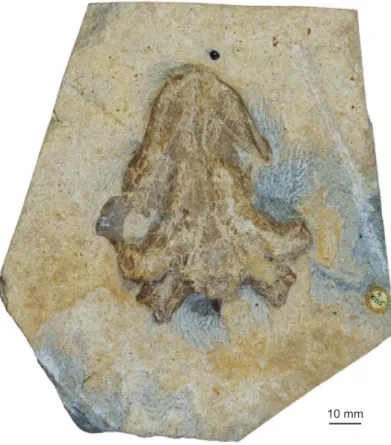
Figure 1.The cranium DORCM G.10715 before full extraction from the matrix. Only the palatal aspect is visible. Note the presence of a lepisosteiform tooth (seeMaterial) just anterior to the cranium.
Dorchester (United Kingdom) the following year. According to the label, the specimen was subsequently prepared in the Department of Zoology of the University of Oxford, but this preparation was only super-ficial. The skull remained embedded in the matrix and only observable in palatal view (Fig. 1). Apart from a mention inMilner (2004), this skull was never described or figured in the literature.
The specimen was borrowed from the DORCM by JA in 2010 and fully prepared in 2016 by Renaud Roch (Jurassica Museum, Porrentruy, Switzerland) using standard techniques. Two scales and three teeth referable to Scheenstia sp. (Lepisosteiformes, Ginglymodi, Actinopterygii) were found in the block of ma-trix during preparation.
Geological setting
DORCM G.10715 was collected in Durlston Bay (near Swanage, Dorset, UK) from fallen blocks immediately south of the Zigzag path on the shore (British National Grid reference: SZ 0342 7790;Fig. 2). Therefore, the exact stratigraphical position of the specimen is ambiguous. However, the strata forming the cliff where the specimen was found correspond to the ’Middle Purbeck Beds’ and beginning of the ’Upper Purbeck Beds’, following the informal denomination (see below). According to the label accompanying the specimen, the block was tentatively assigned to the Chief Beef Beds based on the lithology of the matrix and knowledge of the sections. The Chief Beef Beds consist of alternating dark shales and bands of hard shell-limestones deposited under brackish conditions (Batten, 2002). DORCM G.10715 was indeed found in a hard limestone rich in shell fragments.
Durlston Bay represents one of the largest exposures of the Purbeck Group along the Dorset coast. The Purbeck Group consists of a succession of limestones and calcareous shales and marls with
occa-Figure 2.Map of the south Dorset coast showing the place where DORCM G.10715 was found (red star).
sional evaporites that were deposited in a marginal setting (Allen and Wimbledon, 1991;Clements, 1993). The climate during this period is interpreted to have been semi-arid and becoming wetter to-ward the top of the sequence (Allen, 1998;Batten, 2002). The Purbeck Group represents a transition between the marine succession of the underlying Portland Limestone Group and the non-marine sedi-ments of the overlying Wealden Group, reflecting a major regression occurring at the end of the Jurassic and onset of the Cretaceous (e.g.,Rawson, 2006).
For long, the Purbeck Group was subdivided into Lower, Middle and Upper Purbeck. This is the only stratigraphical data we have for most of the fossils collected in the nineteenth century (e.g.,Milner, 2004). The Purbeck Group is now divided into two formations: the oldest Lulworth Formation and the youngest Durlston Formation (Townson, 1975). Ensom (1985)at Worbarrow Tout andClements (1993)at Durl-ston Bay formalized the nineteenth century tradition to finely separate the Purbeck Group into 15 or so members, each of them consisting of several beds (Fig. 3). Later,Westhead and Mather (1996)argued that these so-called members were difficult to follow at the regional scale. Therefore, these authors pro-posed to divise the Purbeck Group into five members and to relegate the traditional members to the bed status, but the latter continue to be used at least locally for the level of stratigraphical detail they provide (Ensom, 2002). There is an agreement among specialists that most of the Purbeck Group is Berriasian (Early Cretaceous) in age, but the exact position of the Jurassic-Cretaceous boundary has been debated. Several studies place the boundary at the base of the ’Cypris’ Freestones (e.g.,Allen and Wimbledon, 1991;Feist et al., 1995), but others have argued that the limit is higher in the Lulworth Formation (see
Ensom, 2002). In any case, DORCM G.10715 can be confidently assigned to the Berriasian.
Anatomical comparisons
Since DORCM G.10715 was tentatively referred to Dorsetochelys typocardium byMilner (2004), it was primarily compared with this taxon based on the original description (Evans and Kemp, 1976) and first hand observation of the holotype and only-known cranium, DORCM G.23. By extension, the new Purbeck cranium was also compared with other pleurosternids, notably Glyptops ornatus and Pleurosternon bul-lockii, based on the primary literature (Gaffney, 1979b;Evans and Kemp, 1975) and first hand observa-tion of the relevant material. For a recent reassessment of non-baenid paracryptodire taxonomy, the reader is referred toJoyce and Anquetin (2019). However, it became clear early on that the cranium DORCM G.10715 shared similarities with other European turtles, notably Thalassochelydia (seeAnquetin et al., 2017). Comparisons were therefore extended to that group as well based on published literature (Gaffney, 1975, 1976;Rieppel, 1980;Anquetin et al., 2015;Püntener et al., 2017), as well as first hand
Figure 3. Stratigraphy of the Purbeck Group in Durlston Bay, Dorset, UK. The old informal subdivision into Lower, Middle and Upper Purbeck is conserved since many fossils lack more precise stratigraphical information. The horizons that are known to have produced substantial turtle material (seeMilner, 2004) are indicated by a turtle shell icon on the right. Beds and bed numbers (DB) followClements (1993). Members followWesthead and Mather (1996). See alsoEnsom (2002).
observation of the available material. Anatomical descriptions in the present study follow the nomen-clature formalized byGaffney (1972, 1979a)as updated byRabi et al. (2013)for the carotid system and
Rollot et al. (2018)for the vidian nerve system.
3D model
In order to complement the photographs and line drawings provided herein and to facilitate future com-parisons, we produced a textured 3D mesh of DORCM G.10715 with a structured-light surface scanner (Artec Space Spider, Artec Group). The 3D model was computed with the dedicated software Artec Studio 13 Professional, and can be freely accessed on OSF (seeSupplementary information).
Phylogenetic analysis
In order to infer the phylogenetic relationships of the new cranium DORCM G.10715, we used one of the most recent global morphological matrix for turtles (Evers and Benson, 2019). In addition to DORCM G.10715, we also scored Hylaeochelys belli and a merged ’DORCM G.10715 + Hylaeochelys belli’ because we think they may represent the same species (seeAlpha taxonomy). The matrix is provided as Supple-mentary information. At first, DORCM G.10715 and Hylaeochelys belli (an exclusively shell-based species) were treated as distinct terminal taxa. A second analysis was performed in which both were merged into a single terminal taxon.
We followed the general analytical procedure detailed byEvers and Benson (2019). Analyses were performed in TNT 1.5 for Windows (Goloboff et al., 2008;Goloboff and Catalano, 2016). The relation-ships of extant taxa were constrained using a molecular backbone following the topology ofPereira et al. (2017). Characters were treated as unordered and equally weighted. Branches were set to collapse if branch length is zero. Proganochelys quenstedti was used as outgroup. Tree searches were performed using New Technology algorithms in TNT with default settings, tree drifting (Goloboff, 1999), parsimony ratchet (Nixon, 1999), and constraints enforced. The initial level of the driven search and the number of times the minimum tree length should be found were both set to 30. The most parsimonious trees (MPTs) were subjected to an additional round of tree bisection and reconnection (TBR). The resulting MPTs were used to compute a strict consensus tree. Consistency index (CI) and retention index (RI) were calculated using the STATS script bundled with TNT. Absolute Bremer support values were calculated in TNT using TBR branch swapping to generate suboptimal trees up to 20 steps longer than the MPTs.
It should be noted that a slightly updated version of Evers and Benson’s (2019) matrix was published more recently (Evers et al., 2019). This update is focussed mostly on chelonioid sea turtles with an in-creased taxon sampling for that group and a revision of postcranial characters related to their marine lifestyle. These modifications would not affect the phylogenetic position of DORCM G.10715. In addition, they resulted in a less resolved consensus among Mesozoic turtles (notably Xinjiangchelyidae, Sinemy-didae, and Macrobaenidae) and a poorly supported sister-group relationship between Angolachelonia (i.e., Thalassochelydia + Sandownidae) and pleurodires (Evers et al., 2019). Therefore, we preferred to use the original matrix ofEvers and Benson (2019)in the present study.
Institutional abbreviations
DORCM, Dorset County Museum, Dorchester, UK. NHMUK, Natural History Museum, London, UK.
Systematic paleontology
TESTUDINATAKlein, 1760(sensuJoyce et al., 2004)
THALASSOCHELYDIAAnquetin et al., 2017
Remark. Thalassochelydians are traditionally considered to be basal pan-cryptodiran turtles (Gaffney and Meylan, 1988;Lapparent de Broin et al., 1996;Joyce, 2007;Anquetin et al., 2015;Zhou and Rabi, 2015). However, some recent phylogenetic analyses have placed them along the stem of crown-group tur-tles (Sterli, 2010;Sterli et al., 2013;Cadena and Parham, 2015;Evers and Benson, 2019). This is in part the result of the instable position of the clade Pan-Pleurodira from one analysis to the next. According toEvers and Benson (2019), whose matrix we herein use to investigate the phylogenetic relationships of DORCM G.10715, Thalassochelydia are sister group to SandownidaeTong and Meylan, 2013forming the clade AngolacheloniaMateus et al., 2009, which is placed as the most crownward clade of stem turtles. However, forcing Thalassochelydia or Sandownidae as stem-group cryptodires or even as stem-group chelonioids only requires two to four additional steps, and forcing one group drags its sister group along maintaining the clade Angolachelonia (Evers and Benson, 2019). Two updated versions of the Evers and Benson’s (2019) matrix place Angolachelonia as sister group to Pleurodira (Evers et al., 2019) and as stem chelonioids (Gentry et al., 2019). Therefore, the position of Thalassochelydia must be considered uncer-tain and prone to change for the time being. Finally, it should also be noted that a recent study suggested that sandownids more likely derived from thalassochelydian ancestors (Evers and Joyce, 2020).
Thalassochelydia indet.
Figure 4
Referred material. DORCM G.10715, a relatively complete, but dorsoventrally crushed cranium. Locality. Durlston Bay, Swanage, Dorset, UK. Probably Chief Beef Beds, Stair Hole Member, Durlston
Formation, Purbeck Group (Berriasian, Early Cretaceous). SeeGeological settingabove.
Remark. DORCM G.10715 cannot be referred to any known species at the moment and could potentially
represent a new taxon. However, there is a reasonably good chance (seeAlpha taxonomy) that this material actually belongs to the shell-based species Hylaeochelys belli (Mantell, 1844) known from the Purbeck and Wealden of England, as recently revised byPérez-García (2012). Therefore, we refrain from establishing a new taxon based on this new skull and wait for more material to conclude on this matter.
Description
General description
DORCM G.10715 consists of a relatively complete, isolated cranium that was severely crushed dorsoven-trally during fossilization (see 3D mesh available asSupplementary information). As preserved, the cra-nial roof contacts the palate, and the entire internal anatomy is unfortunately lost to us. The squamosals as well as most of the jugals and quadratojugals are missing (Fig. 4). The ventral aspect of the skull was exposed when discovered and subjected to weathering (Fig. 1), notably the pterygoids and the triturating surface. The dorsal surface of the cranium is better preserved. The skull roof is smooth and lacks any sign of the ornamentation typically found in all non-baenid paracryptodires for which the skull is known, in-cluding the pleurosternids Glyptops ornatus, Pleurosternon bullockii, and Dorsetochelys typocardium (Evans and Kemp, 1975, 1976;Gaffney, 1979b;Pérez-García, 2012;Joyce and Anquetin, 2019). As preserved, the skull dimensions are as follows: length (midline) = 76.37 mm; length (max) = 78.12 mm; width (at the level of the processus articularis of the quadrates) = 60.34 mm.
Dermal roofing elements
Nasal
The complete skull roof was pushed down during the dorsoventral compression of the skull and now rests on the dorsal surface of the palatal region. Similarly, the roof of the nasal cavity got separated from the dorsal processes of the maxillae and now lies on the floor of the cavity (Fig. 4A–B). The dorsal margin of the apertura narium externa, seemingly formed by the nasal bones, was greatly damaged in the process. Only fragments of the posterior part of the nasals are visible anterior to the prefrontals, but the exact limit between these elements is elusive. It is unknown whether the nasals contacted the maxillae laterally, but this is highly probable. There is evidence for a nasal-frontal contact beneath the prefrontal (seeFrontalbelow).
Prefrontal
As a result of the dorsoventral crushing that occurred during fossilization, the anterior and anterolat-eral contacts of the prefrontals with the nasals and the maxillae are poorly preserved. Latanterolat-erally, the prefrontals form the anterodorsal margin of the orbits. Posteriorly, the prefrontals have a mostly trans-verse suture with the frontals. As seen from the right prefrontal, these bones meet one another on the midline along their entire length and are somewhat quadrangular in shape (Fig. 4A–B). This is clearly dif-ferent from the condition in the pleurosternids Glyptops ornatus, Pleurosternon bullockii and Dorsetochelys
A
B
D
C
10 mm
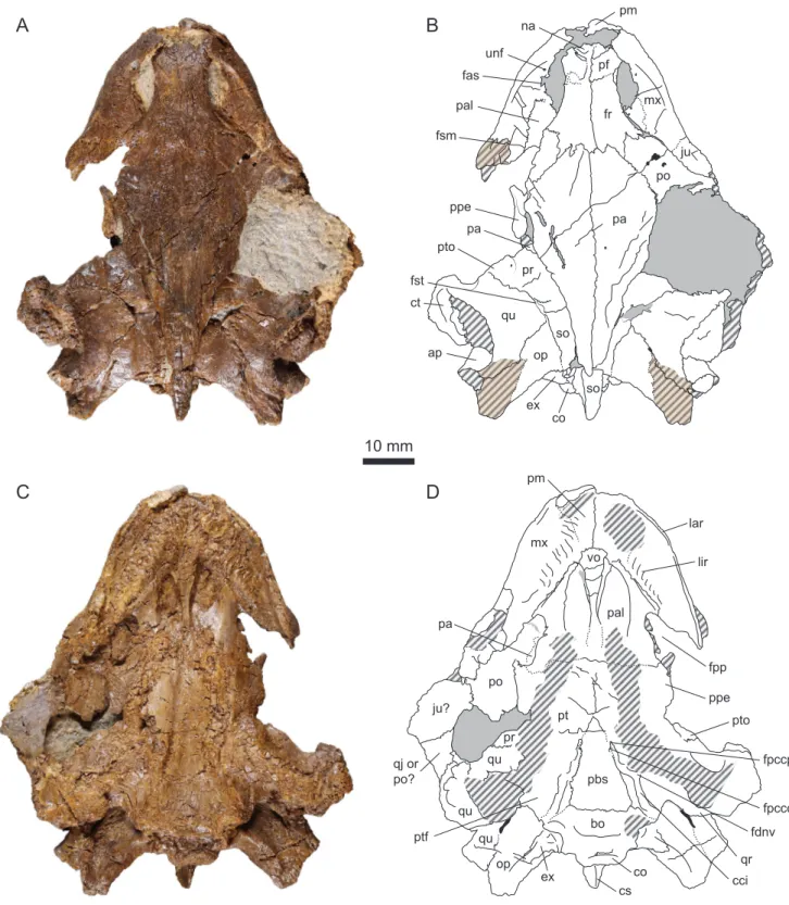
pm pf fr mx pal unf fsm fas na ju po pa ppe pa pr qu pto fst ct so op ap ex co so pm mx vo lar lir pal fpp pa po ju? qj or po? pr qu qu ppe pt pbs bo qu op ex co cs pto fpccp fpccc fdnv cci ptf qrFigure 4.DORCM G.10715, Purbeck Group (Berriasian), Swanage, Dorset, UK. (A) Photograph of the specimen in dorsal view; (B) Interpretative drawing in dorsal view; (C) Photograph of the specimen in ventral view; (D) Interpretative drawing in ventral view. Matrix is figured in gray. Hatchings represent heavily eroded areas. Sutural surfaces are indicated in light brown and hatchings. The shade difference between the dorsal and ventral sides is natural. Abbreviations: ap, antrum postoticum; bo, basioccipital; cci, canalis caroticus internus; co, condylus occipitalis; cs, crista supraoccipitalis; ct, cavum tympani; ex, exoccipital; fas, foramen alveolare superius; fdnv, foramen distalis nervi vidiani; fpccc, foramen posterius canalis carotici cerebralis; fpccp, foramen posterius canalis carotici palatinum; fpp, foramen palatinum posterius; fr, frontal; fsm, foramen supramaxillare; fst, foramen stapedio-temporale; ju, jugal; lar, labial ridge; lir, lingual ridge; mx, maxilla; na, nasal; op, opisthotic; pa, parietal; pal, palatine; pbs, parabasisphenoid; pf, prefrontal; pm, premaxilla; po, postorbital; ppe, processus pterygoideus externus; pr, prootic; pt, pterygoid; ptf, pterygoid fossa; pto, processus trochlearis oticum; qj, quadratojugal; qr, quadrate ridge; qu, quadrate; so, supraoccipital; unf, unnamed foramen; vo, vomer.
typocardium in which the prefrontals have limited dorsal exposure along the orbit margin and never meet in the midline (Evans and Kemp, 1975, 1976;Gaffney, 1979b). The medial part of the left prefrontal is damaged and reveals that the frontals extend anteriorly beneath the prefrontals to meet the nasals (see
Frontalbelow). This morphology where the medial part of the prefrontal consists in a thin sheet of bone that covers the anterior medial process of the frontal dorsally without preventing a frontal-nasal contact underneath recalls the condition in the thalassochelydian plesiochelyid turtles Plesiochelys etalloni, Ple-siochelys planiceps and Portlandemys mcdowelli (Gaffney, 1976), although not all thalassochelydians show this morphology (Rieppel, 1980;Anquetin et al., 2015).
Frontal
The frontals are complete and well preserved in DORCM G.10715 (Fig. 4A–B). In dorsal view, they con-tact the prefrontals anteriorly, the postorbitals posterolaterally, and the parietals posteromedially. The anterior suture with the prefrontal is mostly transverse. The posterior sutures with the postorbital and parietal are strongly interdigitating. The frontals meet one another medially along their entire length. Laterally, they form the dorsal margin of the orbits. The damage of the medial part of the left prefrontal reveals that the frontals extend anteriorly beneath the prefrontals to meet the nasals. As noted above, a similar morphology was described in some, but not all, thalassochelydians (Gaffney, 1976;Anquetin et al., 2015). Detecting this condition is not always easy and it cannot be excluded that it is present in other basal pan-cryptodires as well. However, it is never present in pleurosternids where a broad anterior process of the frontals clearly separates the relatively small prefrontals in dorsal view.
Parietal
Only the anterior and medial parts of the parietals are preserved in DORCM G.10715 (Fig. 4A–B). As pre-served, the parietals contact the frontals anteriorly along a transverse, strongly interdigitating suture, and the postorbital anterolaterally. Posteroventrally, the parietals also have an overlapping contact with the crista supraoccipitalis formed by the supraoccipital. The other usual contacts of the parietals in the ethmoid region and in the roof of the cavum cranii are not preserved due to the dorsoventral crushing of the specimen. The medial margin of the upper temporal emargination is partly preserved on both sides. On the right side, the margin reaches the level of the foramen stapedio-temporale. On the left side, the medial margin of the upper temporal emargination can be followed as far anteriorly as the processus trochlearis oticum. DORCM G.10715 is therefore characterized by an extensive upper temporal emargina-tion that probably revealed much of the dorsal surface of the otic chamber in dorsal view. However, the exact shape of the emargination is unknown. In pleurosternids, the upper temporal emargination is at best poorly developed (Evans and Kemp, 1975, 1976;Gaffney, 1979b;Joyce and Anquetin, 2019).
Jugal
The jugal is badly preserved in DORCM G.10715. The left jugal is completely missing, only a sutural scar on the dorsal surface of the maxilla shows where the jugal articulated (Fig. 4A). Of the right jugal, only the anteroventral part in the floor of the orbit is preserved in anatomical position, still sutured to the maxilla. There is apparently no developed medial process on the jugal. The contribution of the jugal to the orbit is unknown. The temporal bar is broken on each side of the cranium, but a flat piece of bone lies where the right temporal bar should have been (Fig. 4C–D). This piece possibly corresponds to the ventral part of the jugal and quadratojugal, although it is now oriented horizontally rather than vertically. If this interpretation is correct, the slightly concave medial margin of this piece would indicate a modest lower temporal emargination. Another possibility is that this piece of bone corresponds to the jugal and postorbital. If this second interpretation is correct, then the slightly concave medial margin would
correspond to the posteroventral margin of the orbit. The first interpretation may appear more likely at first as it requires less rotation of the flat piece of bone during dorsoventral compression, but the second interpretation is supported by the shiny aspect of the visible bone surface which would indicate that this is the internal face of the bones that now faces ventrally.
Quadratojugal
Little can be said about this bone. A small portion of the right quadratojugal may be preserved, but this remains uncertain (seeJugalabove).
Postorbital
Most of the postorbital is missing in DORCM G.10715 (Fig. 4A–B). On the left side, only the anteriormost part of the bone is preserved. On the right side, a slightly larger portion of the postorbital is preserved and forms the posterodorsal margin of the orbit. A large area of matrix in the right temporal region could not be removed during preparation without compromising the stability of the fossil. The posterior part of the right postorbital is hidden beneath this matrix, but can be seen in ventral view posterolateral to the foramen palatinum posterius (Fig. 4C–D). The postorbital contacts the frontal anteromedially and the parietal medially. The other contacts of the bone are obscure. It is unknown if and how much the postorbital contributes to the margin of the upper temporal emargination.
Palatal elements
Premaxilla
The premaxillae are poorly preserved in DORCM G.10715. Only the midline suture between the two premaxillae can be seen clearly (Fig. 4C–D). However, it is apparent that the premaxillae contact the maxillae laterally and the vomer posteriorly. Foramina praepalatinum were likely present, but this cannot be confirmed due to poor preservation. Dorsally, the premaxillae form the ventral margin of the apertura narium externa (Fig. 4A–B).
Maxilla
The ventral aspect of the maxillae is broken and eroded in several places (Fig. 4C–D). The maxilla contacts the premaxilla and vomer anteromedially, the palatine medially, and the jugal posterodorsally. The up-per jaw is relatively short compared to the length of the skull. It is U-shaped with moderately diverging rami and a broad, well-rounded anterior part. The triturating surface is broad and its width increases anteriorly. The labial ridge is thin and damaged, which makes it impossible to infer its height. The lin-gual ridge is broad and serrated, although we do not know whether these serrations were also apparent on the rhamphotheca. It is widest and highest at the level of the anteriormost ventral exposure of the vomer. It is vaguely sinusoidal in outline: concave anterolaterally for most of its length, the ridge cur-vature shifts to concave posteromedially on the premaxilla (Fig. 4C–D). The lingual ridge is apparently lowest on the premaxilla. The trough between the labial and lingual ridges is shallow. Its width varies with the curvature of the lingual ridge. Glyptops ornatus and Pleurosternon bullockii have relatively narrow triturating surface with a low, simple lingual ridge (Evans and Kemp, 1975;Gaffney, 1979b). Compared to these two taxa, the triturating surface of Dorsetochelys typocardium is broader and widens slightly posteriorly. Dorsetochelys typocardium is further characterized by a relatively poor development of the lingual ridge and an unusually extensive contribution of the palatine to the triturating surface (Evans and Kemp, 1976). Thalassochelydians generally have high labial and lingual ridges separated by a deep
trough (Gaffney, 1976;Rieppel, 1980;Anquetin et al., 2015). DORCM G.10715 is clearly different from any of the aforementioned taxa.
In dorsal view, the maxilla forms a large portion of the floor of the fossa orbitalis (Fig. 4A–B). Only the base of the ascending process is preserved and the contacts this process probably had with the nasal and prefrontal dorsally cannot be confirmed. The anterior border of each maxilla is broken, but there is no doubt that the bone formed the lateral margin of the apertura narium externa. The lateral part of the foramen alveolaris superior can be seen medially at the base of the ascending process of the maxilla. This foramen faces dorsomedially and probably opened in the foramen orbito-nasale (not visible here). On the left side, a smaller, but well defined foramen opens slightly anterior to the foramen alveolaris anterior on the floor of the orbit (Fig. 4A–B). The same foramen was probably also present on the right side, but the area is damaged. The nature of this foramen remains uncertain, but a similar foramen was described in several thalassochelydians as well as in the stem-group chelonioid Toxochelys, although the distribution and homology of this feature remains to be investigated (Gaffney, 1976;Anquetin et al., 2015).
Vomer
As usual the vomer forms the medial septum of the apertura narium interna (Fig. 4C–D). The sutures of the vomer with other elements are poorly preserved, with exception of the dorsal suture with the palatines that can be seen relatively easily in some areas. Anteroventrally, the vomer widens and contacts the premaxillae and maxillae, but this area is poorly preserved (e.g., the foramina praepalatinum cannot be seen). Posterodorsally, the vomer flattens and widens to form part of the palatal roof. There, the vomer appears to contact the pterygoids as well as the palatines.
Palatine
In ventral view, the palatines contact the vomer medially, the maxillae anterolaterally, and the pterygoids posteriorly (Fig. 4C–D). The suture with the pterygoids is poorly preserved but visible by intermittence. Although this area is not very well preserved, it appears that the palatines are fully separated from one another in ventral view as a result of a contact between the vomer and the pterygoids. This is also the condition observed in most thalassochelydians, except Jurassichelon oleronensis (Rieppel, 1980) and per-haps Plesiochelys planiceps (Gaffney, 1975, 1976). Anteromedially, the palatines form the arched roof of the nasal canals and apertura narium interna. Laterally, the palatines form an anterolateral process that braces the medial margin of the triturating surface. In contrast to the assertion ofMilner (2004), the palatine does not contribute to the triturating surface in DORCM G.10715, which clearly differentiates this specimen from the remarkable condition observed in Dorsetochelys typocardium (Evans and Kemp, 1976). Posterolaterally, the palatine forms the anterior and medial margins of a large and kidney-shaped fora-men palatinum posterius that remains open posterolaterally due to the absence of a contact between the pterygoid and maxilla/jugal. Paracryptodires in general have a relatively small foramen palatinum posterius that is closed posterolaterally by a broad bar of bone (Evans and Kemp, 1975, 1976;Gaffney, 1979b;Joyce and Lyson, 2015). In contrast, an open foramen palatinum posterius is relatively rare among turtles. It is known only in some thalassochelydians (Plesiochelys spp. and Jurassichelon oleronensis) and some protostegids (e.g.,Gaffney, 1976;Hirayama, 1998;Kear and Lee, 2006;Joyce, 2007;Cadena and Parham, 2015;Anquetin et al., 2017;Evers et al., 2019). Protostegids are generally considered to be early pan-chelonioids, but they have been repeatedly hypothesized to be closely related to thalassochelydians (e.g.,Joyce, 2007). Modern sea turtles lack a foramen palatinum posterius altogether.
Palatoquadrate elements
Quadrate
The severe dorsoventral compression of DORCM G.10715 strongly affected the quadrate, notably the middle ear, ethmoid region, and processus articularis (see 3D mesh available asSupplementary infor-mation). On the dorsal surface of the otic chamber, the quadrate contacts the prootic anteromedially, the supraoccipital medially, and the opisthotic posteromedially (Fig. 4A–B). The brief contact between the quadrate and supraoccipital prevents the prootic and opisthotic to meet in dorsal view. Postero-laterally, the quadrate also had a large contact with the squamosal. This element is now lost and the position of the suture with quadrate is difficult to place. In this area, the squamosal simply lies above the quadrate and leaves little marks on the surface of the latter when disarticulated. The quadrate forms the lateral half of the foramen stapedio-temporale and processus trochlearis oticum. The latter structure is modest in development and remarkable in being quite oblique in orientation.Evers and Benson (2019)
noted that some turtles, including most thalassochelydians, have a processus trochlearis oticum limited to the medial part of the otic chamber and framed laterally by a deep recess. This condition is apparently also present in DORCM G.10715 (Fig. 4A–B). The cavum tympani and antrum postoticum were both ap-parently well developed. The incisura columellae auris probably remained open posteroventrally. The ventral part of the quadrate is poorly preserved, being both crushed dorsoventrally and much eroded (Fig. 4C–D). However, it should be noted that a strong ridge is present on the posterior surface of the pro-cessus articularis of the quadrate. This ridge prolongates the posterior margin of the pterygoid laterally on the posterior surface of the processus articularis and appears to bend ventrally on the more lateral parts of the processus articularis. This ridge strongly reminds the well-developed, ventrally-infolding ridge present in thalassochelydians (Gaffney, 1976;Anquetin et al., 2015). A similar, though less pro-nounced ridge is also present in sandownids (Evers and Benson, 2019; Evers and Joyce, 2020). As noted byEvers and Benson (2019)for thalassochelydians and sandownids, the processus articularis of the quadrate is oriented strongly posteriorly relative to the sagittal plane.
Pterygoid
The ventral aspect of the pterygoids is much eroded. The pterygoid contacts the vomer (probably) and palatine anteriorly, the quadrate posterolaterally (suture not preserved), the basioccipital posteromedi-ally, the parabasisphenoid mediposteromedi-ally, and the other pterygoid anteromedially (Fig. 4C–D). It was reported that Pleurosternon bullockii and Glyptops ornatus lack a midline contact of the pterygoids due to a long ventral exposure of the parabasisphenoid that would reach the vomer anteriorly (Evans and Kemp, 1975;
Gaffney, 1979b), but this may be a preservational artifact (Joyce and Anquetin, 2019). Anterolaterally, the pterygoid forms a well-developed processus pterygoideus externus. The vertical plate of the pro-cessus pterygoideus externus is triangular in outline with a low anterior part and a high posterior part (Fig. 4A–B). In ventral view, the anterior part of the processus pterygoideus externus forms an antero-laterally oriented process that defines the posterolateral margin of the foramen palatinum posterius (Fig. 4C–D). As noted above, there is no contact between the pterygoid and the maxilla and/or jugal in this area, and the foramen palatinum posterius remains open posterolaterally.
As preserved, the canalis caroticus internus runs in an open gutter on the ventral surface of the cra-nium (Fig. 4C–D). It is unknown whether the canal was at least partly floored in life, or if it remained open. In any case, the canalis caroticus internus was superficial (i.e., running close to or on the bone surface). This is clearly reminiscent of the condition in some thalassochelydians, such as Plesiochelys etal-loni, Plesiochelys bigleri, and Jurassichelon oleronensis (Gaffney, 1976;Rieppel, 1980;Anquetin et al., 2015;
some thalassochelydians such as Portlandemys spp. and Neusticemys neuquina (Gaffney, 1976;Anquetin et al., 2015;González Ruiz et al., 2020).Anquetin et al. (2015)andRaselli and Anquetin (2019)provided a thorough survey of this feature in thalassochelydians. Posteriorly, the canalis caroticus internus is lo-cated only on the pterygoid a short distance lateral to the suture with the parabasisphenoid. At about mid-length, the canalis caroticus internus bends medially and then runs along the parabasisphenoid-pterygoid suture up to the split between the two branches of the internal carotid artery (see Paraba-sisphenoid). The foramen distalis nervi vidiani seems to open in the pterygoid just lateral to the mid-length bending of the canalis caroticus internus. The pterygoid fossa is relatively shallow and triangular in outline, contrasting with the much narrowed pterygoid fossa observed in Dorsetochelys typocardium.
Braincase elements
Supraoccipital
The supraoccipital contacts the prootic anteroventrally, the quadrate laterally (short contact), and the opisthotic posterolaterally (Fig. 4A–B). The contacts with the parietal anterodorsally and the exoccipital posteroventrally are not directly observable due to the strong dorsoventral compression of the cranium. Only the posterodorsal part of the crista supraoccipitalis can be observed. The crista does not extend much posteriorly, reaching only as far as the opisthotics (it can be surmised that the squamosals reached further posteriorly). The part of the crista supraoccipitalis that is not covered dorsally by the parietals forms a narrow, triangular horizontal plate that prolongates the skull roof posteriorly.
Exoccipital
The exoccipitals are severely affected by the dorsoventral compression of the cranium that resulted in a contact between the crista supraoccipitalis and condylus occipitalis. The exoccipital apparently con-tacted the supraoccipital mediodorsally, the opisthotic dorsolaterally, and the basioccipital ventromedi-ally (Fig. 4). The only preserved suture is the one with the opisthotic in the roof of the cavum acustico-jugulare. Two foramina nervi hypoglossi are visible on the left side, but only one is preserved on the right side. Below this point, the preservation of the exoccipitals is extremely poor. Therefore, it is unknown whether the exoccipital contacted the pterygoid as in many cryptodires (Gaffney, 1979a). The extent to which the exoccipitals contribute to the condylus occipitalis is also uncertain.
Basioccipital
The basioccipital contacts the parabasisphenoid anteriorly along a seemingly transverse suture (poorly preserved), the pterygoid anterolaterally along a broad and oblique suture, and the exoccipital dorsally (Fig. 4C–D). The tubercula basioccipitale are poorly preserved. Compared to Dorsetochelys typocardium, the basioccipital has a shorter ventral exposure and a broader contact with the pterygoid.
Prootic
In DORCM G.10715, the prootic can only be observed on the dorsal surface of the otic chamber (Fig. 4A–B). The rest of the bone, notably in the ethmoid region, is not accessible. As preserved, the prootic contacts the parietal anteromedially, the quadrate laterally and anteroventrally, and the supraoccipital posteriorly. As noted above, a brief contact of the quadrate and supraoccipital posterior to the foramen stapedio-temporale prevents the prootic and opithotic to meet on the dorsal surface of the otic chamber. As usual, the prootic forms the medial half of the foramen stapedio-temporale and processus trochlearis oticum. The latter structure is modest in development, remarkable in being oriented quite obliquely relative to the transverse axis, and limited to the medial part of the otic chamber (seeQuadrate).
Opisthotic
The opisthotic contacts the supraoccipital medially, the quadrate anterolaterally, and the exoccipital pos-teriorly (suture not preserved dorsally). At first it may appear that the opisthotic as an extensive dorsal exposure, but this is due to the disarticulation and loss of the squamosal on both sides (Fig. 4A–B). As noted above, there is no anterior contact with the prootic on the dorsal surface of the otic chamber. Due to the postmortem dorsoventral crushing of the cranium, the processus interfenestralis of the opisthotic is not preserved. The processus paroccipitalis is dorsoventrally flattened (seemingly the natural condi-tion) and forms an acute posterior ridge. A similar morphology is found in a wide array of turtles including Sandownidae, Trionychia, Pleurodira, Pleurosternidae, and several stem-group turtles (Evers and Ben-son, 2019).
Parabasisphenoid
In ventral aspect, the parabasisphenoid contacts the pterygoid laterally and the basioccipital posteriorly (Fig. 4C–D). The bone is triangular in outline with a concave ventral surface. This concavity is more pro-nounced posteriorly along the suture with the basioccipital. The anterior part of the superficial canalis caroticus internus runs on the parabasisphenoid-pterygoid suture and is formed by both elements (see
Pterygoid). The preservation is not perfect, but the split between the cerebral and palatine branches of the internal carotid artery can still be observed in the anteriormost part of the canalis caroticus internus. The foramen posterius canalis carotici cerebralis opens mediodorsally in the canalis caroticus internus and is formed by the parabasisphenoid. The palatine branch continues anteriorly for a short distance and enters the foramen posterius canalis carotici palatinum formed mostly (if not exclusively) by the pterygoid. As noted above, the morphology of the internal carotid sytem in DORCM G.10715 clearly re-minds the condition observed in some thalassochelydians, such as Plesiochelys etalloni, Plesiochelys bigleri, and Jurassichelon oleronensis (Gaffney, 1976;Rieppel, 1980;Anquetin et al., 2015;Püntener et al., 2017). This point is developed further in theDiscussionbelow. Unfortunately, the morphology of the paraba-sisphenoid inside the cranium is not accessible.
Phylogenetic relationships
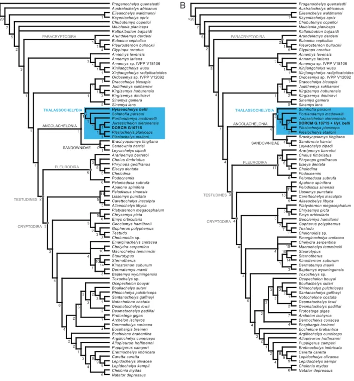
In our first analysis, the cranium DORCM G.10715 and the shell-based species Hylaeochelys belli were treated as separate terminal taxa. This analysis resulted in four most parsimonious trees (MPTs; Tree Length = 1540 steps; Consistency Index = 0.260; Retention Index = 0.653). The strict consensus tree (TL = 1546 steps; CI = 0.259; RI = 0.651) corresponds fairly well to the topology obtained byEvers and Benson (2019)for their analysis with unordered characters (Fig. 5A). The differences are: the support of the clade Paracryptodira, the resolution of the relationships among Xinjianchelyidae, and the improve resolution of the relationships among the clades Sinemydidae + Macrobaenidae and Thalassochelydia. DORCM G.10715 and Hylaeochelys belli both fall within Thalassochelydia. Hylaeochelys belli is resolved as the most basal thalassochelydian, whereas DORCM G.10715 is recovered as the sister-group of the Late Jurassic Jurassichelon oleronensis. The inclusion of DORCM G.10715 and Hylaeochelys belli in the matrix tends to have a negative effect on Bremer support values in the central portions of the consensus tree (i.e., from Xinjiangchelyidae to Trionychia) compared to the reference analysis ofEvers and Benson (2019).
In our second analysis, the cranium DORCM G.10715 and the shell-based species Hylaeochelys belli were merged into a single terminal taxon, reflecting our suggestion that they may indeed represent the same species (seeAlpha taxonomy). This second analysis also resulted in four MPTs (TL = 1541 steps; CI = 0.260; RI = 0.653). The topology of the strict consensus tree (TL = 1547 steps; CI = 0.259; RI = 0.651) is exactly the same as the one obtained during our first analysis (Fig. 5B). The merged DORCM G.10715 +
Figure 5.Phylogenetic relationships of DORCM G.10715 and Hylaeochelys belli. (A) Strict consensus tree (TL = 1546 steps; CI = 0.259; RI = 0.651) of four MPTs with DORCM G.10715 and Hylaeochelys belli treated as distinct terminal taxa; (B) Strict consensus tree (TL = 1547 steps; CI = 0.259; RI = 0.651) of four MPTs with DORCM G.10715 and Hylaeochelys belli merged into a single terminal taxon. Numbers below nodes correspond to Bremer support values.
Hylaeochelys belli is recovered at the same position as the isolated cranium DORCM G.10715 before, that is as the sister-group of Jurassichelon oleronensis. Merging Hylaeochelys belli with DORCM G.10715 has a positive effect on Bremer support values compared to our first analysis. Most of the Bremer values are the same as the one obtained by the reference analysis ofEvers and Benson (2019). Notable discrep-ancies include the slightly lower support for the clade Sinemydidae + Macrobaenidae and the clade that unites them to other turtles, and the slightly increased support for the clade Thalassochelydia.
In our two analyses, the clade Angolachelonia is supported by the same eight unambiguous synapo-morphies as in the reference analysis (Evers and Benson, 2019): the foramen stapedio-temporale is concealed in dorsal view (ch. 18.0); the posterior surface of the quadrate bears a strong ridge ventral to the incisura columellae auris (ch. 83.1); the articular process of the quadrate is directed strongly pos-teriorly (ch. 84.1); the medial contact of the pterygoids is longer than the parabasisphenoid at midline (ch. 94.0); the pterygoid contacts the medial edge of the mandibular condyle when seen in ventral view (ch. 103.1); the posterior margin of the coronoid has a deep notch (ch. 181.1); the anterior part of the coronoid bears a foramen leading to the fossa meckelii (ch. 182.1); the coronoid process is relatively high and dorsally or posterodorsally pointed (ch. 184.1). In our second analysis, the clade Thalassochelydia is supported by the same eight unambiguous synapomorphies as the one obtained byEvers and Benson (2019): the processus trochlearis oticum is limited to the medial part of the otic chamber and a deep recess is present lateral to it (ch. 81.1); presence of a fossa on the posterodorsal surface of the floor of the supratemporal fossa (ch. 113.1); the foramina anterius canalis carotici cerebralis are located close together (ch. 142.1); the splenial is present (ch. 185.0); costals lack lateral ossification (ch. 210.2); rib-free peripherals are present (ch. 211.1); vertebrals 2–4 are significantly broader than pleurals (ch. 224.0); cen-tral cervical articulations are not formed (ch. 278.0). In our first analysis, the presence of the shell-based Hylaeochelys belli renders most of these characters ambiguous and the clade Thalassochelydia is only sup-ported by the presence of rib-free peripherals (ch. 211.1) and the vertebrals 2–4 broader than pleurals (ch. 224.0), explaining the reduced support for the clade in this analysis. Finally, in both of our analyses, the clade formed by Jurassichelon oleronensis and DORCM G.10715 (or DORCM G.10715 + Hylaeochelys belli) is supported by the following unambiguous synapomorphies: the medial contact of the pterygoids is equal to or shorter than the parabasisphenoid at midline (ch. 94.1); the presence of a poorly developed crista supraoccipitalis (ch. 110.0); the internal carotid arterial system is partially embedded, a foramen posterius canalis carotici interni is present, and the split into palatine and cerebral arteries happens at the fenestra caroticus and is thus ventrally exposed (ch. 147.1). It should be noted for the record that thalassochelydians are particularly difficult to score for the latter character due to intraspecific variability and preservation bias (Anquetin et al., 2015).
Discussion
Beta taxonomy
When it was still partly embedded in matrix (Fig. 1), DORCM G.10715 was provisionally referred to Dorse-tochelys delairi (now DorseDorse-tochelys typocardium) based on a short list of tentative observations: palate of similar size and shape, "the palatines contribute to the triturating surface, the pterygoids meet an-teriorly ahead of the foreshortened basisphenoid and there is an abbreviated pterygoid-basisphenoid contact" (Milner, 2004). The description above disagrees with this first assignment. The triturating sur-face of DORCM G.10715 is relatively short and U-shaped, and the palatines do not contribute much to it. The large, kidney-shaped, and posterolaterally open foramen palatinum posterius of the new cranium is also clearly different from the relatively small, rounded, and closed foramen observed in Dorsetochelys typocardium (Evans and Kemp, 1976). The fact that the pterygoids meet anteriorly to the parabasisphe-noid, therefore reducing the length of the contact between these elements, is actually only remarkable within the context of non-baenid paracryptodires by comparison with Pleurosternon bullockii and Glyptops ornatus (but seeJoyce and Anquetin, 2019), but not when turtles are considered more globally.
Furthermore, DORCM G.10715 exhibits several characteristics that clearly exclude it from Paracryp-todira. The skull roof surface is smooth and lacks any indication of the ornamentation typically found in non-baenid paracryptodires and basal baenids (seeJoyce and Lyson, 2015;Joyce and Anquetin, 2019).
The prefrontals of DORCM G.10715 have a relatively large exposure on the skull roof and they meet on the midline, whereas they are reduced to small lappets in most paracryptodires and even absent from the skull roof in derived forms. The upper temporal emargination is relatively extensive in DORCM G.10715, contrasting with the at-most modest emargination seen in non-baenid paracryptodires.
DORCM G.10715 actually shows a number of features that support a close relationships with Thalas-sochelydia. The most obvious of these features is the presence of a relatively large and posterolater-ally open foramen palatinum posterius. This is found only in some thalassochelydians and some early pan-chelonioids (e.g.,Gaffney, 1976;Joyce, 2007;Evers and Benson, 2019). The presence of a well-developed ridge on the posterior surface of the processus articularis of the quadrate is also a strong argument. This prominent ridge on the quadrate below the incisura columallae auris was first noted by
Parsons and Williams (1961)in Solnhofia andGaffney (1976)in Plesiochelys and Portlandemys, but it is more generally present in all thalassochelydians for which this particular area of the cranium is known (Anquetin et al., 2015). More recently, it was noted that a somewhat similar, albeit less pronounced, ridge was also present in sandownids and possibly also a couple of additional taxa (Evers and Benson, 2019). The ridge present in DORCM G.10715 is incompletely preserved but clearly reminds the condition and development observed in thalassochelydians. Other similarities between DORCM G.10715 and tha-lassochelydians include the presence of a small foramen in the maxilla along the anteroventral margin of the orbit, a nasal-frontal contact below the prefrontals, and possibly the configuration of the canalis caroticus internus (but see below). In addition to the previous, new characters supporting Angolachelonia (Thalassochelydia + Sandownidae) or Thalassochelydia more specifically were recently proposed by Ev-ers and Benson (2019). As in angolachelonians, the articular process of the quadrate is directed strongly posteriorly in DORCM G.10715, and as in thalassochelydians (not exclusive), the processus trochlearis oticum is limited to the medial part of the otic chamber and bordered laterally by a deep recess. The presence of all of these features in DORCM G.10715 is enough to firmly place this specimen within Tha-lassochelydia, as unsurprinsignly confirmed by the phylogenetic analysis (see above;Fig. 5).
We believe the interrelationships of Thalassochelydia to be poorly understood for the moment. In our phylogenetic analysis, the sister group relationships between DORCM G.10715 (or DORCM G.10715 + Hylaeochelys belli) and Jurassichelon oleronensis is only supported by three "reversals" to plesiomorphic conditions, namely a midline contact of the pterygoids that is equal or shorter than the length of the parabasisphenoid, a poorly developed crista supraoccipitalis, and a partly embedded internal carotid system with the split into cerebral and palatine branches still exposed ventrally (see above). Therefore, we will refrain from any comment on these relationships herein and only retain the clear placement of DORCM G.10715 within Thalassochelydia.
Alpha taxonomy
Although DORCM G.10715 shares many features with thalassochelydians, it is also distinct from any known species in that group. The peculiar morphology of the upper triturating surface (seeMaxilla
above) and the kidney-shaped foramen palatinum posterius clearly set DORCM G.10715 apart from the rest (Fig. 4). However, we intentionally refrain from naming a new species based on this specimen be-cause we believe there is a great chance that it actually represents the cranium of the shell-based species Hylaeochelys belli, which is found in the same layers. This is why DORCM G.10715 is left in open nomen-clature herein (Thalassochelydia indet.).
Thanks to intensive fossil collection since the nineteenth century, we can consider the Purbeck turtle fauna to be relatively well known. FollowingMilner (2004)andPérez-García (2012, 2014), four distinct species can be recognized in this assemblage. Pleurosternon bullockii and Dorsetochelys typocardium, both known by a cranium and numerous shells, are paracryptodires. "Helochelydra" anglica is known only by
fragmentary shell remains and is a representative of Helochelydridae, a group of terrestrial stem turtles characterized by a distinctive cranial anatomy (seeJoyce, 2017). DORCM G.10715 does not belong to any of these three species. The fourth Purbeck turtle, Hylaeochelys belli, is known by numerous well-preserved shells from the Purbeck and Wealden Groups of southern England (Milner, 2004;Pérez-García, 2012). Unfortunately, none is associated with cranial material. Distinguished by a smooth shell with very wide vertebral scales and the absence of mesoplastra, Hylaeochelys belli was repeatedly proposed to be a representative, or at least a close relative, of the Late Jurassic Plesiochelyidae (Lydekker, 1889;Nopcsa, 1928;Hirayama et al., 2000;Lapparent de Broin, 2001;Milner, 2004;Karl et al., 2007;Pérez-García, 2012), a conclusion supported by the phylogenetic analysis proposed herein since Hylaeochelys belli is recovered as a basal Thalassochelydia when scored as a distinct terminal taxon (see above;Fig. 5).
Since both DORCM G.10715 and the shell-based Hylaeochelys belli co-occur in the Purbeck Group and appear to be representatives of Thalassochelydia, a group usually considered to disappear at the end of the Jurassic (e.g.,Bardet, 1994;Anquetin et al., 2017), we believe that there is a reasonably good chance that they actually represent the same species. Although we lack more definitive evidence for the mo-ment, it should be noted that the shell corresponding to the cranium DORCM G.10715 can be estimated to be around 38–40 cm in length (supposing the cranium represents about 20% of the carapace length as common in these turtles), which matches fairly well with the size range of Hylaeochelys belli (Milner, 2004;
Pérez-García, 2012). Furthermore, when DORCM G.10715 and Hylaeochelys belli are merged into a single terminal taxon (see our second phylogenetic analysis) they fit snugly into the phylogenetic hypothesis of
Evers and Benson (2019)without causing much disruption in terms of topology and character distribu-tion. This should definitely not be taken as hard evidence, but it somewhat suggests that this association makes sense.
A
B
10 mm
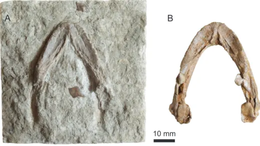
Figure 6.Two undescribed mandibles, Purbeck Group (Berriasian), Swanage, Dorset, UK. (A) NHMUK OR21974x in dorsal view; (B) NHMUK OR44815 in dorsal view. The mandibles are lit mainly from the right to accentuate relief. Mentioned byLydekker (1889)andMilner (2004).
Finally, it should be reminded that two undescribed turtle mandibles from the Purbeck Group in Swan-age are housed in the Natural History Museum in London: NHMUK OR21974x and NHMUK OR44815 (Lydekker, 1889;Milner, 2004). Unfortunately, we saw these mandibles before conducting the present study. However, based on our observations at the time and photographs of the specimens, we believe we can make a few pertinent comments here. The morphology of the triturating surface is remarkable and
indicates that the two mandibles belong to the same taxon (Fig. 6). The anterior outline of the mandibles is rather rounded. The triturating surface is relatively short and broad, with low lingual and labial ridges. The lingual ridges become more pronounced anteriorly and do not meet one another on the symphysis, rather bordering a narrow sagittal gutter. This morphology matches fairly well that of DORCM G.10715 (Fig. 4) and we are relatively confident that they can all be assigned to the same taxon, which is pos-sibly Hylaeochelys belli. Interestingly, the more complete of these two mandibles (NHMUK OR44815) possesses a well-developed processus coronoideus projecting posteriorly, an angolachelonian feature (non-exclusive) according toEvers and Benson (2019).
Notes on the internal carotid arterial system in Paracryptodira
The configuration of the internal carotid arterial system and more specifically the position of the fora-men posterius canalis carotici interni has been given a great significance in terms of turtle systematics (Albrecht, 1976;Gaffney, 1979a). Paracryptodires are usually depicted as having a foramen posterius canalis carotici interni located halfway along the parabasisphenoid-pterygoid suture and facing ventrally. The split between the two branches of the internal carotid artery (that is when the palatine branch is present; seeRollot et al., 2018) occurs within the bones and is therefore not visible in ventral view (Gaffney, 1979a;Joyce and Lyson, 2015;Joyce and Anquetin, 2019).
The previous description applies fairly well to baenids, but less so to non-baenid paracryptodires. This became plainly apparent to us when we revised the latter while describing DORCM G.10715. In Pleurosternon bullockii and Glyptops ornatus, the internal carotid artery penetrates the cranium at the level of the fused and still visible basipterygoid process, which lies in a shallow depression along the parabasisphenoid-pterygoid suture. In both of these species, the authors describe and illustrate a short sinus that extends posterolaterally from the foramen posterius canalis carotici cerebralis and crosses the medial part of the basipterygoid process (Evans and Kemp, 1975;Gaffney, 1979b). Just lateral to the foramen posterius canalis carotici cerebralis and anterolateral to the previously described sinus lies the small foramen posterius canalis carotici palatinum (not readily observable in Pleurosternon bullockii). In other words, it would seem that the internal carotid artery runs for a short length in a sinus (equivalent to a ventrally open canalis caroticus internus) along the parabasisphenoid-pteryoid suture, then splits into a palatine and cerebral branches before really entering the cranium through the two corresponding foramina. The split of the internal carotid artery is therefore exposed in ventral view in these two species and there is no proper foramen posterius canalis carotici interni, because the internal carotid artery does not travels through a canal before splitting into its cerebral and palatine branches, only through a short, ventrally open sinus.
In Dorsetochelys typocardium, the internal carotid artery runs in a ventrally open groove extending from the posterior margin of the pterygoid to the anterior half of the parabasisphenoid-pterygoid suture and there is no visible basipterygoid process (Evans and Kemp, 1976). This groove corresponds to a ventrally open canalis caroticus internus. For most of its length, the groove follows the suture between the paraba-sisphenoid and pterygoid, but the posteriormost part of the groove bends laterally and lies only on the pterygoid. In the most anterior part of the groove the cerebral branch of the internal carotid artery en-ters the basisphenoid through the foramen posterius canalis carotici cerebralis (Evans and Kemp, 1976). Although the original description only mentions this foramen, our personal observations confirm that a small foramen posterius canalis carotici palatinum formed mostly by the pterygoid can be seen just anterolateral to the foramen for the cerebral branch. Therefore, here again, there is no proper foramen posterius canalis carotici interni and the split between cerebral and palatine branches is exposed in ven-tral view. More importantly, the condition in Dorsetochelys typocardium is strikingly reminiscent of what was described in a number of thalassochelydians, including DORCM G.10715 (this study), Plesiochelys
etalloni, Plesiochelys bigleri, and Jurassichelon oleronensis (Gaffney, 1976;Rieppel, 1980;Anquetin et al., 2015;Püntener et al., 2017). Ironically,Hirayama et al. (2000)used this similitude to suggest that the cranium of Dorsetochelys typocardium (their Dorsetochelys delairi) could be the cranium of Hylaeochelys belli, a form they considered to have plesiochelyid affinities.
The cranium is known in only two other non-baenid paracryptodires (Joyce and Anquetin, 2019). In the Late Jurassic Uluops uluops, the internal carotid artery also travels in a ventrally open groove running along the parabasisphenoid-pterygoid suture (Carpenter and Bakker, 1990). The authors clearly com-pared this condition to that of Plesiochelys etalloni and Jurassichelon oleronensis, as discussed byRieppel (1980). The material of Uluops uluops is currently being properly described by Yann Rollot and colleagues, but we can already expect the arrangement of the internal carotid arterial system to be more similar to what we have in Dorsetochelys typocardium than in baenids. The Late Cretaceous Compsemys victa is char-acterized by a highly derived cranial anatomy (Lyson and Joyce, 2011). It appears that a proper and relatively large foramen posterius canalis carotici interni is formed halfway along the suture between the much reduced parabasisphenoid and the pterygoid. So the condition in Compsemys victa is probably more similar to baenids, although it should be noted that the foramen posterius canalis carotici interni faces posteriorly instead of ventrally in this species.
The above discussion highlights that the morphology of the internal carotid arterial system in non-baenid paracryptodires is much more variable than usually considered. We do not believe that the simili-tude between the condition in the paracryptodire Dorsetochelys typocardium (possibly also Uluops uluops) and some thalassochelydians indicates close relationships. It more probably represents either a more generalized ancestral state or a similar transitional step toward more derived arrangements of the in-ternal carotid arterial systems in both groups. For simplicity, we omitted to mention xinjianchelyids, sinemydids and macrobaenids here, but they also represent transitional forms with their own specific arrangement of the internal carotid arterial system (Rabi et al., 2013). A more comprehensive revision of the existing variation in these early turtles is needed and will probably greatly impact the way characters of the internal carotid arterial system are interpreted for systematic purposes and coded in phylogenetic analyses. A possible outcome of such a revision is that future phylogenetic analyses may fail to retrieve a monophyletic Paracryptodira.
Conclusion
The Late Jurassic deposits of western and central Europe have a predominantly marine origin. Turtles from these deposits are mostly referred to the clade Thalassochelydia, a diversified group of coastal ma-rine turtles known almost exclusively from that part of the world (Anquetin et al., 2017). Thalassochely-dians are generally believed to go extinct at the end of the Jurassic following the drastic reduction of their preferred habitats (Bardet, 1994). Until now, no fossil turtle has been unambiguously assigned to the Thalassochelydia after the Jurassic (Anquetin et al., 2017).
The onset of the Early Cretaceous in western and central Europe is marked by a transition to more proximal and then fully continental depositional settings, as illustrated by the succession of the Purbeck and Wealden facies. Turtle faunas from these deposits are dominated by freshwater and terrestrial forms, notably paracryptodires and helochelydrids (Milner, 2004). As exemplified by Hylaeochelys belli (see above), some Cretaceous turtles have been proposed to be close relatives of thalassochelydians based on a general resemblance of the shell, but these tentative conclusions were never supported by unequivocal morphological arguments (notably based on the cranial anatomy).
In the present contribution, we show that the cranium DORCM G.10715 from the Purbeck of Swan-age (Dorset, England) can be confidently referred to Thalassochelydia. This is the first conclusive
evi-dence that thalassochelydians survived the Jurassic-Cretaceous boundary and associated environmen-tal changes in Europe. We also suggest that DORCM G.10715 is probably the skull of Hylaeochelys belli, although we lack conclusive evidence on that point. The fact that the Purbeck material is associated with marginal deposits and that the species Hylaeochelys belli persists into the more frankly continental Wealden Group could suggest that thalassochelydians became adapted to new environmental condi-tions after the Jurassic. But we cannot exclude that this may be a preservational artefact due to the lack of continental deposits in the Late Jurassic.
Acknowledgements
The authors want to warmly thank Jenny Cripps (DORCM) for the loan of the specimen described herein and permission to carry out preparation. Renaud Roch (Jurassica Museum) skillfully prepared the spec-imen. Hugo Martín Abad (Universidad Autónoma, Madrid) is thanked for the identification of the lep-isosteiform Scheenstia. The following people provided access to specimen in their care: Carl Mehling and Gene Gaffney (American Museum of Natural History), Mathew Lowe (Cambridge University Museum of Zoology), Jenny Cripps (DORCM), Silvan Thüring (Naturmuseum Solothurn), Loïc Costeur (Naturhis-torisches Museum Basel), Paul Barrett and Sandra Chapman (NHMUK), Heinz Furrer and Christian Klug (Paläontologisches Institut und Museum, Universität Zürich). Walter Joyce (University of Fribourg), Yann Rollot (University of Fribourg), and Irena Raselli (Jurassica Museum) are thanked for helpful discussions. A previous version of this article greatly benefited from comments by Igor Danilov and Serjoscha Evers, as well as Hans-Dieter Sues (Editor).
Additional information
Funding
JA was funded by a grant from the Swiss National Science Foundation (SNF 205321_175978).
Competing interests
The authors declare they have no personal or financial conflict of interest relating to the content of this study. JA is one of the founder and current managers of PCI Paleo, but he was not involved in the peer review evaluation of this work.
Author contributions
JA conceived the study, completed the description, analyzed and interpreted the data, prepared fig-ures, and wrote the manuscript. CA performed the initial description, prepared figfig-ures, wrote an initial draft, and revised the manuscript.
Data availability
The 3D data, the updated phylogenetic matrix, and the individual figure files have been deposited and made public on OSF (doi: 10.17605/OSF.IO/95P78).