Development of a Collagen Gel Sandwich Hepatocyte Bioreactor for
Detecting Hepatotoxicity of Drugs and Chemicals
by D6ra Farkas
B.S. Chemical Engineering (1998) Massachusetts Institute of Technology
SUBMITTED TO THE BIOLOGICAL ENGINEERING DIVISION
IN PARTIAL FULFILLMENT OF THE REQUIREMENTS FOR THE DEGREE OF
DOCTOR OF PHILOSOPHY IN TOXICOLOGY at the
MASSACHUSETTS INSTITUTE OF TECHNOLOGY
JUNE, 2004
© 2004 Dora Farkas. All rights reserved.
The author hereby grants to MIT the permission to reproduce and to distribute publicly paper and electronic copies of this thesis document in whole or in part
Signature of Author:
Biological Engineering Division May, 2004 Certified by:-r
- - 0
-Accepted by: _
Professor Steven R. Tannenbaum
,,, Thesis Supervisor
2
/ Professor Alan J. Grodzinsky
Committee in Graduate Studies
I7,
-This doctoral thesis has been examined by a committee of the Biological Engineering Division as follows:
Professor Ram Sasisekharan: Chairman
Professor Steven R. Tannenbaum:
Thesis Supervisor /
Professor Linda Griffith: _ g_ Z _ ,,
Development of a Collagen Gel Sandwich Hepatocyte Bioreactor for
Detecting Hepatotoxicity of Drugs and Chemicals
by D6ra Farkas
Abstract
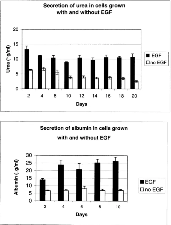
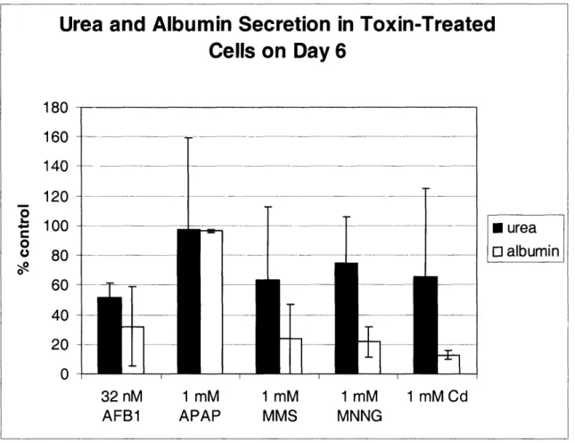
Understanding the hepatotoxicity of drugs and chemicals is essential for progress in academic research, medical science and in the development of new pharmaceuticals. Studying hepatotoxicity in vitro is a challenging task because hepatocytes, the metabolically active cells of the liver, are very difficult to maintain in culture. After just 24 hours, the cells detach from the plate and die, and even if they survive they usually do not express the metabolic functions which they have in vivo. It has been observed by others that culturing hepatocytes between two layers of collagen type I maintains in vivo-like morphology and also many drug metabolizing enzymes for weeks. In spite of the research examining drug metabolism in collagen sandwiches, there are very few studies evaluating this system for investigating hepatotoxicity. We cultured primary rat hepatocytes in the collagen sandwich configuration and our goal was to optimize this system for long-term studies and to examine toxicity of a variety of hepatotoxins. By measuring secretions of urea and albumin, and P4501A activity, we determined the optimal cell density to be 50,000 cells/cm2. We also evaluated the need for epidermal growth factor (EGF) in our cultures, by comparing urea and albumin secretions in cultures grown with and without EGF. The cultures without EGF had significantly less secretion of both urea and albumin just two days after plating. Therefore, we decided to include EGF in the medium. The toxins we examined were aflatoxin B1, acetaminophen, carbon tetrachloride, N-methyl-N'-nitro-N-nitrosoguanidine (MNNG), methyl methane sulfonate (MMS), cadmium, vinyl acetate and dimethylformamide (DMF). The cells were sensitive to aflatoxin B1, MMS, MNNG and cadmium. However, they were immune to acetaminophen, carbon tetrachloride, vinyl acetate and DMF. Our Western Blots showed that CYP1A, 2B and 3A were maintained in the culture for a week, but CYP2E1 was lost gradually over time. CYP2E1 is also the primary metabolic enzyme for acetaminophen, carbon tetrachloride and DMF. Thus, it is possible that the lack of toxicity is due to the loss of the enzyme responsible for the metabolism of these compounds. Immunity to vinyl acetate suggests that carboxylesterase is also lost in culture, since this enzyme is the one which converts vinyl acetate to acetaldehyde. The metabolism of acetaminophen was also examined with liquid chromatography and mass spectrometry. Liquid chromatography showed that acetaminophen is metabolized primarily to the sulfate and glucuronide metabolites. In order to investigate whether the glutathione adduct was formed, we synthesized the adduct and determined its retention time with liquid chromatography and its fragmentation pattern with mass spectrometry. We isolated the fraction with the same retention time from the medium of
conditioned medium from the hepatocytes to investigate the secreted protein profile, which could potentially be used to find toxicity biomarkers. We were able to remove most of the albumin from the medium using an immuno-affinity column containing anti-albumin antibodies bound to protein A-agarose beads. Our proteomic analysis combined gel electrophoresis with Qstar LC-MS/MS, and we were able to identify 493 proteins. Of those 493 proteins, 59 were plasma proteins, 234 were intracellular proteins and 21 were extracellular matrix proteins. The other 179 proteins were classified as miscellaneous, and they included 13 embryonic proteins, 40 central system proteins, 42 proteins from organs other than the liver and the central nervous system, and the remaining 84 proteins had unknown functions. Several of these proteins have only been identified in stellate and endothelial cells previously, suggesting the presence of nonparenchymal cells in the culture. Furthermore, we also identified many structural matrix proteins, such as laminin-12, fibronectin precursor and five types of collagen, showing that hepatocytes in the sandwich secrete the necessary matrix proteins for adhesion. In summary, we have shown that the collagen sandwich is a suitable system for hepatotoxicity studies as well as proteomic sanalysis.
Thesis supervisor: Steven R. Tannenbaum
Note: The term "collagen" is used in this thesis to refer to collagen type I, unless otherwise noted.
Acknowledgments
First and foremost, I would like to thank my advisor, Professor Steven Tannenbaum, for being a very dedicated and enthusiastic mentor. Steve gave me the opportunity to work on a very exciting project, which allowed me to grow in many scientific areas and has helped me to focus my choices for future career goals. I would like to especially thank Steve for always making time to discuss my research and for being very patient with me as I transitioned from a chemical engineering background into toxicology. Steve has also been supportive of my extracurricular interests, such as being a tutor in an undergraduate dormitory and attending classes at the Sloan Business School and the Museum of Fine Arts. During the years I spent in Steve's lab I have grown as a person as well as a scientist.
I would also like to thank my thesis committee members, Professors Linda Griffith, David Schauer and my committee chair Professor Ram Sasisekharan. Professor Griffith has been a mentor for me since my sophomore year as a chemical engineering student and she played a key role in my decision to pursue my graduate studies in the biological engineering field. I am also indebted to Professor Schauer for taking time out of his busy schedule to discuss my research with me, and for allowing me to use his cell culture facility for my experiments. I would also like to thank Professor Sasisekharan for being a great teacher in Tox.103, and for motivating my interest in the extra-cellular matrix, which led to some very interesting research in the last part of my thesis.
I am also very grateful to the Wogan lab for allowing me to use their cell culture facility. A special acknowledgement goes to Laura Trudel, and for sharing her bag of tricks with me when my experiments did not work, and for her insights for preventing repetitive strain injury. Her suggestions came in quite useful when I had to pipet many hundreds of times a day, and especially during the writing of this thesis!
I am also thankful to Pete Wishnok for his patience with me as I learned the necessary analytical skills for the proteomics and drug metabolism projects. Pete has not only been a great mass spectrometry teacher, but he has also motivated my interest in cultural events in Boston and exotic chili recipes! The success of my proteomics project is due in large part to the work of Vadiraja Bhat, who has mastered the method of sample cleanup and preparation. Vadiraj has also been a great friend, and I would like to acknowledge him for the many insightful discussions and for teaching me so much about proteomics. A big thank you goes to Man Ho Choi, who patiently worked out the analytical methods for the proteomics project and for the analysis of acetaminophen metabolites. I am also indebted to Paul Skipper, Rosa Libermann and Prithi Rao for time they spent analyzing my samples and for their helpful discussions.
I would also like to thank many other past and present members of the Tannenbaum Lab. Dr. Saraswathi Mandapati was one of my first teachers in proteomics, and her work has contributed very significantly to the research in this thesis. Ajit Dash has been very supportive during the last part of my thesis and has even lent me some of his cultures for microscopy studies. Anna Stevens has kept me company during late nights experiments and her sense of humor definitely made working late in lab very fun and entertaining! Teresa Wright and Chuck Thompson were my first cell culture teachers during the early years of the collagen sandwich, and I am very thankful for their patience
whom I could always count on for proofreading, literature search, and many cool tricks on the computer. Hong Bin Yu has been my personal Chinese coach, who helped me to impress my father-in-law, as well as many other native Chinese speakers! I would also like to thank Patty Sun, Ji-Eun Kim, Kevin Leach, Bill Connors and Joe Glogowski for their support and discussions during my research. I am also indebted to previous lab members, including Can Ozbal, Jinping Gan, Jaquin Niles, Joe Lee, George Nikov and Gerald Cozier for their support during the early years of my graduate studies.
A very big thank you goes to Amy Francis and Olga Parkin. Amy and Olga are incredibly efficient at getting things done. Whether I needed to order something, to get something fixed, to find information, or if I needed moral support, I could always count on them. Thank you Olga and Amy for always being there to cheer me up, and for creating such a welcoming environment!
I am also indebted to the Biotechnology Process Engineering Center (BPEC) for providing me with fresh hepatocytes on a regular basis and for their microscope facilities. I would like to thank past and present members of the lab, including, Emily Larson, Jessica Hart, Mark Powers, Anand Sivaram, Katie Wack and Carolyn Baker. I would also like to acknowledge the Whitehead Institute Biological Imaging Facility for the microscope facility. I would like to especially thank Nicki Watson for being a very patient and incredibly knowledgeable teacher.
I am also grateful to many other professors in the department, especially John Essigmann and James Sherley. Professor Essigmann was my housemaster when I was an undergraduate in New House (and later as a tutor there) and he played a very important role in my decision to apply to the Division of Toxicology for graduate studies. I would also like to acknowledge James Sherley, for setting aside time to discuss my research and for his enthusiasm during my graduate years.
The writing of this thesis would not have been possible without the support of the Adaptive Technology for Information and Computing (ATIC) lab. I would like to acknowledge the staff of the ATIC lab especially Kathy Cahill, Mary Ziegler and Kathleen Monagle, for allowing me to use their facilities so I could write my thesis in a safe, ergonomic and very fun environment! Thank you again, for being so supportive of my thesis, and for working with me to get through numerous computer troubles.
Last but not least I would like to thank my family. My husband, Leaf, has been supportive of me since our senior year in high school. His sense of humor, patience and creative energy has always lifted my spirits up and motivated me during my undergraduate and graduate years. Thank you, Leaf, for always believing in me. I would also like to acknowledge Leaf's parents and siblings for being so excited about my research and for being patient and supportive of me during tight deadlines.
The biggest thank you goes to my family in Hungary. I would first like to thank my brother, Adam, for being so excited about my studies and research, and for making me feel like a very special big sister. I would not be where I am today without the support of my parents, Jnos and Zsuzsanna Farkas. They have taught me discipline and a love for math, science and the arts at a very early age. Their foresight and sacrifices have opened up opportunities for me, which were unimaginable to them or their families. Their enthusiasm and encouragement have motivated me to realize my dreams academically and personally. In recognition of their love and support, I would like to dedicate this thesis to my parents.
Table of Contents
THIS DOCTORAL THESIS HAS BEEN EXAMINED BY A COMMITTEE OF THE
BIOLOGICAL ENGINEERING DIVISION AS FOLLOWS: ... 2
CHAPTER 1: IN VITRO METHODS TO STUDY HEPATOTOXICITY: A LITERATURE REVIEW ... 9
1.0 ABSTRACT ... 10
1.1 INTRODUCTION ... 11
1.2 IN VITRO MODELS OF THE LIVER ... 14
1.2.1 THE ISOLATED PERFUSED LIVER ... 15
1.2.2 LIVER SLICES ... 16
1.2.3 STATIC: CELL-CULTURES ... ... 18
Hepatotoxicity studies in primary hepatocytes ... 21
H epatoxicity in hum an cell lines ... 25
1.2.4 THREE.-DIMENSIONAL AND FLOW-THROUGH BIOREACTORS ... 28
1.3 THE "-OMICS" REVOLUTION ... 30
1.3.1 GENOMICS ... 30
Toxicogenom ics in vivo ... 32
Toxicogenom ics in vitro ... 33
1.3.2 PROTEOMICS ... 34
1.3.3 METABONOMICS ... 38
1.3.4 SYSTEMS BIOLOGY ... ... 39
1.4 CONCLUSIONS ... 41
CHAPTER 2: DEVELOPMENT OF A COLLAGEN SANDWICH BIOREACTOR FOR HEPATOTOXICITY STUDIES ... ... 44
2.0 ABSTRACT ... 45
LIST OF FIGURES ... 46
2.1 INTRODUCTION ... 47
2.2 MATERIALS AND METHODS ... 51
2.2.1 CELL CULTURE ... 51
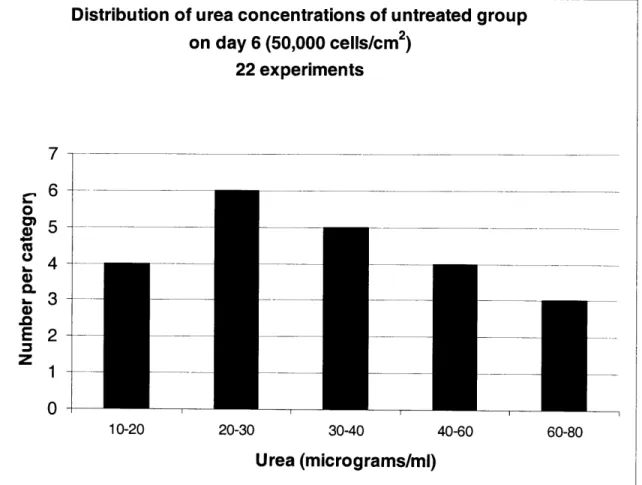
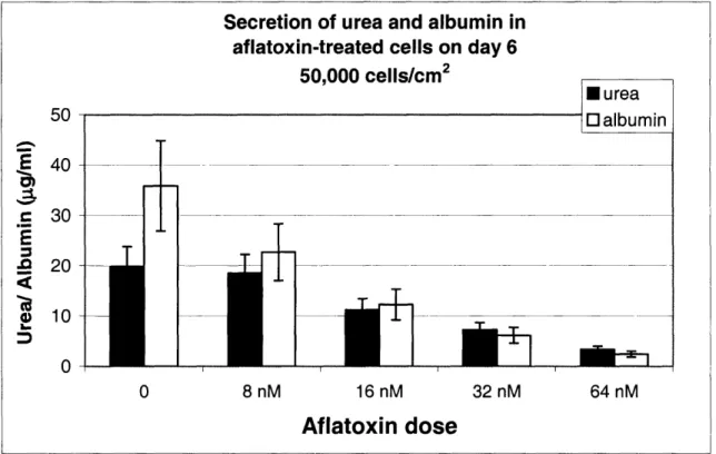
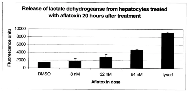
2.2.2 ASSAYS FOR MEASURING CELL VIABILITY ... 5 1 2.2.3 LIGHT MICROSCOPY ... ... 54 2.2.4 ToxIN TREATMENT ... 55 2.2.5 METAB()LISM OF ACETAMINOPHEN ... ... 56 2.3 RESULTS ... 57 2.3.1 CELL CULTURE ... 57 2.3.2 TOXICnY DATA ... 59
Toxicity of direct-acting compounds and acetaminophen - ... 59
2.3.3 COMPARING HIGH AND LOW METAL MEDIUM . ... 60
2.3.4 METABOLISM OF ACETAMINOPHEN ... ... 61
2.4 DISCUSSION ... 61
2.5 CONCLUSIONS ... 67
CHAPTER 3: CHARACTERIZING THE PROTEOME OF CONDITIONED MEDIUM FROM
RAT HEPATOCYTES CULTURED IN COLLAGEN SANDWICHES ... 86
3.0 ABSTRACT ... 87
LIST OF FIGURES ... 88
3.1 INTRODUCTION ... 89
3.2 METHODS AND MATERIALS ... 92
3.2.1 IDENTIFICATION OF ACUTE-PHASE PROTEINS ... 92
Sam ple preparation... 92
W estern blotting ... 93
3.2.2 CHARACTERIZATION OF THE SECRETED PROTEIN PROFILE ... 93
Silver staining - (performed by Saraswathi Mandapati) ... 93
Albumin removal (with Vadiraja Bhat) ... 94
Mass spectrometry ... 96
Data Analysis ... 97
3.3 RESULTS ... 97
3.3.1 DOWN- REGULATION OF ACUTE-PHASE PROTEINS ... 97
3.3.2 CHARACTERIZATION OF THE SECRETED PROTEOME ... 98
Depletion of albumin from conditioned medium... 98
Protein Profile ... 99
Silver Staining ... 100
3.4 DISCUSSION ... 100
3.4.1 DOWN-REGULATION OF ACUTE-PHASE PROTEINS ... 100
3.4.2 CHARACTERIZING THE SECRETED PROTEIN PROFILE ... 101
Plasm a proteins ... 102
Extracellular matrix and membrane proteins ... ... 103
Miscellaneous proteins... 105
3.5 CONCLUSIONS ... 107
FIGURES ... 109
CHAPTER 4: SUMMARY AND CONCLUSIONS ... 119
REFERENCES ... 124
APPENDIX I: DETAILED PROTOCOLS ... 141
APPENDIX II: DETAILED METABOLIC PATHWAYS FOR AFLATOXIN B1 AND ACETAMINOPHEN ... 149
CHAPTER 1: In vitro methods to study hepatotoxicity: A literature
review
1.0 Abstract
Understanding the hepatotoxicity of drugs and chemicals is essential for progress in the pharmaceutical industry, medical science and academic research. The study of
hepatotoxicity is complicated by the difficulty of culturing hepatocytes in vitro due to a lack of understanding of the humoral and matrix requirements of these cells. A variety of
in vitro models of the liver have been developed, such as perfused livers, liver slices,
static cell cultures and three-dimensional perfused bioreactors. The static cell culture is the most commonly used system for hepatotoxicity studies. In this review we present the role of each system in the study of drug metabolism and toxicity. We will also discuss the application of primary cells and human cell lines in toxicity studies. Since the static cell culture has been used in so many studies, we will just highlight some of the
hepatotoxicity studies with model. Finally, we will discuss the impact of genomics, proteomics and metabonomics on hepatotoxicity studies, specifically how these approaches have helped to understand differential expression under toxic conditions.
1.1 Introduction
Developing new systems and methods for studying liver toxicity has important applications in medical science, academic research and the pharmaceutical industry. The liver is one of the major detoxifying organs in the body and it is especially susceptible to foreign agents, such as drugs and environmental pollutants, and their metabolites [1]. For this reason, the liver is the primary organ targeted during early toxicity screening studies and nonclinical safety assessment. It has been shown that 40% of the failures during preclinical trials are due to toxicity [2]. Furthermore, only one out of five drugs, which reach clinical trials, also reach the market place, and many of them fail due to hepatotoxicity. This has serious financial implications for the pharmaceutical industry, since the cost of bringing a drug to the market today is estimated to be $800 million. In fact, even though investment in drug development has tripled in the last 10 years to $30 billion, there are fewer drugs coming to the market. This phenomenon is thought to be due to increasing costs of developing drugs and to withdrawal of drugs during the post-marketing period because of adverse drug reactions. Thus, developing a system which would eliminate compounds earlier in the pipeline would be very beneficial [2].
Based on a survey between 1961 and 1992 from France, Germany, UK and USA, 23 out of 181 market withdrawals due to safety were because of hepatotoxicity [3]. Hepatoxicity during post-marketing also leads modification of labeling and stricter guidelines for prescriptions, limiting the market potential of the drug. In fact, development of hepatitis is a common side-effect of drugs. According to Zimmerman et al.[4] 10% of hepatitis is caused by drugs, and in patients over 50, exposure to drugs
accounts for 40% of hepatitis. Additionally, 15-25% of fulminant hepatic failure is due to adverse reaction to drugs.
In spite of the data generated by previous pharmaceutical and clinical research it is still not trivial to estimate the appropriate doses in humans based on animal studies. One reason is that there are significant species differences in the drug metabolism enzymes, as well as in the kinetics of the metabolic reactions. There are also significant polymorphisms in drug-metabolizing enzymes among humans. For example, West African populations have CYP2D6 variants, and some populations of Australian descent have variants of CYP2D6, CYP2C9 and CYP2E1 [5]. Furthermore, many instances of hepatotoxicity are due to an immune-mediated iodisyncratic response, which would be almost impossible to predict based on animal models.
In order to understand the metabolism of novel compounds, there has been a growing interest in the development of in vitro systems for studying hepatotoxicity. Hepatocytes, the metabolically active cells in the liver, are one of the most challenging cells to maintain in vitro. When plated on plastic or collagen-coated dishes, they have been shown to detach and lose their metabolic capacities within 24 hours. The needs of hepatocytes in vitro are not well-understood, but it is known that hepatocyte-specific
gene regulation is influenced by both soluble and insoluble factors [6]. Soluble factors include hormones, and cytokines, and insoluble factors refer to cell-cell and cell-matrix contacts. There is currently no universally accepted protocol for maintaining hepatocytes, since as we will discuss, most factors can either stimulate regeneration or differentiation, but not both. In fact, the factors, which lead to hepatocyte differentiation usually suppress differentiation, and vice-versa. For example, long-term cultures of hepatocytes usually
requires a mitogenic compound, such as epidermal growth factor (EGF), which stimulates DNA synthesis. However, it has also been shown that addition of EGF to hepatocyte cultures leads to a down-regulation of the metabolic enzymes, specifically CYP1A, CYP2B [7;8], CYP2C11 [9] and CP3A [10]. Cell density also has a profound influence on cell proliferation and metabolism. Plating cells at low density increases cell proliferation, but reduces the expression and inducibilty of metabolic enzymes [11]. Therefore, when designing in vitro systems for detecting hepatotoxicity, these parameters need to optimized so that cells can be sustained without losing metabolic functions. Once the system has been designed, one still has to establish the experimental endpoints, which would be most relevant to the particular compound. Optimizing in vitro testing of compounds also involves determining the appropriate doses and the relevant time-points.
As we will discuss, the parameters of in vitro systems also depend on the type of toxin studied. While the variety of toxins is so complex, that no strict categorization can be applied, they are usually separated according to whether or not they need to endogenously activated for toxicity. The compounds, which are metabolically activated usually cause toxicity through formation of electrophiles, nucleophiles, free radicals or macromolecular binding [12]. Several commercial assays have been developed to measure toxicity, including measuring for leakage of intracellular enzymes, such as lactate dehydrogenase (LDH) and aspartate amino transferase (AST) as well as reduction of the tetrazolium salts, XTT and MTT. Furthermore, secretion of urea and albumin are also used as markers to asses the health of hepatocytes. Although these commercial assays can be used to measure cell viability, detection of toxicity should also include experimental endpoints known to be relevant to the mechanism of toxicity for a specific
compound. We will discuss some compound-specific endpoints, such as DNA-adducts and protein nitrotyrosine adducts.
We will also review the influence of the "-omics" revolution (genomics, proteomics and metabonomics) on the study of hepatoxicity. Application of genomics and proteomics technologies in toxicology have opened up the field of "toxicogenomics" and "toxicoproteomics." These technologies have facilitated very rapid collection of data to analyze differential gene, protein and metabolite expression, potentially enhancing the development of diagnostics and therapeutics. We will also discuss how the integration this data with systems biology has the potential to facilitate screening of drugs and prediction of metabolism and toxicity.
1.2 In vitro models of the liver
The in vitro systems, which are most commonly used to study drug metabolism and liver toxicity are (a) perfused livers, (b) liver slices, (c) static cell cultures, (d) flow-through bioreactors, and (e) cell-free systems such as microsomes, liver cytosol fractions and S9 fractions. Although perfused livers and liver slices have been used to study hepatotoxicity, static cell cultures are the most wide-spread system for toxicity studies. We will discuss many of the important parameters in cell cultures, including composition of the medium and the extracellular matrix, cell-density and the effects of co-culturing with nonparenchymal cells.
1.2.1 The isolated perfused liver
The isolated perfused liver is considered to be the closest system to in vivo experiments, since it maintains the three-dimensional architecture, the bile flow and the hemodynamics of the liver. Besides its role as an in vitro model, it has also been used in clinical settings to treat patients with hepatic coma [13;14]. Although it is expensive to maintain and it can only be used for a few hours, the isolated perfused liver has been used for various hepatotoxicity studies, especially for compounds, which could affect the bile flow. For example, the toxicity of the immunosuppressant drug, cyclosporine, has been studied in isolated perfused livers from rats. It has been shown that this drug can lead to significant liver injury, including cholestasis, and leakage of lactate dehydrogenase and glutamate dehydrogenase [15]. The isolated perfused liver has also been useful in studying the effects of metals, such as cadmium, mercury and copper [16]. These metals were found to decrease the bile flow, but they also decreased ATP content, glutathione content and increase the release of lactate dehydrogenase. Furthermore, copper also led to significant lipid peroxidation, as measured by malondialdehyde content. Release of oxidized glutathione (GSSG) into the bile has been used by several groups as a marker of oxidative stress by aromatic amines and nitroarenes, and it has been concluded that the carcinogenic doses are probably not sufficient to cause acute injury or oxidative stress in the liver [17;18]. The toxicity acetaminophen has also been studied in this system in order to determine how decreased metabolism due to hypothyrodism affects toxicity by this compound. Interestingly, it has been found that hypothyrodism protects the liver from injury by acetaminophen, even when the expression of CYP2E1 is the same as in the control groups [19]. In summary, the isolated perfused liver can be a useful system
for hepatotoxicity studies especially for studying changes in bile flow. As the above studies show, this system has also been used to study oxidative damage and release of intracellular enzymes. While the isolated perfused liver can be a good model for the bile flow and the three-dimensional architecture of the liver, it is very short-lived and it is probably best-suited for compounds which are expected to have toxic effects within a few hours.
1.2.2 Liver slices
Liver slices have been prepared since 1923 [20] and they have been used in a variety of toxicity studies because they maintain the cell-cell and cell-matrix interactions found in liver. The disadvantage of this system in comparison with the isolated perfused liver is that it cannot be used to analyze bile flow. It is also short-lived, primarily due to poor oxygen and nutrient diffusion from the medium. Liver slices are expected to be viable for a few hours, although some groups have found them to be metabolically active for two days [ 13;21].
Liver slices from rats, guinea pigs, male cynomolgus monkeys, and humans have been used to study unscheduled DNA synthesis (UDS) after exposure to the mutagens aflatoxin B 1 (AFB1), 2-acetylaminofluorene (2-AAF) and 6-aminochrysene (6-AC) in order to determine species differences in response to these toxins [22]. UDS was induced by all three compounds in rats, guinea pigs, and humans. Although UDS was observed in monkeys by AFB1 and 6-AC, 2-AAF did not induce DNA synthesis possibly due to a lack of CYP1.A2 in this species. A comparison of AFB 1 metabolism between humans and rats was also carried out by Heinonen et al. [23], who showed that the rates of metabolism were similar among rats and humans, but there were significant inter-indivual
differences among the three human liver slices observed. This is an interesting result, since it suggests that species differences in metabolism of alfatoxin B1 in liver slices might be less significant than inter-individual differences within the same species.
Environmental pollutants such as cadmium and the herbicide, paraquat, have also been examined in liver slices. The toxicity of cadmium was investigated by Chan et al. [24], who demonstrated that pre-induction with zinc sulphate protected the slices from a decrease in glutathione concentration. The protective effect of zinc sulphate against cadmium toxicity has also been observed in hepatocytes by Moffat et al. [25]. Paraquat has been shown cause significant toxicity within six hours of exposure around the central vein [26]. The role of superoxide in the toxicity of this compound was examined by showing that addition of a superoxide dismutase inhibitor increased lactate dehydrogenase leakage. Inflammation caused by exposure to paraquat was also visualized by histological examination of the liver slices.
Studies of tissue slices are not limited to liver slices. Toxicity caused by a number of halogenated compounds, including the liver toxin carbon tetrachloride, has been examined in slices of rat liver, kidney spleen and testes in order to examine how well the LD50 correlated with doses shown to cause lipid peroxidation [27]. Interestingly,
the doses only correlated for a few of the compounds, suggesting that lipid peroxidation alone is not sufficient for lethality. In summary, these studies show that liver slices have been applied in a number of different hepaotoxicity studies, and can be useful for studying DNA damage, oxidative damage and histology.
1.2.3 Static cell-cultures
Static cell cultures are currently the most popular in vitro systems for studying
hepatotoxicity. The most commonly cultured cells are freshly isolated hepatocytes, since cryopreserved cells and cell lines do not express the necessary metabolic enzymes. However, cryopreserved cells and cell lines can be induced to express many of the cytochrome P450's, by growing the cells in the presence of phenobarbital, dexamethasone, rifampicin and 3-methylcholanthrene [28;29]. We will discuss the role of human cell lines in toxicity studies in a later section.
In order to facilitate adhesion of cells to the surface, most cultures are plated on dishes coated with extracellular matrix proteins, such as collagen, fibronectin and laminin. Culturing hepatocytes on these structural matrix proteins has been found to also maintain cell morphology and the metabolic functions much better and for longer than plating them on plastic. Culturing hepatocytes in a sandwich configuration is even more suitable for maintaining metabolically active cells, and hepatocytes in this configuration have also been shown to maintain biliary secretion. The most common matrices for sandwich cultures are collagen type I and Matrigel, an extract derived from the basesement membrane of the Engelbroth- Holm-Sworm mouse sarcoma [30]. Both collagen-collagen and collagen-Matrigel sandwiches have been shown to maintain morphology and albumin secretion for several weeks [31], and they are also inducible for both CYP1A2 and CYP3A4 in systems with human hepatocytes [32]. Collagen-Matrigel
sandwiches have been shown to have some advantages over collagen-collagen sandwiches, such as expression of the gap junction protein connexin 32 and the expression of the epidermal growth factor receptor[31] . However, the disadvantages of
Matrigel are that it is significantly more costly than collagen, and there are many batch-to-batch variations. Thus, most studies in a sandwich culture have been carried out in collagen-collagen sandwiches. Cells in a collagen sandwich have been shown to maintain
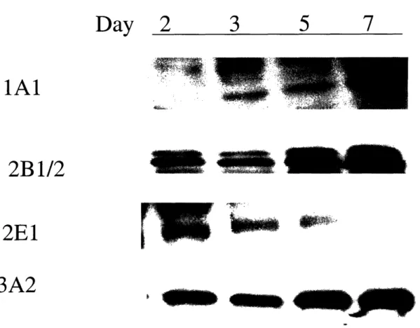
their polygonal morphology [33-35] and remain viable in culture for up to six weeks. In addition, they maintain CYP1A and CYP3A, sulfo-and glucuronsyltransferases, as well as glutathione S-transferase [36;37]. In fact, hepatocytes in the sandwich configuration have been shown to maintain biliary exretion [38-40] and this efflux has been show to be inhibited in a dose-dependent manner by cyclosporine A, a known blocker of bile secretion.
The composition of medium is also a very important factor in the maintenance of long-term cultures. Components in the medium can influence regeneration of hepatocytes as well as up-regulation or suppression of metabolic enzymes. In the absence of serum, long-term cultures of hepatocytes require insulin, as well as a mitogen such as EGF [41]. While EGF has been shown to maintain viable hepatocytes for weeks, it has also been shown to significantly decrease CYP2C11 [9] and CP3A [10] activities in hepatocytes and specifically CYP1A and CYP2B in collagen sandwiches [7;8]. This effect can lead to lack of toxicity from compounds metabolized by these enzymes. In contrast, Phenobarbital suppressed hepatocyte growth and secretion of structural matrix proteins, while maintaining expressions of CYP1A, 2B and 2E1 [42]. Thus, the choice of medium should be influenced by the type of application, such as the life-time of the culture system, and which toxins will be tested in the system.
parenchymal cells play very important roles in the survival and differentiation of hepatocytes as well as in the mediation of toxicity. Endothelial cells line the sinusoidal wall and can play a role in the reorganization of hepatocytes in culture. They also secrete cytokines, nitric oxide and matrix components. Kupffer cells are the hepatic macrophages and they mediate inflammation and innate immunity through secretion of signaling molecules and antigen presentation. In the healthy liver, stellate cells are responsible for storing vitamin A and maintaining the extracellular matrix. When the liver is injured, stellate cells transform into myofibroblast cells to mediate the inflammatory response [43]. Co-culturing of nonparenchymal cells is challenging because the medium and matrix composition has to satisfy the needs of two different cell types. Co-cultures with endothelial cells has been achieved by Hirose et al. [44] and Harimoto et al.[45] and they observed that co-cultures maintenained cell junctions, secretion of extracellular matrix and hepatocyte morphology better than hepatocytes grown alone. Co-culture with stellate cells can also stimulate DNA synthesis [46] and maintain CYP450 activities [47]. Hepatocyte function can also be stimulated with conditioned medium from nonparenchymal cells. Kang et al. [48] exposed Matrigel and collagen gel sandwiches to conditioned medium from a culture of nonparenchymal cells and found an increase in DNA synthesis as well as an improvement in hepatocyte morphology. As these studies show, hepatocyte function in culture can be altered by a variety of matrix components, medium components and non-parenchymal cells, and the design of hepatotoxicity experiments with hepatocytes needs to take all these factors into consideration.
Hepatotoxicity studies in primary hepatocytes
It would be impossible to give a complete overview of all the hepatotoxicity studies with primary hepatocytes, because they have been used in a variety of toxicity studies. Most studies use hepatocytes from rat and mice, although we did find some references with primary human hepatocytes. In this section, we will highlight some examples of hepatotoxicity studies in primary hepatocytes with model toxins, particularly with acetaminophen and carbon tetrachloride, in order to demonstrate some of the advantages and disadvantages of this system.
Acetaminophen, an over-the-counter analgesic, has caused significant concern because of its easy accessibility and its history as a common suicidal agent. It has been used as a model toxin to study acute liver failure and the role and of biotransformation in hepatotoxicity [4]. Acetaminophen (APAP) can be metabolized by CYP1A2, CYP3A4 and CYP2El and converted to the reactive N-acetylbenzoquinone imine (NABQI), but in humans this conversion occurs primarily by CYP2E1 [49]. NABQI is detoxified by conjugation with glutathione, but after the glutathione is depleted NABQI can bind to cytosolic and mitochondrial proteins [50]. In cultured mouse hepatocytes 5 mM APAP led to oxidant stress and ultimately necrosis, but this toxicity was attenuated by pretreating the cells with N-acetylcysteine, a precursor of glutathione [51]. APAP has also been shown to cause DNA strand breaks, although the exact mechanism for this is not known. It has been shown recently that addition of the antioxidants, N-acetylcysteine, polyphenol silibin and ot-tocopherol, protected primary rat hepatocytes from APAP-induced DNA strand breaks, but not APAP-APAP-induced toxicity [52]. The role of oxidant
production at the doses which caused DNA damage. Experiments with acetaminophen have also been useful in determining the effect of growth factors on toxicity. The growth factors EGF and HGF are known to suppress metabolic enzymes, but they have also been
shown to offer protection against APAP toxicity in rat hepatocytes [53].
It is well-known that consumption of ethanol together with APAP leads to hepatotoxicity, due to the induction of CYP2E1 by alcohol [12;54]. Interestingly, caffeine has also been shown to increase APAP hepatotoxicity in mice and rats [55;56]. DiPetrillo et al. [57] investigated the effect of caffeine on APAP toxicity in cultured rat hepatocytes. They observed that ethanol treatment alone did not increase APAP toxicity, although it
did induce CYP2E and CYP3A. However, when ethanol-pretreated hepatocytes were exposed to caffeine, there was a significant increase in APAP toxicity. In addition,
caffeine increased the formation of the glutathione adduct (an indirect measure of NABQI formation) three-fold compared to cells exposed to just ethanol and APAP. These results could have important implications for human consumption of APAP with caffeine, which is found in many beverages and medications.
Carbon tetrachloride, which was used previously as a dry cleaning agent and refrigerant, has also been widely studied due to its severe hepatotoxic effects. It is converted to a free radical after undergoing reductive deholgenation by cytochrome P450's to form a carbon-centered free radical [58], that initiates lipid peroxidation and
leads to morphological changes. Hepatocyte cultures lend themselves very well to microscopic investigations, allowing for detailed examination of structural changes after toxin treatment. While light microsopy has been used widely to study gross microscopic changes, the development of more advanced microscopy methods such as
epiflourescence microscopy, scanning electron microscopy, and laser-scanning confocal microscopy have significantly enhanced morphological studies. These techniques have made it possible to perform immunologic detection, to visualize ultrastructural changes and to recreate a cell or even a tissue section in three dimensions.
Toxicity following carbon tetrachloride treatment was investigated in hepatocyte cultures with light microscopy and it was observed that carbon tetrachloride leads to significant morphological alterations, which result from the peroxidation of polyunsaturated fatty acids in cellular membranes and affect the endoplasmic reticulum, the Golgi apparatus, the plasma membrane and the mitochondria [59;60]. Carbon tetrachloride is also known to be a fast-acting toxin and in vivo administration results in morphological changes appearing within 15 minutes after exposure. Thus, carbon tetrachloride is a good model compound for studying morphological changes in hepatocyte culture. In fact, carbon tetrachloride has been observed to cause significant ultrastructural changes in membranes 5 minutes after treatment. Scanning and transmission electron microscopy which magnify cells many thousand-fold can be used to visualize the effects of carbon tetrachloride, which include blebbing of the plasma membrane, smoothing out and degranulation of the rough endoplasmic reticulum 24 hours after treatment [59].
Microcystin-LR, a potentially lethal toxin produced by aquatic organisms, has been shown to exert its toxicity by inhibiting serine/threonine protein phosphatases, specifically types 1, 2A and 3. Similarly to carbon tetrachloride, microcystin has been shown to lead to lipid peroxidation, as well as production of reactive oxygen species and
microscopy, it has been shown that microcystin exposure in hepatocytes leads to cell shrinkage, nuclear condensation, collapse of the actin cytoskeleton and cell blebbing, suggesting that microcystin causes cell death through apoptosis [62;63].
Confocal laser scanning microscopy (CLSM) combined with fluorescent detection of monochlorobimane (a lipophilic glutathione-specific probe, which is only fluorescent 'when bound to thiols) has also been applied to measure intracellular glutathione distribution [64]. This new technique has used to monitor depletion of glutathione by inhibitors such as buthionine sulfoximine, and known glutathione depletors, such as menadione, in rat hepatocytes. Since glutathione plays an important role in detoxification, this new method could be used to study the role of glutathione in the metabolism of novel compounds.
CLSM has also been used to evaluate the viability of cryopreserved hepatocytes. McKay et al. [65] used a vital stain carboxyfluorescein diacetate, which is deacetylated by intracellular esterases to form a fluorescent carboxyfluorescein (CF). CF cannot diffuse through membranes and is retained inside the cells. CLSM imaging showed higher CF retention for cells cultured in Williams' medium, versus Chee's medium, prior to freezing. Interestingly, the imaging suggested better retention of CF inside organelles, suggesting that the membranes of the organelles are less sensitive to freezing than the cell membranes, which is an important consideration when using cryopreserved cells for cell culture.
Since hepatocytes in culture do not express all the metabolic enzymes, in vitro experiments are sometimes combined with in vivo experiments by pretreating animals with inducers before hepatocyte isolation. It has been shown that carbon tetrachloride
toxicity is potentiated in rat hepatocytes isolated from animals treated with phenobarbital. In addition, the levels of total CYP450 were very close to those in vivo [66]. On the other hand, pretreating the animals with a protective agent such as alpha-tocopherol, significantly protected hepatocytes from necrosis due to carbon tetrachloride compared to cells from untreated rats. Lipid peroxidation, which is usually measured by monitoring malondialdehyde formation with thin layer chromatography, was also found to be
significantly reduced in hepatocytes from treated rats [67].
Isolation of hepatocytes from treated rats has also been used for studying the anticancer drug, Yondelis. This drug, has been shown to be effective in treating breast and ovarian cancer, but it also causes hepatotoxicity in rats [68]. However, it has been observed that pretreatment of rats with dexamethasone, protects against hepatotoxicity, probably due to the upregulation of CYP3A, which metabolizes the drug. However, toxicity of Yondelis was not ameliorated in hepatocytes isolated from dexamethasone-pretreated rats. Thus, upregulation of the enzymes is not the only mechanism responsible for the protection against hepatotoxicity, suggesting that in order to study drugs with unknown mechanism, in vivo experiments are necessary for complete understanding of toxicity pathways.
Hepatoxicity in human cell lines
The HepG2 hepatoma cell line is one of the most popular human cell lines for hepatoxicity studies. This cell lines has been used to study the mechanisms of toxicity of a variety of toxins such as 7H-Dibenzo[c,g]carbazole (DBC) and aflatoxin B1 [69]. DBC, N-heterocyclic aromatic hydrocarbon, has been classified as carcinogenic and
hepatotoxic in several species. By exposing HepG2 cells to DBC it has been shown that at doses lower than 10 zM, DBC forms DNA adducts and leads to apoptotic cell death. At doses higher than 80 tM, there were fewer DNA adducts, and necrotic cell death was apparent from cell swelling and loss of membrane integrity. In contrast to the biphasic death curve of DBC, exposure to AFB 1 resulted in a concentration-dependent increase in apoptosis.
The cytotoxicity of ethanol and arachidonic acid was studied in transduced HepG2 cells expressing CYP2E1 [70]. Toxicity by both compounds led to apoptosis, as shown by activation of caspases I and III. Furthermore, apoptosis was prevented by transfection with a plasmid containing Bcl-2. Thus, HepG2 can be used as a model cell line for studying toxicity and apoptosis by hepatotoxins. HepG2 cells have also been used to study the toxicity of pharmaceuticals such as troglitazone [71], an antihyperglycemic agent, and diclofenac [72], an anti-inflammatory drug, both of which have been shown to cause adverse hepatic reactions in patients. Troglitazone was found to cause significant mitochondrial damage as revealed by ultrastructural analysis, as well as a decrease in ATP levels and mitochondrial membrane potential. Diclofenac was actually more toxic to freshly isolated hepatocytes than to HepG2 cells, probably due to drug-metabolizing capacity of primary cells. Although diclofenac was shown to lead to mitochondrial damage in HepG2 cells, their decreased sensitivity in comparison to fresh hepatocytes suggests that HepG2 cells are better suited for testing drugs not requiring metabolism. However, as mentioned previously, HepG2 cells can be transduced to express certain CYP450's, making it possible to test toxicity of indirect toxins.
Another human hepatoma cell line (HLE), was engineered to express 2E1[73]. It was observed that a 3mM dose of CC14caused almost 90% death in engineered cell lines
but only a 20% death in the non-engineered controls. It was also shown that the gene expression of the heat shock protein HSP70, a marker of oxdative stress, increased more in the engineered cell lines than in the control cells after CC14exposure. Mace et al.[74]
engineered several human cell lines from THLE, a non-tumorigenic human cell line. These cells retained most of their phase II enzymes, but lacked the cytochrome P450's. The engineered cell lines were transfected with human CYP's 1A2, 2A6, 2B6 and 3A4. The sensitivity of these cell lines to aflatoxin Bi was, 125-, 2-, 2, and 15-fold higher than in the non-engineered cells. This result is consistent with in vivo metabolism studies since it is known that in humans CYP1A2 is the primary enzyme and CYP3A4 is the secondary enzyme responsible for the metabolism of aflatoxin. The DNA adducts were also quantified and it was found that nanomolar doses caused DNA adduct formation. The primary adducts were AFB 1-N7 guanine, -pyrimidyl, and -diol. In addition, all three cell lines showed a dose-dependent accumulation in S-phase but they all had different abilities to recover from the S-phase [75]. These results have biological significance, since the most common lesions in vivo are the N7-guanine and the pyrimidyl, also known as AFB 1 -formamidopyrimidine (FAPY) [76].
Recently, a new human cell line, BC2, was derived from HepG2 and it has been shown that this cell line can maintain a number of drug-metabolizing enzymes, such as CYP1A1/2, 2E1l and 3A4, for up to five weeks in culture [77]. This cell line has also been shown to be sensitive to several toxins including acetaminophen and acetylsalicylic acid [78].
1.2.4 Three-dimensional and flow-through bioreactors
Although static hepatocyte cultures are inexpensive and practical, they lack the three-dimensional cell-cell contacts and the constant flow of oxygen and nutrients, which are present in the liver. In order to remedy this issue, several perfused bioreactors were developed. The earliest flow-through bioreactors were hollow fiber bioreactors, which are used today to sustain patients with acute liver failure [79], but they have also been tested as in vitro models of drug metabolism. Hollow fibers have been shown to maintain hepatocyte morphology such as bile canaliculi and intercellular junctions even seven days after culture [80]. Nyberg et al. [81] cultured gel-entrapped rat hepatocytes perfused in a hollow fiber reactor and showed that the hepatocytes in this system maintained several metabolic activities, including sulfation, glucuronidation, P4503A activity. Powers et al. [82;83] have developed a perfused three-dimensional bioreactor, consisting of primary rat hepatocytes seeded onto channels etched into a silicon scaffold. After seeding, the cells rearranged themselves into tissue-like structures, and it was observed that pre-aggregation of cells into spheroids prior to seeding improved morphogenesis. This system was also able to maintain rates of albumin and urea secretion for 15 days after isolation. There are also a few limited studies on the application of perfused systems for drug metabolism studies. Bader et al. [84] developed a flow-through collagen sandwich bioreactor with primary rat hepatocytes. They have shown that the metabolic profile of the antihypertensive drug, urapidil, includes all the metabolites found in vivo, suggesting this system can be used for drug metabolism studies.
In addition to all the considerations in the design static culture experiments, design of perfused reactors needs to take several addition parameters into account. The shear stress associated with the flow can lead to cell detachment, thus it can be advantageous to coat the surfaces with a matrix protein such as collagen to enhance cell adhesion. This strategy also ensures cell adhesion to the desired areas [83]. On the other hand, some shear stress can be beneficial, since it has been shown to stimulate metabolic functions and morphogenesis [85;86]. Furthermore, the flow of the medium needs to be controlled, in order to ensure an even distribution of oxygen and nutrients in the system. One of the considerations with metabolism studies is that the perfusion introduces significantly more liquid volume, which dilutes the concentration of drugs and their metabolites. This dilution factor complicates the detection of metabolites in the effluent medium, especially if an on-line detection system is desired. Therefore, design of perfused bioreactors for metabolism and hepatotoxicity studies should consider the parameters mentioned above to ensure more physiological conditions and maximum sensitivity.
1.2.5 Subcellular fractions
Subcellular fractions, such as microsomes, are widely used for studying metabolic activation of drugs. Microsomes, the packets of enzymes on the endoplasmic reticulum, contain the P450's and the necessary cofactors for metabolism of drugs[13]. Micrsomes are particularly useful for comparing species differences in the bioactivation of drugs by P450's. Human-derived liver microsomes have also been used to study the metabolisms
It was observed that CYP1A2 contributed to over 95% of the AFB1 activation. This result is physiologically relevant DNA binding studies also showed that CYP1A2 was responsible for 95% of the AFB1 adduct formation [88].
Other subcellular factions such as liver cytosol and the S9 fraction have also been used for drug metabolism studies. The liver cytosolic fraction is isolated by differential centrifugation of a liver homogenate [89]. The advantage of this fraction is that it contains phase II enzymes such glutathione S-transferase and N-acetyltransferase. Although additional cofactors, such as acetyl coenzyme A, are necessary for this system, liver cytosolic fractions are available commercially and are easy to use. The S9 fraction of the liver contains the cytosols and the microsomes, thus it combines the metabolic capacities of the two systems. However, the S9 fraction also requires additional cofactors for phase II activities, as well as an NADPH-regenerating system to maintain the CYP450 enzyme activities.
1.3 The "-omics" revolution
1.3.1 Genomics
The genomics revolution, which began with the genome sequencing project, aims at identifying all the genes involved in biological processes. The identification of the complete genome sequences of multiple organisms made it possible to engineer DNA microarrays, which can measure gene expression of hundreds or even thousands of genes simultaneously. DNA microarrays consist of a solid support with oligonucleotides or
cDNA complementary to the sequences under investigation. The microarrays are incubated with fluorescently labeled samples, and hybridization is detected using fluorescence. The data shows the up or down-regulation of the genes and allows for comparison of gene expression levels between different biological samples. Boess et al. [90] evaluated gene expression in two cell lines, hepatocytes in a conventional monolayer, hepatocytes in a sandwich and liver slices. Overall, liver slices were the most similar to the in vivo liver, but even this system has decreased expression of several genes, including many of the drug metabolizing genes. It was also observed that gene expression changes most dramatically right after isolation and then stabilizes during the culture. This has implications for drug metabolism studies, since this data suggests that in
vitro systems have significantly lower metabolic activities, even though some metabolic
capacity is maintained throughout the life of culture.
Using a special toxicological microarray, which contained 900 genes, Baker et al. [91] studied the temporal changes (between 4 and 72 hours) in genes relevant to toxicological studies and confirmed the studies with RT/PCR. Clustering analysis showed that there was a time-dependent dedifferentiation response of the hepatocytes. Although many of the cytochrome P450's were down-regulated, their gene expression changed at different time-points. The expression of the phase II enzymes was more varied with UDP-glucuronosyl transferase and GST pi being upregulated, while the expressions of sulfotransferase and GST alpha were repressed.
DNA microarrays been also applied in toxicological studies to identify the pathways involved in toxicity mechanisms, opening up the fields of toxicogenomics The aim of toxicogenomics to identify a global gene expression profile in order to determine
the differential gene expression between two samples [2]. Toxicogenomics has been used to study differential gene expression caused by hepatotoxins including aflatoxin B1, 3-methylcholanthrene and dimethylnitrosamine.
Toxicogenonics in vivo
Gerhold et al.[92] exposed rats to several inducers of CYP450 genes, namely 3-methylcholanthrene (3MC), phenobarbital, dexamethasone or clofibrate and measured the expression of several drug metabolizing genes including CYP's, glutathione transferases,
sulfotransferases, and drug transporters genes. It was observed that that the responses of the genes agreed with the published observations. For example, 3MC induced CYP1A1/2 and CYP2B1 and several phase II enzymes including UGT1A6, GSTA1, GSTA2 and GSTM1, all of which have been observed in previous research to be induced by 3MC in rat hepatocytes.
Ellinger-Ziegelbauer et al. [93] compared the expression profiles induced in rats by four genotoxic hepatocarcinogens: dimethylnitrosamine, 2-nitrofluorine, aflatoxin B1 and 4-(methylnitrosamino)- 1 l-(3-pyridyl)- 1 -butanone. As expected, the expression profiles among the four different toxins showed several similarities including up-regulation of DNA damage, oxidative stress and fibrosis response genes. Furthermore, there was a significant increase in the tumor suppressor protein p53. It is encouraging, that compounds which are known to act through the same pathways lead to similar changes in the gene expression profiles in the rat liver. Similar results were found by Waring et al. [94] who tested 15 different hepatotoxins including carbon tetrachloride, dimethylformamide and arsenic, and analyzed the histopathologic and gene expression caused by these agents. Treatment with these agents resulted in significant liver injury
including cirrhosis, hypertrophy and also hepatocellular damage such as necrosis and DNA damage. The gene expression results correlated well with the histopathological results, suggesting that genomics analysis can be used to evaluate hepatotoxicity changes
in vivo.
Toxicogenomzics in vitro
In vitro systems have decreased gene expression compared to the liver, but
experimental data suggests that they are still suitable for toxicogenomics studies. Waring et al. [95] treated rat hepatocytes with several hepatotoxins such as carbon tetrachloride and dimethylformamide in order to study differential gene expression. The changes in gene expression in the hepatocytes correlated well with the histopathological changes observed in the in vivo experiment. Furthermore, compounds with similar toxicity mechanisms led to overlapping changes in gene expression. These changes were not identical, however, suggesting that each compound might have a unique genomic signature.
Most microarrays contain many thousands of targets, therefore special microarrays have been developed for particular applications, which contain a smaller set of genes. De Longueville et al [96] used a special microarray containing 59 genes, which were thought- to be relevant to toxicology. The chip included the CYP450's, stress response genes and genes involved in apoptosis. Eleven toxins, divided into 4 categories (CYP 450 inducers, necrosis inducers, cholestasis inducers and steatosis inducers) were tested on primary rat hepatocytes. Changes in gene expression correlated well with the known mechanisms of toxicity for these compounds, and clustering analysis showed that
The toxicogenomics of carbon tetrachloride and ethanol were investigated in HepG2 cells in order to determine similarities and differences in the differential gene expression [97]. It was shown that carbon tetrachloride up-regulated genes involved in extracellular transport and cell signaling, such as apolipoprotein A-II and IL-6, and ethanol down-regulated genes involved in stress response and metabolism, such as DNA mismatch repair protein PMS2 and mutY. Changes in genes involved in the cell cycle was common to both compounds, such as the down-regulation of MAP kinase 3 and c-myc binding protein MM-1.
In order to compare toxicogenomics in vitro to in vivo, Jessen et al. [98] tested five different hepatotoxins in rats and rat liver slices. Qualitatively, there was 80% concordance between the two systems but the in vitro systems had a much lower response, suggesting that in vitro experiment might not give the complete global genomics profile.
1.3.2 Proteomics
Proteomics, defined as the high-throughput separation, display and identification of proteins, is a developing field which has the potential to enhance understanding of biological processes by identifying the full range of proteins in biological samples [99-101]. One of the major advantages of using proteins as biomarkers is that they are secreted in body fluids and many of them change during a disease state. With the advances in technology, especially in mass spectrometry and liquid chromatography, it is now possible to quickly characterize proteins in biological samples and to identify some possible targets for screening and diagnostics.
The methods for proteomics analysis are still under development, but most protocols rely upon separation of proteins by gel electrophoresis, digesting the proteins with a proteolytic enzyme such as trypsin, and identifying the peptide masses with mass spectrometry. The most common ionization techniques for identifying peptides are matrix assisted laser desorption/ionization (MALDI) and electrospray ionization (ESI). MALDI instruments, which are usually coupled to time-of-flight (TOF) spectrometers, are especially well-suited for peptide mass fingerprinting. However, this approach usually requires the identification of at least four peptides per protein [102]. For protein mixtures, which usually require LC-MS or LC-MS/MS, ESI is the standard method of ionization. The development of the nanospray around 1999 [103] allowed injections in the range of 20 nl/min, which made it possible to perform several stages of tandem mass spectrometry from sample quantities as small as 1 ld [104]. In general, the detection limit for proteins and peptides is between femtomoles and picomoles, however the sensitivity depends on the purity of the sample and the instrument.
Identification of proteins in biological samples usually requires the removal of high-abundance proteins, such as serum albumin and immunoglobulins, before gel electrophoresis [105;106]. The removal of these proteins is usually achieved by immunoaffinity columns, and has allowed for more sensitive and higher resolution of proteins in serum [106;107]. The sensitivity of the proteomics analysis is also influenced by the sample collection method. Witzmann et al.[108] showed that one can obtain a higher yield of cellular proteins when the proteins are directly solubilized in the cell culture, rather than extracting them after cell scraping. In fact, they found the
The identification of proteins involved in disease or toxicological processes has opened up the new field of "toxicoproteomics," with the potential of discovering proteins, which can be used as markers of toxicity in biological samples. Biological fluids such as serum, urine, spinal fluid and even exhaled breath can be used as proteomics samples to diagnose and prevent disease [104]. One of the diseases that proteomics aims to study is cancer. Comparison of normal tissues with tumor tissues has already revealed a number of differences in protein expression. Heat shock proteins, in particular, have been found to be differentially expressed in liver tumors [109], oral tongue squamous cell carcinoma
[110] and ductal carcinoma of the breast [ 111].
It is well-known that growth factors in the medium can significantly change the expression of several proteins, especially those involved in differentiation and regeneration. Chevalier et al. [112] analyzed the protein profiles in primary hepatocytes treated with either epidermal growth factor (EGF) or the peroxisome proliferator, nafenopin, in the presence of tumor necrosis factor alpha (TNF-cX). Both EGF and nafenopin stimulate DNA replication, but through different pathways. The proteomic profiles in response to these agents were quite different, and some of the proteins induced by nafenopin did not involve the peroxisomes at all. These types of analyses can help to understand the mechanisms of growth control in hepatocyte cultures.
Proteomics analysis has also been used to gain a better understanding of the effects of toxin exposure. Ruepp et al. [113] characterized the protein profile in the mouse liver after acetaminophen exposure and found a significant decrease in protein abundance in several mitochondrial proteins, such as the heat shock proteins, just 15 minutes after exposure. Large doses of acetaminophen have been found to bind
covalently to proteins, particularly the cysteine residues, which leads to significant centrilobular necrosis [114]. Oxidant stress mediated by nitric oxide has also been shown to be involved in acetaminophen toxicity, which can be detected by measuring nitrotyrosine adducts in proteins [114;115]. Qiu et al. [116] investigated the target proteins for acetaminophen binding by treating mice with radio-labeled acetaminophen, isolating liver proteins and separating the proteins with two-dimensional gel electrophoresis. Using MALDI mass spectrometry, they identified 20 proteins, including glutathione peroxidase, aldehyde dehydrogenase and glutathione S-transferase, which were radiolabeled, suggesting that these proteins formed acetaminophen adducts. The effects on protein expression of several other hepatotoxins was also investigated by Man et al.[117], who found that the steroid cyproterone acetate and dexamethasone both upregulate haptoglobin, al-antitrypsin and carboxylesterase in rat livers. Such analyses could therefore be used to determine whether toxins act through shared pathways.
Fountoulakis et al. [118] identified several proteins, which changed in the liver after rats were dosed with carbon tetrachloride. They found that two stress proteins, catalase and uricase, were up-regulated, while (x2-macroglobulin and senescence marker protein decreased. Interestingly, the changes in the gene expression were different, with an increase in DNA damage and stress reponse genes and a decrease in some of the P450's. While the changes in gene and protein expression are consistent with the mechanism of toxicity, they do not correlate, probably due to different regulation of protein and gene expressions. It is also possible that the proteomic and the genomic analyses did not give a global profile, and many of the affected genes and proteins were not identified.