Comparison of the Absorbable Polymer Sirolimus-Eluting Stent
(MiStent) to the Durable Polymer Everolimus-Eluting Stent
(Xience) (from the DESSOLVE I/II and ISAR-TEST-4 Studies)
The MIT Faculty has made this article openly available. Please share
how this access benefits you. Your story matters.
Citation Lansky, Alexandra J. et al. “Comparison of the Absorbable
Polymer Sirolimus-Eluting Stent (MiStent) to the Durable Polymer Everolimus-Eluting Stent (Xience) (from the DESSOLVE I/II and ISAR-TEST-4 Studies).” The American Journal of Cardiology 117, 4 (February 2016): 532–538 © 2016 Elsevier Inc
As Published http://dx.doi.org/10.1016/J.AMJCARD.2015.11.044
Publisher Elsevier BV
Version Author's final manuscript
Citable link http://hdl.handle.net/1721.1/117348
Terms of Use Creative Commons Attribution-NonCommercial-NoDerivs License
A Matched Comparison of the Bioabsorbable Polymer
Sirolimus Eluting Stent (MiStent) to the Durable Polymer
Everolimus Eluting Stent (Xience) in the DESSOLVE I/II
and ISAR-TEST-4 studies
Alexandra J. Lansky MD1, Adnan Kastrati MD2, Elazer R. Edelman MD, PhD3, Helen
Praise PhD1, Vivian G. Ng MD1, John Ormiston MD4, William Wijns MD5, Robert A.
Byrne MD2
1Yale University School of Medicine, New Haven, CT, USA; 2Deutsches Herzzentrum
München, Technische Universität München, Munich, Germany; 3Massachusetts
institute of Technology, Harvard Medical School, MA, USA; 4Mercy Angiography,
Auckland, New Zealand; 5Cardiovascular Center Aalst, OLV Hospital, Aalst, Belgium
Short Title: Improved outcomes with bioabsorbable polymer stent Total Word Count: 6300
Address for correspondence:
Alexandra J. Lansky, MD
Associate Professor of Medicine
Director Heart and Vascular Clinical Research Program Yale University School of Medicine
PO Box 208017
New Haven, CT 06520-8017 Fax: 203-737-6118
Phone: 203-737-2142
Key-words: Angiographic follow-up; bioabsorbable polymer; drug-eluting stents;
ABSTRACT (max 193 words)
Aims: We compared the outcomes of a novel thin strut cobalt chromium absorbable
polymer sirolimus-eluting stent (APSES; MiStent) to the durable polymer everolimus-eluting cobalt chromium stent (EES; Xience).
Methods and Results: A propensity-matched analysis was performed comparing
data from the DESSOLVE I and II studies, evaluating the APSES to the EES arm of the ISAR-TEST-4 study. Target Lesion Failure (TLF) and its components were evaluated at 12 months and annually to 3 years. 805 patients (APSES =153; EES=652) were included with propensity matching in 204 patients (APSES =102; EES=102). APSES compared to EES had lower TLF at 1 year (3% vs. 10%, p=0.038) driven by a difference in TLR (1% vs. 6%, p=0.05), with no difference in target vessel MI
(p=0.14) or stent thrombosis (p=0.31). At 3 years, TLF (5.0% vs. 13.5%, p=0.02) and TLR (2% vs. 8.4%, p=0.05) remained lower with APSES. By landmark analysis, there was no significant difference in TLF between 1 and 3 years.
CONCLUSIONS: In a propensity-matched analysis, the APSES demonstrated reduced
TLF and TLR rates at 1 year and 3 years compared to the durable polymer EES., with minimal accrual of events between 1 and 3 years.
CONDENSED ABSTRACT (100 words max)
We compared the 3-year clinical outcomes of a novel thin strut cobalt chromium absorbable polymer sirolimus-eluting stent (APSES; MiStent) in a propensity matched analysis of 204 patients from the DESSOLVE I and II and the ISAR-TEST-4 studies. Target lesion failure (TLF) was lower at 12 months with APSES compared to EES (p=0.038) with no difference in stent thrombosis. At 3 years, TLF (p=0.02) and TLR (p=0.05) remained lower with APSES, with no additional stent thrombosis. TLF rates were similar between 1 and 3 years. These promising results may be MiStent design specific and need confirmation in a randomized clinical trial.
ABBREVIATIONS
APSES: Absorbable polymer sirolimus-eluting stent DES: Drug eluting stent
DESSOLVE: DES with Sirolimus and a Bioabsorbable Polymer for the Treatment of Patients with De Novo Lesions in the Native Coronary Arteries
EES: Everolimus-eluting cobalt chromium stent
ISAR-TEST-4: Intracoronary Stenting and Angiographic Results: Test Efficacy of 3 Limus Eluting Stents-4
LLL: Late lumen loss
LOCF: last observation carried forward MACE: major adverse cardiac events MI: myocardial infarction
MLD: minimal lumen diameter TLR: Target lesion revascularization TVR: Target vessel revascularization
INTRODUCTION
Drug-eluting stents (DES) have markedly reduced revascularization rates after percutaneous coronary interventions compared to bare metal stents (1). Despite these benefits, durable polymer DES exhibit delayed vessel healing, hypersensitivity reactions and neoatheroma formation, resulting in delayed restenosis and repeat revascularization as well as late and very late stent thrombosis (2,3).
Bioabsorbable polymer coatings degrade over months to years and allow delivery of an antiproliferative drug until the polymer disappears leaving behind a bare metal stent. Limiting the duration of polymer exposure to the endothelial wall is
intuitively attractive as this limits the inflammatory exposure to the duration
necessary to deliver the anti-proliferative drug, thus offering potential for improved late safety and efficacy in comparison with durable polymer stents (4).
The absorbable polymer sirolimus-eluting stent (APSES, MiStent; Micell
Technologies, Durham, North Carolina) is a thin strut cobalt-chromium stent coated with crystalline sirolimus in a bioabsorbable polymer (see Figure 1). The
combination of crystalline sirolimus within the bioabsorbable polymer enables the deposition of drug into the surrounding tissue and prolonged elution at a controlled rate, providing therapeutic tissue concentrations of sirolimus up to 9 months post-implantation, without an initial burst of drug release. The coating is cleared from the stent in 45 to 60 days – leaving behind a bare metal stent – and is absorbed into the tissue within 90 days. However comparative efficacy data against benchmark durable polymer DES remains scant.
MiStent APSES with the durable polymer everolimus-eluting stent (EES; Xience , Abbot Vascular, Abbott Park, IL) using pooled data from 3 randomized trials and propensity score-matching to account for baseline differences in patient risk.
METHODS
Patient population and study design
This analysis included patients enrolled in the DESSOLVE I and II trials (DES with Sirolimus and a Bioabsorbable Polymer for the Treatment of Patients with De Novo Lesions in the Native Coronary Arteries) (5,6) who received a MiStent APSES and a contemporary cohort of patients assigned to the EES from the ISAR-TEST-4 trial (Intracoronary Stenting and Angiographic Results: Test Efficacy of 3 Limus Eluting Stents-4) (7). Full details of the 3 trials have been published. DESSOLVE I was the First-in-Human experience with the APSES, enrolling 30 patients at 5 centerswith symptomatic coronary artery disease with stable or unstable angina pectoris and lesions with >50% diameter stenosis, amenable to coverage with a < 23-mm long stent in vessel sizes of 2.5 to 3.5 mm in diameter (5). Patients in consecutive groups of 10 underwent repeat angiography, intravascular ultrasound, and optical
coherence tomography at 4, 6, or 8 months, and all patients had repeat angiography, intravascular ultrasound and optical coherence tomography at 18 months of follow-up. The primary endpoint was angiographic in-stent late lumen loss. DESSOLVE II included 184 patients at 26 centers, randomized in a 2:1 manner to APSES or the
zotarolimus-eluting stent (ZES; Endeavor Sprint, Medtronic Vascular, Santa Rosa, CA). Patients were included if they had stable or unstable angina pectoris, a single,
de novo, type A, B1 or B2 lesion of >50% diameter stenosis in a 2.5 to 3.5 mm
diameter native coronary artery that could be covered with a ≤30 mm long stent (6).
Total occlusions, in-stent restenosis, highly calcified or thrombotic lesions and lesions located at major bifurcations or in highly tortuous vessels were excluded from the study. The primary efficacy hypothesis was superiority of in-stent late lumen loss (LLL) of APSES compared to ZES. The ISAR-TEST 4 trial was a
randomized clinical trial with broad inclusion criteria, enrolling 2,603 patients at 2 clinics in Munich, Germany. Patients were randomized to either bioabsorbable polymer (N=1,299) or durable polymer DES (N=1,304); patients treated with durable polymer stents were randomly allocated to Xience EES (N=652) or SES (Cypher, Cordis, Miami Lakes, FL)(N=652) (7). We included only the EES arm of ISAR-TEST-4 in this analysis. The primary endpoint was the composite of cardiac death, target vessel-related myocardial infarction (MI), ortarget lesion
revascularization (TLR). A detailed comparison of inclusion and exclusion criteria for the DESSOLVE and ISAR-TEST-4 studies is reported in Supplementary Table 1. All patients were prescribed treatment with standard guideline-recommended dual antiplatelet therapy for 12 months.
Clinical Endpoints:
Clinical endpoint measures were collected prospectively within each trial using standard definitions, and endpoints in this analysis are a combination of each study’s protocol-defined endpoints (detailed in Supplementary Table 2). The primary clinical endpoint measure for this analysis was target lesion failure (TLF)
defined as the composite of cardiac death, target-vessel MI and clinically indicated TLR. Secondary clinical endpoints include the patient-oriented composite of major adverse cardiac events defined as death, MI and clinically-indicated target vessel revascularization (TVR). In addition component endpoints are reported and include the following: all death, cardiac death, all MI, target vessel MI, clinically indicated TLR and TVR. Target vessel failure was defined as cardiac death, target-vessel MI, and target vessel revascularization. Stent thrombosis was adjudicated according to Academic Research Consortium criteria (8). An independent Clinical Event
Committee adjudicated all events up to 3-year follow-up for each trial (DESSOLVE I and II: Harvard Clinical Research Institute, Boston, MA, USA; ISAR-TEST-4:
ISARESEARCHCenter, Munich, Germany). Definitions of endpoints were similar across
the three trials.
Angiographic Endpoints
In the DESSOLVE I trial, follow-up angiography was performed in consecutive groups of 10 patients at 4, 6, and 8 months, and in all patients at 18 months. In DESSOLVE II angiographic follow-up was performed at 9 months and in ISAR TEST-4 between 6 and 8 months. Because of the variable angiographic follow-up times between trials, angiographic measures are reported as “early” defined between 4 and 9 months or “late” between 18 and 24 months. An overall angiographic measure is provided for all patients with angiographic follow-up based on a last observation carried forward (LOCF) principle, which includes the latter of the early or late angiographic follow-up data. Independent angiographic core laboratories assessed
all angiographic endpoints (DESSOLVE I and II: Yale Cardiovascular Research Group, Yale University, New Haven, CT; ISAR-TEST-4 ISARESEARCHCenter, Munich,
Germany) using the same software (CMS version 7.1/7.2, Medis Medical Imaging Systems, Leiden, The Netherlands). Image acquisition was protocol-guided. Standard definitions were used to assess angiographic endpoints. These included minimal lumen diameter (MLD, mm), percent diameter stenosis (DS, %), late lumen loss (LLL, mm) defined as the difference in MLD from post-procedure to follow-up, binary restenosis defined as the frequency of patients with >50% diameter stenosis at follow-up, change (∆) in MLD from early to late follow-up, change in %DS from early to late follow-up, change in LLL from early to late follow-up, and change in binary restenosis between early and late follow-up. All angiographic measures are reported in-stent and in-lesion defined as the stented segment and 5mm on either edge of the stent.
Statistical Analysis
The population for analysis and propensity score modeling was defined using the following rules: (1) only patients with single vessel intervention who received the study stent were included; (2) patients presenting with acute MI were excluded; and (3) patients with total occlusions, thrombus, bifurcation lesions requiring side branch intervention, and ostial lesions were excluded.
Treatment groups were matched via propensity scores. A logistic regression was fit with treatment (APSES vs. EES) as the dependent variable against the following baseline covariates: age, sex, diabetes, smoking, hypertension,
hypercholesterolemia, prior MI, prior PCI, prior bypass, and angina status (stable vs. non-stable), as well as whether or not the lesion required more than one stent and target vessel location (LAD, LCX, RCA), reference vessel diameter (RVD), lesion length, ACC/AHA classification, and moderate/severe calcification. The logistic regression model fit was assessed via Hosmer-Lemeshow. Patients were matched using the “greedy” algorithm with the maximum distance set at 0.1. A box plot of propensity scores before (but after applying exclusion criteria) and after matching was examined (Supplementary Figure 1).
All statistical analyses were performed using SAS statistical software and all statistical tests were conducted at the two-sided, 0.05 significance level. For categorical variables, the number and percentage within each category of the parameter are presented. For continuous variables, the mean, standard deviation, minimum and maximum values are presented. In the matched sample, data were compared between groups using methods appropriate for the matched (correlated) nature of the data. The primary outcome variable, TLF, is presented as Kaplan-Meier estimates and compared between groups via marginal hazard ratios (9) and 95% confidence intervals from the Cox proportional hazards regression using robust sandwich estimates of the variance. The assumption of proportionality was tested using the method of Lin, et al (10).
As late angiographic follow-up was not planned/conducted in the DESSOLVE II study, sensitivity analyses were carried out to assess the robustness of the results to late angiographic follow-up for the primary outcome as follows: (1) all patients were censored at the time of their actual late angiographic follow-up. Patients
without late angiographic follow-up were analyzed as usual and censored at their last known follow-up if prior to 3-years; (2) it was assumed that the covariates “early angiographic follow-up” and “late angiographic follow-up” proportionally affect the hazard of an event. Early and late angiographic follow-up were included in the model for TLF and treated as time-updated covariates for this analysis.
RESULTS
A total of 805 patients (APSES=153; EES=652) were included in the overall analysis. Propensity score matching was performed in 204 patients (APSES =102; EES=102).
Baseline Characteristics
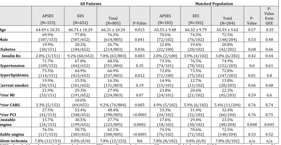
Baseline patient characteristics between APSES and EES in the overall population are displayed in Table 1. There were significant differences between the groups. After matching, characteristics were well balanced between the groups (Table 1). Mean age of the matched population was 66.5 years, 21% were diabetics, 24% had unstable angina. Overlap of the propensity scores was excellent as demonstrated by the box plots (Supplementary Figure 1). The propensity model fit was good as assessed by Hosmer-Lemeshow (P>0.05). The assumption of proportionality was not violated.
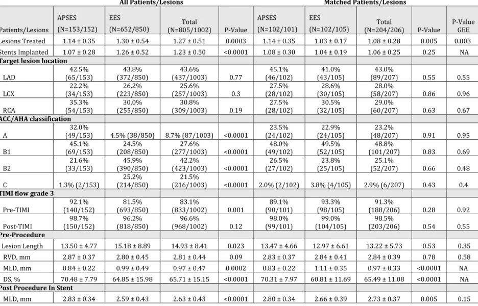
Baseline lesion characteristics between APSES and EES in the overall population are displayed in Table 2. There were significant differences between the groups. After matching, characteristics were well balanced between the groups (Table 2). The number of stents implanted and procedure success per patient was similar, but the
maximum deployment pressure was higher in the EES group. The pre-procedure MLD was larger and the post-procedure MLD was smaller in the EES group (p<0.0001) resulting in a higher final in-stent and lesion diameter stenosis in the EES compared to the APSES group (Table 2).
Angiographic Outcomes
Results of angiographic up are shown in Table 3. Early angiographic follow-up between 4-9 months occurred in 87% of all APSES and 77% of all EES patients and 89% of matched APSES and 77% of matched EES patients, with a 44-day longer mean follow-up in the APSES group (p<0.0001). The follow-up in-lesion MLD was larger in APSES, however the in-stent and in-lesion LLL and binary restenosis were not significantly different between treatment groups. Late angiographic follow-up between 18 and 24 months was completed in 18% of all APSES and 51% of all EES patients and 16% of matched APSES and 54% of matched EES patients, with a 200 day longer mean follow-up in the EES group (p<0.0001). No differences in late lumen loss or restenosis were observed, except for less change in % diameter stenosis in-lesion (14% ± 6% vs. 22% ± 8%, p=0.001)and in-stent (4% ± 7% vs. 13% ± 9%, p=0.0002)with APSES compared with EES. With all angiographic data carried forward, the results in LLL and restenosis are similar between groups despite a significant delay in angiographic follow-up in the EES group (Table 3).
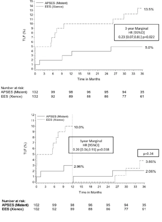
In the matched population, APSES had significantly lower TLF (3% vs. 10%,
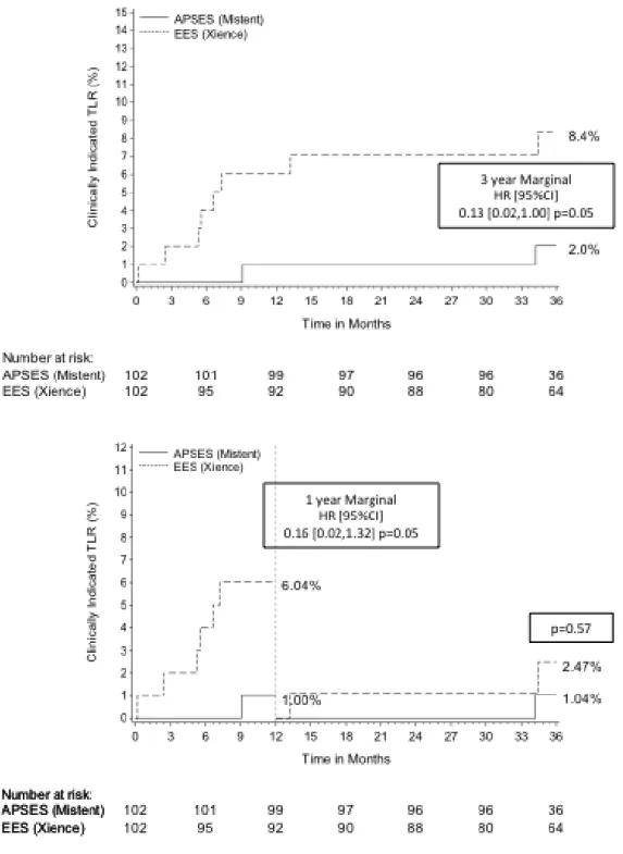
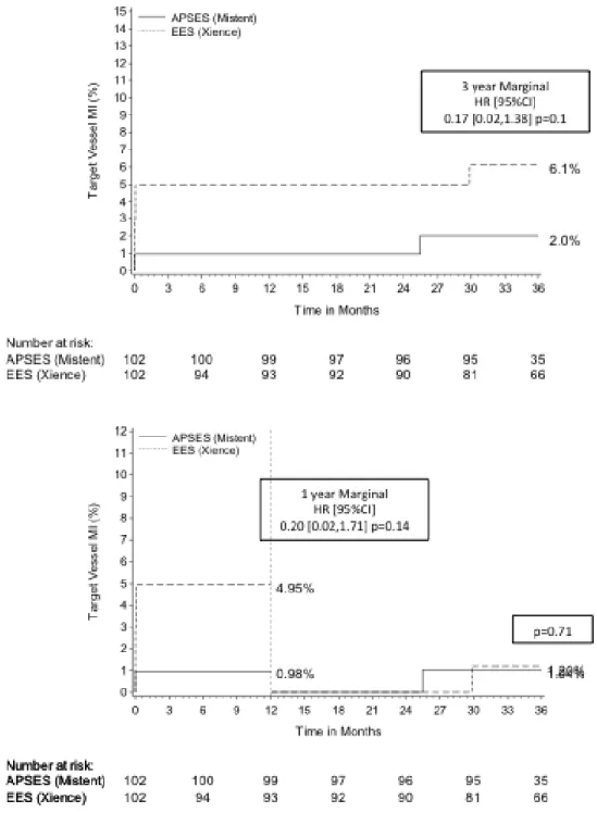
p=0.038; Figure 2) and TLR (1% vs. 6%, p=0.05; Figure 3) outcomes at 1 year, with no significant differences in target vessel MI (1% vs. 5%, p=0.14; Figure 4) or definite/probable stent thrombosis (0% vs. 1%; p=0.31) compared with EES. At 3 years, TLF (5.0% vs. 13.5%, p=0.02; Figure 2) remained significantly lower with APSES compared to EES, driven by a lower TLR rate (2.0% vs. 8.4%, p=0.05;
Figure 3). At 3 years, there were no differences in cardiac death (2.0% vs. 2.1%,
p>0.99) or target vessel myocardial infarction between the groups (2.0% vs. 6.1%; p=0.14; Figure 4), and there were no additional late or very late stent thrombosis in either group (0% vs. 1.0%, p=0.31). The landmark analysis demonstrates no
significant difference in TLF or other outcomes in the matched analysis between 1 and 3 years (Figures 2-4).
Results for sensitivity analyses were similar for the primary outcome, TLF. When all patients were censored at the time of their actual late angiographic follow-up, TLF remained significantly lower with APSES (5% vs. 12%, p=0.032). Additionally, when “early” and “late” angiographic follow-up are included in the Cox regression, the conclusions are the same. While late angiographic follow-up was related to an increase in the likelihood of an event (HR 11.1 [1.39, 88.4]; p=0.023), it had little effect on the relationship with treatment (HR 3.81 [1.26, 11.48]; p=0.017).
DISCUSSION
In this propensity-matched analysis of pooled data from 3 clinical trials, the thin strut cobalt chromium MiStent APSES showed significantly lower rates of TLF at 1
year and at 3 years compared with the durable polymer Xience EES. These differences were driven by a significantly lower TLR rate at both time points. Landmark analysis demonstrated that differences in TLF between APSES and EES occurred within the first year and was maintained at 3 years with minimal accrual beyond 1 year, and no difference in events between groups were observed from 1 to 3 years. Definite and probable stent thrombosis was low in both groups with no additional ST events from 1 to 3 years. In view of the dearth of randomized trial data comparing outcomes with both stents, our findings provide preliminary evidence of the comparable safety and effectiveness of the MiStent APSES versus the leading benchmarked Xience EES in a well-matched patient population with 3 year follow-up.
The thin strut MiStent APSES releases sirolimus in its crystalline form from a bioabsorbable polylactide-coglycolic acid polymer coating (approximately 3-5 μm thick on the luminal and 10-15 μm thick on the abluminal stent surfaces) (11). Based on our data, the achieved drug-release kinetic may confer benefit within the first year compared to the benchmark durable polymer EES; moreover, this benefit appears to be sustained between 1 and 3 years. The early benefit of the MiStent APSES may result from the unique combination of crystalline sirolimus within the bioabsorbable polymer, which enables the deposition of drug into the surrounding tissue with prolonged elution at a controlled rate. This unique design provides therapeutic tissue concentrations of sirolimus up to 9 months post-implantation, without an initial burst of drug release (12). Unlike other bioabsorbable polymer
DES, the MiStent polymer coating is cleared from the stent in 45 to 60 days and absorbed into the tissue within 90 days; long before complete drug elution. This unique prolonged drug elution resulting from the crystalline formulation of sirolimus (9 months) combined with a shorter polymeric absorption (3 months) may be associated with reduced inflammation (12), and may well account for the observed lower rates of TLF and TLR at 1 year compared to the durable polymer EES platform observed in our study.Another important differentiating factor are the release kinetics of Mistent APSES, which lack the early drug release burst described with conventional DES (13), and may mitigate an early dose related exaggerated vascular inflammatory effect.
A number of large-scale randomized clinical trials evaluating bioabsorbable polymer DES compared to the durable polymer EES have been recently reported and
demonstrated generally comparable results in TLF at 9 to 12 months. The CENTURY II trial evaluated a thin strut cobalt-chromium stent releasing sirolimus from a poly-DL-lactic acid (PDLLA) and polycaprolactone co-polymer, which degrades over 3-4 months (14). The 9-month TLF rate of the bioabsorbable polymer DES was similar (4.4% vs. 4.9%, p=0.66) compared to EES. The BIOSCIENCE randomized trial (15) evaluated a thin strut cobalt-chromium poly-L lactic acid polymer that degrades over 12–24 months. The 12 months TLF rates (6.7% vs. 4.1%) were non-inferior to the EES. Similar results were seen in the smaller BIOFLOW-II trial (16). The EVOLVE II randomized trial evaluated a thin-strut platinum-chromium stent platform that delivers everolimus from a bioabsorbable poly(DL-lactide-coglycolide) (PLGA)
polymer applied to the abluminal surface; this device has near synchronous drug release (90 days) and polymer absorption (120 days) (17). At 12 months, TLF rates (6.7% vs. 6.2%, p=0.83) were non-inferior to the durable polymer Promus Element EES.
The high performance of bioabsorbable polymer DES out to 12 months is encouraging. A number of large meta-analyses comparing durable and
bioabsorbable DES have confirmed similar outcomes at 12 months compared to durable polymer DES (18-21). Comparable outcomes versus benchmark durable polymer DES at this time point should be a sine qua non for their adoption. Unlike the results of these larger series and randomized trials, our matched analysis shows reduced TLF rates with Mistent relative to the durable polymer comparator.
The hypothesized benefit of bioabsorbable polymer DES is expected to become manifest first at late follow-up. In this respect, although the durable polymer EES demonstrates high efficacy at 9-12 months, accrual of events beyond 12 months has been described to occur at a rate of 2-3% per year in a large series of durable
polymer DES (22). However, clear demonstration of long-term benefit with other bioabsorbable polymer DES remains to be shown. In the 3-year follow-up of the original ISAR TEST 4 trial, continued accrual of events occurred in both the durable and bioabsorbable polymer DES groups with no significant differences in clinical events discernable at 3 years with the biodegradable DES (23). Moreover, although long-term follow-up from the LEADERS trial as well as a pooled analysis including
data from ISAR-TEST 3 and ISAR-TEST 4 showed improved late outcomes with bioabsorbable polymer DES the comparator stent in these analysis was the early generation Cypher SES (4,24). Indeed in a recent analysis of the final 5-year results from ISAR-TEST 4, we showed similar long-term results between bioabsorbable polymer DES and durable polymer EES (25).
Whether our clinical findings reflect a design-specific advantage of the APSES is difficult to determine from our matched analysis and warrants confirmation in a randomized clinical trial. One cannot exclude that methodologic issues inherent to the design of our study may have played an important role. However, a matched propensity analysis allows us to assign a mechanistic basis to performance superiority. That one device is proven to have fewer target lesion and vessel failures, and with time less recoil acutely, less thrombosis in the subacute setting, less intimal hyperplasia and restenosis late not only removes ambiguity as to which device is better but explains why; in other words there is less lesion failure with the one because every design feature is superior on every aspect of vascular repair. This mechanistic superiority rather than simple clinical superiority emerges most when devices are constructed differently – the Mistent and Xience devices fall into this comparison as they are different in many respects. The MiCell APSES shows not simply superior acute performance that persists but rather continued incremental superiority in comparison to the EES Xience, offering the possibility that Mistent is superior for each of the fundamental elemental aspects of the vascular response to stenting – vasomotor response, thrombosis, cell migration and proliferation and
tissue hyperplasia. Ultimately, our data suggests that the unique design properties of the MiStent APSES may offer early and sustained clinical benefit.
Limitations
The present study has some important limitations. First, a propensity matched analysis cannot completely correct for baseline confounding factors between the groups. Therefore, we cannot exclude the possibility that our results are due at least in part to residual unmeasured confounding. Second, the number of patients
included is modest and this limits the ability of our study to detect differences between the groups especially in relation to rarely occurring clinical events. Third, interpretation of angiographic results is limited by the fact that quantitative
coronary angiographic analysis was performed by different core labs for the DESSOLVE I and II studies in comparison with ISAR-TEST 4. However, the
definitions of endpoints were similar, and the same software packages were used for analysis.
Conclusions
This propensity-matched analysis comparing the MiStent APSES to the durable polymer Xience EES showed significantly lower TLF rates at 1 year and at 3 years with MiStent APSES. Differences in TLF occurred within the first year and were maintained at 3 years, with minimal accrual of events beyond 1 year in both groups. Definite and probable stent thrombosis was low in both groups with no additional
events from 1 to 3 years. These results warrant confirmation in a randomized clinical trial.
Funding: This study was support in part by Micell Technologies Impact on daily practice:
This propensity-matched analysis comparing the MiStent APSES to the durable polymer Xience EES showed significantly lower TLF rates at 1 year and at 3 years with MiStent APSES. These data suggest that the unique design properties of the MiStent APSES may offer early and sustained clinical benefit compared to Xience EES.
Conflict of Interest:
Dr. Eldeman reports financial interest and is a paid consultant in the company. The other authors report no conflict of interest
REFRENCES
1. Cassese S, Byrne RA, Tada T et al. Incidence and predictors of restenosis after coronary stenting in 10 004 patients with surveillance angiography. Heart 2014;100:153-9.
2. Finn AV, Nakazawa G, Joner M et al. Vascular responses to drug eluting stents: importance of delayed healing. Arterioscler Thromb Vasc Biol 2007;27:1500-10.
3. Byrne RA, Joner M, Kastrati A. Polymer coatings and delayed arterial healing following drug-eluting stent implantation. Minerva Cardioangiol 2009;57:567-84.
4. Stefanini GG, Byrne RA, Serruys PW et al. Biodegradable polymer drug-eluting stents reduce the risk of stent thrombosis at 4 years in patients undergoing percutaneous coronary intervention: a pooled analysis of individual patient data from the ISAR-TEST 3, ISAR-TEST 4, and LEADERS randomized trials. Eur Heart J 2012;33:1214-22.
5. Ormiston J, Webster M, Stewart J et al. First-in-human evaluation of a bioabsorbable polymer-coated sirolimus-eluting stent: imaging and clinical results of the DESSOLVE I Trial (DES with sirolimus and a bioabsorbable polymer for the treatment of patients with de novo lesion in the native coronary arteries). JACC Cardiovasc Interv 2013;6:1026-34.
6. Wijns W, Vrolix M, Verheye S et al. Randomised study of a bioabsorbable polymer-coated sirolimus-eluting stent: results of the DESSOLVE II trial. EuroIntervention 2014.
7. Byrne RA, Kastrati A, Kufner S et al. Randomized, non-inferiority trial of three limus agent-eluting stents with different polymer coatings: the Intracoronary Stenting and Angiographic Results: Test Efficacy of 3 Limus-Eluting Stents (ISAR-TEST-4) Trial. Eur Heart J 2009;30:2441-9.
8. Cutlip DE, Windecker S, Mehran R et al. Clinical end points in coronary stent trials: a case for standardized definitions. Circulation 2007;115:2344-51. 9. Wei L, Lin D, Weissfeld L. Regression analysis of multivariate incomplete
failure time data by modeling marginal distributions. J Am Stat Assoc 1989;84:1065-1073.
10. Lin D, Wei L, Ying Z. Checking the cox model with cumulative sums of Martingale-based residuals. Biometrika 1993;80:557-572.
11. Attizzani GF, Bezerra HG, Ormiston J et al. Serial assessment by optical coherence tomography of early and late vascular responses after implantation of an absorbable-coating Sirolimus-Eluting stent (from the first-in-human DESSOLVE I trial). Am J Cardiol 2013;112:1557-64.
12. Attizzani GF, Bezerra HG, Chamie D et al. Serial Evaluation of Vascular Response After Implantation of a New Sirolimus-Eluting Stent With Bioabsorbable Polymer (MISTENT): an optical coherence tomography and histopathological study. J Invasive Cardiol 2012;24:560-8.
13. Wang Q, Pierson W, Sood P et al. Pharmacokinetic sub-study in the SPIRIT III Randomized and Controlled Trial of XIENCE V everolimus eluting coronary stent system. J Interv Cardiol 2010;23:26-32.
14. Saito S, Valdes-Chavarri M, Richardt G et al. A randomized, prospective, intercontinental evaluation of a bioresorbable polymer sirolimus-eluting coronary stent system: the CENTURY II (Clinical Evaluation of New Terumo Drug-Eluting Coronary Stent System in the Treatment of Patients with Coronary Artery Disease) trial. Eur Heart J 2014;35:2021-31.
15. Pilgrim T, Heg D, Roffi M et al. Ultrathin strut biodegradable polymer sirolimus-eluting stent versus durable polymer everolimus-eluting stent for percutaneous coronary revascularisation (BIOSCIENCE): a randomised, single-blind, non-inferiority trial. Lancet 2014;384:2111-22.
16. Windecker S, Haude M, Neumann FJ et al. Comparison of a Novel Biodegradable Polymer Sirolimus-Eluting Stent With a Durable Polymer Everolimus-Eluting Stent: Results of the Randomized BIOFLOW-II Trial. Circ Cardiovasc Interv 2015;8:e001441.
17. Keriakes D. A prospective randomized investigation of a novel bioabsorbable polymer-coated Everolimus coronary stent- Primary outcomes of the EVOLVE II Trial. presented late breaking trial American Heart Association 2014.
18. Kang SH, Park KW, Kang DY et al. Biodegradable-polymer drug-eluting stents vs. bare metal stents vs. durable-polymer drug-eluting stents: a systematic review and Bayesian approach network meta-analysis. Eur Heart J 2014;35:1147-58.
19. Bangalore S, Toklu B, Amoroso N et al. Bare metal stents, durable polymer drug eluting stents, and biodegradable polymer drug eluting stents for
coronary artery disease: mixed treatment comparison meta-analysis. BMJ 2013;347:f6625.
20. Lupi A, Rognoni A, Secco GG et al. Biodegradable versus durable polymer drug eluting stents in coronary artery disease: insights from a meta-analysis of 5,834 patients. Eur J Prev Cardiol 2014;21:411-24.
21. Nakazawa G, Shinke T, Ijichi T et al. Comparison of vascular response between durable and biodegradable polymer-based drug-eluting stents in a porcine coronary artery model. EuroIntervention 2014;10:717-23.
22. Dangas GD, Serruys PW, Kereiakes DJ et al. Meta-analysis of everolimus-eluting versus paclitaxel-everolimus-eluting stents in coronary artery disease: final 3-year results of the SPIRIT clinical trials program (Clinical Evaluation of the Xience V Everolimus Eluting Coronary Stent System in the Treatment of Patients With De Novo Native Coronary Artery Lesions). JACC Cardiovasc Interv 2013;6:914-22.
23. Byrne RA, Kastrati A, Massberg S et al. Biodegradable polymer versus permanent polymer drug-eluting stents and everolimus- versus sirolimus-eluting stents in patients with coronary artery disease: 3-year outcomes from a randomized clinical trial. J Am Coll Cardiol 2011;58:1325-31.
24. Serruys PW, Farooq V, Kalesan B et al. Improved safety and reduction in stent thrombosis associated with biodegradable polymer-based biolimus-eluting stents versus durable polymer-based sirolimus-eluting stents in patients with coronary artery disease: final 5-year report of the LEADERS (Limus
Eluted From A Durable Versus ERodable Stent Coating) randomized, noninferiority trial. JACC Cardiovasc Interv 2013;6:777-89.
25. Kufner S, Byrne RA, Valeskini M et al. Five-year outcomes from a trial of three limus-eluting stents with different polymer coatings in patients with coronary artery disease: final results from the ISAR-TEST 4 randomised trial. EuroIntervention 2014.
FIGURES LEGENDS
Figure 1. MiStent Design
Figure 2. Kaplan Meier estimates of 3-year target lesion failure (top) and 1 year landmark analysis for target lesion failure (bottom)
Figure 3. Kaplan Meier estimates of 3-year ischemia driven lesion
revascularization (top) and 1 year landmark analysis for target lesion revascularization (bottom)
Figure 4. Kaplan Meier estimates of 3-year target vessel myocardial infarction (top) and 1 year landmark analysis for target vessel myocardial infarction (bottom)
TABLES:
Table 1. Baseline patient characteristics
All Patients Matched Population
APSES (N=153) EES (N=652) (N=805) Total P-Value APSES (N=102) EES (N=102) (N=204) Total Value P-Value from GEE Age 64.49 ± 10.35 66.74 ± 10.29 66.31 ± 10.34 0.015 65.55 ± 9.48 66.32 ± 9.79 65.93 ± 9.62 0.57 0.55 Male (107/153) 69.9% (507/652) 77.8% (614/805) 76.3% 0.041 (72/102) 70.6% (76/102) 74.5% (148/204) 72.5% 0.53 0.48 Diabetes (30/151) 19.9% (184/652) 28.2% (214/803) 26.7% 0.036 (22/100) 22.0% (20/102) 19.6% (42/202) 20.8% 0.68 0.66 Insulin Rx 2.0% (3/151) 9.2% (60/652) 7.8% (63/803) 0.003 2.0% (2/100) 3.9% (4/102) 3.0% (6/202) 0.42 0.44 Hypertension (109/152) 71.7% (442/652) 67.8% (551/804) 68.5% 0.35 (74/101) 73.3% (78/102) 76.5% (152/203) 74.9% 0.6 0.61 Hyperlipidemia (114/151) 75.5% (423/652) 64.9% (537/803) 66.9% 0.012 (72/100) 72.0% (75/102) 73.5% (147/202) 72.8% 0.81 0.8 Current smoker (30/151) 19.9% (101/652) 15.5% (131/803) 16.3% 0.19 (15/101) 14.9% (13/102) 12.7% (28/203) 13.8% 0.66 0.68 Prior MI (33/151) 21.9% (191/652) 29.3% (224/803) 27.9% 0.07 (24/101) 23.8% (21/102) 20.6% (45/203) 22.2% 0.59 0.6 Prior CABG 3.3% (5/152) (69/652) 10.6% 9.2% (74/804) 0.005 4.9% (5/102) 5.9% (6/102) 5.4% (11/204) 0.76 0.74 Prior PCI (42/153) 27.5% (348/652) 53.4% (390/805) 48.4% <0.0001 (34/102) 33.3% (32/102) 31.4% (66/204) 32.4% 0.76 0.75 Unstable angina (24/153) 15.7% (199/652) 30.5% (223/805) 27.7% 0.0002 (18/102) 17.6% (30/102) 29.4% (48/204) 23.5% 0.048 0.045 Stable angina (117/153) 76.5% (383/652) 58.7% (500/805) 62.1% <0.0001 (76/102) 74.5% (72/102) 70.6% (148/204) 72.5% 0.53 0.52
Silent ischemia 7.8% (12/153) 0.0% (0/0) 7.8% (12/153) NA 7.8% (8/102) 0.0% (0/0) 7.8% (8/102) n/a n/a
APSES=absorbable polymer sirolimus-eluting stent; EES=everolimus-eluting stent; MI= myocardial infarction; CABG=coronary artery bypass grafting; PCI= percutaneous coronary intervention
Table 2. Baseline lesion and angiographic characteristics
All Patients/Lesions Matched Patients/Lesions
Patients/Lesions APSES (N=153/152) EES (N=652/850) (N=805/1002) Total P-Value APSES (N=102/101) EES
(N=102/105) (N=204/206) Total P-Value P-Value GEE Lesions Treated 1.14 ± 0.35 1.30 ± 0.54 1.27 ± 0.51 0.0003 1.14 ± 0.35 1.03 ± 0.17 1.08 ± 0.28 0.005 0.003 Stents Implanted 1.07 ± 0.28 1.26 ± 0.52 1.23 ± 0.50 <0.0001 1.08 ± 0.30 1.04 ± 0.19 1.06 ± 0.25 0.25 NA
Target lesion location
LAD (65/153) 42.5% (372/850) 43.8% (437/1003) 43.6% 0.77 (46/102) 45.1% (43/105) 41.0% (89/207) 43.0% 0.55 0.55 LCX (34/153) 22.2% (223/850) 26.2% (257/1003) 25.6% 0.3 (28/102) 27.5% (30/105) 28.6% (58/207) 28.0% 0.86 0.96 RCA (54/153) 35.3% (255/850) 30.0% (309/1003) 30.8% 0.19 (28/102) 27.5% (32/105) 30.5% (60/207) 29.0% 0.63 0.67 ACC/AHA classification A (49/153) 32.0% 4.5% (38/850) 8.7% (87/1003) <0.0001 (24/102) 23.5% (24/105) 22.9% (48/207) 23.2% 0.91 0.95 B1 (69/153) 45.1% (208/850) 24.5% (277/1003) 27.6% <0.0001 (49/102) 48.0% (52/105) 49.5% (101/207) 48.8% 0.83 0.69 B2 (33/153) 21.6% (390/850) 45.9% (423/1003) 42.2% <0.0001 (27/102) 26.5% (25/105) 23.8% (52/207) 25.1% 0.66 0.48 C 1.3% (2/153) (214/850) 25.2% (216/1003) 21.5% <0.0001 2.0% (2/102) 3.8% (4/105) 2.9% (6/207) 0.43 0.4
TIMI flow grade 3
Pre-TIMI (140/152) 92.1% (693/850) 81.5% (833/1002) 83.1% 0.001 (90/101) 89.1% (98/105) 93.3% (188/206) 91.3% 0.28 0.92 Post-TIMI (150/152) 98.7% (818/850) 96.2% (968/1002) 96.6% 0.12 (99/101) 98.0% (104/105) 99.0% (203/206) 98.5% 0.54 0.55 Pre-Procedure Lesion Length 13.50 ± 4.77 15.18 ± 8.89 14.93 ± 8.41 0.023 13.47 ± 4.66 12.97 ± 6.61 13.22 ± 5.73 0.53 0.35 RVD, mm 2.87 ± 0.37 2.80 ± 0.45 2.81 ± 0.44 0.09 2.83 ± 0.37 2.84 ± 0.41 2.84 ± 0.39 0.78 0.58 MLD, mm 0.84 ± 0.22 0.99 ± 0.49 0.97 ± 0.47 0.0002 0.83 ± 0.22 1.11 ± 0.35 0.97 ± 0.33 <0.0001 NA DS, % 70.48 ± 7.79 64.85 ± 15.98 65.71 ± 15.15 <0.0001 70.31 ± 7.97 60.81 ± 11.69 65.49 ± 11.08 <0.0001 NA
Post Procedure In Stent
DS, % 3.19 ± 7.29 11.83 ± 6.30 10.51 ± 7.17 <0.0001 3.05 ± 7.64 10.92 ± 6.10 7.02 ± 7.93 <0.0001 <0.0001
Post Procedure In-Lesion
MLD, mm 2.54 ± 0.38 2.25 ± 0.51 2.30 ± 0.50 <0.0001 2.52 ± 0.40 2.29 ± 0.48 2.40 ± 0.46 0.0003 0.0009 DS, % 13.47 ± 7.55 23.60 ± 11.44 22.04 ± 11.52 <0.0001 13.20 ± 7.61 23.70 ± 10.20 18.50 ± 10.42 <0.0001 <0.0001
APSES=absorbable polymer sirolimus-eluting stent; DS= diameter stenosis; EES=everolimus-eluting stent; MLD= minimal luminal diameter; RVD= reference vessel diameter; TIMI= thrombolysis in myocardial infarction
Table 3: Follow-up angiographic results
All Patients/Lesions Matched Patients/Lesions
APSES (N=153) EES (N=652) (N=805) Total P-Value APSES (N=102) EES
(N=102) (N=204) Total P-Value P-Value GEE
Early 4-9 months (N=134) (N=499) (N=633) (N=92) (N=78) (N=170) Days to follow-up 248.19 ± 46.63 208.93 ± 67.22 217.25 ± 65.39 <0.0001 254.74 ± 42.92 210.62 ± 65.26 234.50 ± 58.47 <0.0001 <0.0001 In-Stent MLD, mm 2.61 ± 0.50 2.36 ± 0.62 2.40 ± 0.61 <0.0001 2.59 ± 0.47 2.49 ± 0.67 2.55 ± 0.57 0.23 NA DS, % 10.47 ± 15.26 19.19 ± 16.93 17.70 ± 16.97 <0.0001 10.34 ± 14.40 15.66 ± 17.71 12.83 ± 16.21 0.031 NA LLL, mm 0.23 ± 0.43 0.23 ± 0.52 0.23 ± 0.51 0.94 0.23 ± 0.37 0.17 ± 0.55 0.20 ± 0.46 0.33 0.29 Restenosis, % 3.7% (5/134) (43/651) 6.6% 6.1% (48/785) 0.21 3.3% (3/92) 6.2% (5/81) 4.6% (8/173) 0.36 0.19 In-lesion MLD, mm 2.40 ± 0.48 2.07 ± 0.58 2.13 ± 0.58 <0.0001 2.40 ± 0.47 2.18 ± 0.60 2.30 ± 0.54 0.006 0.033 DS, % 18.04 ± 13.59 29.20 ± 16.14 27.30 ± 16.28 <0.0001 17.34 ± 12.68 26.28 ± 16.53 21.53 ± 15.24 <0.0001 0.057 LLL 0.15 ± 0.41 0.19 ± 0.53 0.18 ± 0.51 0.4 0.14 ± 0.36 0.11 ± 0.53 0.13 ± 0.45 0.73 0.24 Restenosis, % 3.7% (5/134) (65/651) 10.0% 8.9% (70/785) 0.021 3.3% (3/92) 7.4% (6/81) 5.2% (9/173) 0.22 NA Late 18-24 months (N=27) (N=332) (N=359) (N=15) (N=53) (N=68) Days to follow-up 540.59 ± 23.42 751.9 ± 189.85 736.01 ± 190.99 <0.0001 548.73 ± 23.94 749.25 ± 194.36 705.02 ± 190.9 0.0002 <0.0001 In-Stent MLD, mm 2.74 ± 0.32 2.31 ± 0.62 2.34 ± 0.61 0.0003 2.66 ± 0.32 2.55 ± 0.44 2.58 ± 0.42 0.37 0.17 DS, % 4.85 ± 6.83 20.97 ± 16.96 19.99 ± 16.96 <0.0001 4.04 ± 7.05 13.42 ± 8.51 11.41 ± 9.04 0.0002 <0.0001 LLL, mm 0.09 ± 0.15 0.29 ± 0.51 0.27 ± 0.49 0.045 0.12 ± 0.13 0.15 ± 0.28 0.14 ± 0.26 0.7 0.53 Restenosis, % 0.0% (0/27) (26/416) 6.3% 5.9% (26/443) 0.18 0.0% (0/15) 0.0% (0/55) 0.0% (0/70) NA NA In-lesion MLD, mm 2.42 ± 0.36 2.09 ± 0.58 2.11 ± 0.57 0.004 2.38 ± 0.31 2.30 ± 0.40 2.31 ± 0.38 0.44 0.26 DS, % 16.05 ± 8.32 28.33 ± 16.07 27.58 ± 15.98 <0.0001 14.21 ± 5.58 22.08 ± 8.36 20.39 ± 8.46 0.001 <0.0001
LLL, mm 0.06 ± 0.26 0.17 ± 0.55 0.16 ± 0.54 0.32 0.05 ± 0.24 0.03 ± 0.41 0.04 ± 0.38 0.89 0.87 Restenosis, % 0.0% (0/27) (35/416) 8.4% 7.9% (35/443) 0.12 0.0% (0/15) 0.0% (0/55) 0.0% (0/70) NA NA LOCF (N=134) (N=511) (N=645) (N=92) (N=78) (N=170) Days to LOCF 321.26 ± 113.09 567.54 ± 299.4 516.38 ± 289.18 <0.0001 315.23 ± 105.42 584.99 ± 294.00 439.00± 252.08 <0.0001 <0.0001 In-Stent MLD, mm 2.61 ± 0.50 2.26 ± 0.67 2.32 ± 0.66 <0.0001 2.59 ± 0.47 2.44 ± 0.64 2.52 ± 0.56 0.08 0.17 DS, % 10.79 ± 15.05 22.23 ± 19.58 20.28 ± 19.36 <0.0001 10.80 ± 14.12 17.13 ± 17.40 13.76 ± 16.01 0.009 NA LLL, mm 0.24 ± 0.43 0.32 ± 0.59 0.31 ± 0.57 0.11 0.24 ± 0.37 0.21 ± 0.55 0.23 ± 0.46 0.71 0.51 Restenosis, % 3.7% (5/134) (62/651) 9.5% 8.5% (67/785) 0.029 3.3% (3/92) 6.2% (5/81) 4.6% (8/173) 0.36 0.19 In-lesion MLD, mm 2.38 ± 0.48 2.04 ± 0.63 2.10 ± 0.62 <0.0001 2.39 ± 0.47 2.20 ± 0.56 2.30 ± 0.52 0.017 0.046 DS, % 18.67 ± 13.40 30.13 ± 18.30 28.17 ± 18.07 <0.0001 18.03 ± 12.45 25.34 ± 15.58 21.46 ± 14.43 0.0008 0.034 LLL, mm 0.16 ± 0.41 0.22 ± 0.61 0.21 ± 0.58 0.28 0.15 ± 0.36 0.09 ± 0.52 0.12 ± 0.44 0.4 0.4 Restenosis, % 3.7% (5/134) (82/651) 12.6% 11.1% (87/785) 0.003 3.3% (3/92) 6.2% (5/81) 4.6% (8/173) 0.36 0.19
APSES=absorbable polymer sirolimus-eluting stent; DS= diameter stenosis; EES=everolimus-eluting stent; LLL=late luminal loss; LOCF=last observation carried forward; MLD= minimal luminal diameter
FIGURES
Figure 2. Kaplan Meier estimates of 3-year target lesion failure and 1-year
Figure 3. Kaplan Meier estimates of 3-year ischemia driven lesion revascularization
Figure 4. Kaplan Meier estimates of 3-year target vessel myocardial infarction and